
Biomarkers of Glyco-Metabolic Control in Hemodialysis Patients: Glycated Hemoglobin vs. Glycated Albumin

 , ,
, ,  and
and
Abstract
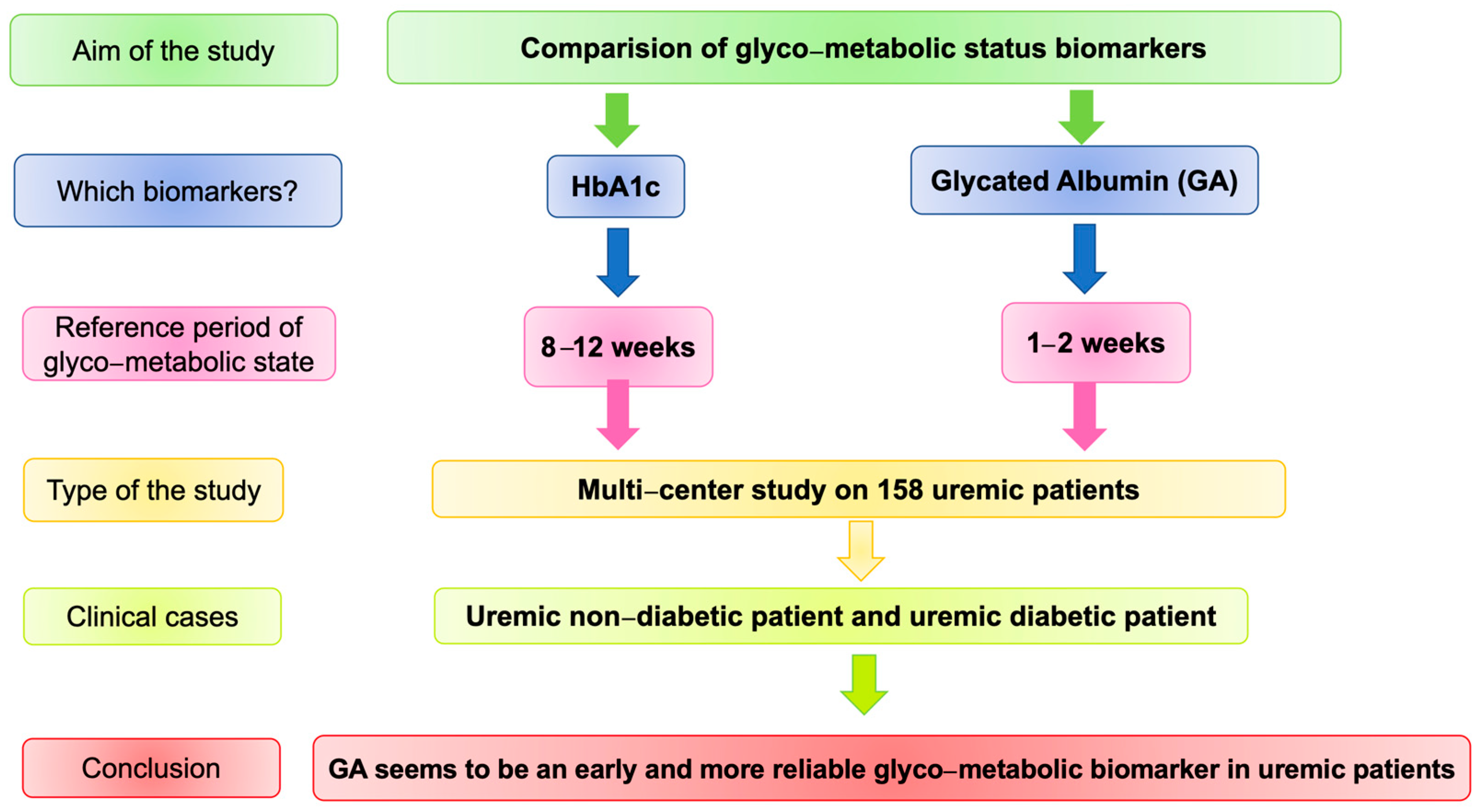
:1. Introduction
2. Materials and Methods
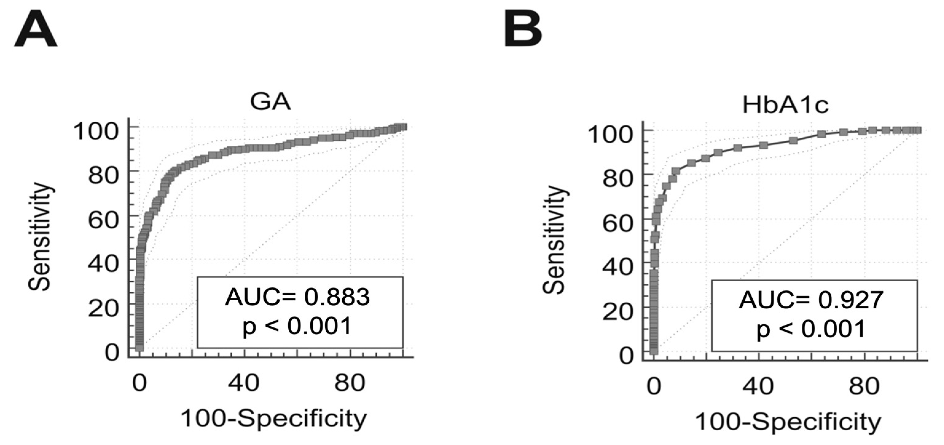
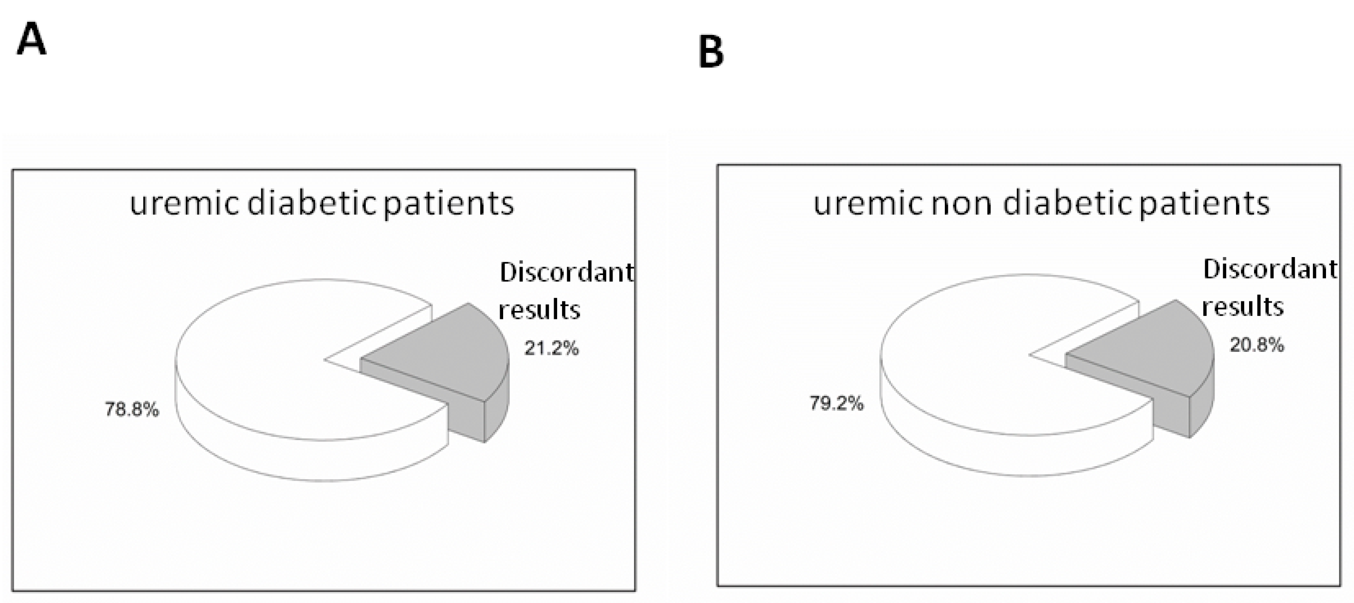
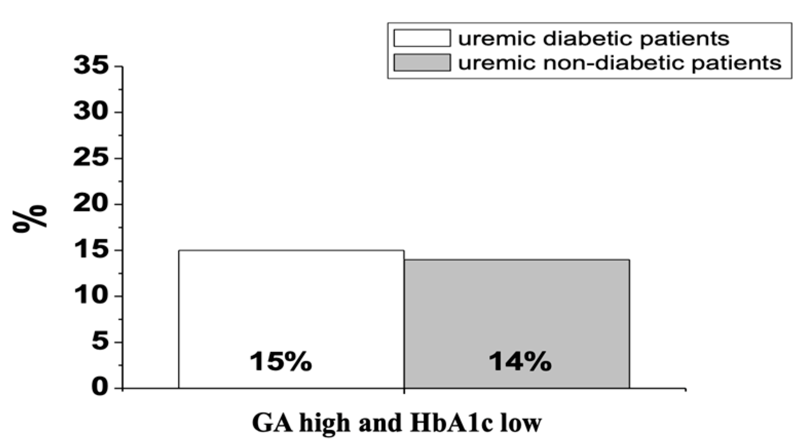
3. Results
4. Discussion
5. Conclusions
Author Contributions
Funding
Institutional Review Board Statement
Informed Consent Statement
Data Availability Statement
Acknowledgments
Conflicts of Interest
References
- Lv, J.C.; Zhang, L.X. Prevalence and Disease Burden of Chronic Kidney Disease. Adv. Exp. Med. Biol 2019, 1165, 3–15. [Google Scholar] [CrossRef]
- Stompor, T.; Adamczak, M.; Masajtis-Zagajewska, A.; Mazanowska, O.; Maziarska, K.; Witkowska, A.; Wiecek, A. Diagnosis and treatment of type 2 diabetes mellitus in patients with chronic kidney disease and eGFR < 60 mL/min—A position statement of the Polish Society of Nephrology Working Group on Metabolic and Endocrine Disorders in Kidney Diseases. Endokrynol. Pol. 2020, 70, 3–14. [Google Scholar] [CrossRef]
- Noce, A.; Canale, M.P.; Capria, A.; Rovella, V.; Tesauro, M.; Splendiani, G.; Annicchiarico-Petruzzelli, M.; Manzuoli, M.; Simonetti, G.; Di Daniele, N. Coronary artery calcifications predict long term cardiovascular events in non diabetic Caucasian hemodialysis patients. Aging Albany N. Y. 2015, 7, 269–279. [Google Scholar] [CrossRef] [PubMed] [Green Version]
- Yamamoto, H. Kidney diseases and metabolic disorders—Basics and applications required for general physicians. Topics: VIII. Anemia and iron metabolism in chronic kidney disease. Nihon Naika Gakkai Zasshi 2015, 104, 960–966. [Google Scholar] [CrossRef] [PubMed] [Green Version]
- Papadopoulou-Marketou, N.; Paschou, S.A.; Marketos, N.; Adamidi, S.; Adamidis, S.; Kanaka-Gantenbein, C. Diabetic nephropathy in type 1 diabetes. Minerva Med. 2018, 109, 218–228. [Google Scholar] [CrossRef] [PubMed]
- Cryer, M.J.; Horani, T.; DiPette, D.J. Diabetes and Hypertension: A Comparative Review of Current Guidelines. J. Clin. Hypertens. Greenwich 2016, 18, 95–100. [Google Scholar] [CrossRef] [PubMed]
- Duru, O.K.; Middleton, T.; Tewari, M.K.; Norris, K. The Landscape of Diabetic Kidney Disease in the United States. Curr. Diab. Rep. 2018, 18, 14. [Google Scholar] [CrossRef] [Green Version]
- Kainz, A.; Hronsky, M.; Stel, V.S.; Jager, K.J.; Geroldinger, A.; Dunkler, D.; Heinze, G.; Tripepi, G.; Oberbauer, R. Prediction of prevalence of chronic kidney disease in diabetic patients in countries of the European Union up to 2025. Nephrol. Dial. Transplant. 2015, 30 (Suppl. 4), iv113–iv118. [Google Scholar] [CrossRef]
- Kong, A.P.; Xu, G.; Brown, N.; So, W.Y.; Ma, R.C.; Chan, J.C. Diabetes and its comorbidities—Where East meets West. Nat. Rev. Endocrinol. 2013, 9, 537–547. [Google Scholar] [CrossRef]
- Ielpo, B.; Pernaute, A.S.; Elia, S.; Buonomo, O.C.; Valladares, L.D.; Aguirre, E.P.; Petrella, G.; Garcia, A.T. Impact of number and site of lymph node invasion on survival of adenocarcinoma of esophagogastric junction. Interact. Cardiovasc. Thorac. Surg. 2010, 10, 704–708. [Google Scholar] [CrossRef] [Green Version]
- Bajaj, S.; Makkar, B.M.; Abichandani, V.K.; Talwalkar, P.G.; Saboo, B.; Srikanta, S.S.; Das, A.; Chandrasekaran, S.; Krishnan, P.V.; Shah, A.; et al. Management of anemia in patients with diabetic kidney disease: A consensus statement. Indian J. Endocrinol. Metab. 2016, 20, 268–281. [Google Scholar] [CrossRef]
- Di Daniele, N.; Di Renzo, L.; Noce, A.; Iacopino, L.; Ferraro, P.M.; Rizzo, M.; Sarlo, F.; Domino, E.; De Lorenzo, A. Effects of Italian Mediterranean organic diet vs. low-protein diet in nephropathic patients according to MTHFR genotypes. J. Nephrol. 2014, 27, 529–536. [Google Scholar] [CrossRef]
- Schinzari, F.; Iantorno, M.; Campia, U.; Mores, N.; Rovella, V.; Tesauro, M.; Di Daniele, N.; Cardillo, C. Vasodilator responses and endothelin-dependent vasoconstriction in metabolically healthy obesity and the metabolic syndrome. Am. J. Physiol. Metab. 2015, 309, E787–E792. [Google Scholar] [CrossRef] [Green Version]
- Whiting, D.R.; Guariguata, L.; Weil, C.; Shaw, J. IDF diabetes atlas: Global estimates of the prevalence of diabetes for 2011 and 2030. Diabetes Res. Clin. Pract. 2011, 94, 311–321. [Google Scholar] [CrossRef]
- Fox, A.; Feng, W.; Asal, V. What is driving global obesity trends? Globalization or “modernization”? Global Health 2019, 15, 32. [Google Scholar] [CrossRef] [Green Version]
- Bocedi, A.; Noce, A.; Marrone, G.; Noce, G.; Cattani, G.; Gambardella, G.; Di Lauro, M.; Di Daniele, N.; Ricci, G. Glutathione Transferase P1-1 an Enzyme Useful in Biomedicine and as Biomarker in Clinical Practice and in Environmental Pollution. Nutrients 2019, 11, 1741. [Google Scholar] [CrossRef] [Green Version]
- Noce, A.; Ferrannini, M.; Fabrini, R.; Bocedi, A.; Dessi, M.; Galli, F.; Federici, G.; Palumbo, R.; Di Daniele, N.; Ricci, G. Erythrocyte glutathione transferase: A new biomarker for hemodialysis adequacy, overcoming the Kt/V(urea) dogma? Cell Death Dis. 2012, 3, e377. [Google Scholar] [CrossRef] [Green Version]
- Krhac, M.; Lovrencic, M.V. Update on biomarkers of glycemic control. World J. Diabetes 2019, 10, 1–15. [Google Scholar] [CrossRef]
- American Diabetes Association. Diagnosis and classification of diabetes mellitus. Diabetes Care 2004, 27 (Suppl. 1), S5–S10. [Google Scholar] [CrossRef] [Green Version]
- Sherwani, S.I.; Khan, H.A.; Ekhzaimy, A.; Masood, A.; Sakharkar, M.K. Significance of HbA1c Test in Diagnosis and Prognosis of Diabetic Patients. Biomark. Insights 2016, 11, 95–104. [Google Scholar] [CrossRef]
- Noce, A.; Fabrini, R.; Dessi, M.; Bocedi, A.; Santini, S.; Rovella, V.; Pastore, A.; Tesauro, M.; Bernardini, S.; Di Daniele, N.; et al. Erythrocyte glutathione transferase activity: A possible early biomarker for blood toxicity in uremic diabetic patients. Acta Diabetol. 2014, 51, 219–224. [Google Scholar] [CrossRef]
- Pieri, M.; Pignalosa, S.; Zenobi, R.; Calla, C.; Martino, F.G.; Menichella, G.; Mancina, F.; Moscato, U.; Nocca, G.; Khashoggi, H.; et al. Reference intervals for HbA1c partitioned for gender and age: A multicenter study. Acta Diabetol. 2016, 53, 1053–1056. [Google Scholar] [CrossRef] [PubMed]
- Ly, J.; Marticorena, R.; Donnelly, S. Red blood cell survival in chronic renal failure. Am. J. Kidney Dis. 2004, 44, 715–719. [Google Scholar] [CrossRef]
- Spencer, N.Y.; Stanton, R.C. Glucose 6-phosphate dehydrogenase and the kidney. Curr. Opin. Nephrol. Hypertens. 2017, 26, 43–49. [Google Scholar] [CrossRef]
- Ayesh Haj Yousef, M.H.; Bataineh, A.; Elamin, E.; Khader, Y.; Alawneh, K.; Rababah, M. Adequate hemodialysis improves anemia by enhancing glucose-6-phosphate dehydrogenase activity in patients with end-stage renal disease. BMC Nephrol. 2014, 15, 155. [Google Scholar] [CrossRef] [Green Version]
- Dessi, M.; Noce, A.; Bertucci, P.; Noce, G.; Rizza, S.; De Stefano, A.; Manca di Villahermosa, S.; Bernardini, S.; De Lorenzo, A.; Di Daniele, N. Plasma and erythrocyte membrane phospholipids and fatty acids in Italian general population and hemodialysis patients. Lipids Health Dis. 2014, 13, 54. [Google Scholar] [CrossRef] [PubMed] [Green Version]
- Santoro, A.; Canova, C. Anemia and erythropoietin treatment in chronic kidney diseases. Minerva. Urol. Nefrol. 2005, 57, 23–31. [Google Scholar]
- Lippi, G.; Targher, G. Glycated hemoglobin (HbA1c): Old dogmas, a new perspective? Clin. Chem. Lab. Med. 2010, 48, 609–614. [Google Scholar] [CrossRef] [PubMed]
- Lippi, G.; Franchini, M.; Salvagno, G.L.; Montagnana, M.; Targher, G.; Guidi, G.C. Determinants of anaemia in the very elderly: A major contribution from impaired renal function? Blood Transfus. 2010, 8, 44–48. [Google Scholar] [CrossRef] [PubMed]
- Gafter-Gvili, A.; Schechter, A.; Rozen-Zvi, B. Iron Deficiency Anemia in Chronic Kidney Disease. Acta Haematol. 2019, 142, 44–50. [Google Scholar] [CrossRef] [PubMed]
- Johnson-Wimbley, T.D.; Graham, D.Y. Diagnosis and management of iron deficiency anemia in the 21st century. Therap. Adv. Gastroenterol. 2011, 4, 177–184. [Google Scholar] [CrossRef] [Green Version]
- St Louis, J.; Valdini, A. Abnormally Low Hemoglobin A1c as Harbinger of Hemoglobinopathy. J. Am. Board Fam. Med. 2019, 32, 923–924. [Google Scholar] [CrossRef] [PubMed]
- Loutradis, C.; Skodra, A.; Georgianos, P.; Tolika, P.; Alexandrou, D.; Avdelidou, A.; Sarafidis, P.A. Diabetes mellitus increases the prevalence of anemia in patients with chronic kidney disease: A nested case-control study. World J. Nephrol. 2016, 5, 358–366. [Google Scholar] [CrossRef]
- Tsai, S.F.; Tarng, D.C. Anemia in patients of diabetic kidney disease. J. Chin. Med. Assoc. 2019, 82, 752–755. [Google Scholar] [CrossRef] [PubMed]
- Gianchandani, R.Y.; Neupane, S.; Iyengar, J.J.; Heung, M. Pathophysiology and Management of Hypoglycemiain End-Stage Renal Disease Patients: A Review. Endocr. Pract. 2017, 23, 353–362. [Google Scholar] [CrossRef] [PubMed]
- Hsiao, C.C.; Tu, H.T.; Lin, C.H.; Chen, K.H.; Yeh, Y.H.; See, L.C. Temporal Trends of Severe Hypoglycemia and Subsequent Mortality in Patients with Advanced Diabetic Kidney Diseases Transitioning to Dialysis. J. Clin. Med. 2019, 8, 420. [Google Scholar] [CrossRef] [PubMed] [Green Version]
- Tesauro, M.; Nistico, S.; Noce, A.; Tarantino, A.; Marrone, G.; Costa, A.; Rovella, V.; Di Cola, G.; Campia, U.; Lauro, D.; et al. The possible role of glutathione-S-transferase activity in diabetic nephropathy. Int. J. Immunopathol. Pharmacol. 2015, 28, 129–133. [Google Scholar] [CrossRef]
- Pastore, A.; Noce, A.; Di Giovamberardino, G.; De Stefano, A.; Calla, C.; Zenobi, R.; Dessi, M.; Di Daniele, N. Homocysteine, cysteine, folate and vitamin B(1)(2) status in type 2 diabetic patients with chronic kidney disease. J. Nephrol. 2015, 28, 571–576. [Google Scholar] [CrossRef] [Green Version]
- Jager, K.J.; Lindholm, B.; Goldsmith, D.; Fliser, D.; Wiecek, A.; Suleymanlar, G.; Ortiz, A.; Massy, Z.; Martinez-Castelao, A.; Agarwal, R.; et al. Cardiovascular and non-cardiovascular mortality in dialysis patients: Where is the link? Kidney Int. Suppl. 2011, 1, 21–23. [Google Scholar] [CrossRef] [Green Version]
- Park, J.; Lertdumrongluk, P.; Molnar, M.Z.; Kovesdy, C.P.; Kalantar-Zadeh, K. Glycemic control in diabetic dialysis patients and the burnt-out diabetes phenomenon. Curr. Diab. Rep. 2012, 12, 432–439. [Google Scholar] [CrossRef]
- Tascona, D.J.; Morton, A.R.; Toffelmire, E.B.; Holland, D.C.; Iliescu, E.A. Adequacy of glycemic control in hemodialysis patients with diabetes. Diabetes Care 2006, 29, 2247–2251. [Google Scholar] [CrossRef] [Green Version]
- Bowry, S.K.; Gatti, E. Impact of hemodialysis therapy on anemia of chronic kidney disease: The potential mechanisms. Blood Purif. 2011, 32, 210–219. [Google Scholar] [CrossRef]
- Maruyama, Y.; Kanda, E.; Kikuchi, K.; Abe, M.; Masakane, I.; Yokoo, T.; Nitta, K. Association between anemia and mortality in hemodialysis patients is modified by the presence of diabetes. J. Nephrol. 2021. [Google Scholar] [CrossRef]
- Abe, M.; Kaizu, K.; Matsumoto, K. Evaluation of the hemodialysis-induced changes in plasma glucose and insulin concentrations in diabetic patients: Comparison between the hemodialysis and non-hemodialysis days. Ther. Apher. Dial. 2007, 11, 288–295. [Google Scholar] [CrossRef]
- Abe, M.; Kalantar-Zadeh, K. Haemodialysis-induced hypoglycaemia and glycaemic disarrays. Nat. Rev. Nephrol. 2015, 11, 302–313. [Google Scholar] [CrossRef]
- Roselli, M.; Guadagni, F.; Buonomo, O.; Belardi, A.; Ferroni, P.; Diodati, A.; Anselmi, D.; Cipriani, C.; Casciani, C.U.; Greiner, J.; et al. Tumor markers as targets for selective diagnostic and therapeutic procedures. Anticancer. Res. 1996, 16, 2187–2192. [Google Scholar]
- Guo, J.; Zheng, H.J.; Zhang, W.; Lou, W.; Xia, C.; Han, X.T.; Huang, W.J.; Zhang, F.; Wang, Y.; Liu, W.J. Accelerated Kidney Aging in Diabetes Mellitus. Oxid. Med. Cell Longev. 2020, 2020, 1234059. [Google Scholar] [CrossRef]
- Noce, A.; Rovella, V.; Marrone, G.; Cattani, G.; Zingaretti, V.; Limongi, D.; D’Agostini, C.; Sorge, R.; Casasco, M.; Di Daniele, N.; et al. Hemodialysis biomarkers: Total advanced glycation end products (AGEs) against oxidized human serum albumin (HSAox). Acta Diabetol. 2019, 56, 1323–1331. [Google Scholar] [CrossRef]
- Choi, H.S.; Han, K.D.; Oh, T.R.; Suh, S.H.; Kim, M.; Kim, C.S.; Bae, E.H.; Ma, S.K.; Kim, S.W. Trends in the incidence and prevalence of end-stage renal disease with hemodialysis in entire Korean population: A nationwide population-based study. Med. Baltim. 2021, 100, e25293. [Google Scholar] [CrossRef]
- Winocour, P.H. Diabetes and chronic kidney disease: An increasingly common multi-morbid disease in need of a paradigm shift in care. Diabet. Med. 2018, 35, 300–305. [Google Scholar] [CrossRef]
- Iseki, K. Gender differences in chronic kidney disease. Kidney Int. 2008, 74, 415–417. [Google Scholar] [CrossRef] [PubMed] [Green Version]
- Noce, A.; Marrone, G.; Di Lauro, M.; Urciuoli, S.; Pietroboni Zaitseva, A.; Wilson Jones, G.; Di Daniele, N.; Romani, A. Cardiovascular Protection of Nephropathic Male Patients by Oral Food Supplements. Cardiovasc. Ther. 2020, 2020, 1807941. [Google Scholar] [CrossRef] [PubMed]
- Halbesma, N.; Brantsma, A.H.; Bakker, S.J.; Jansen, D.F.; Stolk, R.P.; de Zeeuw, D.; de Jong, P.E.; Gansevoort, R.T.; PREVEND Study Group. Gender differences in predictors of the decline of renal function in the general population. Kidney Int. 2008, 74, 505–512. [Google Scholar] [CrossRef] [Green Version]
- Mo, Y.; Ma, X.; Li, H.; Ran, X.; Yang, W.; Li, Q.; Peng, Y.; Li, Y.; Gao, X.; Luan, X.; et al. Relationship between glycated albumin and glycated hemoglobin according to glucose tolerance status: A multicenter study. Diabetes Res. Clin. Pract. 2016, 115, 17–23. [Google Scholar] [CrossRef]
- Hoshino, J.; Abe, M.; Hamano, T.; Hasegawa, T.; Wada, A.; Ubara, Y.; Takaichi, K.; Nakai, S.; Masakane, I.; Nitta, K. Glycated albumin and hemoglobin A1c levels and cause-specific mortality by patients’ conditions among hemodialysis patients with diabetes: A 3-year nationwide cohort study. BMJ Open Diabetes Res. Care 2020, 8, e001642. [Google Scholar] [CrossRef]


| Total | Men | Women | |
|---|---|---|---|
| N (%) | 160 | 102 (63.8) | 58 (36.2) |
| Age; mean ± SD, years | 64.1 ± 12.6 | 64 ± 13 | 64 ± 11 |
| BMI; mean ± SD, kg/m2 | 24.8 ± 3.5 | 25 ± 3.5 | 24.4 ± 3.4 |
| Diabetic uremic patients; N (%) | 60 (37.5) | 46 (77) | 14 (23) |
| Non-diabetic uremic patients; N (%) | 98 (61.3) | 55 (56) | 43 (44) |
| IGT; N (%) | 2 (1.2) | 1 (50) | 1 (50) |
| Patient | Diabetic * | Non-Diabetic |
|---|---|---|
| Sex | M | M |
| Age (years) | 75 | 72 |
| Type of dialysis | Convective technique | Diffusive technique |
| Type and dosage of EPO | Epoetin-α 4000 IU × 2/week | No therapy |
| BMI (kg/m2) | 23.8 | 21.4 |
| Patient | Diabetic | Non-Diabetic | Normal Range Values |
|---|---|---|---|
| Glycemia mg/dL (T0) | 99 | 137 | 80–100 mg/dL |
| Glycemia mg/dL (T1) | 151 | 160 | |
| Glycemia mg/dL (T2) | 161 | 120 | |
| Glycemia mg/dL (T3) | 95 | 105 | |
| Glycemia mg/dL (T4) | 80 | 126 | |
| HbA1c mmol/mol/Hb g/dL (T0) | 34/10.5 | 33/10.7 | HbA1c: <38 mmol/mol (Normal) 39–47 mmol/mol (Pre-diabetes) >48 mmol/mol (Diabetes) |
| HbA1c mmol/mol/Hb g/dL (T1) | 38/9.6 | 35/10.6 | |
| HbA1c mmol/mol/Hb g/dL (T2) | 36/9.8 | 35/10.8 | |
| HbA1c mmol/mol/Hb g/dL (T3) | 30/10.2 | 35/11.0 | |
| HbA1c mmol/mol/Hb g/dL (T4) | 35/11.1 | 36/11.3 | |
| GA %/A g/dL (T0) | 14.3/4.5 | 17.2/5.8 | GA ≤ 15% |
| GA %/A g/dL (T1) | 16.5/4.8 | 16.0/5.7 | |
| GA %/A g/dL (T2) | 16.3/5.0 | 16.4/5.8 | |
| GA %/A g/dL (T3) | 14.6/5.0 | 16.4/5.3 | |
| GA %/A g/dL (T4) | 14.9/5.1 | 15.4/5.5 |
Publisher’s Note: MDPI stays neutral with regard to jurisdictional claims in published maps and institutional affiliations. |
© 2021 by the authors. Licensee MDPI, Basel, Switzerland. This article is an open access article distributed under the terms and conditions of the Creative Commons Attribution (CC BY) license (https://creativecommons.org/licenses/by/4.0/).
Share and Cite
Martino, F.G.; Vitillo, M.; Pieri, M.; Marrone, G.; Gangeri, F.; Ansali, F.; Dessì, M.; Bernardini, S.; Di Daniele, N.; Noce, A. Biomarkers of Glyco-Metabolic Control in Hemodialysis Patients: Glycated Hemoglobin vs. Glycated Albumin. Medicina 2021, 57, 712. https://0-doi-org.brum.beds.ac.uk/10.3390/medicina57070712
Martino FG, Vitillo M, Pieri M, Marrone G, Gangeri F, Ansali F, Dessì M, Bernardini S, Di Daniele N, Noce A. Biomarkers of Glyco-Metabolic Control in Hemodialysis Patients: Glycated Hemoglobin vs. Glycated Albumin. Medicina. 2021; 57(7):712. https://0-doi-org.brum.beds.ac.uk/10.3390/medicina57070712
Chicago/Turabian StyleMartino, Francesca Gabriela, Marina Vitillo, Massimo Pieri, Giulia Marrone, Fabio Gangeri, Ferruccio Ansali, Mariarita Dessì, Sergio Bernardini, Nicola Di Daniele, and Annalisa Noce. 2021. "Biomarkers of Glyco-Metabolic Control in Hemodialysis Patients: Glycated Hemoglobin vs. Glycated Albumin" Medicina 57, no. 7: 712. https://0-doi-org.brum.beds.ac.uk/10.3390/medicina57070712