
Secondary Amenorrhea and Infertility Due to an Inhibin B Producing Granulosa Cell Tumor of the Ovary. A Rare Case Report and Literature Review


 ,
, 
Abstract
:1. Introduction
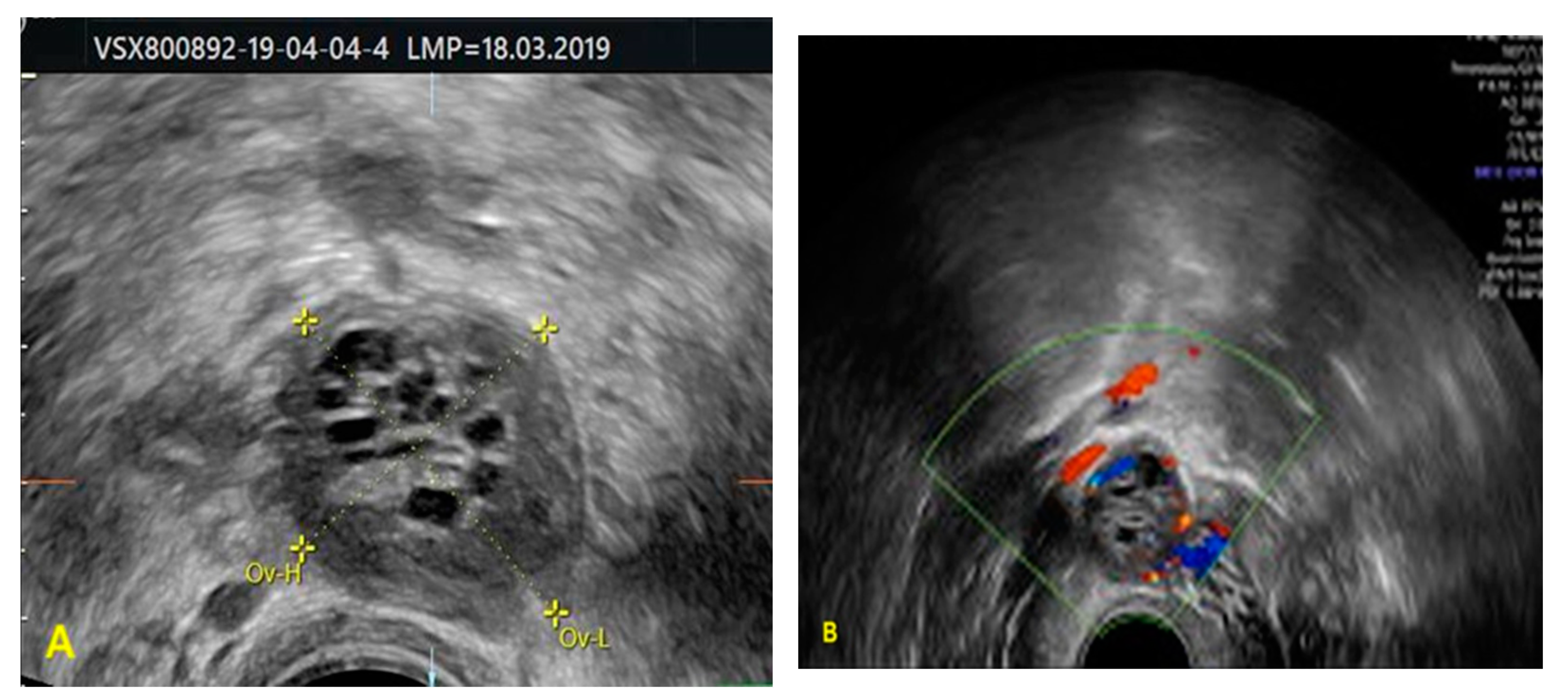
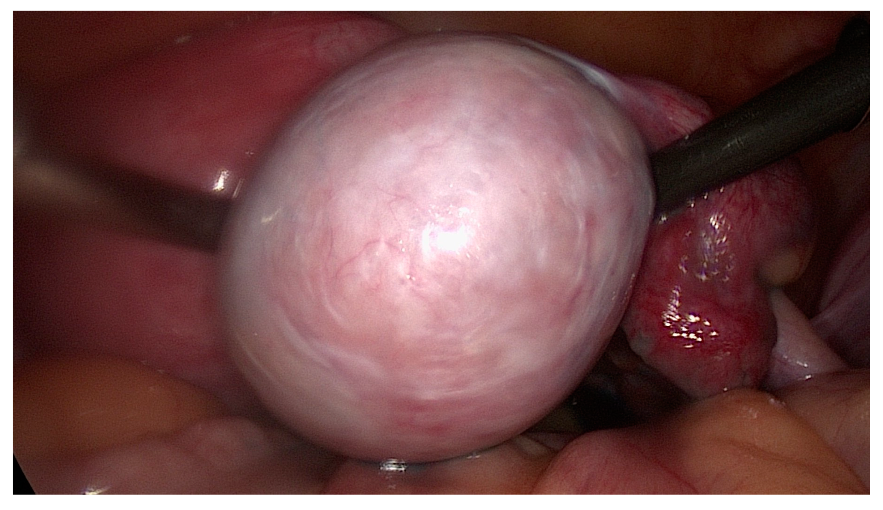
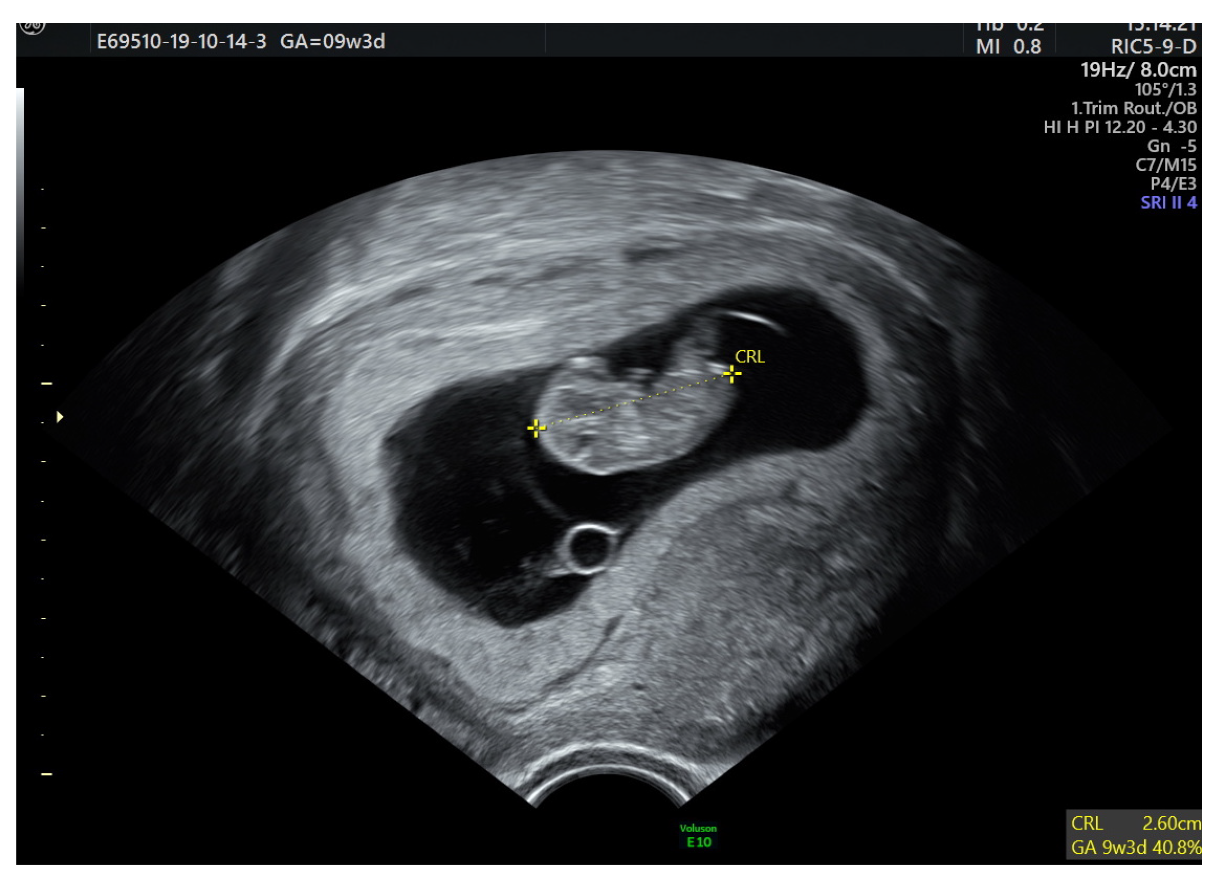
2. Case Report
3. Discussion and Conclusions

| Authors | Year | Journal | Cases | FSH | LH | Estradiol | Inhibin B |
|---|---|---|---|---|---|---|---|
| Lappöhn et al. [13] | 1989 | N Engl J Med | 2 cases | low | normal | NA | high |
| Lappöhn et al. [13] | 1989 | N Engl J Med | 1 case | low | low | NA | high |
| Sakamoto et al. [24] | 1998 | Acta Obstet. Gynecol. Scand. | 1 case | low | normal | NA | high |
| Krishnan et al. [25] | 2003 | Human Reproduction | 1 case | low | normal | normal | high |
| Kurihara et al. [19] | 2004 | J Obstet Gynaecol Res. | 1 case | low | normal | normal | high |
| Nasu et al. [15] | 2007 | International J of Clinical Oncology | 1 case | normal | high | normal | NA |
| Agha-Hosseini et al. [6] | 2008 | Taiwan J Obstet Gynecol | 1 case | low | low | normal | high |
| Tasci et al. [26] | 2015 | Turkish J of Gynecological Oncology | 2 cases | low | normal | normal | high |
| Ran et al. [16] | 2017 | J of Ovarian Research | 1 case | normal | high | low | NA |
| Okawa et al. [27] | 2020 | J of the Endocrine Society | 1 case | low | normal | low | high |
Author Contributions
Funding
Institutional Review Board Statement
Informed Consent Statement
Conflicts of Interest
References
- Gica, N.; Mustata, L.; Botezatu, R.; Chirculescu, R.; Cigaran, R.; Gica, C.; Peltecu, G. Management of Borderline Ovarian Tumors: Series of Case Report and Review of the Literature. Indian J. Surg. 2020, 83, 617–624. [Google Scholar] [CrossRef]
- Thomakos, N.; Biliatis, I.; Koutroumpa, I.; Sotiropoulou, M.; Bamias, A.; Liontos, M.; Rodolakis, A. Prognostic factors for recurrence in early stage adult granulosa cell tumor of the ovary. Arch. Gynecol. Obstet. 2016, 294, 1031–1036. [Google Scholar] [CrossRef] [PubMed]
- Kottarathil, V.D.; Antony, M.A.; Nair, I.R.; Pavithran, K. Recent advances in granulosa cell tumor ovary: A review. Indian J. Surg. Oncol. 2013, 4, 37–47. [Google Scholar] [CrossRef] [Green Version]
- Färkkilä, A.; Haltia, U.M.; Tapper, J.; McConechy, M.K.; Huntsman, D.G.; Heikinheimo, M. Pathogenesis and treatment of adult-type granulosa cell tumor of the ovary. Ann. Med. 2017, 49, 435–447. [Google Scholar] [CrossRef] [PubMed]
- Lee, I.H.; Choi, C.H.; Hong, D.G.; Song, J.Y.; Kim, Y.J.; Kim, K.T.; Lee, K.W.; Park, I.S.; Bae, D.S.; Kim, T.J. Clinicopathologic characteristics of granulosa cell tumors of the ovary: A multicenter retrospective study. J. Gynecol. Oncol. 2011, 22, 188–195. [Google Scholar] [CrossRef] [Green Version]
- Agha-Hosseini, M.; Aleyaseen, A.; Safdarian, L.; Kashani, L. Secondary amenorrhea with low serum luteinizing hormone and follicle-stimulating hormone caused by an inhibin A- and inhibin B-producing granulosa cell tumor. Taiwan J. Obstet Gynecol. 2009, 48, 72–75. [Google Scholar] [CrossRef] [Green Version]
- Gica, N.; Panaitescu, A.M.; Iancu, G.; Botezatu, R.; Peltecu, G.; Gica, C. The role of biological markers in predicting infertility associated with non-obstructive endometriosis. Ginekol. Pol. 2020, 91, 189–192. [Google Scholar] [CrossRef]
- Pectasides, D.; Pectasides, E.; Psyrri, A. Granulosa cell tumor of the ovary. Cancer Treat. Rev. 2008, 34, 1–12. [Google Scholar] [CrossRef] [PubMed]
- Dridi, M.; Chraiet, N.; Batti, R.; Ayadi, M.; Mokrani, A.; Meddeb, K.; Yahiaoui, Y.; Raies, H.; Mezlini, A. Granulosa Cell Tumor of the Ovary: A Retrospective Study of 31 Cases and a Review of the Literature. Int. J. Surg. Oncol. 2018, 2018, 4547892. [Google Scholar] [CrossRef] [Green Version]
- Pautier, P.; Lhomme, C.; Culine, S.; Duvillard, P.; Michel, G.; Bidart, J.M.; Droz, J.P. Adult granulosa-cell tumor of the ovary: A retrospective study of 45 cases. Int. J. Gynecol. Cancer 1997, 7, 58–65. [Google Scholar] [CrossRef]
- Ko, S.F.; Wan, Y.L.; Ng, S.H.; Lee, T.Y.; Lin, J.W.; Chen, W.J.; Kung, F.T.; Tsai, C.C. Adult ovarian granulosa cell tumors: Spectrum of sonographic and CT findings with pathologic correlation. AJR Am. J. Roentgenol. 1999, 172, 1227–1233. [Google Scholar] [CrossRef] [Green Version]
- National Comprehensive Cancer Network. NCCN Clinical Practice Guidelines in Oncology. Version 1. 2021. Available online: https://www.nccn.org/guidelines/guidelines-detail?category=1&id=1453 (accessed on 12 June 2021).
- Lappöhn, R.E.; Burger, H.G.; Bouma, J.; Bangah, M.; Krans, M.; de Bruijn, H.W. Inhibin as a marker for granulosa-cell tumors. N. Engl. J. Med. 1989, 321, 790–793. [Google Scholar] [CrossRef] [PubMed]
- Petraglia, F.; Hartmann, B.; Luisi, S.; Florio, P.; Kirchengast, S.; Santuz, M.; Genazzani, A.R. Low levels of serum inhibin A and inhibin B in women with hypergonadotropic amenorrhea and evidence of high levels of activin A in women with hypothalamic amenorrhea. Fertil. Steril. 1998, 70, 907–912. [Google Scholar] [CrossRef]
- Nasu, K.; Fukuda, J.; Yoshimatsu, J.; Takai, N.; Kashima, K.; Narahara, H. Granulosa cell tumor associated with secondary amenorrhea and serum luteinizing hormone elevation. Int. J. Clin. Oncol. 2007, 12, 228–230. [Google Scholar] [CrossRef] [PubMed]
- Ran, S.; Yu, Q.; Deng, S.; Xu, L. Luteinizing hormone elevation in ovarian granulosa cell tumor: A case report and review of the literature. J. Ovarian Res. 2017, 10, 30. [Google Scholar] [CrossRef] [PubMed]
- Cheung, C.K.; Leung, P.C.K. Molecular biology of gonadotropinreleasing hormone (GnRH)-I, GnRH-II, and their receptors in humans. Endocr. Rev. 2005, 26, 283–306. [Google Scholar] [CrossRef]
- Donovan, L.E.; Brain, P.H.; Duggan, M.A. Isolated luteinizing hormone (LH) elevation in a woman with secondary amenorrhea: A clue to the diagnosis of an inhibin B-producing thecoma and insights into the influence of inhibin B on LH. Fertil. Steril. 2010, 94 (Suppl. 3), 1097.e9–1097.e12. [Google Scholar] [CrossRef]
- Kurihara, S.; Hirakawa, T.; Amada, S.; Ariyoshi, K.; Nakano, H. Inhibin-producing ovarian granulosa cell tumor as a cause of secondary amenorrhea: Case report and review of the literature. J. Obstet. Gynaecol. Res. 2004, 30, 439–443. [Google Scholar] [CrossRef]
- Crew, K.D.; Cohen, M.H.; Smith, D.H.; Tiersten, A.D.; Feirt, N.M.; Hershman, D.L. Long natural history of recurrent granulosa cell tumor of the ovary 23 years after initial diagnosis: A case report and review of the literature. Gynecol. Oncol. 2005, 96, 235–240. [Google Scholar] [CrossRef]
- Gainder, S.; Kaur, J.; Siwatch, S.; Gupta, N. Adult Granulosa Cell Tumor: A Sinister Differential for Clomiphene-resistant Infertility. J. Hum. Reprod. Sci. 2018, 11, 190–192. [Google Scholar] [CrossRef]
- Schumer, S.T.; Cannistra, S.A. Granulosa cell tumor of the ovary. J. Clin. Oncol. 2003, 21, 1180–1189. [Google Scholar] [CrossRef]
- Babarović, E.; Franin, I.; Klarić, M.; Ferrari, A.M.; Karnjuš-Begonja, R.; Eminović, S.; Ostojić, D.V.; Vrdoljak-Mozetič, D. Adult Granulosa Cell Tumors of the Ovary: A Retrospective Study of 36 FIGO Stage I Cases with Emphasis on Prognostic Pathohistological Features. Anal. Cell Pathol. 2018, 2018, 9148124. [Google Scholar] [CrossRef] [PubMed] [Green Version]
- Sakamoto, S.; Sakamoto, M.; Nagai, A.; Obayashi, S.; Kubota, T.; Aso, T. Increased serum level of inhibin causes amenorrhea in a granulosa cell tumor patient. Acta Obstet. Gynecol. Scand. 1998, 77, 243–244. [Google Scholar]
- Krishnan, A.; Murdock, C.; Allard, J.; Cisar, M.; Reid, E.; Nieman, L.; Segars, J. Pseudo-isolated FSH deficiency caused by an inhibin B-secreting granulosa cell tumour: Case report. Hum. Reprod. 2003, 18, 502–505. [Google Scholar] [CrossRef] [PubMed] [Green Version]
- Tasci, T.; Kimyon Comert, G.; Karalok, A.; Ureyen, I.; Turan, T.; Tulunay, G. Secondary amenorrhea with low serum follicle stimulating hormone caused by granulosa cell tumor. Turk. J. Gynecol. Oncol. 2015, 18, 26–28. [Google Scholar]
- Okawa, E.R.; Gardner, R.; Feltmate, C.; Hirsch, M.S.; Craig, J.W.; Chan, Y.M. An Unusual Cause of Secondary Amenorrhea in an Adolescent: Expanding the Differential. J. Endocr. Soc. 2020, 4, 159. [Google Scholar] [CrossRef]
- Park, J.Y.; Jin, K.L.; Kim, D.Y.; Kim, J.H.; Kim, Y.M.; Kim, K.R.; Nam, J.H. Surgical staging and adjuvant chemotherapy in the management of patients with adult granulosa cell tumors of the ovary. Gynecol. Oncol. 2012, 125, 80–86. [Google Scholar] [CrossRef] [PubMed]


Publisher’s Note: MDPI stays neutral with regard to jurisdictional claims in published maps and institutional affiliations. |
© 2021 by the authors. Licensee MDPI, Basel, Switzerland. This article is an open access article distributed under the terms and conditions of the Creative Commons Attribution (CC BY) license (https://creativecommons.org/licenses/by/4.0/).
Share and Cite
Gică, C.; Cigăran, R.-G.; Botezatu, R.; Panaitescu, A.M.; Cimpoca, B.; Peltecu, G.; Gică, N. Secondary Amenorrhea and Infertility Due to an Inhibin B Producing Granulosa Cell Tumor of the Ovary. A Rare Case Report and Literature Review. Medicina 2021, 57, 829. https://0-doi-org.brum.beds.ac.uk/10.3390/medicina57080829
Gică C, Cigăran R-G, Botezatu R, Panaitescu AM, Cimpoca B, Peltecu G, Gică N. Secondary Amenorrhea and Infertility Due to an Inhibin B Producing Granulosa Cell Tumor of the Ovary. A Rare Case Report and Literature Review. Medicina. 2021; 57(8):829. https://0-doi-org.brum.beds.ac.uk/10.3390/medicina57080829
Chicago/Turabian StyleGică, Corina, Ruxandra-Gabriela Cigăran, Radu Botezatu, Anca Maria Panaitescu, Brîndușa Cimpoca, Gheorghe Peltecu, and Nicolae Gică. 2021. "Secondary Amenorrhea and Infertility Due to an Inhibin B Producing Granulosa Cell Tumor of the Ovary. A Rare Case Report and Literature Review" Medicina 57, no. 8: 829. https://0-doi-org.brum.beds.ac.uk/10.3390/medicina57080829