
A State-of-the-Art Review on Core–Shell Pigments Nanostructure Preparation and Test Methods


Department of Building, School of Design and Environment, National University of Singapore, Singapore 117566, Singapore
*
Authors to whom correspondence should be addressed.
Micro 2021, 1(1), 55-85; https://0-doi-org.brum.beds.ac.uk/10.3390/micro1010006
Submission received: 15 June 2021
/
Revised: 1 July 2021
/
Accepted: 5 July 2021
/
Published: 9 July 2021
(This article belongs to the Section Microscale Materials Science)
Abstract
:Uses of novel technologies for improving the durability and lifespan of the construction materials have emerged as viable solutions toward the sustainable future wherein the coating industry plays a significant role in economy growth and better livelihoods. Thus, the continual innovation of various technologies to introduce diverse market products has become indispensable. Properties of materials like color stability under UV, elevated temperatures and aggressive environments, and skid and abrasion resistance are the main challenges faced by commercial coating materials, leading to more demand of natural materials as sustainable agents. Lately, nanostructured core–shell pigments with unique compositions have widely been utilized in composite materials to enhance their properties. Core–shell particles exhibit smart properties and have immense benefits when combined with building materials. Based on these facts, we comprehensively overviewed the state-of-the-art research of core–shell nanomaterials in terms of their preparation and performance evaluation methods, as well as feasible applications. The first part of this article discusses effective shell materials, including most common silica and titanium oxides. In addition, nanotechnology enabling the production and patterning of low-dimensional materials for widespread applications is emphasized. The second part deals with various potential core materials used to achieve core–shell nanostructures. The third part of this paper highlights some interesting mechanisms of core–shell structures in the modified systems that display high stability, durability, efficiency, and eco-friendliness. Finally, different applications of these core–shell nanostructures are underscored together with their test methods to evaluate their performances.
1. Introduction
In recent years, synthetically colored pigments have been introduced in various market products. Consequently, they have been the subject of extensive scientific investigation. The most common uses of pigments are in paints, varnishes, printing inks, plastics and textiles, leather decoration, building materials, rubber, paper, ceramic glazes, and so forth [1,2]. The durability of pigments refers to their capacity to withstand weathering processes and avoid deterioration when exposed to an external environment [3]. Studies have revealed that efficient uses of energy for construction materials production can protect the environment [4]. Thus, the development of durable and sustainable pigments has become fundamental in the construction industry. Diverse methods have been introduced to improve the durability of pigments, especially the core–shell approach for smart/functional materials preparation [5,6,7,8].
Earlier, different chemical techniques have been developed to prepare high-quality core–shell nanostructures. The multicomponent materials were shown to have better properties due to their diverse compositions and structures, making them versatile for widespread applications [9,10,11,12]. Owing to their distinct properties, core–shell structures have received intense research attention. These structures are more advantageous than other composite materials. For example, they can strengthen or generate new chemical and physical capabilities, facilitate the structural integrity desirable for self-maintenance, stop the core from breaking up into large particles, and ensure effective dispersion. In addition, they offer other benefits, like typical multifunctional compositions and structures. A higher performance can be achieved in the presence of synergy between the shells and cores [13].
The properties of materials obtained from core–shell particles can be finely tuned, making them fundamentally interesting in the fields of science and technology [1,14,15]. In every core/shell particle, there is a core structural domain cloaked by a shell domain. Various types of materials can constitute the core and shell domains, such as metals, polymers, and inorganic solids. It is easy to modify the structure, size, and composition of these particles to customize their mechanical, optical, magnetic, thermal, catalytic, electrical, and electro-optical attributes. To create hollow spheres and reduce the expenditure of these precious materials, core/shell morphology can be employed, in which the materials with relatively cheaper cores are coated with expensive materials [16,17].
Nanoparticles are defined as particles with diameters below 0.1 µm, and such particles are attractive for diverse functional applications. In essence, these nanoparticles can be considered as smart materials due to their distinctive properties. Nanoscale systems are more advantageous than microscale, macroscale, and bulk materials because of their large surface area-to-volume ratios and quantum size effects [18,19]. The development of new and advanced synthesis methods has enabled the production of symmetrical (spherical) and various other shaped nanoparticles (e.g., cube, prism, hexagon, wire, rod, and tube) [20,21,22]. Nonetheless, the preparation and characterization of differently shaped nanoparticles have only been performed recently. Some reports have indicated that it is relatively easier to make non-spherical nanoparticles than other shapes [23,24,25]. It is important to note that the properties of these nanoparticles are both size- and shape-dependent. For instance, some properties of magnetic nanocrystals (including blocking temperatures, permanent magnetization, and magnetic saturation) are determined by their particle size, whereas the coercivity of such a system is decided by their shape due to its impact on the surface anisotropy. For this reason, nanoparticles are often used to improve the pigment performance [26].
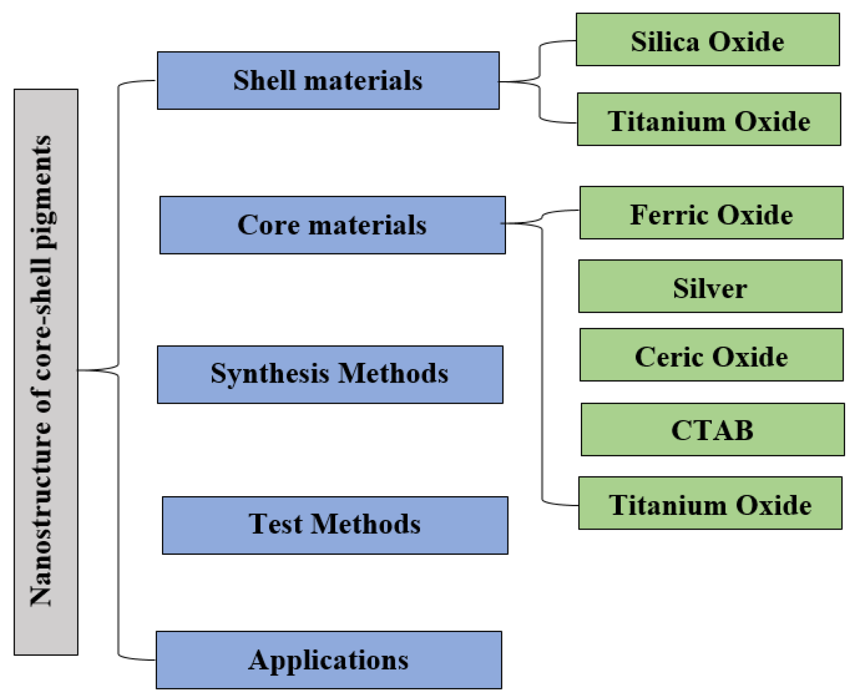
Nanotechnology has developed rapidly in recent years, and consequently, core–shell nanoparticles (NPs) have emerged as the key functional material. Currently, many researchers have been investigating core–shell NPs with different functional compositions. They find a wide range of applicability in various fields, including biomedicine, electronics, optics, catalysis, and pharmaceuticals [27]. Core–shell nanoparticles have distinct physiochemical traits and, thus, attract substantial research interests [22,28]. The key advantages of core–shell nanoparticles are enhanced levels of protection, encapsulation, and controlled release. Different types of core/shell nanoparticles have been identified and applied for various practical purposes. However, it remains challenging to classify all the available core/shell nanoparticles based on their industrial applications due to their wide varieties. Some studies were conducted on the pigments made from core–shell nanoparticles to outline their production methods and the nature of their core and shell materials, as well as their uses. Considering the immense benefits of core–shell nanostructures, this paper will analyze the key features and properties of such nanosystems. The features of inorganic materials and their fabrication methods and common uses are underscored. First, a brief overview of the different methods for the production of these particles is underlined. Next, different classes of existing core–shell materials and their uses are highlighted. Later, various latest techniques for the synthesis of core–shell pigment nanocapsules, followed by the use of core–shell nanoparticles in road paints, are explored. Finally, the paper is concluded, followed by some recommendations for further research and development within this field. Figure 1 provides the overall structure of this paper.
2. Core–Shell Nanoparticle: Synthesis Approach and Importance
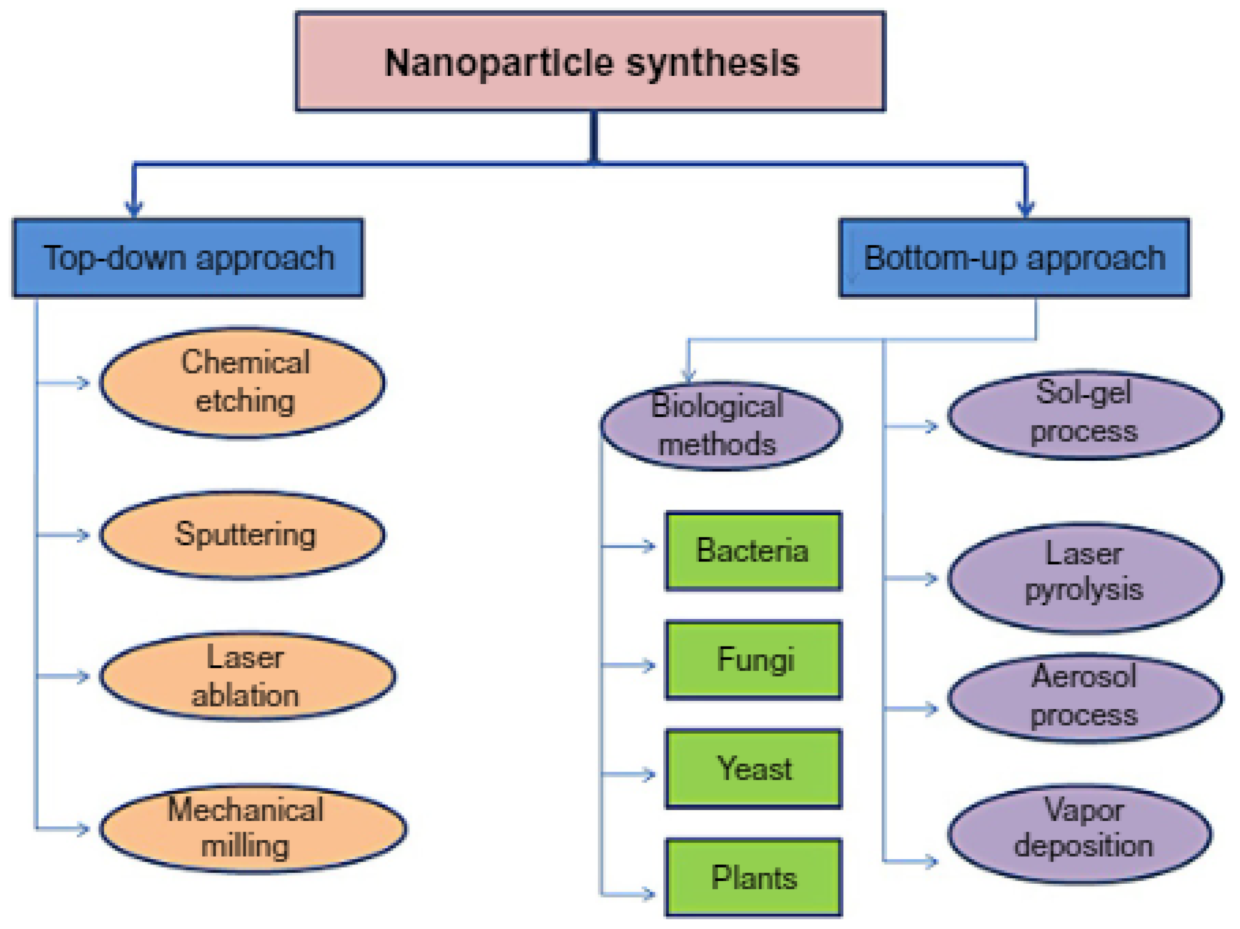
Nanotechnology refers to the atomic-level manipulation of materials that can be performed by combining the engineering, chemical, and biological approaches. Several techniques can be used to synthesize nanoparticles, like chemical, biological, physical, and even hybrid approaches (Figure 2). Generally, two key types of approaches are used for the production of nanostructures: “top-down” and “bottom-up”. In the former (“top-down”) approach, traditional workshops and microfabrication techniques are used in addition to externally controlled equipment to cut, mill, shape, and mold materials into their desired forms. The most common top-down methods are the lithographic techniques (e.g., UV, electron or ion beam, optical near-field scanning, laser beam processing, and scan probing) and mechanical methods (i.e., machining, grinding, and polishing) [29,30,31,32]. Conversely, the “bottom-up” approaches focus on the manipulation of the chemical properties of molecules, forcing them to self-assemble into a new and more beneficial form with emergent traits. The most commonly employed bottom-up approaches include chemical synthesis, laser-induced assembly, chemical vapor deposition, colloidal aggregation, self-assembly, and film deposition and growth [33,34]. Both approaches have strengths and weaknesses. Nonetheless, one key advantage of the bottom-up approach is that it can create much smaller particles and may be more cost-effective than the top-down approach due to its absolute precision, minimal energy loss, and complete control over the whole process. With regards to the synthesis of core/shell nanoparticles, ultimate control is needed to create a uniform coating around the core materials while the particles are forming. In this respect, a bottom-up approach would be more appropriate. In addition, a hybrid approach can also be used. For example, a top-down approach may be used to make the core, and a bottom-up approach can be subsequently employed to maintain the shell’s thickness. As water droplets can serve as nanoreactors, the use of microemulsion is also recommended to regulate the size and thickness of shells.
Nowadays, several researchers are turning their attention towards core/shell nanostructures because of their great benefits in a raft of fields, including chemistry, electronics, pharmaceuticals, optics, biomedicine, and catalysis. Additionally, these nanoparticles are highly functional and show specialized properties, wherein the characteristics of the core or shell materials can be entirely different. It is possible to modify these properties by altering the materials or the core-to-shell ratio [35]. Interestingly, the reactivity and thermal stability of the core materials can be changed by modifying the shell coating, thereby enhancing the stability and dispersion of the core particles. This, in turn, results in unique properties of each material employed, wherein the resultant product has an inherent ability to modify the surface functions based on the environment when implemented [36]. Some benefits of coating the core particles include the facilitation of surface modifications; enhanced functionality, stability, and dispersion of core particles; the significant reduction of expensive material consumption; and controlled release of the core.
Figure 2.
The synthesis of nanoparticles using different approaches [37].
Figure 2.
The synthesis of nanoparticles using different approaches [37].

3. Materials Based Shell Component
Core–shell nanoparticles are made up of multiple materials, including metals and biomolecules, with one forming the central core and the other covering the core in the form of a shell. Core–shell nanostructures have excellent thermal and chemical stabilities, high levels of solubility, low toxicity, and greater permeability toward certain target cells, making them immensely useful for a variety of uses. Micro-nanoscale core/shell particles have distinct compositions that make then unique compared to other types of particles. They effectively bring together the properties of the materials used in the core and shell, as well as the smart properties generated through the individual component materials. Of late, increasing research attention has been paid to the production and use of core–shell structures [38], especially in the pigment industry. To be more specific, core–shell materials have widely been applied to improve the durability of pigments (Table 1). To meet the ongoing demand, diverse organic and inorganic materials have been produced to achieve the optimum core–shell nanostructure. For example, Cao et al. [39] examined inorganic–organic hybrid pigments using an inorganic mixture of precipitated SiO2 and TiO2. Mesoporous soft template synthesis has been used to fabricate a dye core@silica shell structure [40]. At this point, it is customary to describe some beneficial inorganic materials used to produce the shells in core–shell structures, particularly SiO2 and TiO2.
3.1. SiO2-Based Shells
The encapsulation of individual pigment nanoparticles using a compact and durable shell is the most common approach. Several researchers have also put forward a reverse silica pigment topology [41,42,43,44,45,46]. Silicon is a very good material to use for core–shell coatings, because it is transparent, inert, and available in abundance. For this reason, it has been selected for use in many pigment encapsulation processes, such as in situ sol–gel reactions (with silica products used as precursors, including potassium/sodium silicate solutions) [47,48], various silanes with fumed silica [49], or tetraethoxysilane (TEOS), which is perhaps the most commonly used silica precursor [50]. A ball-milling treatment using silica nanoparticles can also be used for this purpose [51]. A layer-by-layer method of pre-synthesizing silica nanoparticles is also popular [52]. Silica is an excellent inorganic material to use for coating core–shell particles and useful in diverse fields, including biotechnology, medicine, and biomedical sensing. Distinct properties of silica, such as its high chemical stability, low cost, and great formability, enable the production of microscale particles from larger spherical particles.
Lu et al. [53] used the sol–gel method to hydrolyze tetraethyl orthosilicate (TEOS) to cover the iron oxide nanoparticles in uniform shells. By changing the sol–gel precursor concentration, the thickness of these silica shells was modified. Fluorescent dyes were added to these silica shells via the process of covalent coupling, in which the organic dyes and sol–gel precursor joined together. Silica (SiO2)-coated ceria (CeO2) nanoparticles were grown [54] via the water-in-oil microemulsion process. The SiO2-covered CeO2 nanoparticles were synthesized using cyclohexane (99%) as the organic phase, wherein reverse micelles on polyoxyethylene (15) cetylether were formed. The surfactant used in the organic solution had a concentration of 0.5 mol/L. Additionally, CeO2 was sourced from cerium nitrate and cerium chloride (cerium nitrate hexahydrate, Ce(NO3)3·6H2O, purity 99% and cerium chloride, CeCl3, purity 99.9%). Subsequently, a liquid phase (4.0 mL, represented as aq.) of cerium ions (Ce(NO3)3 aq. and CeCl3 aq.) was added to the organic surfactant solution with the concentration of 0.14 mol/L (100 mL). The resultant mixture was magnetically stirred at 50 °C, allowing the formation of a microemulsion solution. Either an ammonium solution (NH4OH aq., 2.70 mol/L, 3.0 mL) or oxalic acid solution ((COOH)2 aq., 1 mol/L, 3.0 mL) was used as the CeO2 precursor agent. The SiO2 was obtained from tetraethylorthosilicate (TEOS; 0.86 mol/l of microemulsion solution) and used to produce a NH4OH solution (2.70 mol/L, 15.0 mL). Then, it was added to the microemulsion with the CeO2 precursor nanoparticles, followed by hydrolysis of the solution for one hour at 50 °C. Throughout the TEOS hydrolysis process, the water-to-surfactant molar ratio was maintained at 23. It was impossible to accurately measure the pH of the microemulsion. An electric pH gauge was used to make an approximation with the meter reading ranging from 11.0 to 11.5. The solution was centrifuged after removing the CeO2 precursor nanoparticles from SiO2, followed by bath in propanol, and then left at 80 °C to dry overnight. Finally, it was calcined for 5 h at 400 °C to eliminate the surfactant.
The chemical liquid deposition technique was used to produce aluminum pigments covered in SiO from the tetraethyl silicate precursor (TEOS) [55]. The SiO2-coated aluminum particles were grown using 10 g of aluminum flakes inserted into 100 mL of pure ethanol followed by ultrasonic treatment for half an hour. A well-dispersed aluminum suspension was obtained, and then, TEOS was added. Subsequently, the aluminum suspension was put into a three-neck flask of 1000-mL capacity and then placed in a water bath before being stirred at a rate of 500 rpm at 40 °C. After, a solution consisting of 28% ammonia was supplemented together with a certain quantity of distilled water, plus 16 mL of pure ethanol. Next, TEOS was diluted using 12 mL of pure ethanol. The used mole ratios of TEOS-to-Al throughout the experiment were 1:44, 1:22, and 1:11. Moreover, the mole ratio of distilled water-to-TEOS was kept constant at 30:1. The solutions discussed above were then placed into an aluminum suspension via two continuous flow pumps at a constant flow rate, with a feeding time of 1 h. The newly produced suspension was then stirred for 5 h at 40 °C. The precipitate was filtrated and then was washed multiple times with pure ethanol. It was then placed in an electric oven at 120 °C for 12 h and at 200 °C for another 2 h for drying.
Ahmed et al. [56] used zinc, calcium, and strontium nitrate solutions to produce ferrite/silica core–shell pigments, wherein ferric nitrate were mixed with silica fume. After the solution was stirred for 60 min at 100 °C, the resultant paste was heated in an oven at 400 °C. Finally, the paste was ground and calcined at 750 °C. The Stöber method was used [57] to grow crystals of SiO2-covered Sr2+-doped red γ-Ce2S3 (Sr2+-doped γ-Ce2S3@c-SiO2) pigments. In this process, amorphous SiO2 (a-SiO2) sol and Sr2+-doped CeO2 were used as precursors. The original materials used in this experiment were Si(OC2H5)4 (TEOS), Sr(NO3)2, Ce(NO3)3·6H2O, (NH4)2CO3, ammonia water, and ethanol. The following process was conducted to acquire the Sr2+-doped CeO2 pigment precursor. First, the placement of Sr(NO3)2 and Ce(NO3)3·6H2O with a Sr2+/Ce3+ mole ratio of 0.15 was incorporated into deionized water to dissolve the particles, producing a homogeneous solution. Second, the precipitant (NH4)2CO3 was added to the solution to produce some white precipitate. Next, the precipitate was filtered and washed several times using water before being left at 80 °C for 12 h to dry. Afterward, it was calcined for 1 h in a muffle furnace at 1000 °C. Finally, it was left to cool down naturally, producing Sr2+-doped CeO2. Later, TEOS was dissolved in a solution containing water and alcohol, whilst ammonia water (25.0 wt%) was used to change the solution’s pH level to 11.00. The solution was then stirred for 0.5 h and kept in an 80 °C water bath for six hours to produce SiO2 sol. The resultant Sr2+-doped CeO2 was added to the SiO2 sol at a Ce3+/Si4+ mole ratio of 0.2, 0.4, 0.6, 0.8, or 1 and stirred for 0.5 h before being left to dry at 80 °C. This produced the precursor Sr2+-doped γ-Ce2S3@c-SiO2 red pigments in powder form. Afterward, this powder was treated in a tubular furnace with a gas flow containing both argon and CS2 (100 mL/min). The vulcanization temperatures of the furnace were changed intermittently (880 to 920 °C) over a period of two hours. Once sulfurization occurred, the powder was subjected to calcination in argon gas at various temperatures (1100 to 1250 °C) for 60 min and left to cool down naturally to room temperature. The process yielded Sr2+-doped γ-Ce2S3@c-SiO2 red pigments, which were later treated with 2-M HCl for 6 h. Finally, the colors of the samples were compared before and after acid etching.
Mjejri et al. [58] fabricated core–shell Zn1−xCoxO@SiO2 nanoparticles with green pigments using SiO2-based shell materials. The main objective was to create particles with high chemical stability and low cytotoxicity. A polyol process was used to form ZnO doped with Co2+, a well-known substitute for chromium-based inorganic pigments. Additionally, to source the zinc and cobalt needed in the experiment, zinc acetate dihydrate (Zn(CH3OO)2·2H2O) and cobalt nitrate (Co(NO3)2·6H2O) were utilized, wherein diethylene glycol (H10C4O3) was used as the template. Upon adding zinc and cobalt precursors into the solution at a stoichiometric proportion, 1 g of Zn1−xCoxO powder (2.924 g) was obtained. Subsequently, the powder was supplemented with 250 mL of H10C4O3. The resultant mixture was then continually stirred for 1 h before being heated at 180 °C to produce a green sol. Next, the precipitate was centrifuged and then washed multiple times in pure ethanol to eliminate the residual organic product. Then, it was oven-dried at 80 °C. Later, tetraethoxysilane (TEOS and Si(OC2H5)4) was subjected to hydrolysis, followed by condensation in an alcohol solution (EtOH). The Stöber experiment was conducted to produce Zn1−xCoxO@SiO2 core shells. In this process, about 100 mg of powder was added to 150 mL of EtOH and 6.4 mL of NH4OH as a hydrolysis–condensation catalyst. To vary the thicknesses of the SiO2 shells, various quantities of TEOS were added slowly to the solution as a prepared suspension. Subsequently, the suspension was kept at room temperature for two hours under constant agitation. Once the solution got matured, a few drops of HCL liquid solution (0.2 M) were added to ensure the complete dissolution of the ZnO cores, producing some porous silica shells.
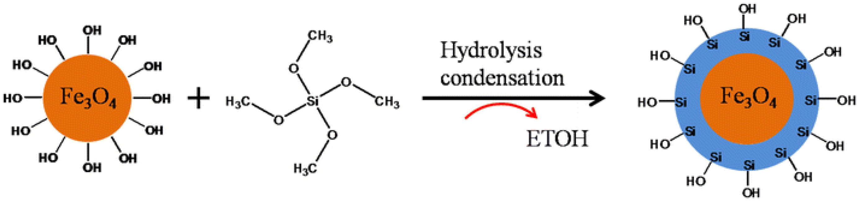
The hydrothermal Stöber techniques were used effectively to produce α-Fe2O3@SiO2 reddish pigments with core–shell structures [28]. The reddish pigments of α-Fe2O3 acted as chromophores and tetraethoxysilane (TEOS, AR) as the source of the silica. First, 0.05 g of α-Fe2O3 and polyvinyl pyrrolidone (PVP-30) were dissolved in 100 mL of deionized water and then subjected to ultrasonication for 15 min. This produced a homogeneous solution, which was stirred at room temperature continually for 12 h. The resultant solution was centrifuged multiple times to collect the PVP-modified α-Fe2O3 following three times bathing in ethanol and deionized water. Later, the as-obtained α-Fe2O3 was mixed in 100 mL of ethanol and subjected to an ultrasound for 5 min until the solution became homogeneous. Afterward, various contents of ammonia (0.5, 1.5, and 2.5 mL) and tetraethoxysilane (0.1, 0.3, and 0.5 mL) were supplemented into the solution with continuous stirring. Table 1 explains the experimental conditions of each sample. The resultant solution was stirred for 6 h at room temperature, forming α-Fe2O3@SiO2 NPs. The as-prepared α-Fe2O3@SiO2 NPs were left for 12 h at 70 °C to dry before being calcined at 1000 °C for 1 h. Figure 3 illustrates the process of fabricating α-Fe2O3@SiO2 pigments with core–shell structures.
Fabjan et al. [48,59] produced multilayered SiO2 core–shell pigments by adding ethoxylated alcohol (EA) and hexadecyl-trimethylammonium bromide (CTAB) to distilled water to fabricate a surfactant solution. The red pigment was then incorporated into the solution to create a pigment dispersion of 1 wt%. Meanwhile, a mild basic solution was used by Yu et al. [60] to coat the SiO2 shells in order to produce α-Fe2O3@SiO2 nanoparticles. First, α-Fe2O3 nanoparticles in spindle shapes were fabricated that served as the core materials. Next, these nanoparticles were covered by silica through the sol–gel technique. The original materials used (obtained from Aldrich) in the process were FeCl3·6H2O (97%), tetraethylorthosilicate (TEOS of 99.99% purity), cetyltrimethylammonium bromide (CTAB, 99%), and NH4OH (28% in water of 99.99% purity). Additionally, deionized water was employed, and the Stöber method was applied to treat the hematite with silica. Then, the silica shells were selectively etched using the NH4OH solution. The resultant silica-coated hematite powder was then placed in distilled water (10 mL), whilst 1 mL and 10 mL of NH4OH solution were poured into the hematite solutions to change the etchants’ concentrations. The resultant mixtures were centrifuged, and the solutions were kept at 80 °C in vacuum to dry. The samples were then subjected to heat treatments at either room temperature or higher temperatures (300 and 1000 °C) for sixty minutes before the physical properties of each sample were compared. This enabled the researcher to assess the coloration and heat stability of each etched sample.
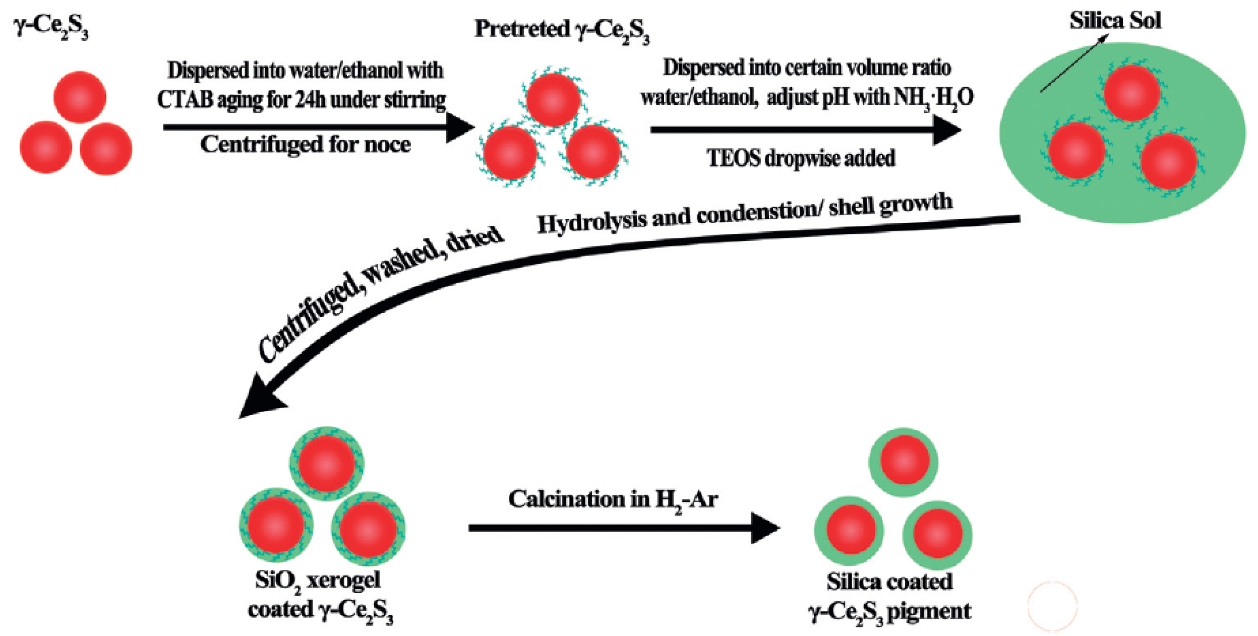
Wang et al. [61] conducted a study to grow ZnS@SiO2 core–shell nanoparticles using a homogeneous deposition method, in which deionized water (1 mL) and ammonia (0.5 mL) were mixed with 30 mL of ZnS (0.5–2 wt%) colloidal solution, and the obtained solution was stirred for 60 min. Subsequently, TEOS (0.5–2 g) was incorporated in the silica-coated material, and the reactions occurred at room temperature over a period of 5 h. After the reactions were completed, the resultant core–shell particles were collected and subjected to centrifugation in order to purify them. Subsequently, the particles were redispersed in water and ethanol multiple times. Lui et al. [62] treated γ-Ce2S3 red pigments with a silica layer using the Stöber technique combined with a hydrogen–argon atmosphere heat treatment (700 °C). Figure 3 shows the experimental procedure for the silica-coated γ-Ce2S3 pigments’ fabrication. In this process, first, untreated γ-Ce2S3@SiO2 pigments (2.0 g) and CTAB (0.2 g) particles were stirred into a solution containing 200 mL of ethanol and 250 mL of deionized water for a period of 24 h. Next, the solution was centrifuged to separate the particles, allowing the collection of pretreated, uncoated γ-Ce2S3@SiO2 pigments. Second, a solution containing water and ethanol was made with an established volume ratio, and then, ammonia water was added to change the pH value to approximately 10.5. The pretreated uncoated γ-Ce2S3@SiO2 pigments were then placed into the solution. Later, TEOS was added to the solution using a continuous flow pump, producing a sol mixture that was stirred for 3 h at 40 °C. Afterward, the sol mixture was centrifuged, washed in ethanol several times, and left in an oven at 70 °C to dry, yielding a SiO2 xerogel-coated γ-Ce2S3. Finally, the SiO2 xerogel-coated γ-Ce2S3 particles were inserted into a sealed double crucible and annealed in a hydrogen–argon gas mixture at 700 °C for 2 h in order to fabricate γ-Ce2S3@SiO2 pigments.
Figure 3.
Experimental process schematic of the preparation of γ-Ce2S3@SiO2 [62].
Figure 3.
Experimental process schematic of the preparation of γ-Ce2S3@SiO2 [62].

Colloidal silica nanoparticles were used in a sol–gel process to coat the natural pigment of hematite (red ocher, α-Fe2O3) [63], wherein the main objective was to produce a long-lasting hematite pigment of the Fe2O3@SiO2 nanocomposite that may be used to preserve artefacts. To achieve this, 2.2 wt% of pure α-Fe2O3 in the silica sol was charged over a prolonged time and continually stirred. The process of coating the Fe2O3 particles with SiNP lasted for 60 min. Finally, a Buchner funnel was used to gather the resultant Fe2O3@SiO2, which was initially washed using absolute ethanol and, then, by deionized water before being left to dry at 135 °C.
All-inclusive analyses of the previous literature revealed that SiO2 as the shell of a metal oxide core decreases the bulk conductivity and increases the suspension stability of the prepared core–shell particles. Moreover, the optical transparency of SiO2 allows a spectroscopic investigation of the core. The SiO2 shell is also helpful in increasing the thermal stability of the core materials. The toxic nature and ease of preparation of SiO2 recommends this material-based shell for the production of high-quality core–shell nanostructures with customized properties.
3.2. TiO2-Based Shells
TiO2 is another type of shell-coating material that can be used to fabricate core–shell nanoparticles. Xue et al. [64] used it to improve the color stability of the resultant product. First, PS@TiO2 and Air@CTiO2 core–shell nanoparticles were created. Then, liquid NaOH (5 wt%) was applied to purify the styrene (St) and methacrylic acids. This process was repeated three times before being placed in a refrigerator. These experiments were conducted using titanium butoxide (TBOT), acetic acid (99%), ethanol, potassium persulfate, sulphuric acid (98%), and hydrogen peroxide (H2O2) (28–30%). In addition, deionized water was purified through a specialized water purification system possessing a resistivity in excess of 18 MΩ·cm. Besides, other analytical-grade reagents were used without any further purification. Subsequently, the soap-free emulsion polymerization and sol–gel methods were used to create PS@TiO2 spheres. Specifically, a hydrolysis reaction between TBOT and ethanol was performed for this purpose. PS (0.8 g) was manufactured in the laboratory and redistributed into an acetonitrile (10 mL) and ethyl alcohol (70 mL) solution. Next, ammonia (0.5 mL) was added to the PS dispersion for half an hour (500 rpm). A solvent containing a specified quantity of TBOT (2 mL), acetonitrile (10 mL), and pure alcohol (10 mL) was added to the PS solution drop by drop and continuously stirred for 2 h. The same process was repeated for 5, 10, and then 15 h, resulting in the formation of PS@TiO2 core−shell nanoparticles. The obtained PS@TiO2 nanoparticles were calcined twice to create APSs of hollow Air@C@TiO2 shells. First, PS@TiO2 nanoparticles were calcined at a rate of 1 °C/min at 500 °C and then stored in a nitrogen atmosphere for 2 h. The solutions were then left to cool down to room temperature, yielding dark Air@C@TiO2 shells. In the next step, as-prepared cupped shells were subject to calcination at a rate of 1 °C/min and at a temperature of 280 °C. The sample was kept under these conditions for 10 min. The remaining products were left to cool down to room temperature naturally inside the furnace. The resultant dyes showed complete spherical Air@C@TiO2 shells with suitable saturation levels. The same conditions were utilized to produce PS@TiO2 spheres of various sizes via the calcination process.
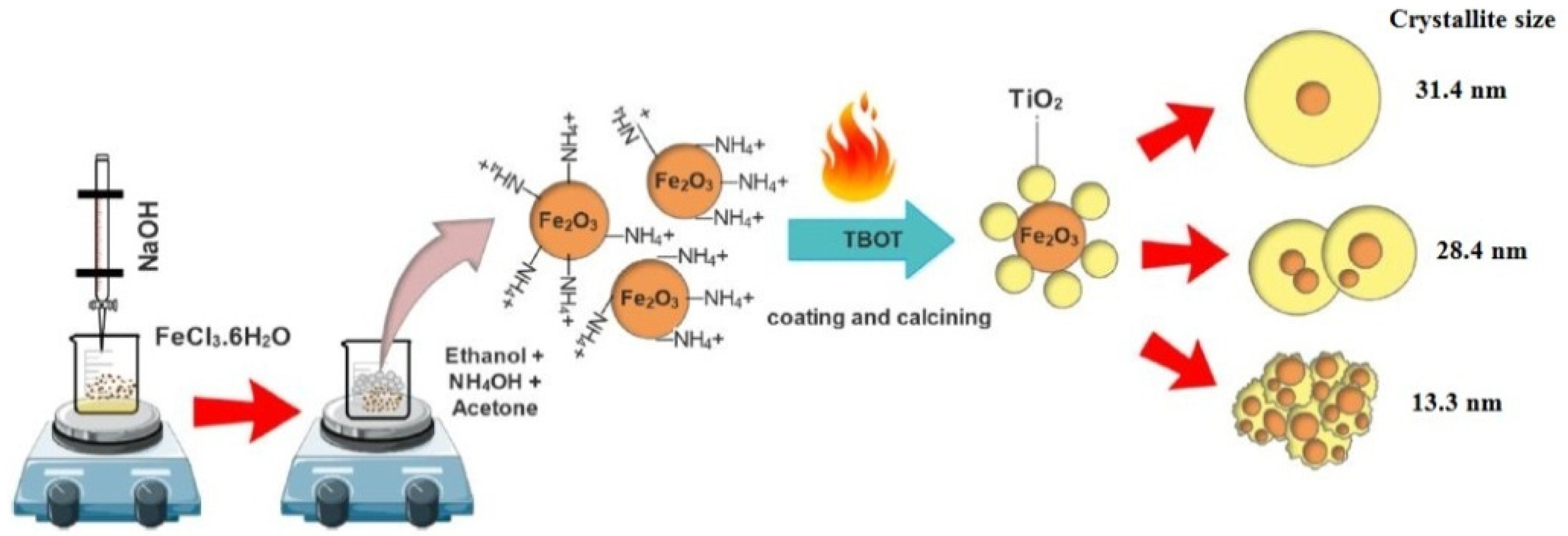
Sadeghi-Niaraki et al. [65] produced Fe2O3@TiO2 nanoparticles using TiO2. A dual-functional nanostructured Fe2O3@TiO2 pigment (sometimes referred to as a cool pigment) with unique photocatalytic properties and NIR reflection was achieved. Fe2O3@TiO2 nanoparticles were created using different proportions of Fe2O3@TiO2 at 0.06, 0.62, and 1.24. Furthermore, a coprecipitation method was used in the presence of Fe3+ precursors to fabricate Fe2O3 nanoparticles. Next, the sol–gel technique was used to coat them in TiO2. To create Fe2O3@TiO2 nanoparticles, various materials like ethanol, acetone, NH3, TBOT, and Fe2O3 were used. Various ratios of Fe2O3:TiO2 (0.02, 0.06, 1.0, 0.62, and 1.24) and different contents of Fe2O3 (0 for 0.05, 0.05, 0.05, 0.5, 1, and 0 g) were used. About 0.05 g of Fe2O3 was dispersed in solutions containing 25 mL of acetone, 25 mL of ethanol, and 0.15 mL of ammonia. To ensure the even dispersion of the solution, it was sonicated for 30 min before being supplemented with 3 mL of TBOT with intense stirring at 600 rpm. The reaction lasted for 2 h, and the remaining products were centrifuged at 3000 rpm and then rinsed with DI water, as well as ethanol. Eventually, they were left to air-dry at 80 °C for 12 h. The resultant Fe2O3@TiO2 nanoparticles were then air-calcined for 2 h at 600 °C. Figure 4 depicts the schematic presentation of the Fe2O3@TiO2 core–shell particle fabrication process.
Dandan et al. [66] used the hydrothermal technique to make homogeneous α-Fe2O3@TiO2 core–shell nanoparticles. In this process, the photocatalytic performance was found to be enhanced when TiO2 was used. To prepare the α-Fe2O3@TiO2 particles, α-FeOOH nanoparticles were subjected to a hydrothermal process. To grow the homogeneous TiO2 shell, TBOT was employed, and a sol–gel reaction was performed for 24 h at 45 °C. A two-stage solution preparation process was performed to produce α-Fe2O3 @amorphous TiO2 core–shell particles. The first stage involved the preparation of α-Fe2O3 nanoparticles using the surfactant-free solvothermal method [67]. In this method, 14.5 mL of deionized water was magnetically stirred to dissolve 0.055 mol of FeCl3·6H2O, producing a yellowish substance. This substance was placed onto a Teflon-lined autoclave capable of holding 20.0 mL and left for 4 h in an electric oven at 180 °C. Once the reaction was finished, the autoclave was turned off and left to cool down to room temperature. The resultant specimen was centrifuged to achieve red precipitate, which was rinsed in distilled water and pure ethanol before being left to dry at room temperature. Then, about 0.02 g of the α-Fe2O3 nanoparticles were placed in 30 mL of ethanol to get a suspension. Next, an ethanol mixture containing tetrabutyl titanate (Ti(BuO)4) (volume ratio VTi(BuO)4/Vethanol = 0.3 mL/10 mL) was added into the suspension and stirred intensely for 2 h. Thereafter, a mixture made from 0.2 mL of water and 5.0 mL of ethanol was added to the solution. Subsequently, the solution was stirred for an extra 3 h at room temperature before being centrifuged to separate the precipitates. Later, the precipitates were washed multiple times with water and pure ethanol and left to air-dry. Finally, the obtained α-Fe2O3@TiO2 core–shell particles were placed in a muffle furnace and heated at 450 °C for 2 h for calcination.
Using a chemical vapor deposition method, Wang et al. [67] produced β-FeOOH nanorod arrays. This also enhanced the process of separating photogenerated electron hole pairs using simulated solar light. First, the FTO substrates coated with β-FeOOH nanorod arrays were placed in the PECVD chamber. Then, a glass flask containing a titanium precursor was attached to the reaction system. To ensure the sufficiency of the vapor pressure of the precursor, the glass flask was heated at 70 °C. Subsequently, the chamber pressure was reduced to less than 0.5 Pa, and 20 sccm of O2 was added to the chamber. The PECVD approach (100 W of plasma power with different durations) was used to produce β-FeOOH nanorod arrays with varying TiO2 shell thicknesses. After the post-annealing treatment for two hours at 750 °C, the Fe2O3@TiO2 core–shell nanorod arrays were obtained. Zhang et al. [68] also used this approach to fabricate α-Fe2O3@TiO2 particles with porous structures, wherein the main objective was to improve the photocatalytic activity of the achieved products.
To create highly dispersed NiTiO3@TiO2 yellow pigments in a core–shell form, the precursors acquired from the Ni2+ present on the surface of TiO2 particles [69,70] were calcined. The first step of this two-stage process involved the controlled hydrolysis of TBOT (tetra butyl titanate). In this process, 300 mL of ethanol and 1.0 mL of KCl solution (0.4 mM) were mixed and stirred for 20 min, and then, 5 mL of TBOT was added with continual stirring for 10 min. After stirring was discontinued, the reactions were completed, and the resultant powder was obtained via centrifugation. The obtained powder was washed multiple times with water and ethanol. The as-prepared white powder was heated in a vacuum oven (60 °C) for 12 h to dry. Second, 0.2 g of as-prepared TiO2 powder was dispersed into 200 mL of distilled water, followed by ultrasonication. Next, Ni (NO3)2·6H2O and CO(NH2)2 were added to the suspension, and the solution was stirred for 30 min. A water bath at 80 °C was then used to heat the suspension following continual stirring for 6 h. After cooling, the suspension was centrifuged to collect the precipitates. The precipitates were then washed multiple times with water and ethanol and stored at 60 °C overnight to produce green powders. Furthermore, the NiTiO3@TiO2 yellow pigments were produced by calcinating it for three hours at different temperatures in the range of 500–950 °C. The samples were labeled as NiTiO3−x@TiO2 (x-range was from 0.25 to 1.0 wt%).

Table 1.
Core–shell nanostructure pigment synthesis methods.
| No. | Core–Shell NPs | Synthesis Method | Synthesis Details (Brief) | Ref. |
|---|---|---|---|---|
| 1 | Fe2O3@SiO2 | Sol–gel method | The sol–gel method to hydrolyze tetraethyl orthosilicate (TEOS) in an attempt to cover iron oxide nanoparticles in uniform shells. By changing the sol–gel precursor concentration, it was possible to modify the thickness of these silica shells. | [53] |
| 2 | CeO2@SiO2 | Sol–gel method | To synthesize the SiO2-covered CeO2 nanoparticles, cyclohexane (99%) was applied as the organic phase in order to perform reverse micelles on polyoxyethylene (15) cetylether. | [54] |
| 2 | Sr2+-doped γ-Ce2S3@c-SiO2 | Stöber method | Amorphous SiO2 (a-SiO2) sol and Sr2+-doped CeO2 were employed as precursors. The original materials used in this experiment were Si(OC2H5)4 (TEOS), Sr(NO3)2, Ce(NO3)3·6H2O, (NH4)2CO3, ammonia water, and ethanol. | [57] |
| 3 | Zn1−xCoxO@SiO2 | Stöber method | A polyol process was used to form ZnO doped with Co2+, a well-known substitute for chromium-based inorganic pigments. Additionally, to source the zinc and cobalt needed in the experiment, zinc acetate dihydrate (Zn(CH3OO)2·2H2O) and cobalt nitrate (Co(NO3)2·6H2O) were employed, with diethylene glycol (H10C4O3) being used as a template. | [58] |
| 4 | Fe2O3@SiO2 | Hydrothermal and Stöber methods | The Stöber method was carried out to fabricate the reddish pigments using α-Fe2O3 as chromophore particles and tetraethoxysilane (TEOS, AR) to source the silica. | [28] |
| 5 | α-Fe2O3@SiO2 | Sol–gel method | The original materials used in the process were FeCl3·6H2O (97%), tetraethylorthosilicate (TEOS 99.99%), cetyltrimethylammonium bromide (CTAB, 99%), and NH4OH (28% in water, 99.99%), which were obtained from Aldrich. Additionally, DI water was employed. | [60] |
| 6 | ZnS@SiO2 | Homogeneous deposition method | ZnS@SiO2 core–shell nanoparticles were produced through a homogeneous deposition method in which deionized water (1 mL) and ammonia (0.5 mL) were placed into a 30-mL colloidal solution of ZnS (0.5–2 wt%). | [61] |
| 7 | γ-Ce2S3@SiO2 | Stöber method | The γ-Ce2S3 red pigments with a silica layer using the Stöber technique combined with hydrogen–argon atmosphere heat treatment (700 °C). | [62] |
| 8 | α-Fe2O3@SiO2 | Sol–gel process | Silica nanoparticles were used in a sol–gel process to coat the natural pigment of hematite (red ocher, α-Fe2O3), the objective of which was to produce a long-lasting hematite pigment of the Fe2O3@SiO2 nanocomposite that could be used to preserve artefacts. To achieve this, 2.2 wt% of pure α-Fe2O3 in the silica sol was charged over a period of time and continually stirred. | [63] |
| 9 | PS@TiO2 and Air@C@TiO2 | Soap-free emulsion polymerization and sol–gel methods | The experiments were conducted using titanium butoxide (TBOT), acetic acid (99%), ethanol, potassium persulfate, sulphuric acid (98%), and hydrogen peroxide (H2O2) (28−30%). | [64] |
| 10 | Fe2O3@TiO2 | Sol–gel method | To create Fe2O3@TiO2 nanoparticles, various materials were used, including ethanol, acetone, NH3, TBOT, and Fe2O3. The following ratios of Fe2O3:TiO2 were also used: 0.02, 0.06, 1.0, 0.62, 1.24, and 0 for 0.05, 0.05, 0.05, 0.5, 1, and 0 g of Fe2O3, respectively. Usually, 0.05 g of Fe2O3 are dispersed in solutions containing 25 mL of acetone, 25 mL of ethanol, and 0.15 mL of ammonia. | [65] |
| 11 | α-Fe2O3@TiO2 | Hydrothermal method/sol–gel method | The α-FeOOH nanoparticles were subjected to a hydrothermal process. Additionally, to create the homogeneous TiO2 shell, a TBOT was employed, and a sol–gel reaction was performed for 24 h at 45 °C. | [66] |
| 12 | β-FeOOH nanorod@TiO2 | Plasma-assisted chemical vapor deposition | A two-stage solution preparation process was then carried out to produce α-Fe2O3 @amorphous TiO2 core–shell particles, the first stage of which involved preparing α-Fe2O3 nanoparticles through the performance of a surfactant-free solvothermal method. | [67] |
| 13 | α-Fe2O3@TiO2 | Plasma-assisted chemical vapor deposition | Similar to the above approach in point 12, the α-Fe2O3@TiO2 particles were fabricated with porous structures, the key objective of which was to improve the photocatalytic activity. | [68] |
| 14 | NiTiO3@TiO2 | Sol–gel method | First stage, 300 mL of ethanol and 1.0 mL of KCl liquid solution (0.4 mM) were mixed and stirred for 20 min; after which, 5 mL of TBOT was added. It was then stirred for 10 min. Secondly, 0.2 gas-prepared TiO2 powder was dispersed into 200 mL of distilled water ultrasonically, and then, Ni (NO3)2·6H2O and CO(NH2)2 were added to the suspension, and the solution was stirred for 30 min. | [69,70] |
4. Core Materials
4.1. Ferric Oxide
The inorganic compound ferric oxide (also known as iron oxide) represented by the formula Fe2O3 is one of the three oxides of iron, along with iron (II) oxide (FeO) (relatively rare compound) and iron (II, III) oxide (Fe3O4), a naturally occurring substance found in mineral magnetite. Fe2O3 is also known as hematite, which is a primary material used to source iron in the steel industry. Generally, iron oxides, including FeO, α-Fe2O3, γ-Fe2O3, and Fe3O4, are applied in cosmetics, electronic materials, pigments, abrasives, and coatings [71,72,73]. α-Fe2O3 is an important inorganic red pigment and is safe to use because of its nontoxic nature [26]. Over the last few years, an increasing number of researchers have been investigating nanoscale magnetic materials due to their wide array of potential applications, including information storage, magnetic refrigeration, separation, magneto-optical solid devices, and magnetic resonance imaging, in addition to many conventional applications (i.e., plastic reinforcement and fluid rheological additives). These nanoparticles are also suitable for making high-performance devices. Various chemical techniques have been used to fabricate these nanoparticles with narrow-sized distributions and tailored morphologies, as well as structures. These techniques include flame-spray pyrolysis, sol–gel, hot-soap, plasma chemical vapor deposition (CVD), microemulsion, and electrospray pyrolysis.
Many researchers [26,74,75,76,77] have used hematite (Fe2O3) to produce Fe2O3@SiO2 pigments. For instance, an α-Fe2O3@SiO2 composite core–shell was produced to serve as a protective coating for pigments [63]. Ri et al. [77] used core–shell technology to enhance the thermal properties of pigments by coating them with Fe2O3 nanoparticles. Silica was used to coat both β-FeOOH and α-Fe2O3 nanoparticles with tetraethylorthosilicate (TEOS), and cetyltrimethylammonium bromide (CTAB) was used as a surface modifier. The physical properties of the two samples were compared. Fe3O4@SiO2 nanoparticles were prepared [76] using the coprecipitation method. In the air atmosphere, a FeCl3 (0.5 M) and FeSO4 (0.5 M) solution with a molar ratio of 1.75 to 1 was produced to make magnetic nanoparticles (MNPs). After this, 10 mL of liquid ammonia was added to the mixture and stirred intensely, and extra ammonia was added until a pH value of 9 was achieved. The solution was then stirred for another 30 min. Lastly, magnetite was used to obtain the precipitates, which were rinsed multiple times with deionized water and alcohol to reduce the pH to 7.0. To adjust the surface of the Fe3O4 particles, MNPs were reacted with sodium citrate. In this process, 1 g of Fe3O4 MNPs was combined with 200 mL of sodium citrate (0.5 M) in a flask. Then, the solution was ultrasonically irradiated for 30 min in order to bring the MNPs together. Next, it was kept at 60.8 °C and stirred whilst being protected by the air atmosphere. Magnetite was then used to collect the precipitates, which were rinsed in acetone to eliminate any remaining sodium citrate. The new MNPs with modified surfaces were dispersed again in 100 mL of deionized water to create the ferrofluids (FF). Subsequently, 2 mL of the FF was placed in 50 mL of deionized water and ultrasonically irradiated for 30 min. The obtained suspension was added to 150 mL of ethanol plus 5 mL of NH3.H2O and stirred intensively at 40 °C. Later, 4 mL of TEOS were added to the solution for 12 h to complete the reaction. Lastly, the solution was filtrated to collect the precipitates. The collected precipitates was rinsed with ethanol and deionized water before being left to dry for 12 h in a vacuum.
Chen et al. [28] used Fe2O3 as the core material to synthesize Fe2O3@SiO2 core–shell particles. A hydrothermal process was used to fabricate the highly dispersed α-Fe2O3 nanoparticles. First, 2.89 g of FeCl3·6H2O and 3.39 g of Na2CO3 were placed in 80 mL of deionized water with constant stirring. This produced an orange solution that was stirred for 15 min before being placed in a stainless-steel autoclave of 100-mL capacity and maintained at 180 °C for 8 h, then left to cool down to room temperature. The obtained mixture was centrifuged to get a red precipitate, which was washed three times in water and deionized water. The sample was then stored at 70 °C for 5 h to let it dry.
Repeated experiments verified that iron oxide can be mixed with other inorganic elements to form core–shell particles. Rahimi et al. [78] used the hydrothermal process to fabricate nano-hollow Zn-Fe2O4 microspheres where the chemical reagents of ZnCl2, FeCl3·6H2O, and ammonium acetate were mixed. In this process, a mechanical stirrer was used to mix the solution containing 70 mL of ethylene glycol, iron (III) chloride, and zinc chloride for further purification. About 2.312 g of NH4Ac was added to the mixture with constant stirring, wherein the solution slowly turned a dark-yellow color. The obtained solution was sonicated for 40 min, followed by oven heating at 215 °C for 4 h in order to produce some black precipitates. Finally, the ZnO nanoparticles were immobilized on the surface of the Zn-ferrite spheres to form Zn-Fe2O4@ZnO core–shell structured spheres, which was achieved through a sol–gel approach.
4.2. Silver
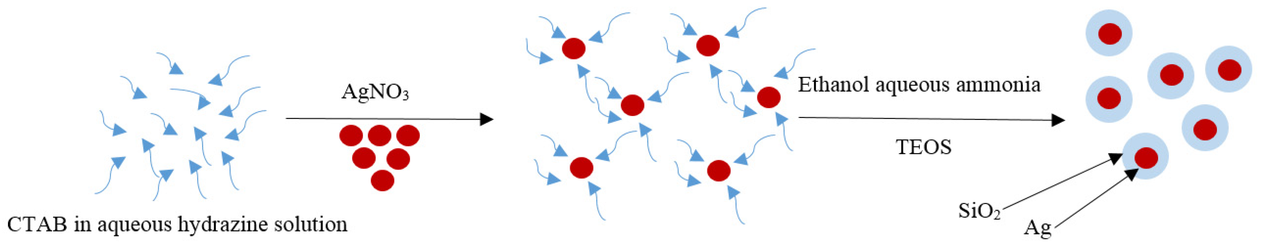
In recent times, many researchers have been focusing on the syntheses and characterizations of various metallic nanoparticles (NPs) to serve as core–shell pigments for diverse industrial applications. These NPs possess many unusual properties, including an enhanced resistance to harsh environments and increased color stability desirable for widespread applications. Silver (Ag) is an example of a metallic nanoparticle that has been widely used as a core–shell composite in the manufacturing industry. In an attempt to identify a material that can be used in marine settings to prevent corrosion, Zhang et al. [79] produced Ag@SiO2 core–shell nanoparticles. In this process, 5 mL of 0.1-M liquid hydrazine was immersed in 400 mL of aqueous solution (0.146 g, C16TMABr) and stirred vigorously at room temperature for 2 min; after which, 10 mL of 0.05-M AgNO3 solution was added to the solution drop by drop to prepare the Ag colloids. This caused the solution to turn a reddish-brown color. Thereafter, 100 mL of ethanol and 4 mL of liquid ammonia (25 wt%) were added to the silver colloids. Finally, 0.5 mL of TEOS was put into the solution and stirred for two more hours to produce a suspension containing monodisperse Ag@SiO2 core–shell nanoparticles. Figure 5 presents the various steps of the preparation.
4.3. Ceric Oxide
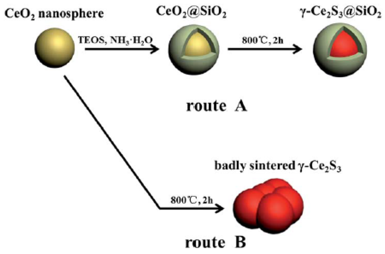
Ceric oxide is a pale-yellow/white powder derived from the rare-earth metal cerium and has the chemical formula CeO2. It an important commercial product and plays a significant role in purifying elements from their ores. In recent years, increasing research focus has been paid to produce CeO2@SiO2 core–shell nanoparticles due to their several benefits [80,81]. Tago et al. [54] implemented a CeO2-based core to fabricate a core–shell nanoparticle. Yu et al. [43] successfully produced different g-Ce2S3@SiO2 pigments through a process of sulfurizing their existing CeO2 coatings with SiO2 shells of varying thicknesses. In this preparation method, 2 g of Ce(NO3)3·6H2O was ultrasonically dissolved in 2 mL of deionized water in order to create the CeO2 nanoparticles, and then, 2 mL of CH3COOH and 60 mL of glycol were added. The whole mixture was stirred for 30 min to produce a homogeneous solution and then placed in a sealed Teflon-lined stainless-steel autoclave for 200 min, followed by a heat treatment at 180 °C. The obtained mixture was centrifuged to separate the precipitates, followed by rinsing with ethanol and water and air-drying at 60 °C. Figure 6 shows the process of coating CeO2 with SiO2 using the sol–gel technique.
Many studies were conducted to wrap the pigments in a clear material to enhance the temperature stability and antioxidant properties of the γ-Ce2S3 pigments. For instance, an amorphous SiO2 (a-SiO2)-coated γ-Ce2S3 (γ-Ce2S3@a-SiO2) was produced [82]. Chen et al. were able to raise the anti-oxidization temperature of the γ-Ce2S3@a-SiO2 pigments to 450 °C [42]. Despite the fact that coating the surface of γ-Ce2S3 pigments with SiO2 can enhance the pigments’ temperature stability, the extent of this enhancement is still limited, which may be due to the low density of the a-SiO2 cladding layer. An alternative to this would be to coat the γ-Ce2S3 pigments with a dense crystal SiO2 (c-SiO2) layer, which would significantly improve the temperature stability of γ-Ce2S3. In this rationale, Li et al. [57] used Ce2S3@SiO2 composites to successfully increase the temperature and acid stability of red pigments.
4.4. Cetrimonium Bromide
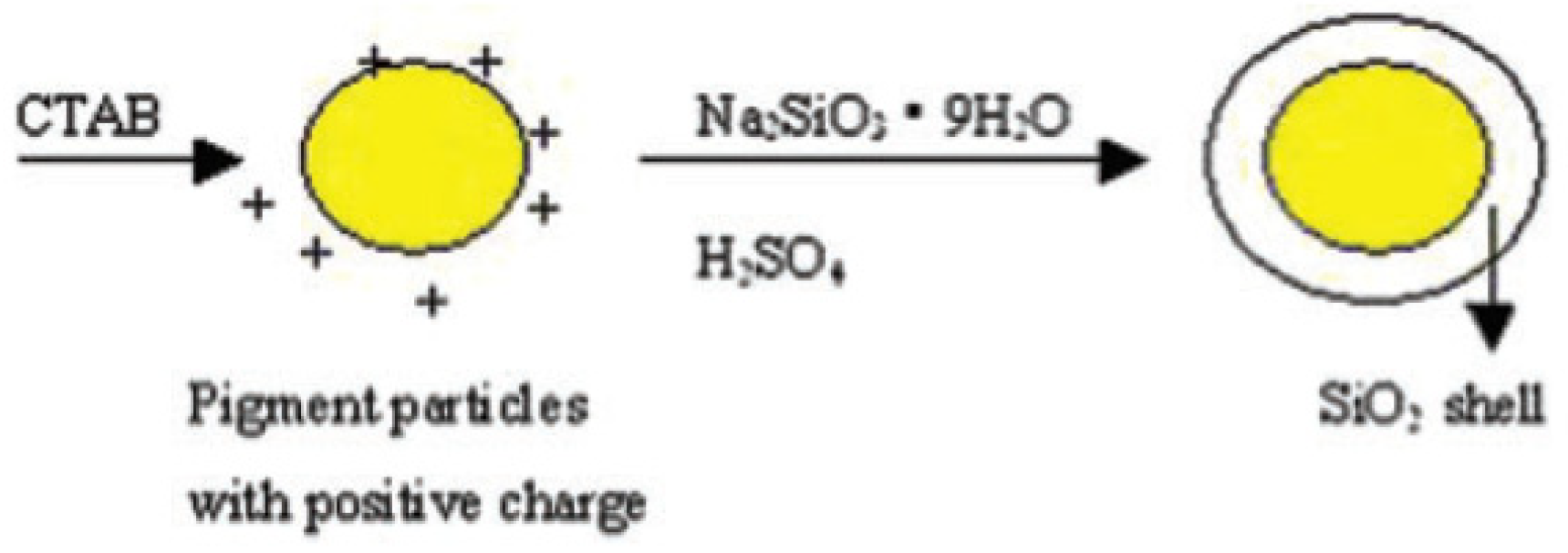
Cetrimonium bromide (CTAB) has been widely applied to fabricate nanoparticles and pigments. An experiment was performed [83] using the electrostatic self-assembly technique to create yellow SiO2/Hansa pigment composite particles in a liquid solution. In this method, cetyltrimethylammonium bromide served as the coupling agent that positively charged the yellow pigment particles. Next, the particles were coated with a thin layer of SiO2 through the hydrolysis of Na2SiO3, which improved their charge load and optical and mechanical properties. The process of coating colloids with silica is generally two-fold. The first step usually involves the adsorbtion of CTAB and growth of the silica shell following the hydrolysis of Na2SiO3.
Figure 7 shows the process of synthesis. First, a glass of 500-mL volume was filled with a solution containing 0.18 g of CTAB and 150 g of water. Then, the mixture was stirred for 10 min at room temperature to form the CTAB solution. About 1.0 g of organic pigment was subsequently added to the CTAB solution and stirred ultrasonically for 60 min to create a stable organic pigment suspension. CTAB thus served as the dispersing and coupling agent. Once the yellow pigments were added to the CTAB solution, the system temperature was increased to 92 °C. Thereafter, 20 mL of Na2SiO3 solution (3.5 g of Na2SiO3·9H2O) and 40 mL of H2SO4 solution (1.2 g of H2SO4) were placed into the pigment emulsion drop by drop, followed by the addition of the Na2SiO3 solution (at a pH of 9). Then, the pH of the H2SO4 solution was increased to 9 to10. It was very important to keep the time interval between the two different drops over 5 s. The entire process took approximately 1 h to complete. Later, the temperature and reaction conditions were kept the same for another 5 h. Finally, the H2SO4 solution was utilized to reduce the pH to 2 to 3 and was kept the same for 2 h. The resulting solution was centrifuged to obtain the precipitates, which were then washed multiple times with pure ethanol and placed in a vacuum at 60 °C for 4 h to dry.
4.5. Titanium
Green pigments made with titanium dioxide or titania (TiO2) are frequently used in the field of plastic production, building materials, glass coatings, and ceramics. Zou and Zhang [84] applied TiO2 as the core material to adjust the color properties of complex TiO2@CoTiO3 green pigments. To synthesize TiO2, 20 mL of TiCl4 was placed on ice blocks and stirred intensively. Once the blocks were fully melted, deionized water was added to dilute the solution to form 1 mL of precursor solution. Then, a dispersant (1 wt% of PEG10000) was placed into the mixture, which was refluxed for 4 h at 100 °C. About 10% of liquid ammonia was added to the solution to modify its pH value. The solution was washed multiple times and dried for 4 h at a temperature of 90 °C in order to collect the resultant TiO2 powder. Subsequently, as-prepared TiO2 powder was reinserted into the distilled water and stirred vigorously to produce 5 wt% of the suspension. After adding cobalt chloride to the mixture, 2 mL of sodium hydroxide solution was applied to increase the pH to 10. This yielded the precipitate, which was filtered and washed multiple times. After drying at 105 °C for 4 h, the powder was calcined at different temperatures (600–1000 °C) for 60 min, producing the final complex pigments.
5. Core–Shell Synthesis Methods
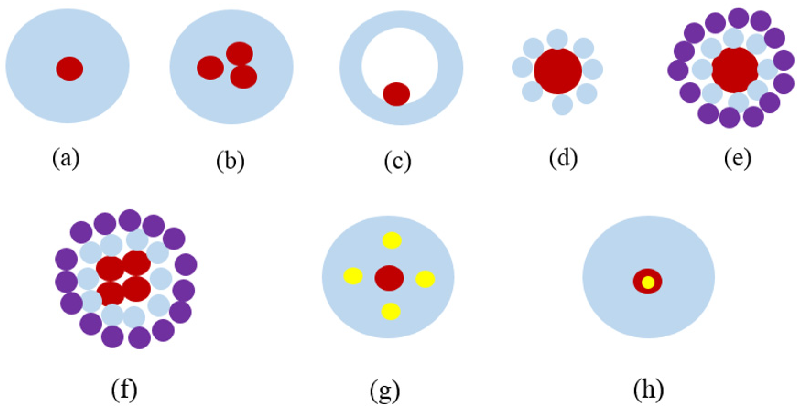
As aforementioned, core–shell particles are particles that consist of a shell and a core, wherein both can be made using the same or different materials [45,85,86]. Figure 8 shows various core/shell particles, wherein the core and shell are marked in different colors. The core can be in the form of a single sphere (Figure 8a) or a collection of many small spheres (Figure 8b). Moreover, there may be a hollow shell with a small sphere inside, which appears in a yolk–shell structure (Figure 8c) [87]. The shell structure may be in the form of a continuous layer (Figure 8a–c), multiple smaller spheres accumulated onto a larger core sphere (Figure 8d,e), or a simple collection of core spheres (Figure 8f) [88]. In fact, the insertion of smaller spheres into the shell can be performed to produce more intricate core/shell structures (Figure 8g) [89]. Alternatively, the same process can be carried out using multiple shells (Figure 8h) [90,91]. Varieties of chemical or physical methods can be used to synthesize core/shell nanoparticles, including wet chemistry and chemical/physical vapor deposition. Generally, multiple steps are involved in core–shell particle production. Typically, core particles are synthesized first; after which, a shell is created around the particle. However, the methods used to achieve these nanoparticles vary according to the type of core and shell materials under consideration [21]. The primary objective of core–shell particle preparation is to bring together the desired properties of diverse materials and structures to produce a synergistic effect, stabilize the active particles, and generate biocompatible properties [92]. Figure 8 illustrates various types of core–shell structures, depending on the type of core and shell materials, preparation method, and applications.
Core–shell nanoparticles have become a fundamental nanomaterial for various industrial usages. During the preparation of core–shell NPs, it is important to identify a simple, quick, and cost-efficient method with the least-possible negative impacts on the environment. To meet this target, several methods have been developed, including the microemulsion, sol–gel, microwave synthesis, multistep reduction, epitaxial growth, electrochemical dealloying, sonochemical processing, and Stöber methods. Alternatively, these methods can be combined to create a hybrid method. Amongst all these approaches, the sol–gel method is most common for core–shell NP preparation. This method is a relatively new approach for preparing nanoparticles that allows for more control over the reaction processes involved in synthesizing solid materials. Homogenous multicomponent systems are easy to acquire, especially homogenous mixed oxides that can be prepared through the mixing of molecular precursor solutions. The sol–gel method is used in materials science to transform small molecules into solid materials. It is often applied to produce metal oxides like SiO2 and TiO2. To achieve this, the monomer must be converted into a colloidal solution (sol), which then serves as a precursor for a combined network consisting of network polymers or discrete particles. Generally, the metal alkoxides are used as precursors. During the chemical reaction, a colloidal solution called a “sol” is produced and gradually forms a gel-like diphasic substance consisting of a liquid and a solid phase. The morphologies of these phases can be in the form of discrete particles or continuous polymer networks. With regards to the colloid, it may be necessary to remove vast amounts of liquid if the volume fraction of the particles (or particle density) is extremely low in order to evoke the gel-like properties. It can be achieved in several ways, wherein the easiest one is to allow sufficient time for the sedimentation to occur. Subsequently, the remaining fluid can be poured away. Conversely, phase separation can be sped up through a process called centrifugation. As a wet chemical process, the sol–gel method is widely applied to produce core–shell nanoparticles [93,94,95].
Microemulsions are isotropic liquid mixtures consisting of water, surfactant, oil, and often, cosurfactants thermodynamically stable and clear in appearance. There may be salt present in the aqueous phase, as well as various other ingredients. The “oil” may actually be an intricate mix of various different hydrocarbons. As opposed to normal emulsions, microemulsions are created by mixing components, and no high shear conditions are required to produce them. There are three main types of microemulsions—namely, direct (oil dispersed in water, o/w), reversed (water dispersed in oil, w/o), and bi-continuous. Microemulsions are ternary systems in which two separate immiscible phases (water and “oil”) occur with a surfactant, and the surfactant molecules can create a monolayer between the oil and water. Additionally, the surfactant molecules have hydrophobic tails that dissolve during the oil phase, whilst hydrophilic head groups dissolve in the liquid phase. To synthesize gold and silver core–shell bimetallic nanoparticles quickly, two-step microwave irradiation is often used. This strategy requires the creation of a bilayer organic barrier around the core, which is achieved using citrate and ascorbic acids, which serve as capping agents. In turn, this enables a well-defined boundary layer to be established between the core and the shell materials. The boundary layer plays a vital role in the process of synthesizing various core–shell particles, which results in the production of modifiable bimetallic NPs with clearly defined core/shell structures. These nanoparticles may have spherical or triangular seed cores.
Another technique that can be used to create core–shell materials such as carbon nanotubes is high-pressure chemical vapor deposition. This method was developed by Nikolaev et al. [96] with the main focus of creating a single-wall carbon nanotube by combing CO with a small quantity of Fe(CO)5 and passing the mixture through a heated reactor. El-Gendy et al. [97] synthesized carbon-coated Fe, Co, Ni, FeRu, CoRu, NiRu, NiPt, and CoPt nanoparticles using high-pressure chemical vapor deposition and manipulating the temperature and pressure inside the reactor. In these experiments, metal–organic precursors known as metallocenes (carbon-rich metal elements) were used. The precursors were inserted into a thermostatic sublimation chamber; after which, argon gas was passed through the chamber to push the vapor into the reactor’s hot zone. The precursors could then break down the nanoparticles in the cooling finger. The precursor entered the gas phase in the hot zone to perform a supersaturation. Subsequently, the nanoparticles were nucleated when the supersaturation was achieved. The extent of a supersaturation can be changed by controlling the temperature inside the sublimation chambers and the pressure/temperature inside the chemical vapor deposition reactor. High pressure increases collisions between gas atoms, which ultimately reduces the rate at which atoms diffuse away from the original location. However, it is important to note that supersaturation cannot be achieved if the diffusion rate is poor, and this will result in single atoms or tiny clusters of atoms being deposited on the cooling finger.
The wet chemical technique was used [98] to synthesize Fe2O3 coated with graphene shells. A solution containing oleic acid and 1-octadecene was heated to 320 °C in a reflux reactor to dissolve purified iron oleate. Subsequently, ethanol and acetone were applied to wash the solution in order to generate iron oxide particles. Additionally, the Stöber process is commonly used to prepare silica (SiO2) particles [99] of controllable and uniform size [100], and these particles can be used for many purposes in the field of material science. This method was proposed by Werner Stöber et al. in 1968 [99] and is still the most common wet chemistry synthetic approach employed to prepare silica nanoparticles [101]. This technique is a sol–gel process, in which a molecular precursor (typically tetraethylorthosilicate) is placed in water and reacted with an alcoholic solution. This causes the newly produced molecules to join together and create bigger structures.
Du et al. [102] used a sol–gel reaction to coat Fe3O4 nanoparticles with a SiO2 shell, yielding a core/shell structure. To synthesize the core/shell nanocomposites, a two-step procedure was used. First, the coprecipitation method was used to acquire Fe3O4 nanoparticles; after which, electrostatic interactions with tetramethylammonium hydroxide (TMAOH) were performed to disperse the particles in a liquid solution. Secondly, the produced SiO2 during the hydrolyzation of tetraethyl orthosilicate (TEOS) was used to cap Fe3O4. Figure 9 presents the sol–gel technique used by Li et al. [103] to produce ZnSiO3/ZnO core–shell nanoparticles. In this experiment, they combined the sol–gel with an annealing technique to synthesize zinc silicate-zinc oxide (Zn2SiO4@ZnO) core–shell nanoparticles with a high band gap energy. First, the concentrations of the Na2SiO3/ZnCl2 precursors were modified to coat ZnO nanoparticles with ZnSiO3 shells of various thicknesses; after which, a low annealing temperature (780 °C) was set. This caused the amorphous ZnSiO3 to react with ZnO and to produce a crystalline Zn2SiO4 shell. The sol–gel technique was also used by Chai et al. [93] to produce core/shell-structured Fe3O4@SiO2 nanoparticles. They employed a two-step process to create silica-coated magnetite (Fe3O4@SiO2) core–shell nanoparticles. Firstly, they used a solvothermal technique to fabricate Fe3O4 nanoparticles. Subsequently, SiO2 produced during the hydrolyzation of tetraethyl orthosilicate was used to coat the Fe3O4 nanoparticles.
A two-step reduction method was used [104] to produce epitaxial Au@Ni core/shell nanocrystals. The first step involved mixing octahedral, triangular and hexagonal platelike, decahedral, and icosahedral crystals; after which, HAuCl4.was reduced in ethylene glycol (EG) and heated in a microwave with polyvinylpyrrolidone (PVP) added as a polymer surfactant. This enabled the core seeds to be produced. After this, oil bath heating was carried out to reduce Ni(NO3)2·6H2O in EG with NaOH and PVP using oil bath heating in order to overgrow the Ni shells on the Au core seeds. Similarly, Fan et al. [105] employed a two-step seed-mediated growth technique using Au cores to test the synthesis of bimetallic core–shell nanocubes in liquid phases. The formation of heterogeneous core/shell structures for four typical noble metals (i.e., Au, Ag, Pd, and Pt) was systematically assessed. The findings of these experiments highlighted the growth modes and general requirements for achieving conformal epitaxial growth, as well as the heterogeneous nucleation and formation of different noble metals. It identified two types of growth for heterogeneous metal shells on gold cores—namely, conformal epitaxial growth (Au@Pd and Au@Ag nanocubes) and heterogeneous nucleation and island growth (Au@Pt nanospheres). Based on their findings, it was concluded that the two metals should have comparable lattice constants, with mismatches being below 5%. Other researchers also confirmed the validity of these findings (Au@Ag (lattice mismatch, 0.2%), Au@Pd (4.7%), and Pt@Pd (0.85%)) [106,107,108].
Tsuji et al. [109] used a one-pot polyol technique to synthesize Ag@Cu core–shell nanoparticles with a high yield. In this method, bubbling Ar gas was used. Chemical reagents such as AgNO3 and Cu(OAc)2·H2O were added to serve as the reagents. First, AgNO3 was reduced in ethylene glycol (EG) to synthesize Ag@Cu particles via a two-step process. To start, Ag cores particles were separated from AgNO3; after which, Cu(OAc)2·H2O was added to create Cu shells. However, this was ineffective and produced no Cu@Ag core–shell particles; rather, Cu/Ag bi-compartmentalized particles were produced. In this work, Ag@Cu particles were prepared using different methods at various reaction temperatures and heating times. It was observed that adding the two reagents in reverse was the most effective way of producing Ag@Cu particles. To start the process, 8 mL of 15.9-mM Cu (OAc)2·H2O in EG and 8 mL of 477-mM poly(vinylpyrrolidone) (PVP; MW: 55,000 monomer units) were placed in EG, and the solution was mixed in a 100-mL three-necked flask. To eliminate all of the oxygen from the solution, Ar gas was bubbled for 10 min at room temperature. The solution was soaked in an oil bath at 180 °C. Subsequently, the temperature was increased to 175 °C, and the solution was left to bubble in Ar for several minutes; after which, 2 mL of 15.7-mM AgNO3 was added to the reagent solution and kept for 20 min at 175 °C. The concentrations of the final yields were 7.0 mM for Cu (OAc)2·H2O, 1.7 mM for AgNO3, and 212 mM for PVP. The influence of different reaction times was tested on the reagent solution to further investigate the Ag@Cu growth process.
A modified Stöber technique was used by Chae et al. [93] to produce Fe3O4@SiO2. A sol–gel method was employed to prepare the Fe3O4@SiO2 nanoparticles. The synthesis method involved the addition of 4 g of Fe3O4 particles and 4 mL of tetraethyl orthosilicate to 40 mL of deionized water, followed by ultrasonication to mix the solution, forming a stable emulsion. Thereafter, the emulsion was inserted into a mixture containing 50 mL of ethanol and 12 mL of NH3·H2O. The resultant solution was stirred for 4 h at room temperature at a rate of 400 rpm, followed by centrifugation to separate the core–shell structure of Fe3O4@SiO2. Figure 10 demonstrates the procedure for Fe3O4@SiO2 synthesis.
Sharma et al. [110] showed that core/shell particles can be fabricated through precipitation without a surfactant. They investigated the impacts of different surfactants and various concentrations on core–shell particle development. To investigate this, anionic and nonionic surfactants were tested. Additionally, fly ash was employed to create nanoTiO2 for shell coating [110]. The main purpose of using surfactants was to enhance nanotitania shells’ adhesion to fly ash core. It was concluded that, to determine the optimum impacts of TiO2 adhesion onto fly ash, various surfactants must be tested. One test was carried out without a surfactant, whilst other tests employed anionic and nonionic surfactants. The particles produced with the addition of an anionic surfactant were found to have better pigment properties and reflectance in the near-infrared area than the one obtained without a surfactant. Thus, such systems were claimed to be used effectively as a pigment in cool coatings. Fly ash was placed in a solution containing 70% ethanol supplemented with an anionic (SDS) or nonionic surfactant (TX-100). Next, titanium isopropoxide was added to the solution, followed by stirring for two hours. After two hours, the solution was dried in the range of 50–600 °C, and the resultant dry powder was collected. An experiment was conducted [111] using the semi-batch emulsion polymerization method to synthesize PUA hybrid emulsion PA/PU with a ratio of 20/80. A 250-mL four-neck glass flask containing a reflux condenser, digital thermometer, nitrogen gas inlet, and mechanical stirrer was used in this synthesis process. To prepare the pre-emulsion, SDS (2.0 g per 100 g of the acrylic and PU content) was dissolved in water. Subsequently, 5.0 g of MMA, 5.0 g of BA, and 0.015 g of AA (0.15 wt% of the overall MMA and BA weights) were added gradually to the emulsifier mixture and constantly stirred. The emulsion was then mixed for another 0.5 h. First, the reactor vessel was charged to achieve 111.3-g PU emulsion dispersion and 10% of the monomers. Then, it was set at 80 °C with constant stirring. Later, 10% of the initiator solution (0.4 g of KPS per 100-g acrylic monomers) was added to the solution and stirred for half an hour. Then, at a constant flow rate, the leftover monomer pre-emulsion and initiator solution was poured into the flask for a total of 4 h while the temperature was increased by 5 °C, keeping the solution at 85 °C for half an hour. The emulsion was then stirred continuously while left to cool. After adding NaHCO3, the pH value was kept within the predetermined range.
In brief, all the developed methods to date have aimed for the green and effective synthesis of core–shell NPs with controllable sizes, compositions, architectures, and properties through rational molecular design and material preparations. Despite substantial advances in the methods of preparation of core–shell nanostructures, the one-step/pot strategy in the preparation of core–shell nanostructures is still in its infancy and remains challenging. For example, the facet control of metal@metal oxide crystalline nanoparticles in a one-step co-reduction is limited compared to those prepared through a two-step seed-coating process.
6. Efficiency and Test Methods
6.1. Structure and Morphology
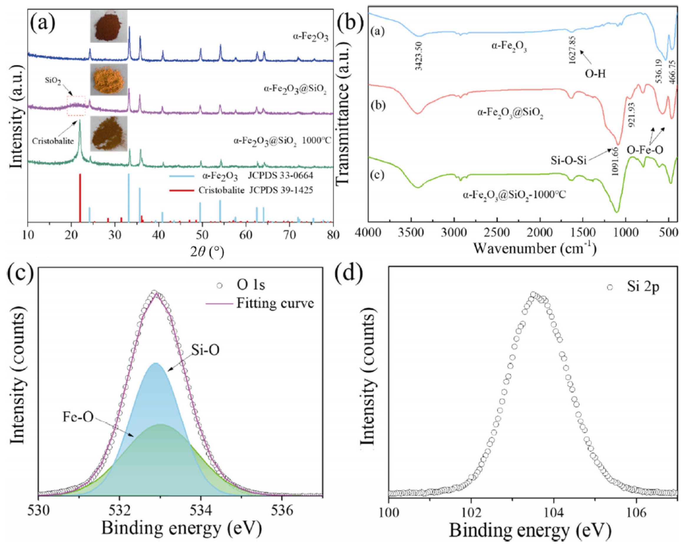
Various characterization tools can be used to evaluate the properties of synthesized core–shell nanoparticles. These techniques include XRD, SEM, TEM, FTIR, BET, LC-MS, XPS, Raman spectroscopy, ultraviolet-visible-NIR absorption spectroscopy, NIR reflectance, and photoluminescence spectroscopy [45,61,63,70,84,86,95,112,113,114,115,116]. For example, XRD, SEM, TEM, FTIR, and XPS tests were carried out [28] to assess the structure, morphology, and chromaticity of α-Fe2O3@SiO2 fabricated pigments. Figure 11a presents the XRD patterns of the reddish pigments α-Fe2O3, α-Fe2O3@SiO2 NPs, and α-Fe2O3@SiO2. The diffraction peak of α-Fe2O3@SiO2 particles were wide (2θ = 15–25°) once the core–shell structures formed, suggesting that there may be amorphous SiO2 present. Moreover, the diffraction peak changed to approximately 22° after calcination at 1000 °C, which suggested that the amorphous silica shell entered into the cristobalite phase to produce a reddish pigment. In addition, the α-Fe2O3 diffraction peak became weaker once the core–shell structure was formed. Figure 11b displays the FTIR results for α-Fe2O3, α-Fe2O3@SiO2 NPs, and α-Fe2O3@SiO2 reddish pigments. The bands at 3423.50 and 1627.85 cm−1 corresponded to the stretching of hydroxyl (–OH), and the bands at 536.19 and 466.75 cm−1 were due to the O–Fe–O bond vibration of α-Fe2O3. After covering α-Fe2O3 in SiO2, bands were developed at 1091.66 and 470 cm−1. The bands were due to the bending and stretching of O–Si–O, indicating that a coating was created on the α-Fe2O3 surface. Furthermore, the intensity of the O–Si–O band became stronger once it was calcined, improving the interaction between the core and shell materials. XPS was used to further assess the reddish pigments, and the results are presented in Figure 11c,d. The high-resolution XPS spectrum showed intense O 1s peaks, verifying the presence of Fe–O bonds and Si–O bonds in the pigment (Figure 11c). Meanwhile, a band was observed at 103.5 eV corresponding to the Si 2p XPS signal, which is commonly seen in pure silica.
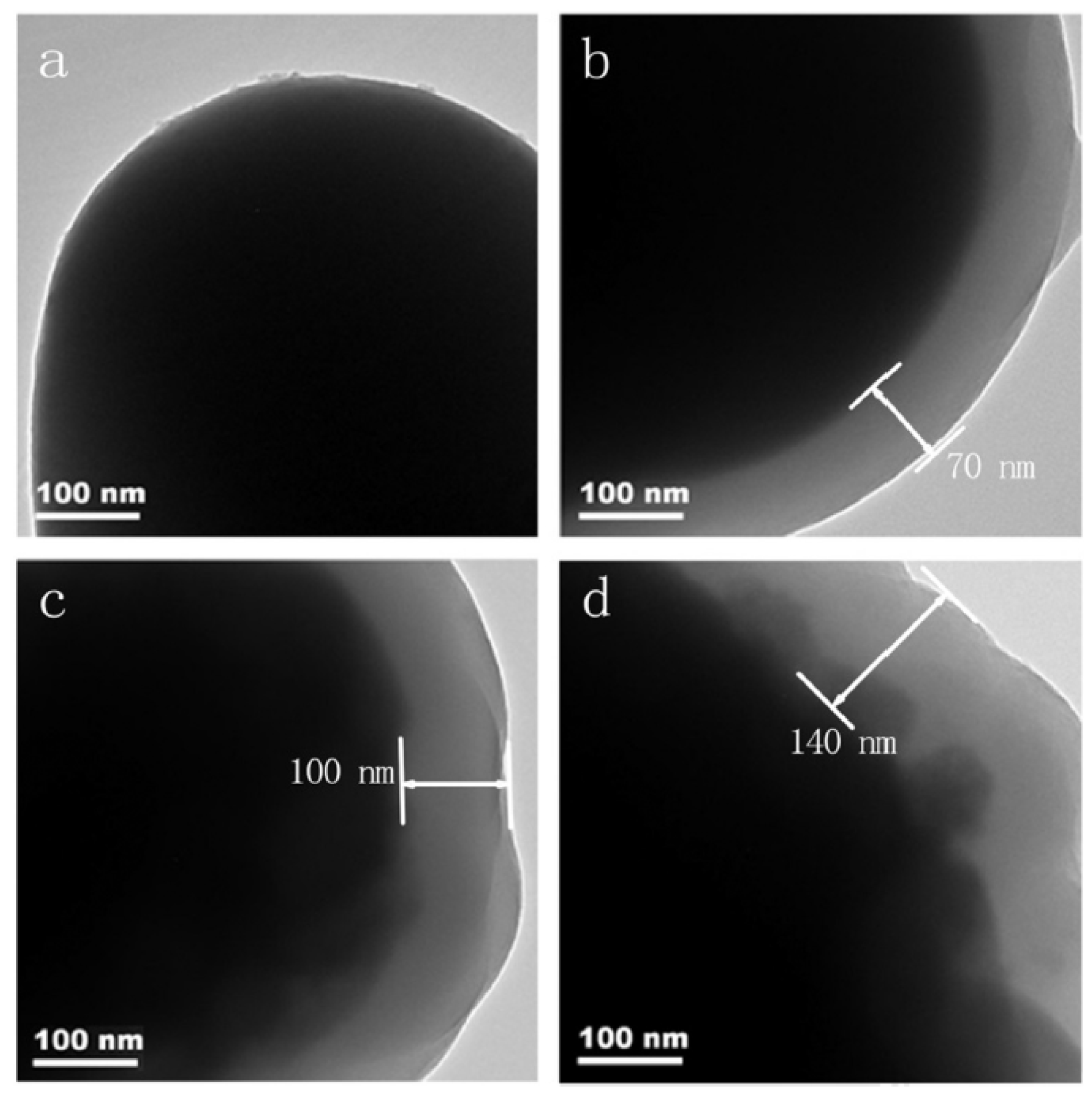
Li et al. [95] recorded the TEM images of synthesized γ-Ce2S3@SiO2 core–shell materials. Figure 12 shows the formation of the silica shell with different coating times. γ-Ce2S3 was covered in a clear layer that was not present on the uncoated samples, thereby supporting the findings of the SEM analysis. Figure 12b–d showed that, with the increase of the coating times, the thickness of the coating layer also increased. For example, the coating layer thicknesses of the particles coated once, twice, and three times were 70 nm, 100 nm and 140 nm, respectively. These disclosures indicated that, by varying the coating times, the thickness of the coating layers can be modified.
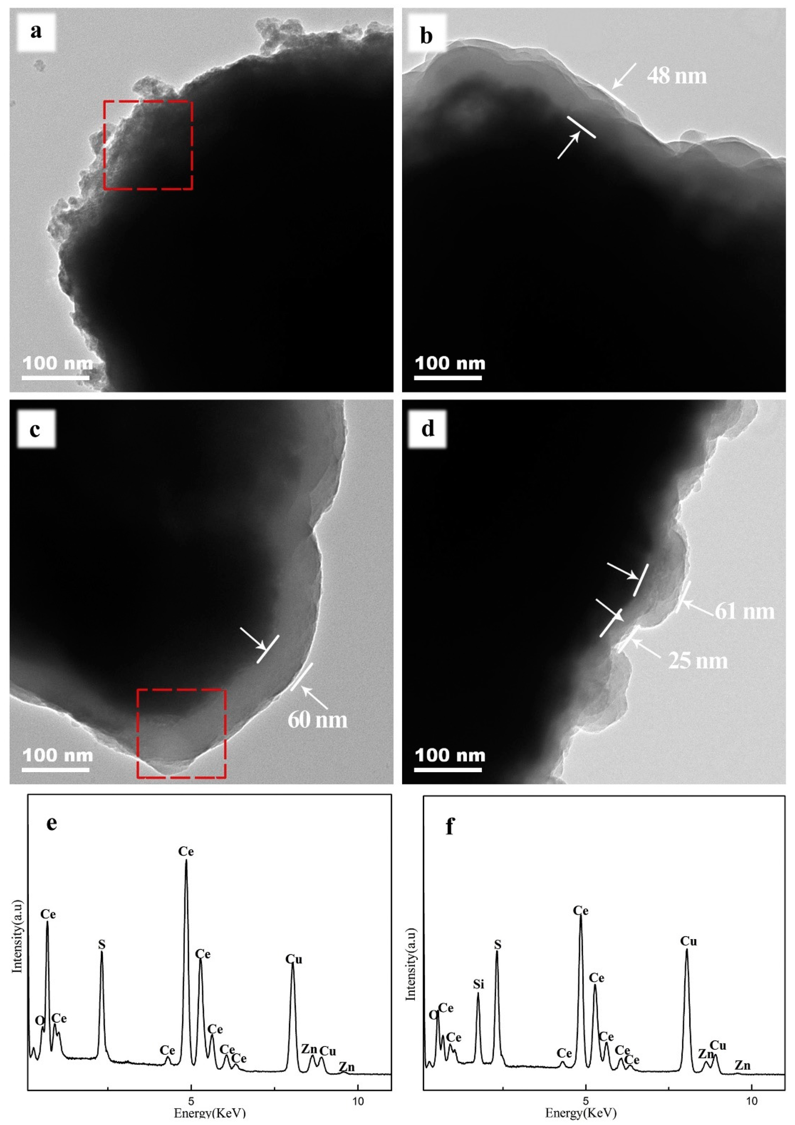
Liu et al. [62] fabricated γ-Ce2S3@SiO2 materials and characterized them using the TEM, FTIR, EDS, and XRD tests to determine their structures and morphologies. First, the TEM image analyses were performed to measure the thickness of SiO2-coated γ-Ce2S3. Figure 13 shows the TEM micrographs of the uncoated γ-Ce2S3 pigments and SiO2 xerogel-coated γ-Ce2S3 prepared with different water-to-ethanol volume ratios. Figure 13a shows that uncoated γ-Ce2S3 pigments take the form of irregularly shaped large chunks, with small particles deposited at the surface. The EDS spectra detected the presence of Zn, meaning that the uncoated γ-Ce2S3 pigments were changed via ZnO, enhancing the color stability and lowering the H2S emissions of the pigments. Figure 13b–d displays all the pigment particles with core–shell structures following the coating process. The EDS spectra (Figure 13f) revealed the presence of Si in the structure. These findings evidently suggested that the coating layer deposited on the surface of γ-Ce2S3 was SiO2 xerogel. Figure 13b,c revealed a relatively uniform shell size when the water-to-ethanol volume ratios of 15:105 (48 nm) and 20:100 (60 nm) were employed. Conversely, the shell thickness seemed to be nonuniform when this ratio was changed to 25:95 (Figure 13d). This was mainly due to the speeding up of TEOS hydrolysis when the water volume was increased. It seems that the silica nuclei and surface nuclei began competing with each other during the coating process, thus resulting in nonuniform shell thicknesses.
6.2. Reflectivity
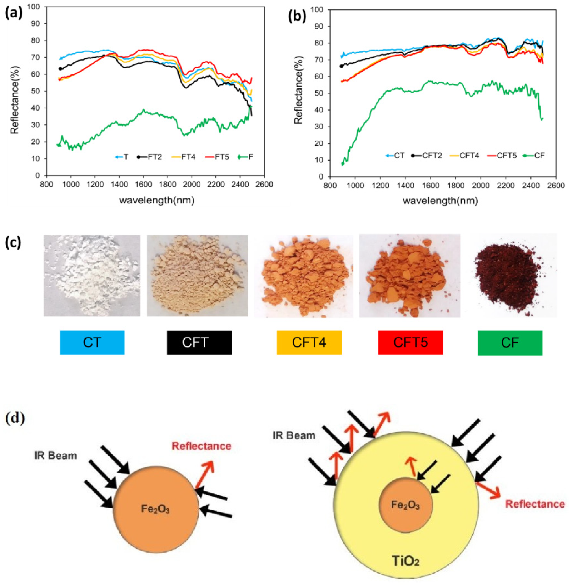
The results showed that the crystallization of Fe2O3 (CF) and TiO2 (CT) highly enhances NIR reflectance. Figure 14 shows the reflectance spectra of CT, CFT2, CFT4, CFT5, and the CF sample [65]. In the CT sample, the crystallinity was increased in the rutile phase, which ultimately enhanced the reflectivity at all wavelengths. Moreover, the crystallization of the sample seems to increase the reflectance values following the calcination process. The observed darker hues of the samples in Figure 14c were due to the Fe2O3 content, reducing the NIR reflectance. The NIR solar reflectance of samples CT, CFT2, CFT4, CFT5, and CF were 76%, 73%, 68.8%, 68.4%, and 39.3%, respectively. Figure 14d shows the mechanism of IR reflectance in the Fe2O3 and Fe2O3–TiO2 particles.
6.3. Toleration of High Temperatures
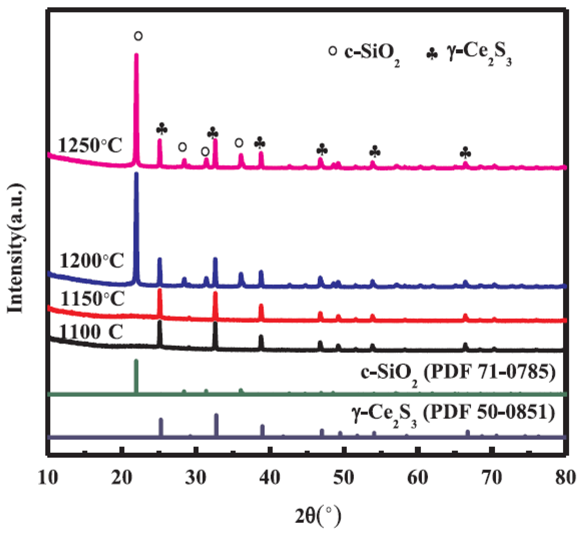
The effects of Ce2S3@SiO2-based core–shell materials on red pigment tolerance at high temperatures were assessed [57]. Figure 15 shows the XRD patterns of γ-Ce2S3@c-SiO2 samples produced at different calcination temperatures. No typical SiO2 diffraction peaks were identified at calcination temperatures between 1100 and 1150 °C. In addition, the diffraction peaks were evidenced in the γ-Ce2S3 crystalline phase, suggesting that SiO2 did not crystallize. Nonetheless, when the temperature was increased to 1200 °C, the typical c-SiO2 diffraction peak started to emerge, indicating that SiO2 started to crystallize at 1200 °C in the Ar gas atmosphere. The intensity of the c-SiO2 diffraction peak was almost constant when the temperature was increased to 1250 °C. At 1200 °C and in an Ar gas atmosphere, c-SiO2 seemed to develop excellent crystallinity. Li et al. [113] carried out an XRD test to analyze the thermal resistance of γ-Ce2S3 red pigments.
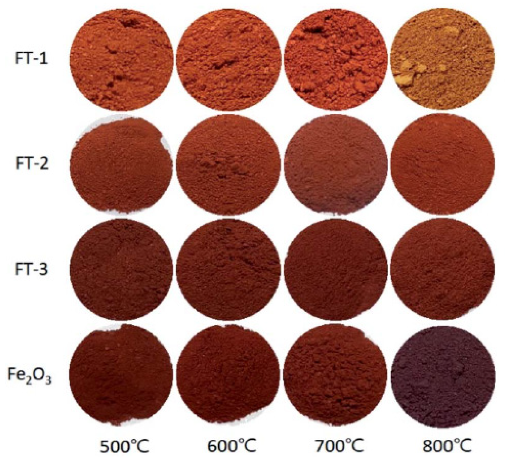
The effect of the Fe2O3-to-TiO2 ratio and calcination temperatures on the color stability of Fe2O3@TiO2-modified red pigments was evaluated [116]. With an increase of the Fe2O3 content with Fe2O3-to-TiO2 ratios of 0.5 (FT-1), 1.0 (FT-2), and 1.5 (FT-3) at the same heat treatment temperature, the color of the Fe2O3@TiO2 powder approached the color of pure Fe2O3. When observed with the naked eye at a fixed Fe2O3@TiO2 ratio, the color of the pigment did not change significantly as the heat treatment temperatures were increased from 500 °C to 700 °C. However, at 800 °C, the FT-2 and FT-3 powder colors showed darker reds than the ones calcined at low temperatures, wherein FT-1 and pure Fe2O3 changed completely to different colors (Figure 16). It was concluded that, to get a red color Fe2O3/TiO2 pigment powder like pure Fe2O3, the weight ratio of Fe2O3 to TiO2 should be 1.0 or higher, and the post-heat treatment temperature should be 700 °C or lower. The XRD test was used to explain the effect of the Fe2O3-to-TiO2 ratio on the improved color stability of the core@shell composite. For pure Fe2O3, the crystallite size was increased linearly with the rise of the calcination temperature. The increase in the Fe2O3 crystallite size in the Fe2O3@TiO2 (FT-2) composite pigment was smaller than that of pure Fe2O3 due to the increase in the calcination temperature, and the crystallite size was decreased at 800 °C. Concisely, the growth of the Fe2O3 crystals depended significantly on the heat treatment temperature when coated on the TiO2 surface, and the Fe2O3 crystals did not grow anymore at 800 °C or higher.
7. Applications of Core–Shell Pigments
Core–shell pigments can be used to decorate or delineate features in public streets, which can ultimately enhance public safety and satisfaction. The development of good infrastructures and clear frameworks can encourage individuals to walk, cycle, and use Personal Mobility Devices (PMDs), which, in turn, lowers the number of short car journeys being made and, thus, promotes a healthier lifestyle. In Figure 17, the various uses of red pigments on pedestrian roads in Singapore are presented. Material failures remains a significant concern throughout the construction industry, despite the improved understanding of its causes and effects. High-performance coatings are typically applied to enhance exterior durability, whilst pigments are usually selected based on a combination of color and performance [114]. High-quality pigments are used almost exclusively in the paint industry, where optimal, uniform particle sizes are crucial, as they impact the gloss, hiding power, lightening capacity, and tinting strength of the paint. What is more, nanoscale pigment particles are required in applications such as coloring and UV coatings on luminescent materials.
Colored asphalt and red concrete are often preferred over conventional materials, since the latter has poor esthetic properties from an architectural design perspective [115]. Significant advancements have been made in nanotechnology and nanomaterials over the last few years, enabling the synthesis of core–shell nanoparticles to create sustainable pigments with high color stability and resistance to harsh environmental conditions. The emergence of new, durable pigments has increased the potential uses of colored concrete and asphalt by creating alternatives that can be used in challenging architectural designs that have high color stability and resistance to abrasions. These features combined with the existing aesthetic and decorative features of traditional colored concrete can create attractive alternative materials.
8. Conclusions
In recent times, sustainable pigments produced via advanced nanotechnologies and core–shell technologies have become useful in construction industries worldwide. An all-inclusive overview of the relevant literature on nanotechnology-based core–shell pigments allowed us to draw the following conclusions:
- The ability to manipulate particle structures at the nanoscale has led to the emergence of a new class of engineered hybrid NPs and core–shell NPs.
- With the rapid development of synthetic chemistry and colloid and interfacial science, as well as devices, many core–shell nanostructures have been successfully fabricated using different strategies.
- All the fabrication processes aim for the green color core–shell NPs with controllable sizes, compositions, architectures, and properties through rational molecular design and appropriate materials (inexpensive and environmentally friendly).
- Using core–shell NPs, it is possible to enhance a pigment’s durability, performance, and sustainability, which is demanded for several applications.
- Owing to their efficiency and distinct traits, SiO2 and TiO2 are the most-recommended materials for shell formation in core–shell nanostructures.
- The TEM, SEM, EDX, FTIR, and XRD tests can reliably be used to measure and evaluate various characteristics of core–shell nanostructures.
- Greener and sustainable synthetic methods can reduce environmental pollution to some extent, while attaining economic growth via green chemistry.
- Each method for core–shell nanostructure growth has some merits and demerits. Thus, it is difficult to recommend a particular method. The recent trends show that the one-step synthesis technique, due to its simplicity and low cost, is more viable for the commercial-scale production of the various core–shell nanostructures required for widespread application.
Author Contributions
Conceptualization, K.W.S. and H.W.K.; methodology, G.F.H.; software, K.W.S.; validation, K.W.S., H.W.K., and G.F.H.; formal analysis, G.F.H.; investigation, K.W.S.; resources, G.F.H.; data curation, K.W.S.; writing—original draft preparation, G.F.H.; writing—review and editing, K.W.S.; visualization, H.W.K.; supervision, K.W.S.; project administration, H.W.K.; and funding acquisition, H.W.K. All authors have read and agreed to the published version of the manuscript.
Funding
This work was supported by funding from the National University of Singapore.
Institutional Review Board Statement
Not applicable.
Informed Consent Statement
Not applicable.
Conflicts of Interest
The authors declare no conflict of interest.
References
- Lin, C.; Li, Y.; Yu, M.; Yang, P.; Lin, J. A facile synthesis and characterization of monodisperse spherical pigment particles with a core/shell structure. Adv. Funct. Mater. 2007, 17, 1459–1465. [Google Scholar] [CrossRef]
- Torres-Cavanillas, R.; Lima-Moya, L.; Tichelaar, F.D.; Zandbergen, H.W.; Giménez-Marqués, M.; Coronado, E. Downsizing of robust Fe-triazole@SiO2 spin-crossover nanoparticles with ultrathin shells. Dalton Trans. 2019, 48, 15465–15469. [Google Scholar] [CrossRef]
- Alexander, M.; Mackechnie, J.; Yam, W. Carbonation of concrete bridge structures in three South African localities. Cem. Concr. Compos. 2007, 29, 750–759. [Google Scholar] [CrossRef]
- Sadeghi-Niaraki, S.; Ghasemi, B.; Ghasemi, E.; Ghahari, M. Preparation of (Fe, Cr)2O3@TiO2 cool pigments for energy saving applications. J. Alloys Compd. 2019, 779, 367–379. [Google Scholar] [CrossRef]
- Soranakom, P.; Vittayakorn, N.; Rakkwamsuk, P.; Supothina, S.; Seeharaj, P. Effect of surfactant concentration on the formation of Fe2O3@SiO2 NIR-reflective red pigments. Ceram. Int. 2021, 47, 13147–13155. [Google Scholar] [CrossRef]
- Dong, X.; Zhang, X.; Yu, X.; Jiang, Z.; Liu, X.; Li, C.; Sun, Z.; Zheng, S.; Dionysiou, D.D. A novel rutile TiO2/AlPO4 core–shell pigment with substantially suppressed photoactivity and enhanced dispersion stability. Powder Technol. 2020, 366, 537–545. [Google Scholar] [CrossRef]
- Yao, B.; Geng, S.; Wang, J.; Wang, L. Synthesis, Characterization, and Optical Properties of Near-Infrared Reflecting Composite Inorganic Pigments Composed of TiO2/CuO Core–Shell Particles. Aust. J. Chem. 2018, 71, 373–379. [Google Scholar] [CrossRef]
- Huseien, G.F.; Shah, K.W.; Sam, A.R.M. Sustainability of nanomaterials based self-healing concrete: An all-inclusive insight. J. Build. Eng. 2019, 23, 155–171. [Google Scholar] [CrossRef]
- Izu, N.; Uchida, T.; Itoh, T.; Shin, W. Decreasing the shell ratio of core–shell type nanoparticles with a ceria core and polymer shell by acid treatment. Solid State Sci. 2018, 85, 32–37. [Google Scholar] [CrossRef]
- Ahmed, N.; Fathi, A.; Mohamed, M.; Abd El-Gawad, W. Evaluation of new core–shell pigments on the anticorrosive performance of coated reinforced concrete steel. Prog. Org. Coat. 2020, 140, 105530. [Google Scholar] [CrossRef]
- Sanchis-Gual, R.; Torres-Cavanillas, R.; Puchau, M.C.; Giménez-Marqués, M.; Coronado, E. Plasmon-assisted spin transition in gold nanostar@spin crossover heterostructures. J. Mater. Chem. C 2021. [Google Scholar] [CrossRef]
- Torres-Cavanillas, R.; Sanchis-Gual, R.; Dugay, J.; Coronado-Puchau, M.; Giménez-Marqués, M.; Coronado, E. Design of Bistable Gold@Spin-Crossover Core–Shell Nanoparticles Showing Large Electrical Responses for the Spin Switching. Adv. Mater. 2019, 31, 1900039. [Google Scholar] [CrossRef]
- Ahmed, N.M.; Mohamed, M.G.; Abd El-Gawad, W.M. Corrosion protection performance of silica fume waste-phosphates core–shell pigments. Pigment Resin Technol. 2018, 47, 261–271. [Google Scholar] [CrossRef]
- Sertchook, H.; Avnir, D. Submicron silica/polystyrene composite particles prepared by a one-step sol-gel process. Chem. Mater. 2003, 15, 1690–1694. [Google Scholar] [CrossRef]
- Zhuang, Z.; Sheng, W.; Yan, Y. Synthesis of monodispere Au@Co3O4 core–shell nanocrystals and their enhanced catalytic activity for oxygen evolution reaction. Adv. Mater. 2014, 26, 3950–3955. [Google Scholar] [CrossRef]
- Zhong, Z.; Yin, Y.; Gates, B.; Xia, Y. Preparation of mesoscale hollow spheres of TiO2 and SnO2 by templating against crystalline arrays of polystyrene beads. Adv. Mater. 2000, 12, 206–209. [Google Scholar] [CrossRef]
- Samal, A.K.; Polavarapu, L.; Rodal-Cedeira, S.; Liz-Marzán, L.M.; Pérez-Juste, J.; Pastoriza-Santos, I. Size Tunable Au@Ag core–shell nanoparticles: Synthesis and surface-enhanced raman scattering properties. Langmuir 2013, 29, 15076–15082. [Google Scholar] [CrossRef] [Green Version]
- Parashar, M.; Shukla, V.K.; Singh, R. Metal oxides nanoparticles via sol-gel method: A review on synthesis, characterization and applications. J. Mater. Sci. Mater. Electron. 2020, 31, 3729–3749. [Google Scholar] [CrossRef]
- Dugay, J.; Evers, W.; Torres-Cavanillas, R.; Giménez-Marqués, M.; Coronado, E.; Van der Zant, H.S. Charge Mobility and Dynamics in Spin-Crossover Nanoparticles Studied by Time-Resolved Microwave Conductivity. J. Phys. Chem. Lett. 2018, 9, 5672–5678. [Google Scholar] [CrossRef] [Green Version]
- Ahmed, J.; Sharma, S.; Ramanujachary, K.V.; Lofland, S.E.; Ganguli, A.K. Microemulsion-mediated synthesis of cobalt (pure fcc and hexagonal phases) and cobalt–nickel alloy nanoparticles. J. Colloid Interface Sci. 2009, 336, 814–819. [Google Scholar] [CrossRef]
- El-Safty, S.A. Synthesis, characterization and catalytic activity of highly ordered hexagonal and cubic composite monoliths. J. Colloid Interface Sci. 2008, 319, 477–488. [Google Scholar] [CrossRef]
- Ghosh Chaudhuri, R.; Paria, S. Core/shell nanoparticles: Classes, properties, synthesis mechanisms, characterization, and applications. Chem. Rev. 2012, 112, 2373–2433. [Google Scholar] [CrossRef]
- Han, W.; Yi, L.; Zhao, N.; Tang, A.; Gao, M.; Tang, Z. Synthesis and shape-tailoring of copper sulfide/indium sulfide-based nanocrystals. J. Am. Chem. Soc. 2008, 130, 13152–13161. [Google Scholar] [CrossRef]
- Lee, H.; Kim, C.; Yang, S.; Han, J.W.; Kim, J. Shape-controlled nanocrystals for catalytic applications. Catal. Surv. Asia 2012, 16, 14–27. [Google Scholar] [CrossRef]
- Libor, Z.; Zhang, Q. The synthesis of nickel nanoparticles with controlled morphology and SiO2/Ni core–shell structures. Mater. Chem. Phys. 2009, 114, 902–907. [Google Scholar] [CrossRef] [Green Version]
- Kim, Y.; Pee, J.-H.; Chang, J.H.; Choi, K.; Kim, K.J.; Jung, D.-Y. Silica effect on coloration of hematite nanoparticles for red pigments. Chem. Lett. 2009, 38, 842–843. [Google Scholar] [CrossRef]
- Liu, R.; Priestley, R.D. Rational design and fabrication of core–shell nanoparticles through a one-step/pot strategy. J. Mater. Chem. A 2016, 4, 6680–6692. [Google Scholar] [CrossRef]
- Chen, S.; Cheng, M.; Lang, Y.; Tian, C.; Wei, H.; Wang, C.-A. Preparation and characterization of monodispersed spherical Fe2O3@SiO2 reddish pigments with core–shell structure. J. Adv. Ceram. 2019, 8, 39–46. [Google Scholar] [CrossRef] [Green Version]
- Dodd, A.C. A comparison of mechanochemical methods for the synthesis of nanoparticulate nickel oxide. Powder Technol. 2009, 196, 30–35. [Google Scholar] [CrossRef]
- Deng, W.; Xia, W.; Li, C.; Tang, Y. Formation of ultra-fine grained materials by machining and the characteristics of the deformation fields. J. Mater. Process. Technol. 2009, 209, 4521–4526. [Google Scholar] [CrossRef]
- Salari, M.; Marashi, P.; Rezaee, M. Synthesis of TiO2 nanoparticles via a novel mechanochemical method. J. Alloys Compd. 2009, 469, 386–390. [Google Scholar] [CrossRef]
- Sasikumar, R.; Arunachalam, R. Synthesis of nanostructured aluminium matrix composite (AMC) through machining. Mater. Lett. 2009, 63, 2426–2428. [Google Scholar] [CrossRef]
- Wang, Y.; Cai, K.; Yao, X. Facile synthesis of PbTe nanoparticles and thin films in alkaline aqueous solution at room temperature. J. Solid State Chem. 2009, 182, 3383–3386. [Google Scholar] [CrossRef]
- Yoo, S.-H.; Liu, L.; Park, S. Nanoparticle films as a conducting layer for anodic aluminum oxide template-assisted nanorod synthesis. J. Colloid Interface Sci. 2009, 339, 183–186. [Google Scholar] [CrossRef] [PubMed]
- Oldenburg, S.; Averitt, R.; Westcott, S.; Halas, N. Nanoengineering of optical resonances. Chem. Phys. Lett. 1998, 288, 243–247. [Google Scholar] [CrossRef]
- Daniel, M.-C.; Astruc, D. Gold nanoparticles: Assembly, supramolecular chemistry, quantum-size-related properties, and applications toward biology, catalysis, and nanotechnology. Chem. Rev. 2004, 104, 293–346. [Google Scholar] [CrossRef] [PubMed]
- Pal, G.; Rai, P.; Pandey, A. Green synthesis of nanoparticles: A greener approach for a cleaner future. In Green Synthesis, Characterization and Applications of Nanoparticles; Elsevier: Amsterdam, The Netherlands, 2019; Volume 1, pp. 1–26. [Google Scholar]
- Galogahi, F.M.; Zhu, Y.; An, H.; Nguyen, N.-T. Core-shell microparticles: Generation approaches and applications. J. Sci. Adv. Mater. Devices 2020, 5, 417–435. [Google Scholar] [CrossRef]
- Cao, L.; Fei, X.; Zhao, H.; Gu, Y. Inorganic–organic hybrid pigment fabricated in the preparation process of organic pigment: Preparation and characterization. Dye Pigments 2015, 119, 75–83. [Google Scholar] [CrossRef]
- Bongur, R.; Le Nouen, D.; Gaslain, F.; Marichal, C.; Lebeau, B.; Guarilloff, P. Red 33 dye co-encapsulated with cetyltrimethylammonium in mesoporous silica materials. Dye Pigments 2016, 127, 1–8. [Google Scholar] [CrossRef]
- Fei, X.; Zhang, T.; Zhou, C. Modification study involving a Naphthol as red pigment. Dye Pigments 2000, 44, 75–80. [Google Scholar]
- Chen, G.; Zhu, Z.; Liu, H.; Wu, Y.; Zhu, C. Preparation of SiO2 coated Ce2S3 red pigment with improved thermal stability. J. Rare Earths 2013, 31, 891–896. [Google Scholar] [CrossRef]
- Yu, S.; Wang, D.; Liu, Y.; Li, Z.; Zhang, X.; Yang, X.; Wang, Y.; Wang, X.; Su, H. Preparations and characterizations of γ-Ce2S3@SiO2 pigments from precoated CeO2 with improved thermal and acid stabilities. RSC Adv. 2014, 4, 23653–23657. [Google Scholar] [CrossRef]
- Guo, Q.; Zhou, Y.; Nie, W.; Chen, P.; Song, L. Synthesis of highly monodispersed CdS/SiO2 core shell nanoparticles and their photocatalytic activities. J. Nanosci. Nanotechnol. 2015, 15, 2364–2370. [Google Scholar] [CrossRef]
- Zhang, Y.; Fan, L.; Chen, H.; Zhang, J.; Zhang, Y.; Wang, A. Learning from ancient Maya: Preparation of stable palygorskite/methylene blue@SiO2 Maya Blue-like pigment. Microporous Mesoporous Mater. 2015, 211, 124–133. [Google Scholar] [CrossRef]
- Zhang, Y.; Zhang, J.; Wang, A. Facile preparation of stable palygorskite/methyl violet@SiO2 “Maya Violet” pigment. J. Colloid Interface Sci. 2015, 457, 254–263. [Google Scholar] [CrossRef] [PubMed]
- Fabjan, E.Š.; Škapin, A.S.; Škrlep, L.; Živec, P.; Čeh, M.; Gaberšček, M. Protection of organic pigments against photocatalysis by encapsulation. J. Sol-Gel Sci. Technol. 2012, 62, 65–74. [Google Scholar] [CrossRef]
- Fabjan, E.Š.; Otoničar, M.; Gaberšček, M.; Škapin, A.S. Surface protection of an organic pigment based on a modification using a mixed-micelle system. Dye Pigments 2016, 127, 100–109. [Google Scholar] [CrossRef]
- Fei, X.; Cao, L.; Liu, Y. Modified CI Pigment Red 170 with a core–shell structure: Preparation, characterization and computational study. Dye Pigments 2016, 125, 192–200. [Google Scholar] [CrossRef]
- Yuan, J.; Zhou, S.; Gu, G.; Wu, L. Encapsulation of organic pigment particles with silica via sol-gel process. J. Sol-Gel Sci. Technol. 2005, 36, 265–274. [Google Scholar] [CrossRef]
- Horiuchi, S.; Horie, S.; Ichimura, K. Core–Shell Structures of Silica-Organic Pigment Nanohybrids Visualized by Electron Spectroscopic Imaging. ACS Appl. Mater. Interfaces 2009, 1, 977–981. [Google Scholar] [CrossRef]
- Yuan, J.; Xing, W.; Gu, G.; Wu, L. The properties of organic pigment encapsulated with nano-silica via layer-by-layer assembly technique. Dye Pigments 2008, 76, 463–469. [Google Scholar] [CrossRef]
- Lu, Y.; Yin, Y.; Mayers, B.T.; Xia, Y. Modifying the surface properties of superparamagnetic iron oxide nanoparticles through a sol-gel approach. Nano Lett. 2002, 2, 183–186. [Google Scholar] [CrossRef]
- Tago, T.; Tashiro, S.; Hashimoto, Y.; Wakabayashi, K.; Kishida, M. Synthesis and optical properties of SiO2-coated CeO2 nanoparticles. J. Nanopart. Res. 2003, 5, 55–60. [Google Scholar] [CrossRef]
- Wang, A.; Ge, C.; Yin, H.; Ren, M.; Zhang, Y.; Zhou, Q.; Wu, Z.; Huo, J.; Li, X.; Jiang, T. Evolution of binary Fe2O3/SiO2 coating layers on the surfaces of aluminum flakes and the pigmentary performances. Powder Technol. 2012, 221, 306–311. [Google Scholar] [CrossRef]
- Ahmed, N.M.; Abd El-Gawad, W.M.; Souaya, E.M. Evaluation of the anticorrosive performance of epoxy coatings containing new core/shell pigments. Pigment Resin Technol. 2018, 7, 116–126. [Google Scholar] [CrossRef]
- Li, Y.; Le, S.; Wang, Z.; Hong, Y.; Li, K.; Pu, Q. Preparation and characterization of the Sr2+-doped γ-Ce2S3@c-SiO2 red pigments exhibiting improved temperature and acid stability. Appl. Surf. Sci. 2020, 508, 145266. [Google Scholar] [CrossRef]
- Mjejri, I.; Mornet, S.; Gaudon, M. From nano-structured polycrystalline spheres with Zn1−xCoxO composition to core–shell Zn1-xCoxO@SiO2 as green pigments. J. Alloys Compd. 2019, 777, 1204–1210. [Google Scholar] [CrossRef]
- Fabjan, E.Š.; Saghi, Z.; Midgley, P.A.; Otoničar, M.; Dražić, G.; Gaberšček, M.; Škapin, A.S. Diketopyrrolopyrrole pigment core@multi-layer SiO2 shell with improved photochemical stability. Dye Pigments 2018, 156, 108–115. [Google Scholar] [CrossRef]
- Yu, R.; An, G.S.; Pee, J.-H.; Kim, Y. Evaluation of Color and Structure of α-Fe2O3 Nanocapsules by Tuning of the SiO2 Morphology. J. Nanosci. Nanotechnol. 2018, 18, 6133–6136. [Google Scholar] [CrossRef] [PubMed]
- Wang, F.; Zhang, X.; Lin, Y.; Wang, L.; Zhu, J. Structural coloration pigments based on carbon modified ZnS@SiO2 nanospheres with low-angle dependence, high color saturation, and enhanced stability. ACS Appl. Mater. Interfaces 2016, 8, 5009–5016. [Google Scholar] [CrossRef] [PubMed]
- Liu, S.-G.; Li, Y.-M.; Wang, Z.-M.; Shen, Z.-Y.; Xie, Z.-X. Enhanced high temperature oxidization resistance of silica coated γ-Ce2S3 red pigments. Appl. Surf. Sci. 2016, 387, 1147–1153. [Google Scholar] [CrossRef]
- Sultan, S.; Kareem, K.; He, L. Synthesis, characterization and resistant performance of α-Fe2O3@SiO2 composite as pigment protective coatings. Surf. Coat. Technol. 2016, 300, 42–49. [Google Scholar] [CrossRef]
- Xue, Y.; Wang, F.; Luo, H.; Zhu, J. Preparation of Noniridescent Structurally Colored PS@TiO2 and Air@C@TiO2 Core–Shell Nanoparticles with Enhanced Color Stability. ACS Appl. Mater. Interfaces 2019, 11, 34355–34363. [Google Scholar] [CrossRef] [PubMed]
- Sadeghi-Niaraki, S.; Ghasemi, B.; Habibolahzadeh, A.; Ghasemi, E.; Ghahari, M. Nanostructured Fe2O3@TiO2 pigments with improved NIR reflectance and photocatalytic ability. Mater. Chem. Phys. 2019, 235, 121769. [Google Scholar] [CrossRef]
- Sun, D.; Cao, Y.; Xu, Y.; Zhang, G.; Sun, Y. Tunable synthesis of core–shell α-Fe2O3/TiO2 composite nanoparticles and their visible-light photocatalytic activity. Chem. Res. Chin. Univ. 2016, 32, 882–888. [Google Scholar] [CrossRef]
- Yang, S.; Xu, Y.; Sun, Y.; Zhang, G.; Gao, D. Size-controlled synthesis, magnetic property, and photocatalytic property of uniform α-Fe2O3 nanoparticles via a facile additive-free hydrothermal route. Cryst. Eng. Commun. 2012, 14, 7915–7921. [Google Scholar] [CrossRef]
- Zhang, X.; Xie, Y.; Chen, H.; Guo, J.; Meng, A.; Li, C. One-dimensional mesoporous Fe2O3@TiO2 core–shell nanocomposites: Rational design, synthesis and application as high-performance photocatalyst in visible and UV light region. Appl. Surf. Sci. 2014, 317, 43–48. [Google Scholar] [CrossRef]
- He, X.; Wang, F.; Liu, H.; Li, J.; Niu, L. Synthesis and coloration of highly dispersed NiTiO3@TiO2 yellow pigments with core–shell structure. J. Eur. Ceram. Soc. 2017, 37, 2965–2972. [Google Scholar] [CrossRef]
- He, X.; Wang, F.; Liu, H.; Li, J.; Niu, L. Fabrication of highly dispersed NiTiO3@TiO2 yellow pigments with enhanced NIR reflectance. Mater. Lett. 2017, 208, 82–85. [Google Scholar] [CrossRef]
- Montes-Hernandez, G.; Pironon, J.; Villieras, F. Synthesis of a red iron oxide/montmorillonite pigment in a CO2-rich brine solution. J. Colloid Interface Sci. 2006, 303, 472–476. [Google Scholar] [CrossRef] [Green Version]
- Legodi, M.A.; de Waal, D. The preparation of magnetite, goethite, hematite and maghemite of pigment quality from mill scale iron waste. Dye Pigments 2007, 74, 161–168. [Google Scholar] [CrossRef]
- Kumari, L.S.; Rao, P.P.; Reddy, M.L. Environment-friendly red pigments from CeO2–Fe2O3–Pr6O11 solid solutions. J. Alloys Compd. 2008, 461, 509–515. [Google Scholar] [CrossRef]
- Hosseini-Zori, M.; Bondioli, F.; Manfredini, T.; Taheri-Nassaj, E. Effect of synthesis parameters on a hematite-silica red pigment obtained using a coprecipitation route. Dye Pigments 2008, 77, 53–58. [Google Scholar] [CrossRef]
- Llusar, M.; Royo, V.; Badenes, J.; Tena, M.; Monrós, G. Nanocomposite Fe2O3–SiO2 inclusion pigments from post-functionalized mesoporous silicas. J. Eur. Ceram. Soc. 2009, 29, 3319–3332. [Google Scholar] [CrossRef]
- Hong, R.-Y.; Li, J.-H.; Zhang, S.-Z.; Li, H.-Z.; Zheng, Y.; Ding, J.-m.; Wei, D.-G. Preparation and characterization of silica-coated Fe3O4 nanoparticles used as precursor of ferrofluids. Appl. Surf. Sci. 2009, 255, 3485–3492. [Google Scholar] [CrossRef]
- Yu, R.; Kim, Y.; Pee, J.-H.; Kim, K.J.; Kim, W. Thermal behavior and coloration study of silica-coated α-Fe2O3 and β-FeOOH nanocapsules. J. Nanosci. Nanotechnol. 2011, 11, 6283–6286. [Google Scholar] [CrossRef]
- Rahimi, R.; Heidari-Golafzani, M.; Rabbani, M. Preparation and photocatalytic application of ZnFe2O4@ZnO core–shell nanostructures. Superlattices Microstruct. 2015, 85, 497–503. [Google Scholar] [CrossRef]
- Zhang, X.-S.; Wang, J.-X.; Xu, K.; Le, Y.; Chen, J.-F. Monodisperse Ag@SiO2 Core–Shell Nanoparticles as Active Inhibitors for Marine Anticorrosion Applications. J. Nanosci. Nanotechnol. 2011, 11, 3481–3487. [Google Scholar] [CrossRef]
- Fang, Q.; Liang, X. CeO2–Al2O3, CeO2–SiO2, CeO2–TiO2 core–shell spheres: Formation mechanisms and UV absorption. RSC Adv. 2012, 2, 5370–5375. [Google Scholar] [CrossRef]
- Liang, X.; Xiao, J.; Chen, B.; Li, Y. Catalytically stable and active CeO2 mesoporous spheres. Inorg. Chem. 2010, 49, 8188–8190. [Google Scholar] [CrossRef] [PubMed]
- Chopin, T.; Guichon, H.; Touret, O. Rare Earth Sesquisulfide Compositions Comprising Alkali/Alkaline Earth Metal Values. U.S. Patent No. 5,348,581, 20 September 1994. [Google Scholar]
- Duan, J.; Feng, Y.; Wang, W.; Li, X. Novel modification method of yellow pigment and its potential application in e-ink. Mater. Res. Innov. 2010, 14, 113–118. [Google Scholar] [CrossRef]
- Zou, J.; Zheng, W. TiO2@CoTiO3 complex green pigments with low cobalt content and tunable color properties. Ceram. Int. 2016, 42, 8198–8205. [Google Scholar] [CrossRef]
- Hayes, R.; Ahmed, A.; Edge, T.; Zhang, H. Core–shell particles: Preparation, fundamentals and applications in high performance liquid chromatography. J. Chromatogr. A 2014, 1357, 36–52. [Google Scholar] [CrossRef] [Green Version]
- Mao, W.-X.; Zhang, W.; Chi, Z.-X.; Lu, R.-W.; Cao, A.-M.; Wan, L.-J. Core–shell structured Ce2S3@ZnO and its potential as a pigment. J. Mater. Chem. A 2015, 3, 2176–2180. [Google Scholar] [CrossRef]
- Liu, J.; Qiao, S.Z.; Chen, J.S.; Lou, X.W.D.; Xing, X.; Lu, G.Q.M. Yolk/shell nanoparticles: New platforms for nanoreactors, drug delivery and lithium-ion batteries. Chem. Commun. 2011, 47, 12578–12591. [Google Scholar] [CrossRef]
- Niu, H.-Y.; Li, W.-H.; Shi, Y.-L.; Cai, Y.-Q. A core–shell magnetic mesoporous silica sorbent for organic targets with high extraction performance and anti-interference ability. Chem. Commun. 2011, 47, 4454–4456. [Google Scholar]
- Insin, N.; Tracy, J.B.; Lee, H.; Zimmer, J.P.; Westervelt, R.M.; Bawendi, M.G. Incorporation of iron oxide nanoparticles and quantum dots into silica microspheres. ACS Nano 2008, 2, 197–202. [Google Scholar] [CrossRef] [PubMed] [Green Version]
- Wang, J.; Yang, N.; Tang, H.; Dong, Z.; Jin, Q.; Yang, M.; Kisailus, D.; Zhao, H.; Tang, Z.; Wang, D. Accurate control of multishelled Co3O4 hollow microspheres as high-performance anode materials in lithiumion batteries. Angew. Chem. 2013, 125, 6545–6548. [Google Scholar] [CrossRef] [Green Version]
- Lai, X.; Li, J.; Korgel, B.A.; Dong, Z.; Li, Z.; Su, F.; Du, J.; Wang, D. General synthesis and gas-sensing properties of multiple-shell metal oxide hollow microspheres. Angew. Chem. Int. Ed. 2011, 50, 2738–2741. [Google Scholar] [CrossRef] [PubMed]
- Wang, D.; Xin, H.L.; Hovden, R.; Wang, H.; Yu, Y.; Muller, D.A.; DiSalvo, F.J.; Abruña, H.D. Structurally ordered intermetallic platinum-cobalt core–shell nanoparticles with enhanced activity and stability as oxygen reduction electrocatalysts. Nat. Mater. 2013, 12, 81–87. [Google Scholar] [CrossRef]
- Chae, H.S.; Kim, S.D.; Piao, S.H.; Choi, H.J. Core-shell structured Fe3O4@SiO2 nanoparticles fabricated by sol-gel method and their magnetorheology. Colloid Polym. Sci. 2016, 294, 647–655. [Google Scholar] [CrossRef]
- Guo, X.; Canet, J.-L.; Boyer, D.; Gautier, A.; Mahiou, R. Sol-gel emulsion synthesis of biphotonic core–shell nanoparticles based on lanthanide doped organic-inorganic hybrid materials. J. Mater. Chem. 2012, 22, 6117–6122. [Google Scholar] [CrossRef]
- Li, Y.-M.; Liu, S.-G.; Song, F.-S.; Wang, Z.-M.; Shen, Z.-Y.; Xie, Z.-X. Preparation and thermal stability of silica layer multicoated γ-Ce2S3 red pigment microparticles. Surf. Coat. Technol. 2018, 345, 70–75. [Google Scholar] [CrossRef]
- Nikolaev, P.; Bronikowski, M.J.; Bradley, R.K.; Rohmund, F.; Colbert, D.T.; Smith, K.; Smalley, R.E. Gas-phase catalytic growth of single-walled carbon nanotubes from carbon monoxide. Chem. Phys. Lett. 1999, 313, 91–97. [Google Scholar] [CrossRef]
- El-Gendy, A.; Ibrahim, E.; Khavrus, V.; Krupskaya, Y.; Hampel, S.; Leonhardt, A.; Büchner, B.; Klingeler, R. The synthesis of carbon coated Fe, Co and Ni nanoparticles and an examination of their magnetic properties. Carbon 2009, 47, 2821–2828. [Google Scholar] [CrossRef]
- Mendes, R.G.; Koch, B.; Bachmatiuk, A.; El-Gendy, A.A.; Krupskaya, Y.; Springer, A.; Klingeler, R.; Schmidt, O.; Büchner, B.; Sanchez, S. Synthesis and toxicity characterization of carbon coated iron oxide nanoparticles with highly defined size distributions. Biochim. Biophys. Acta BBA Gen. Subj. 2014, 1840, 160–169. [Google Scholar] [CrossRef]
- Stöber, W.; Fink, A.; Bohn, E. Controlled growth of monodisperse silica spheres in the micron size range. J. Colloid Interface Sci. 1968, 26, 62–69. [Google Scholar] [CrossRef]
- Bogush, G.; Tracy, M.; Zukoski Iv, C. Preparation of monodisperse silica particles: Control of size and mass fraction. J. Non-Cryst. Solids 1988, 104, 95–106. [Google Scholar] [CrossRef]
- Drašar, P. The Sol-Gel Handbook: Synthesis, Characterization and Applications; David, L., Marcos, Z., Eds.; Wiley: Hoboken, NJ, USA, 2016. [Google Scholar]
- Du, G.-H.; Liu, Z.; Xia, X.; Chu, Q.; Zhang, S. Characterization and application of Fe3O4/SiO2 nanocomposites. J. Sol. Gel. Sci. Technol. 2006, 39, 285–291. [Google Scholar] [CrossRef]
- Li, Z.; Wanjala, B.; Cernigliaro, G.; Nawrocki, D.; Gu, Z. Synthesis of Zn2SiO4@ZnO core–shell nanoparticles and the effect of shell thickness on band-gap transition. Mater. Chem. Phys. 2020, 240, 122144. [Google Scholar] [CrossRef]
- Tsuji, M.; Yamaguchi, D.; Matsunaga, M.; Ikedo, K. Epitaxial growth of Au@Ni core- shell nanocrystals prepared using a two-step reduction method. Cryst. Growth Des. 2011, 11, 1995–2005. [Google Scholar] [CrossRef]
- Fan, F.-R.; Liu, D.-Y.; Wu, Y.-F.; Duan, S.; Xie, Z.-X.; Jiang, Z.-Y.; Tian, Z.-Q. Epitaxial growth of heterogeneous metal nanocrystals: From gold nano-octahedra to palladium and silver nanocubes. J. Am. Chem. Soc. 2008, 130, 6949–6951. [Google Scholar] [CrossRef] [PubMed]
- Wu, Y.; Jiang, P.; Jiang, M.; Wang, T.-W.; Guo, C.-F.; Xie, S.-S.; Wang, Z.-L. The shape evolution of gold seeds and gold@silver core–shell nanostructures. Nanotechnology 2009, 20, 305602. [Google Scholar] [CrossRef]
- Tsuji, M.; Yamaguchi, D.; Matsunaga, M.; Alam, M.J. Epitaxial Growth of Au@Cu Core- Shell Nanocrystals Prepared Using the PVP-Assisted Polyol Reduction Method. Cryst. Growth Des. 2010, 10, 5129–5135. [Google Scholar] [CrossRef]
- Habas, S.E.; Lee, H.; Radmilovic, V.; Somorjai, G.A.; Yang, P. Shaping binary metal nanocrystals through epitaxial seeded growth. Nat. Mater. 2007, 6, 692–697. [Google Scholar] [CrossRef]
- Tsuji, M.; Hikino, S.; Tanabe, R.; Yamaguchi, D. Synthesis of Ag@Cu Core–Shell Nanoparticles in High Yield Using a Polyol Method. Chem. Lett. 2010, 39, 334–336. [Google Scholar] [CrossRef]
- Sharma, R.; Tiwari, S. Synthesis of fly ash based core–shell composites for use as functional pigment in paints. In Proceedings of the AIP Conference Proceedings 2016, Jaipur, Rajasthan, India, 24–25 October 2015; pp. 020083-1–020083-6. [Google Scholar]
- Zhang, J.; Li, X.; Shi, X.; Hua, M.; Zhou, X.; Wang, X. Synthesis of core–shell acrylic-polyurethane hybrid latex as binder of aqueous pigment inks for digital inkjet printing. Prog. Nat. Sci. Mater. Int. 2012, 22, 71–78. [Google Scholar] [CrossRef] [Green Version]
- Guan, L.; Fan, J.; Zhang, Y.; Guo, Y.; Duan, H.; Chen, Y.; Li, H.; Liu, H. Facile preparation of highly cost-effective BaSO4@BiVO4 core–shell structured brilliant yellow pigment. Dye Pigments 2016, 128, 49–53. [Google Scholar] [CrossRef]
- Li, Y.; Gao, Y.; Wang, Z.; Shen, Z.; Hong, Y.; Song, F. Synthesis and characterization of aluminum-based γ-Ce2S3 composite red pigments by microemulsion method. J. Alloys Compd. 2020, 812, 152100. [Google Scholar] [CrossRef]
- Ahmed, N.M.; Abdel-Fatah, H.T.M.; Youssef, E.A. Corrosion studies on tailored Zn·Co aluminate/kaolin core–shell pigments in alkyd based paints. Prog. Org. Coat. 2012, 73, 76–87. [Google Scholar] [CrossRef]
- Jang, H.-S.; Kang, H.-S.; So, S.-Y. Color expression characteristics and physical properties of colored mortar using ground granulated blast furnace slag and White Portland Cement. KSCE J. Civ. Eng. 2014, 18, 1125–1132. [Google Scholar] [CrossRef]
- Lee, H.J.; Jung, K.Y.; Kim, Y.S. Nanostructured Fe2O3/TiO2 composite particles with enhanced NIR reflectance for application to LiDAR detectable cool pigments. RSC Adv. 2021, 11, 16834–16840. [Google Scholar] [CrossRef]
Figure 1.
Flow chart of core–shell pigment materials, synthesis, test methods, and applications. CTAB: Cetrimonium bromide ([(C16H33)N(CH3)3]Br).
Figure 1.
Flow chart of core–shell pigment materials, synthesis, test methods, and applications. CTAB: Cetrimonium bromide ([(C16H33)N(CH3)3]Br).

Figure 4.
Schematic illustration of the Fe2O3@TiO2 core–shell particles’ preparation and formation [65].
Figure 4.
Schematic illustration of the Fe2O3@TiO2 core–shell particles’ preparation and formation [65].

Figure 5.
Schematic illustration for Ag/SiO2 core–shell nanoparticle synthesis.

Figure 6.
Formation of g-Ce2S3@SiO2 nanoparticles [43].
Figure 6.
Formation of g-Ce2S3@SiO2 nanoparticles [43].

Figure 7.
Mechanism of composite electrophoresis particle formation [83].
Figure 7.
Mechanism of composite electrophoresis particle formation [83].

Figure 8.
Schematic representation of different types of core–shell particles (a) single sphere (b) several small spheres (c) yolk-shell (d) small spheres onto small cores (e) small spheres onto big core (f) core spheres (g) smaller spheres into the shell (h) multiple shells.
Figure 8.
Schematic representation of different types of core–shell particles (a) single sphere (b) several small spheres (c) yolk-shell (d) small spheres onto small cores (e) small spheres onto big core (f) core spheres (g) smaller spheres into the shell (h) multiple shells.

Figure 9.
Sol–gel method for the fabrication of core–shell particles [103].
Figure 9.
Sol–gel method for the fabrication of core–shell particles [103].

Figure 10.
The Stöber method for Fe3O4@SiO2 nanoparticle synthesis [93].
Figure 10.
The Stöber method for Fe3O4@SiO2 nanoparticle synthesis [93].

Figure 11.
(a) XRD patterns and (b) FTIR spectra of different samples; high-resolution XPS spectra of (c) O 1s and (d) Si 2p for α-Fe2O3@SiO2 pigments calcined at 1000 °C [28].
Figure 11.
(a) XRD patterns and (b) FTIR spectra of different samples; high-resolution XPS spectra of (c) O 1s and (d) Si 2p for α-Fe2O3@SiO2 pigments calcined at 1000 °C [28].

Figure 12.
TEM images of (a) uncoated γ-Ce2S3 and (b) once, (c) twice, and (d) three times-coated γ-Ce2S3@SiO2 core–shell particles [95].
Figure 12.
TEM images of (a) uncoated γ-Ce2S3 and (b) once, (c) twice, and (d) three times-coated γ-Ce2S3@SiO2 core–shell particles [95].

Figure 13.
TEM images and EDS patterns of SiO2 xerogel-coated Ce2S3 prepared with different water-to-ethanol ratios. (a) S0, (b) S1, (c) S2, (d) S3, (e) EDS spectra of S0, and (f) EDS spectra of S2 [62].
Figure 13.
TEM images and EDS patterns of SiO2 xerogel-coated Ce2S3 prepared with different water-to-ethanol ratios. (a) S0, (b) S1, (c) S2, (d) S3, (e) EDS spectra of S0, and (f) EDS spectra of S2 [62].

Figure 14.
Reflectance spectra of the (a) fabrication of TiO2 (T), Fe2O3@TiO2 with 0.02 ratio of Fe2O3 to TiO2 (FT2), Fe2O3@TiO2 with 0.62 ratio of Fe2O3 to TiO2 (FT4), Fe2O3@TiO2 with 1.24 ratio of Fe2O3 to TiO2 (FT5), and Fe2O3 (F) samples. (b) The crystallite of TiO2 (CT), Fe2O3@TiO2 with size 31.4 nm (CFT2), Fe2O3@TiO2 with size 28.4 nm (CFT4), Fe2O3@TiO2 with size 13.3 nm (CFT5), and Fe2O3 (CF) samples. (c) Photographs of the CT, CFT2, CFT4, CFT5, and CF samples. (d) The proposed mechanism of IR reflectance in Fe2O3 and the Fe2O3@TiO2 composites [65].

Figure 15.
XRD patterns of the γ-Ce2S3@c-SiO2 samples at different sintering temperatures in an Air gas atmosphere [57].
Figure 15.
XRD patterns of the γ-Ce2S3@c-SiO2 samples at different sintering temperatures in an Air gas atmosphere [57].

Figure 16.
The effect of calcination temperature on the color stability of Fe2O3@TiO2 [116].
Figure 16.
The effect of calcination temperature on the color stability of Fe2O3@TiO2 [116].

Figure 17.
Red pigments applied on Singapore’s cyclist and pedestrian roads.

Publisher’s Note: MDPI stays neutral with regard to jurisdictional claims in published maps and institutional affiliations. |
© 2021 by the authors. Licensee MDPI, Basel, Switzerland. This article is an open access article distributed under the terms and conditions of the Creative Commons Attribution (CC BY) license (https://creativecommons.org/licenses/by/4.0/).
Share and Cite
MDPI and ACS Style
Shah, K.W.; Huseien, G.F.; Kua, H.W. A State-of-the-Art Review on Core–Shell Pigments Nanostructure Preparation and Test Methods. Micro 2021, 1, 55-85. https://0-doi-org.brum.beds.ac.uk/10.3390/micro1010006
AMA Style
Shah KW, Huseien GF, Kua HW. A State-of-the-Art Review on Core–Shell Pigments Nanostructure Preparation and Test Methods. Micro. 2021; 1(1):55-85. https://0-doi-org.brum.beds.ac.uk/10.3390/micro1010006
Chicago/Turabian StyleShah, Kwok Wei, Ghasan Fahim Huseien, and Harn Wei Kua. 2021. "A State-of-the-Art Review on Core–Shell Pigments Nanostructure Preparation and Test Methods" Micro 1, no. 1: 55-85. https://0-doi-org.brum.beds.ac.uk/10.3390/micro1010006