Article

Role of the Sortase A in the Release of Cell-Wall Proteinase PrtS in the Growth Medium of Streptococcus thermophilus 4F44

1
CALBINOTOX, Université de Lorraine, F-54000 Nancy, France
2
CNRS, LRGP, Université de Lorraine, F-54000 Nancy, France
3
CNRS, Inria, LORIA, Université de Lorraine, F-54000 Nancy, France
4
Hazard Substance Analysis Division, Gwangju Regional Office of Food and Drug Safety, Gwangju 10031, Korea
*
Author to whom correspondence should be addressed.
Co-senior authors.
Submission received: 2 October 2021 / Revised: 4 November 2021 / Accepted: 13 November 2021 / Published: 18 November 2021

Abstract

:
Growth of the lactic acid bacterium Streptococcus thermophilus in milk depends on its capacity to hydrolyze proteins of this medium through its surface proteolytic activity. Thus, strains exhibiting the cell envelope proteinase (CEP) PrtS are able to grow in milk at high cellular density. Due to its LPNTG motif, which is possibly the substrate of the sortase A (SrtA), PrtS is anchored to the cell wall in most S. thermophilus strains. Conversely, a soluble extracellular PrtS activity has been reported in the strain 4F44. It corresponds, in fact, to a certain proportion of PrtS that is not anchored to the cell wall but rather is released in the growth medium. The main difference between PrtS of strain 4F44 (PrtS4F44) and other PrtS concerns the absence of a 32-residue imperfect duplication in the prodomain of the CEP, postulated as being required for the maturation and correct subsequent anchoring of PrtS. In fact, both mature (without the prodomain at the N-terminal extremity) and immature (with the prodomain) forms are found in the soluble PrtS4F44 form along with an intact LPNTG at their C-terminal extremity. Investigations we present in this work show that (i) the imperfect duplication is not implied in PrtS maturation; (ii) the maturase PrtM is irrelevant in PrtS maturation which is probably automaturated; and (iii) SrtA allows for the PrtS anchoring in S. thermophilus but the SrtA of strain 4F44 (SrtA4F44) displays an altered activity.

1. Introduction

The lactic acid bacterium S. thermophilus is widely used in the manufacturing of domestic and industrial fermented dairy products [1], and has obtained the “Qualified Presumption of Safety” and “Generally Recognized as Safe” (GRAS) designations. It belongs to the Streptococcus genus which is mainly pathogenic or commensal [2,3].
S. thermophilus is auxotrophic for certain amino acids and needs to get them from its environment [4,5], whereas milk, the only known habitat of this bacterium, mainly contains proteins and very few immediately assimilable peptides and amino acids. Hence, S. thermophilus needs them for its growth to reach a high cell density of a functional surface proteolytic system [6,7]. It consists of the CEP PrtS, able to break down caseins into peptides, which are then internalized through specific transporters and hydrolyzed by cytoplasmic peptidases [8,9]. The prtS gene has been probably acquired from a bacterium close to Streptococcus suis, displaying the CEP SspA, which is very similar to PrtS (95% identities) [10,11].
PrtS is a subtilisin-like serine protease [12], synthetized and secreted as a pre-proproteinase. Its maturation remains unknown but could be achieved by peptidyl-prolyl isomerases [13]; as for the CEP PrtP and SpeB of Lactococcus lactis and S. pyogenes, they are maturated by PrtM and PrsA (homologous of PrtM), respectively [14,15]. PrtS is also usually anchored to the cell wall through its LPNTG motif located at its C-terminal extremity. This anchoring is thought to be achieved by the sortase A (SrtA), as the other surface proteins possess the LPXTG motif in Gram-positive bacteria. Sortases are ubiquitous in Gram-positive bacteria. Six classes (A to F) are distinguished, the most well-known being class A with SrtA of Staphylococcus aureus (for recent review, see [16]. In pathogen bacteria, sortases are then responsible for the anchoring of surface proteins involved in biofilm formation and/or their pathogenicity, and strategies of the inhibition of their activity are currently being developed [17,18].
After their synthesis as pre-proteins, the Sec-secretion machinery supports the LPXTG surface proteins of Gram-positive bacteria to export them through the cytoplasmic membrane. A signal peptidase cleaves the N-terminal signal peptide and proteins interact with the membrane by their C-terminal extremity. Then, the transpeptidase SrtA, thought to be located at the pole/septum with the Sec secretion system, anchors these proteins to the wall through LPXTG recognition [19,20]. Finally, it must be noted that (i) SrtA contains a non-cleaved signal peptide at its N-terminal extremity for both its exporting and holding onto the plasmic membrane, and (ii) the catalytic domain (triad Cys208, His142, and Arg216) is located in its C-terminal extremity.
Peptidyl-prolyl isomerases (PPIases, foldases, or maturases) maturate certain LPXTG-proteins, such as CEP [13], by catalyzing the cis-trans isomerization of a peptide bond located upstream a prolyl residue of the prodomain. In the case of the CEP SpeB of S. pyogenes, such a reaction has been postulated as essential for the stabilization of the pre-protein in a sec-pathway-dependent secretion-competent conformation; for its correct exportation; and for subsequent anchoring [21,22].
Chang et al. [23] detected two forms of PrtS in the strain 4F44: one anchored to the wall and an extracellular soluble one. Both forms are devoid of the 32 amino acids’ imperfect duplication in the prodomain PP, which is thought to be required for PrtS maturation [11], and the soluble form displays an intact LPNTG motif at its C-terminal extremity [23]. Two hypotheses were then proposed. The first implies that because of the absence of the 32 amino acids imperfect duplication in the PP domain, maturation of PrtS is incorrect, leading to an incorrect folding of the CEP and then to its imperfect anchoring, similarly to the SpeB of S. pyogenes. The second hypothesis relies on a partial deficiency of the anchoring activity of SrtA of strain 4F44 (SrtA4F44) in spite of only six substitutions between SrtA4F44 and SrtALMD-9 (sortase A of the strain LMD-9). Indeed, one of the six, the Ile218 substitution, could be important for the recognition of the LPNTG motif [24]. Therefore, this study aims to address the cause of PrtS4F44 release in the extracellular medium.

2. Materials and Methods

2.1. Bacterial Strains and Culture Conditions

The strains and plasmids used in this study are presented in the Table 1. Strains were stored in reconstituted skim milk 10% (m/v) at −80 °C. They were precultured in reconstituted skim milk and then introduced at 1% into M17 medium with lactose 20 g L−1 (LM17) [25]. For the transformation experiments, the strains were first cultivated in LM17 before being inoculated in a chemically defined medium with 20 g L−1 of lactose [26]. The incubation temperature was 42 °C. For the LMD-9srtA, LMD-9prtS, and LMD-9prtM mutants, 5 μg mL−1 of erythromycin was added to the LM17 medium, while for the mutants LMD-9srtALMD-9, LMD-9srtA4F44, LMD-9prtS4F44, and LMD-9srtA:Ile218→Val218, the medium was supplemented with 20 μg mL−1 of streptomycin and 300 μg mL−1 of spectinomycin. The growth of the strains was assessed for measuring the pH of the medium and the optical density (OD) at 600 nm.

2.2. DNA Extraction, PCR Amplification, Electrophoresis, and Sequencing Conditions

Plasmid DNAs were isolated from Escherichia coli using the Miniprep Kit (Fermentas, Villebon-sur-Yvette, France) according to the manufacturer’s instructions. Genomic DNAs were extracted as previously described [27]. Primer3plus software was used to design primers, which were then synthesized by Eurogentec (Seraing, Belgium). Sequences of primers and the sizes of amplicons are reported in Tables S1–S3 (Supplementary Data). Polymerase chain reactions (PCRs) were achieved according to the supplier’s recommendations (Fermentas, Villebon-sur-Yvette, France) in a Mastercycler proS thermocycler (Eppendorf, Hambourg, Germany). Cycle conditions were: 95 °C for 5 min, 35 cycles of 3 steps (95 °C for 30 s; hybridization at appropriate temperatures (Tables S1–S3); 72 °C for 1 min kb−1), and 10 min at 72 °C. For mutant construction, high fidelity Phusion DNA polymerase (Fermentas, Saint Rémy-lès-Chevreuse, France) was used for the amplification of each fragment (final DNA concentration of 5 µg mL−1; extension time 30 s kb−1). For overlapping (OL) PCR, DNA fragments required for the constructions were pooled in equal amount (final concentration of 5 µg mL−1). The mixture also contained 0.5 µmol mL−1 of each primer (forward complementary with 5′ end of the first fragment and reverse complementary with 3′ end of the last fragment); high fidelity Phusion DNA polymerase 4U, each dNTP of 0.2 µmol mL−1; and 5X Phusion HF buffer of 4 µL. The program used was: 2 min at 95 °C, 35 cycles of 3 steps (95 °C 30 s, hybridization for 30 s at the annealing temperatures, and 72 °C 30 s kb−1), and finally 72 °C for 10 min.
The high pure PCR product purification kit (Roche Applied Science, Meylan, France) was used to purify PCR products, taking as an eluent either the elution buffer of the kit (for OL PCRs) or ultra-pure water (for sequencing reactions).
PCR products were separated by electrophoresis on 1% (w/v) agarose gel in 0.5× TAE buffer [28] at 100 V. The molecular weight markers used were 1 kb and 100 bp DNA ladders (Fermentas). Sequencing was performed by Beckman Coulter Genomics (Essex, UK) with the Sanger method [29].

2.3. Mutant Constructions and Natural Transformation

The srtA (locus: STER_RS06195; NCBI reference sequence: NC_008532.1) and prtM (locus: STER_RS02415; NCBI reference sequence NC_008532.1) genes of the S. thermophilus wild-type (WT) strain LMD-9 have been replaced by a cassette carrying the erythromycin resistance gene ery, leading to LMD-9srtA and LMD-9prtM mutants using the same strategy. Thus, the upstream (prtM-UpX or srtA-UpX) and downstream (prtM-DownX or srtA-DownX) fragments of gene prtM or srtA were amplified by PCR, as well as by the ery gene, which was located on plasmid pG+host 9. The three overlapped PCR amplicons (prtM-UpX–ery–prtM-DownX, or srtA-UpX–ery–srtA-DownX) obtained were used to create the recombinant fragment by overlapping PCR, which was then introduced into natural competent cells of S. thermophilus LMD-9, allowing for obtainment after its integration through a double crossing-over event, specifically either the LMD-9prtM mutant or LMD-9srtA mutant. Primers sequences used to amplify the different fragments are listed in Table S1 (Supplementary data). The ery gene of mutant LMD-9srtA had been replaced by the allele srtA4F44 (taxon: 1308; GenBank: GU459010.1) or srtALMD-9, whereas the ery gene of mutant LMD-9prtS had been replaced by the allele prtS4F44, leading to mutants LMD-9srtA4F44, LMD-9srtALMD-9, and LMD-9prtS4F44. For that, the upstream (UpX-srtA or UpX-prtS) or downstream (DownX-srtA or DownX-prtS) fragments of gene srtA or prtS, as well as the srtALMD-9 gene, were amplified from the genomic DNA of the LMD-9 strain. The genes srtA4F44 and prtS4F44 were amplified from the genomic DNA of the 4F44 strain and the spec gene (conferring resistance to spectinomycin) was amplified from plasmid pSET4S. According to the mutant anticipated, four appropriate overlapped fragments (UpX-srtA, srtA4F44/srtALMD-9, spec, DownX-srtA or UpX-prtS, prtS4F44, spec, DownX-prtS) were used to create suitable recombinant fragments, which were then introduced into competent cells of mutant LMD-9srtA or LMD-9prtS to produce, after a double crossing-over event, the mutants LMD-9srtA4F44, LMD-9srtALMD-9, and LMD-9prtS4F44. Primer pairs used for the different amplifications are also indicated in Table S1 (Supplementary data).
The strategy of the construction of mutant LMD-9srtA:Ile218→Val218 is presented in Figure A1 (Appendix A). Competent cells of LMD-9, LMD-9srtA, and LMD-9prtS were prepared as described previously [26,31] to introduce, by natural transformation, the recombinant DNA fragments obtained by OL PCRs.

2.4. Detection of Extracellular Proteinase Activity

WT strains LMD-9 and 4F44, and mutants LMD-9srtA, LMD-9srtALMD-9, LMD-9srtA4F44, and LMD-9srtA:Ile218→Val218 were cultured in 200 mL of LM17 until OD600 nm = 1. Supernatants of centrifuged (3900 g–10 min) cultures were filtered and then stored at 4 °C. Bacterial pellets were washed in Tris-HCl buffer (100 mmol L−1, pH 7), then resuspended in 20 mL of the same buffer and kept at 4 °C. The free (supernatant) or bound (pellet) PrtS activity was revealed using the Suc-Ala-Ala-Pro-Phe-pNA substrate (Sigma, St. Louis, MO, USA) [23]. PrtS activity was expressed in arbitrary units, each measure being realized at least in triplicates. Activity values greater than one can be compared since they were established during the same assay and therefore in the same experimental condition for the strains LMD-9prtS4F44 and LMD-9prtS; LMD-9 and LMD-9prtM; LMD-9srtA, LMD-9, and LMD-9srtALMD-9; and LMD-9srtA4F44 and 4F44. (Table 1) The free activity of PrtS was also demonstrated by zymography with caseins as substrates [7].
Molecular modeling simulations were run on a bi-processor AMD Dual Core 280 with 2.4 GHz. Docking and scoring simulations were performed using the LibDock algorithm [34] and the Consensus Score modules of the program-package Discovery Studio version 3.5 (Accelrys, Inc., San Diego, CA, USA), respectively. All molecular mechanics calculations were performed with the CHARMm force field [35]. The Protein Data Bank entry 3FN5 corresponding to the SrtA (Spy1154) of the S. pyogenes serotype M1 strain SF370 was used as the input structure [24]. This was made of two chains per unit cell (length: a—39.8 Å, b—59.46 Å, and c—65.11 Å; angles: α—90°, β—101.96°, and γ—90°). These two chains contained 187 residues corresponding to the catalytic domain. The first 18 residues corresponded to the His-tag region expediting the purification of the protein. The chain A included a 4-(2-Hydroxyethyl)-1-Piperazine Ethanesulfonic Acid (HEPES) entity as an inhibitor. The chain B was picked as the input structure for all simulations. In our study, the first 18 residues due to the crystallization step were withdrawn from chain B. The resulting structure Sp-SrtASF370∆86 was used as a template to design homology models for St-SrtA4F44∆90 and St-SrtALMD-9∆90. In order to investigate the proteinase PrtS “sorting pattern” binding modes within the SrtA active site, two ligands were designed, namely LPNTG and Ace-QLPNTGEND-NMe. Their possible binding modes within the active site of the SrtA systems (Sp-SrtASF370∆86, St-SrtA4F44∆90, and St-SrtALMD-9∆90) were studied through docking simulations using the LibDock algorithm. This method allows the identification of low-energy binding modes of ligands based on polar and apolar interactions sites (hotspots). The SrtA hotspots were defined as the catalytic site which encloses the catalytic residues. To prevent potential interactions, water molecules were removed from the cavity. The structure of the ligand complies following polar and apolar interaction sites of the receptor. Using this docking procedure guarantees that only the highest scoring poses (30 to 100 poses) are kept. The Libdock scoring functions based on simple pair-wise score calculations were used in all simulations. Finally, poses were assessed considering the position and orientation of the sorting pattern within the catalytic cavity and its proximity to the catalytic triad.

3. Results and Discussion

3.1. The Imperfect Duplication of 32 Amino Acid Residues in the Prodomain of PrtSLMD-9 Is Not Involved in the Anchoring of PrtS to the Cell Wall of S. thermophilus

Delorme et al. [11] postulated that the imperfect duplication of 32 amino acid residues (residues 63 to 90) in the prodomain PP of PrtS of the strain LMD-9 (PrtSLMD-9; locus: STER_RS04165; NCBI reference sequence: NC_008532.1) is responsible for the anchoring and/or maturation of this CEP. To evaluate this, the LMD-9prtS4F44 mutant strain was constructed by integrating the prtS allele of the 4F44 strain (prtS4F44 (taxon: 1308; GenBank: GU459009.1)), encoding a CEP devoid of such duplication, in the LMD-9prtS mutant strain [31]. The proteolytic activity was evaluated at the cells’ surface and in the growth supernatant of this mutant as well as the mutant LMD-9prtS, served as a negative control. Followed by the growth of both strains in LM17 to an OD600nm of 1, cells were harvested by centrifugation and both the filtered supernatants and the cells were subsequently incubated with the synthetic substrate Suc-Ala-Ala-Pro-Phe-pNA. As anticipated, a strong proteolytic activity was noticed at the surface of the cells of LMD-9prtS4F44 but not in the filtered supernatant, whereas no such activity was reported for the negative control (Table 1). Hence, it is concluded that any mutation in the sequence of PrtS4F44, particularly the absence of the imperfect duplication of 32 amino acid residues in its prodomain PP, is not responsible for the liberation of PrtS4F44 into the extracellular medium of strain 4F44.

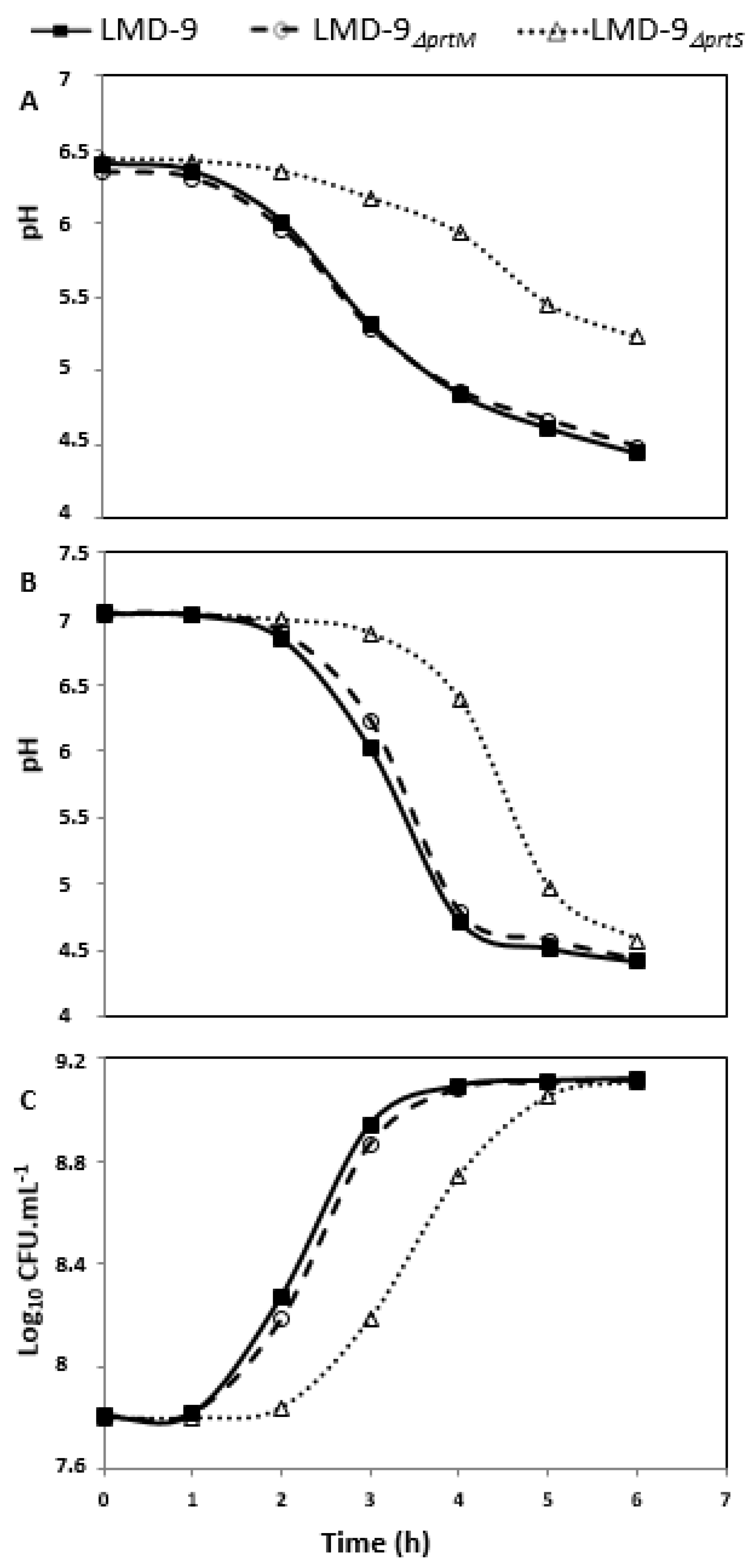
3.2. Role of Maturases in the Maturation of PrtS in S. thermophilus LMD-9

An exploration of data banks allowed for identification of four genes that encode PPIases (prtM, tig or ropA, ppiA, and pplB) in sequenced genomes of S. thermophilus. Among them, two maturases, PplB and PpiA, belong to the cyclophilin family, whereas PrtM and RopA belong to the parvulin and FKBP (FK506 binding protein) families, respectively. An analysis of these four genes in the 4F44 strain revealed that their deduced proteins are identical with the corresponding ones of the LMD-9 strain, except the PrtM protein (taxon: 1308; locus: AMM43147; GenBank: KT809299.1), which displays four differences (D30N, V75A, A78V, and A242T). Hence, to determine its implication in PrtS maturation, the LMD-9prtM mutant strain was constructed by deleting the prtM gene in the LMD-9 strain.
The LMD-9, LMD-9prtS, and LMD-9prtM strains were grown in milk to evaluate the effect of prtM deletion on the growth performance of the mutant. This approach is an indirect measure of the PrtS proteolytic activity considering the relationship between the PrtS activity and S. thermophilus capacity to grow in milk [6,7]. The results revealed that the LMD-9prtS mutant showed a delayed growth both in LM17 and milk media contrary to strains LMD-9 and LMD-9prtM, which showed a similar growth behavior (Figure 1). In a similar manner, the proteolytic activity was observed only at the cells’ surface of LMD-9 and LMD-9prtM (Table 1), and not in the culture supernatants.
Consequently, the lack of the putative PrtM maturase in the LMD-9prtM strain did not result in any growth retardation, nor in a decrease in PrtS activity and/or its release into the extracellular medium; it is most probable that the PrtM maturase was not responsible for the PrtS maturation. Several factors support the hypothesis that PrtS could undergo automaturation. First, unlike thee prsA of S. pyogenes and prtM of L. lactis, which are located upstream of the speB and prtP genes, respectively and co-transcribed, the prtM of S. thermophilus is not located near PrtS [9,12,14,36]. Second, when Chang et al. [23] established the N-terminal sequence of the soluble PrtS form of the 4F44 strain, they detected the N-terminal sequence corresponding to the proenzyme (with prodomain PP) as well as the N-terminal sequence of the mature form (without prodomain PP) of PrtS. The same situation was also detected for anchored PrtS forms present at the cells’ surface of the LMD-9 strain [31]. It was also noticed that the PrtS proenzyme form disappeared progressively in favor of the mature form (unpublished results) preceding a prolonged incubation or high concentration preservation. The fact that PrtS could be automaturated is not inimitable in LAB, e.g., regarding CEP PrtB of Lb. bulgaricus [37]. Finally, the fact that the inactivation of prtM did not lead to any release of PrtS in the growth medium, associated with the absence of difference between the three other known maturases (PpiA, PplB, and RopA), ruled out the hypothesis of a link between maturation via PrtM especially, correct folding, and anchoring.

3.3. SrtA Is Responsible for the Anchoring of PrtS to the Cell Wall of S. thermophilus and Is Deficient in Strain 4F44

The hypothesis that, despite few numbers of substitutions found between SrtA4F44 and SrtALMD-9, the extracellular liberation of PrtS4F44 could result from a partial deficiency of SrtA4F44 implies first providing evidence that SrtA is actually responsible for the PrtS anchoring to the cell wall of S. thermophilus as in other Gram-positive bacteria. Hence, the LMD-9srtA mutant strain was constructed by replacing the srtA gene by an erythromycin resistance gene. Afterwards, a complemented mutant was constructed by reintegrating the srtALMD-9 gene to the LMD-9srtA mutant to verify whether the phenotype of wild type LMD-9 strain could be restored (LMD-9srtALMD-9).
The synthetic substrate Suc-Ala-Ala-Pro-Phe-pNA was used to search proteolytic activity in the filtered growth medium of the LMD-9srtA mutant, complemented mutant, and WT strain (Table 1). It was detected only in the LMD-9srtA mutant (1020 mAU of PrtS activity; Table 1). The same supernatants were then analyzed by a casein-zymogram to detect PrtS (Figure 2A). Three caseinolytic bands (170, 154, and 115 kDa) previously shown to correspond to PrtS [23] were observed (Figure 2A) in the supernatant of mutant LMD-9srtA contrary to WT and LMD-9srtALMD-9. This corroborates the results obtained using synthetic substrate Suc-Ala-Ala-Pro-Phe-pNA. Unexpectedly, the LMD-9srtA strain displayed a surface PrtS activity (Table 1) despite of srtA deletion. To determine whether this resulted from electrostatic or other low-force interactions of PrtS with the cell surface, cells of this mutant and of strains 4F44 and LMD-9srtALMD-9, used as controls, were suspended in Tris HCl (100 mmol/L, pH 7) buffer and incubated from 0 to 70 h at 4 °C. The cells’ surface-bound PrtS activity was determined at t0 and t70 using a casein-zymogram (Figure 2) and the synthetic substrate Suc-Ala-Ala-Pro-Phe-pNA. After 70 h of incubation, the surface PrtS activity of strains 4F44, LMD-9, and LMD-9srtALMD-9 was close to that obtained at t0, while that of the LMD-9srtA strain appeared to be undetectable. The PrtS activity was exclusively found in the incubation buffer of this strain (Figure 2C). These last results proved that the SrtA of S. thermophilus is responsible for the anchoring of PrtS to the cell wall and probably the other proteins possessing a LPXTG motif. Such a phenotype has already been described in a St. aureus mutant that was defective in the anchoring of surface LPXTG proteins because of a mutation in the srtA gene. The deletion of this gene resulted in the liberation of surface LPXTG proteins, thereby leading to a decreased virulence of the bacterium [38]. In a similar manner, in the Streptococcus genus, another study demonstrated that SrtA is a key player responsible for the anchoring of surface LPXTG proteins of S. pyogenes [39].
To further investigate whether SrtA4F44 is partially defective, the LMD-9srtA4F44 mutant was constructed by introducing the srtA4F44 allele into the genome of the LMD-9srtA strain. Hereafter, the soluble PrtS activity was examined in filtered growth supernatants of the strains LMD-9srtA4F44 and 4F44 by using both the Suc-Ala-Ala-Pro-Phe-pNA substrate (Table 1) and a casein-zymogram (Figure 2B). Through analyses being realized in the conditions, levels of PrtS activity could be compared (see Section 2). Twenty-six percentages of total PrtS activity of the mutant LMD-9srtA4F44 were found in its growth supernatant, i.e., a proportion similar to that of strain 4F44 (37%). In addition, the incubation of the cells of this mutant in Tris-HCl (100 mmol L−1, pH 7) buffer during 70 h did not result in a significant increase of extracellular PrtS activity. Therefore, despite the few numbers of substitutions found between the SrtA of strains LMD-9 and 4F44, our results provide genetic proof of the dysfunctioning of SrtA4F44, which leads to the anchoring to the cell wall of the majority of PrtS molecules and to the release of a fraction of PrtS in the growth medium of the 4F44 strain, as postulated by Chang et al. [23]. Indeed, the fact that after 70 h of incubation of the cells of 4F44 and LMD-9srtA4F44 strains in Tris-HCl buffer the surface PrtS activity remained similar to the initial one strongly suggests that it corresponds to PrtS molecules correctly anchored to the cell wall. Besides, even if the total PrtS activity (bound plus free) appeared in our assays to be higher in strain 4F44 than in strain LMD-9 (8860 mAU against 4194, Table 1), the PrtS release cannot be attributed to a higher expression of its gene in strain 4F44, leading to the saturation of SrtA activity and ultimately to a leakage of non-anchored PrtS molecules in the external environment. Indeed, in the mutant LMD-9srtA4F44, the gene prtS undergoes the same regulation like in the wild-type LMD-9 strain, as suggested by the PrtS activity levels observed in this mutant (Table 1). Therefore, no saturation of the anchoring activity of sortase SrtA is expected and SrtA4F44, which is expressed in this mutant, should anchor all PrtS molecules.

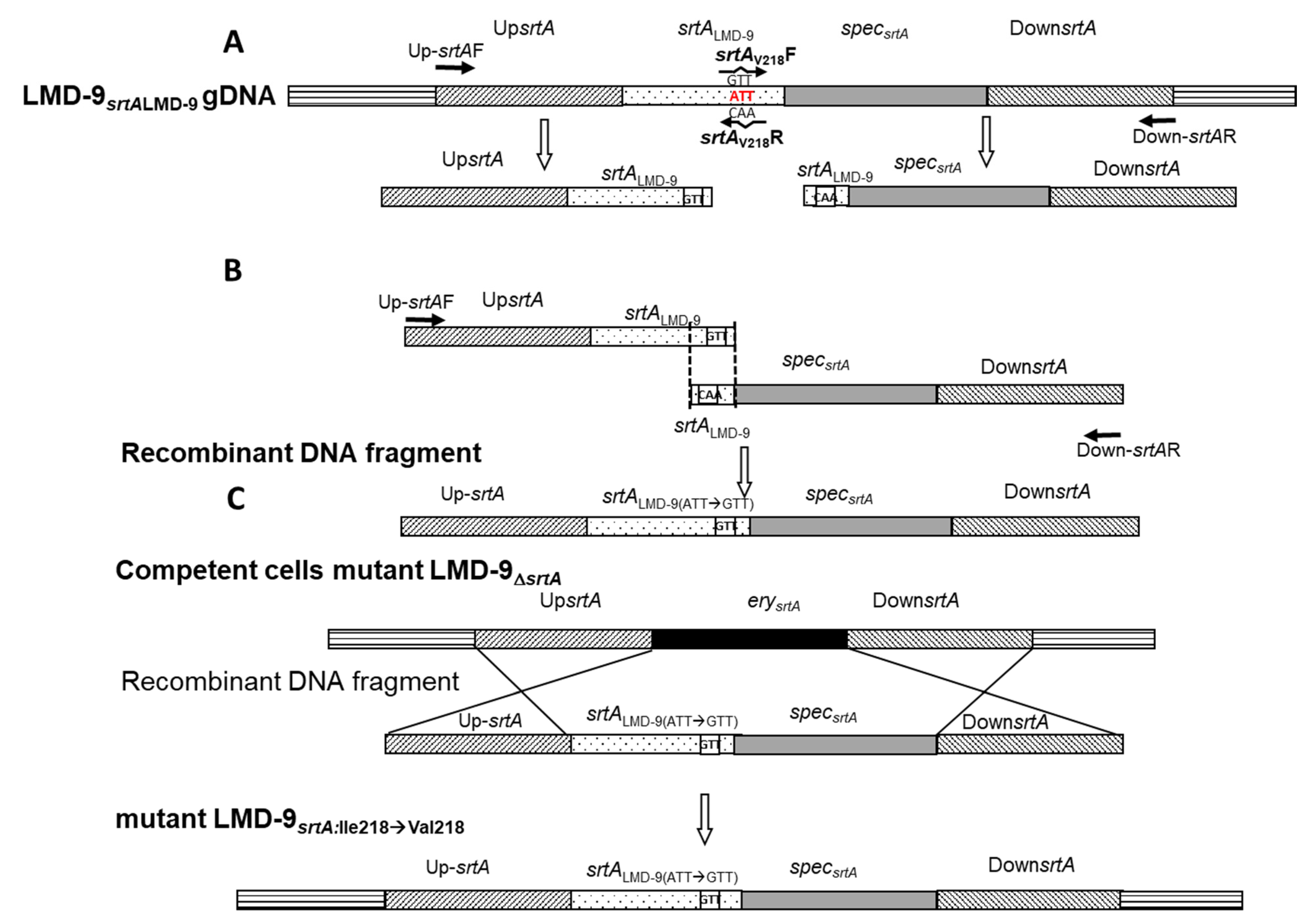
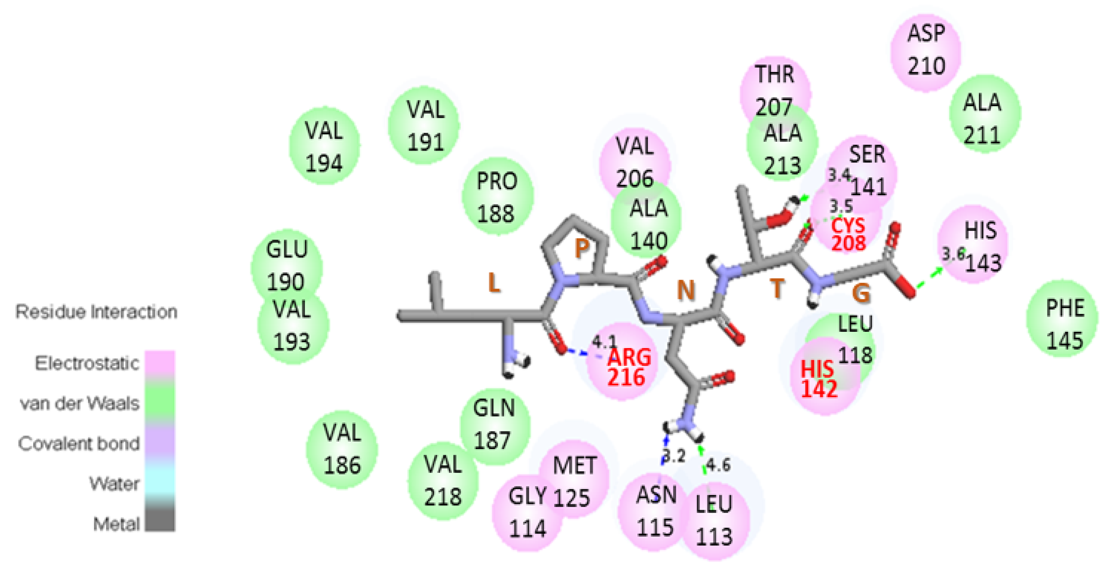
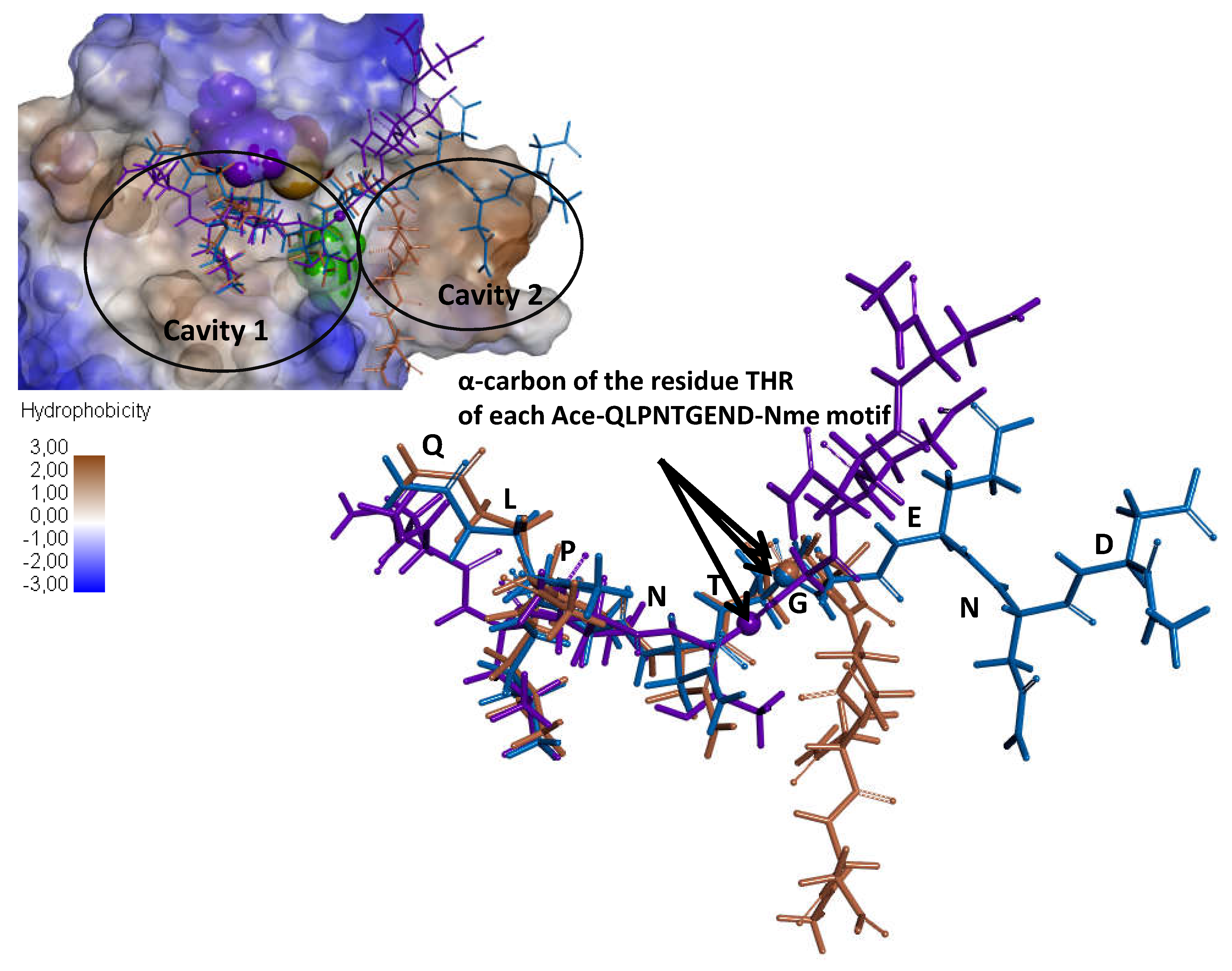
3.4. Substitution of the Ile 218 Residue Is Not Responsible for the Deficiency of SrtA4F44

As we knew the LPNTG motif was present at the C-terminal extremity of the extracellular soluble form of PrtS4F44 [23], molecular modeling simulations were performed to determine whether the binding mechanism of the LPNTG motif to the SrtA catalytic site was at least partially altered in SrtA4F44.
Since S. pyogenes and S. thermophilus belong to the same genus, their respective sortases A were assumed to have similar mechanisms. Thus, the structural models of SrtALMD-9 (St-SrtALMD-9∆90) and SrtA4F44 (St-SrtA4F44∆90) were built from the already defined structure of S. pyogenes SF370 SrtA (Sp-SrtASF370∆86) [24]. In order to build the models with the same amino acid residues as in the Sp-SrtASF370∆86, it was necessary to delete the first 90 residues of St-SrtALMD-9∆90 and SrtA4F44 (St-SrtA4F44∆90). The percentage of identity/similarity between the C-terminal domain of Sp-SrtASF370∆86 and St-SrtA4F44∆90, and Sp-SrtASF370∆86 and St-SrtALMD-9∆90 were found to be sufficient (between 70.6% to 97.5% and 87.1% to 99.4%, respectively) to use Sp-SrtASF370∆86 as a structural pattern for constructing the St-SrtA4F44∆90 and St-SrtALMD-9∆90 ones. Hence, surimposition of the three structures led to RMSD values below 0.16 Å considering all the 169 Cα atoms of the residues 87 to 249. No significant structural difference was observed between the structures, as St-SrtA4F44∆90 and St-SrtALMD-9∆90 models displayed the characteristic structure of sortases, i.e., the eight-stranded β-barrel fold and a long hydrophobic cleft corresponding to the catalytic cavity located at the center of the protein (Figure 3). It was assumed that the residues Cys208, His142, and Arg216 compose the catalytic triad (Figure 3). The orientation of these residues is consistent with the model of reverse protonation that has been proposed in biochemical studies of the sortase A of St. aureus and S. pyogenes [24,40].
These homology models were then used to study possible binding modes of the LPNTG pattern within the sortase catalytic cavity. Enzyme/substrate complexes were generated using docking simulations with two substrates: the LPNTG pattern and a longer pattern, specifically the Ace-QLPNTGEND-Nme pattern. Unfortunately, no significant difference was observed between the complexes Sp-SrtASF370∆86: LPNTG/Ace-QLPNTGEND-Nme; St-SrtALMD-9∆90: LPNTG/Ace-QLPNTGEND-Nme; and St-SrtA4F44∆90: LPNTG/Ace-QLPNTGEND-Nme (Supplementary Data and Figure A2 and Figure A3 in Appendix A). Consequently, these results strongly suggested that none of the four substitutions distinguishing St-SrtA4F44∆90 from St-SrtALMD-9∆90 could account for the defective activity of the SrtA4F44.
To definitively rule out the hypothesis of an eventual role of the substitution of Ile218 by Val218 in the deficiency of StrA4F44 and to validate the modeling prediction, the residue Ile218 of strain LMD-9srtALMD-9 was replaced by a valine residue (Figure A1). The absence of extracellular PrtS activity (searched using the Suc-Ala-Ala-Pro-Phe-pNA substrate) in the supernatant of this strain confirms the non-involvement of Ile218 substitution by Val218 in the deficiency of StrA4F44.
To conclude we showed that (i) the 32 amino acid residues’ imperfect duplication located in the prodomain of certain PrtS, such as PrtSLMD-9, was not essential for the correct maturation and subsequent anchoring of PrtS; (ii) the maturase PrtM, homologous to maturases of other CEP, was not responsible for the maturation of PrtS and neither for its correct anchoring to the cell surface; and (iii) SrtA was responsible for the anchoring of PrtS to the cell wall and (iv) SrtA of strain 4F44 was partially defective despite the low number of dissimilar residues (six substitutions), which differentiates it from that of the LMD-9 strain and probably through a subtle mechanism not yet elucidated perhaps because of a lack of a structural model, including the N-terminal part of SrtA.

Supplementary Materials

The following are available online at https://0-www-mdpi-com.brum.beds.ac.uk/article/10.3390/microorganisms9112380/s1, Table S1: Primers and resulting PCR or Overlapping PCR products used for mutant construction, Table S2: Primers used for the sequencing of PPIases genes, Table S3: Primers used for the verification of mutant sequences.

Author Contributions

Conceptualization, A.D.-M., C.P., E.R, G.H. and O.K.C.; methodology, A.A.A., B.M., C.H., C.P., E.R, G.H. and X.L.; validation, A.A.A. and E.R.; investigation, A.A.A., A.D.-M., B.M., C.H., C.P., E.R. and M.G.; data curation, C.P., E.R., M.G. and Z.H.; writing—original draft preparation, A.A.A., A.D.-M., C.H., C.P. and E.R.; writing—review and editing, A.D.-M., L.M., M.G. and Z.H.; visualization, A.A.A., A.D.-M., C.H., C.P. and E.R.; supervision, A.D.-M., C.H., C.P. and E.R.; project administration, A.A.A., C.H., C.P. and E.R.; funding acquisition, A.D.-M. and C.P. All authors have read and agreed to the published version of the manuscript.

Funding

This research received no external funding.

Institutional Review Board Statement

Not applicable.

Informed Consent Statement

Not applicable.

Acknowledgments

We would like to thank the Cabinet Decisions SARL Togo for the fellowship accorded to Ahoefa Ablavi AWUSSI to prepare her thesis.

Conflicts of Interest

The authors declare no conflict of interest.

Appendix A

Figure A1. Strategy to replace the ATT codon specifying the Ile218 residue of SrtALMD-9 by a GTT codon corresponding to a Valine residue. Two PCRs were carried out using genomic DNA of the LMD-9srtALMD-9 (LMD-9srtALMD-9-g DNA) mutant to obtain (i) an upstream DNA fragment of the srtA gene (UpsrtA) and the first 675 nucleotides of the same gene, and (ii) a DNA fragment containing the second part of the srtA gene (87 nucleotides), the spec gene (confers resistance to spectinomycin), and a downstream sequence of the srtA gene (DownsrtA) (A). These two overlapped PCR fragments were then used to make the recombinant DNA fragment by overlapping PCR (B). The recombinant DNA amplicon was introduced into competent cells of the LMD-9srtA mutant, thereby permitting obtainment after its integration through a double crossing-over event, specifically the mutant LMD-9srtA:Ile218→Val218 (C). Primer sequences used to amplify different fragments are presented in Table S1.
Microorganisms 09 02380 g0a1
Figure A2. Interaction between the LPNTG ligand and amino acid residues of St-SrtA4F44∆90. The numbers represent the distance between atoms of the ligand and residues of the cavity of sortase A. Red letters indicate residues of the catalytic triad. The residues Gln187, Glu190, Val194, Ala211, and Val218 correspond to the residues Ala187, Arg190, Ile194, Ile211, and Ile218 of Sp-SrtASF370∆86, while residue Val218 corresponds to the residue Ile218 of St-SrtALMD-9.
Microorganisms 09 02380 g0a2
Figure A3. Superimposition of the main binding modes of ligand Ace-QLPNTGEND-Nme within the sortase A (here SrtA4F44∆90) catalytic cavity. Ligands denoted here correspond to the best ligand obtained for each complex (Sp-SrtASF370∆86:Ace-QLPNTGEND-Nme; SrtALMD-9∆90:Ace-QLPNTGEND-Nme; and SrtA4F44∆90:Ace-QLPNTGEND-Nme) according to (i) the distance between the S atom of the corresponding sortase A Cys208 residue and the alpha carbon of the threonine residue of the ligand, and (ii) the position of the ligand within the cavity. Thus, the brown ligand corresponds to the ligand of complex Sp-SrtASF370∆86:Ace-QLPNTGEND-Nme, the purple one to SrtALMD-9∆90:Ace-QLPNTGEND-Nme, and the blue one to SrtA4F44∆90:Ace-QLPNTGEND-Nme.
Microorganisms 09 02380 g0a3

References

  1. Cui, Y.; Xu, T.; Qu, X.; Hu, T.; Jiang, X.; Zhao, C. New insights into various production characteristics of Streptococcus thermophilus strains. Int. J. Mol. Sci. 2016, 17, 1701. [Google Scholar] [CrossRef]
  2. Jensen, A.; Valdórsson, O.; Frimodt-Møller, N.; Hollingshead, S.; Kilian, M. Commensal streptococci serve as a reservoir for β-lactam resistance genes in Streptococcus pneumoniae. Antimicrob. Agents Chemother. 2015, 59, 3529–3540. [Google Scholar] [CrossRef] [Green Version]
  3. Krzyściak, W.; Pluskwa, K.K.; Jurczak, A.; Kościelniak, D. The pathogenicity of the Streptococcus genus. Eur. J. Clin. Microbiol. Infect. Dis. 2013, 32, 1361–1376. [Google Scholar] [CrossRef] [Green Version]
  4. Garault, P.; Letort, C.; Juillard, V.; Monnet, V. Branched-chain amino acid biosynthesis is essential for optimal growth of Streptococcus thermophilus in milk. Appl. Environ. Microbiol. 2000, 66, 5128–5133. [Google Scholar] [CrossRef] [PubMed] [Green Version]
  5. Pastink, M.I.; Teusink, B.; Hols, P.; Visser, S.; de Vos, W.M.; Hugenholtz, J. Genome-scale model of Streptococcus thermophilus LMG18311 for metabolic comparison of lactic acid bacteria. Appl. Environ. Microbiol. 2009, 75, 3627–3633. [Google Scholar] [CrossRef] [PubMed] [Green Version]
  6. Dandoy, D.; Fremaux, C.; Henry de Frahan, M.; Horvath, P.; Boyaval, P.; Hols, P.; Fontaine, L. The fast milk acidifying phenotype of Streptococcus thermophilus can be acquired by natural transformation of the genomic island encoding the cell-envelope proteinase PrtS. Microb. Cell Factories 2011, 10 (Suppl. S1), S21. [Google Scholar] [CrossRef] [Green Version]
  7. Galia, W.; Perrin, C.; Genay, M.; Dary, A. Variability and molecular typing of Streptococcus thermophilus strains displaying different proteolytic and acidifying properties. Int. Dairy J. 2009, 19, 89–95. [Google Scholar] [CrossRef]
  8. Rodríguez-Serrano, G.M.; García-Garibay, J.M.; Cruz-Guerrero, A.E.; Gómez-Ruiz, L.; Ayala-Niño, A.; Castañeda-Ovando, A.; González-Olivares, L.G. Proteolytic system of Streptococcus thermophilus. J. Microbiol. Biotechnol. 2018, 28, 1581–1588. [Google Scholar] [CrossRef]
  9. Savijoki, K.; Ingmer, H.; Varmanen, P. Proteolytic systems of lactic acid bacteria. Appl. Microbiol. Biotechnol. 2006, 71, 394–406. [Google Scholar] [CrossRef] [PubMed]
  10. Bonifait, L.; de la Cruz Dominguez-Punaro, M.; Vaillancourt, K.; Bart, C.; Slater, J.; Frenette, M.; Gottschalk, M.; Grenier, D. The cell envelope subtilisin-like proteinase is a virulence determinant for Streptococcus suis. BMC Microbiol. 2010, 10, 42. [Google Scholar] [CrossRef] [Green Version]
  11. Delorme, C.; Bartholini, C.; Bolotine, A.; Ehrlich, S.D.; Renault, P. Emergence of a cell wall protease in the Streptococcus thermophilus population. Appl. Environ. Microbiol. 2010, 76, 451–460. [Google Scholar] [CrossRef] [Green Version]
  12. Fernandez-Espla, M.D.; Garault, P.; Monnet, V.; Rul, F. Streptococcus thermophilus cell wall-anchored proteinase: Release, purification, and biochemical and genetic characterization. Appl. Environ. Microbiol. 2000, 66, 4772–4778. [Google Scholar] [CrossRef] [PubMed] [Green Version]
  13. Fischer, G.; Schmid, F.X. The mechanism of protein folding. Implications of in vitro refolding models for de novo protein folding and translocation in the cell. Biochemistry 1990, 29, 2205–2212. [Google Scholar] [CrossRef] [PubMed]
  14. Ma, Y.; Bryant, A.E.; Salmi, D.B.; Hayes-Schroer, S.M.; McIndoo, E.; Aldape, M.J.; Stevens, D.L. Identification and characterization of bicistronic speB and prsA gene expression in the Group A Streptococcus. J. Bacteriol. 2006, 188, 7626–7634. [Google Scholar] [CrossRef] [Green Version]
  15. Vos, P.; van Asseldonk, M.; van Jeveren, F.; Siezen, R.; Simons, G.; de Vos, W.M. A maturation protein is essential for production of active forms of Lactococcus lactis SK11 serine proteinase located in or secreted from the cell envelope. J. Bacteriol. 1989, 171, 2795–2802. [Google Scholar] [CrossRef] [Green Version]
  16. Bhat, A.H.; Nguyen, M.T.; Asis Das, A.; Ton-That, H. Anchoring surface proteins to the bacterial cell wall by sortase enzymes: How it started and what we know now. Curr. Opin. Microbiol. 2021, 60, 73–79. [Google Scholar] [CrossRef]
  17. Zrelovs, N.; Kurbatska, V.; Rudevica, V.; Leonchiks, A.; Fridmanis, D. Sorting out the superbugs: Potential of sortase A inhibitors among other antimicrobial strategies to tackle the problem of antibiotic resistance. Antibiotics 2021, 10, 164. [Google Scholar] [CrossRef]
  18. Freund, C.; Dirk Schwarzer, D. Engineered Sortases in Peptide and Protein Chemistry. ChemBioChem 2021, 22, 1347–1356. [Google Scholar] [CrossRef]
  19. Hu, P.; Bian, Z.; Fan, M.; Huang, M.; Zhang, P. Sec translocase and sortase A are colocalised in a locus in the cytoplasmic membrane of Streptococcus mutans. Arch. Oral Biol. 2008, 53, 150–154. [Google Scholar] [CrossRef]
  20. Kline, K.A.; Kau, A.L.; Chen, S.L.; Lim, A.; Pinkner, J.S.; Rosch, J.; Nallapareddy, S.R.; Murray, B.E.; Henriques-Normark, B.; Beatty, W.; et al. Mechanism for sortase localization and the role of sortase localization in efficient pilus assembly in Enterococcus faecalis. J. Bacteriol. 2009, 191, 3237–3247. [Google Scholar] [CrossRef] [Green Version]
  21. Cole, J.N.; Aquilina, J.A.; Hains, P.G.; Henningham, A.; Sriprakash, K.S.; Caparon, M.G.; Nizet, V.; Kotb, M.; Cordwell, S.J.; Djordje-vic, S.P.; et al. Role of group A Streptococcus HtrA in the maturation of SpeB protease. Proteomics 2007, 7, 4488–4498. [Google Scholar] [CrossRef]
  22. Lyon, W.R.; Caparon, M.G. Trigger Factor-Mediated Prolyl Isomerization Influences Maturation of the Streptococcus pyogenes Cysteine Protease. J. Bacteriol. 2003, 185, 3661–3667. [Google Scholar] [CrossRef] [Green Version]
  23. Chang, O.K.; Perrin, C.; Galia, W.; Saulnier, F.; Miclo, L.; Roux, E.; Driou, A.; Humbert, G.; Dary, A. Release of the cell-envelope pro-tease PrtS in the growth medium of Streptococcus thermophilus 4F44. Int. Dairy J. 2012, 23, 91–98. [Google Scholar] [CrossRef]
  24. Race, P.R.; Bentley, M.L.; Melvin, J.A.; Crow, A.; Hughes, R.K.; Smith, W.D.; Sessions, R.B.; Kehoe, M.A.; McCafferty, D.G.; Ban-field, M.J. Crystal structure of Streptococcus pyogenes sortase A. implications for sortase mechanism. J. Biol. Chem. 2009, 284, 6924–6933. [Google Scholar] [CrossRef] [Green Version]
  25. Terzaghi, B.E.; Sandine, W.E. Improved Medium for Lactic Streptococci and Their Bacteriophages. Appl. Microbiol. 1975, 29, 807–813. [Google Scholar] [CrossRef]
  26. Gardan, R.; Besset, C.; Guillot, A.; Gitton, C.; Monnet, V. The oligopeptide transport system is essential for the development of natural competence in Streptococcus thermophilus strain LMD-9. J. Bacteriol. 2009, 191, 4647–4655. [Google Scholar] [CrossRef] [Green Version]
  27. Fischer, G.; Decaris, B.; Leblond, P. Occurrence of deletions, associated with genetic instability in Streptomyces ambofaciens, is independent of the linearity of the chromosomal DNA. J. Bacteriol. 1997, 179, 4553–4558. [Google Scholar] [CrossRef] [Green Version]
  28. Sambrook, J.F.; Russell, D.W. Molecular Cloning: A Laboratory Manual, 3rd ed.; Cold Spring Harbor Laboratory Press: New York, NY, USA, 2001; Volume 1–3. [Google Scholar]
  29. Sanger, F.; Nicklen, S.; Coulson, A.R. DNA sequencing with chain-terminating inhibitors. Proc. Natl. Acad. Sci. USA 1977, 74, 5463–5467. [Google Scholar] [CrossRef] [Green Version]
  30. Makarova, K.; Slesarev, A.; Wolf, Y.; Sorokin, A.; Mirkin, B.; Koonin, E.; Pavlov, A.; Pavlova, N.; Karamychev, V.; Polouchine, N.; et al. Comparative genomics of the lactic acid bacteria. Proc. Natl. Acad. Sci. USA 2006, 103, 15611–15616. [Google Scholar] [CrossRef] [Green Version]
  31. Lecomte, X.; Gagnaire, V.; Briard-Bion, V.; Jardin, J.; Lortal, S.; Dary, A.; Genay, M. The naturally competent strain Streptococcus thermophilus LMD-9 as a new tool to anchor heterologous proteins on the cell surface. Microb. Cell Factories 2014, 13, 82. [Google Scholar] [CrossRef] [Green Version]
  32. Maguin, E.; Prévost, H.; Ehrlich, S.D.; Gruss, A. Efficient insertional mutagenesis in lactococci and other gram-positive bacteria. J. Bacteriol. 1996, 178, 931–935. [Google Scholar] [CrossRef] [Green Version]
  33. Takamatsu, D.; Osaki, M.; Sekizaki, T. Construction and characterization of Streptococcus suisEscherichia coli shuttle cloning vectors. Plasmid 2001, 45, 101–113. [Google Scholar] [CrossRef]
  34. Diller, D.J.; Merz, K.M. High throughput docking for library design and library prioritization. Proteins 2001, 43, 113–124. [Google Scholar] [CrossRef]
  35. MacKerell, A.D.; Bashford, D.; Bellott, M.; Dunbrack, R.L.; Evanseck, J.D.; Field, M.J.; Fischer, S.; Gao, J.; Guo, H.; Ha, S.; et al. All-Atom Empirical Potential for Molecular Modeling and Dynamics Studies of Proteins. J. Phys. Chem. B 1998, 102, 3586–3616. [Google Scholar] [CrossRef]
  36. Vitikainen, M.; Lappalainen, I.; Seppala, R.; Antelmann, H.; Boer, H.; Taira, S.; Savilahti, H.; Hecker, M.; Vihinen, M.; Sarvas, M.; et al. Structure-function analysis of PrsA reveals roles for the parvulin-like and flanking N- and C-terminal domains in protein folding and se-cretion in Bacillus subtilis. J. Biol. Chem. 2004, 279, 19302–19314. [Google Scholar] [CrossRef] [Green Version]
  37. Germond, J.-E.; Delley, M.; Gilbert, C.; Atlan, D. Determination of the Domain of the Lactobacillus delbrueckii subsp. bulgaricus cell surface proteinase PrtB involved in attachment to the cell wall after heterologous expression of the prtB Gene in Lactococcus lactis. Appl. Environ. Microbiol. 2003, 69, 3377–3384. [Google Scholar] [CrossRef] [Green Version]
  38. Mazmanian, S.K.; Liu, G.; Ton-That, H.; Schneewind, O. Staphylococcus aureus sortase, an enzyme that anchors surface proteins to the cell wall. Science 1999, 285, 760–763. [Google Scholar] [CrossRef]
  39. Barnett, T.C.; Scott, J.R. Differential recognition of surface proteins in Streptococcus pyogenes by two sortase gene homologs. J. Bacteriol. 2002, 184, 2181–2191. [Google Scholar] [CrossRef] [Green Version]
  40. Mazmanian, S.K.; Ton-That, H.; Schneewind, O. Sortase-catalysed anchoring of surface proteins to the cell wall of Staphylococcus aureus. Mol. Microbiol. 2001, 40, 1049–1057. [Google Scholar] [CrossRef] [Green Version]
Figure 1. Growth of S. thermophilus LMD-9, LMD-9prtM, and LMD-9prtS (a negative control) in milk (A) or in LM17 (B,C), evaluated by measuring either the extracellular pH (A,B) or OD600 (C).
Microorganisms 09 02380 g001
Figure 2. SDS-PAGE zymogram analysis to detect proteolytic activity in the extracellular growth medium of S. thermophilus LMD-9, LMD-9srtA, LMD-9srtALMD-9 (A), and 4F44, LMD-9srtA4F44 (B), and the proteolytic activity liberated in the incubation buffer (t = 0 and t = 70 h) of the LMD-9srtA strain (C).
Microorganisms 09 02380 g002
Figure 3. Putative structure of St-SrtA4F44∆90. The catalytic triad (in CPK, His142 in green, Arg216 in blue, and Cys208 in red, and its thiolate group in brown) position and the amino acid residues important (according to Race et al. [24]) for the positioning of the LPNTG motif within the cavity (in CPK teal) are indicated. The LPNTG motif is also shown within the cavity of SrtA4F44∆90. The structure of Sp-SrtASF370∆86 and St-SrtALMD-9∆90 are analogous to that of 4F44. The residue Val218 corresponds to the residue Ile218 in St- SrtALMD-9∆90, while the residues Val194 and Val218 correspond to residues Ile214 and Ile218 in Sp-SrtASF370∆86. For simplicity, the catalytic cavity has been divided into cavity 1 and cavity 2.
Microorganisms 09 02380 g003
Table 1. Characteristics of Streptococcus thermophilus strains and plasmids used in this study and PrtS activity.
Strains\PlasmidsOriginReferencessrtA AllelePresence (+)/Absence (−)
of the prtS Gene
PrtS Activity in mAU
Cell
Surface
Growth
Supernatant
Streptococcus thermophilus wild-type
LMD-9Yogurt[30]srtALMD-9+31100
4F44Cheese[23]srtA4F44+56203240
Streptococcus thermophilus mutants
LMD-9srtALMD-9LMD-9Present studysrtALMD-9+31000
LMD-9srtALMD-9Present studyΔ+26001020
LMD-9srtA4F44LMD-9Present studysrtA4F44+31241070
LMD-9prtSLMD-9[31]srtALMD-900
LMD-9prtS4F44LMD-9Present studysrtALMD-9+30930
LMD-9prtM
LMD-9srtA:Ile218Val218
LMD-9
LMD-9
Present study
Present study
srtALMD-9
srtA:Ile218Val218
+
+
3114
Present
0
0
PlasmidsRelevant markers and characteristics
pG+host9-[32]ErmR *, pWV01 derivative, with thermoresistant replication function
pSET4s-[33]SpecR *, replication function of Pg + host3 and pUC19
* SpecR and ErmR: resistance to spectinomycin and erythromycin, respectively; Δ: deletion of the srtA, prtS, or prtM gene; and mAU: milli arbitrary units.
Publisher’s Note: MDPI stays neutral with regard to jurisdictional claims in published maps and institutional affiliations.

