Article

Evidence of Borrelia theileri in Wild and Domestic Animals in the Kafue Ecosystem of Zambia

1
Division of International Research Promotion, International Institute for Zoonosis Control, Hokkaido University, N 20 W 10, Kita-ku, Sapporo 001-0020, Japan
2
Wildlife Diseases Unit, Department of Veterinary Services, Ministry of Fisheries and Livestock, Lusaka P.O. Box 50060, Zambia
3
Division of Collaboration and Education, International Institute for Zoonosis Control, Hokkaido University, N 20 W 10, Kita-ku, Sapporo 001-0020, Japan
4
Laboratory of Wildlife Biology and Medicine, Department of Environmental Veterinary Sciences, Faculty of Veterinary Medicine, Hokkaido University, N 18 W 9, Kita-ku, Sapporo 060-0818, Japan
5
Technical Office, International Institute for Zoonosis Control, Hokkaido University, N 20 W 10, Kita-ku, Sapporo 001-0020, Japan
6
International Collaboration Unit, International Institute for Zoonosis Control, Hokkaido University, Sapporo 001-0020, Japan
7
Laboratory of Parasitology, Department of Disease Control, Faculty of Veterinary Medicine, Hokkaido University, N 18 W 9, Kita-ku, Sapporo 060-0818, Japan
8
Division of Molecular Pathobiology, International Institute for Zoonosis Control, Hokkaido University, N 20 W 10, Kita-ku, Sapporo 001-0020, Japan
9
One Health Research Center, Hokkaido University, N 20 W 10, Kita-ku, Sapporo 001-0020, Japan
10
Department of Paraclinical Studies, School of Veterinary Medicine, The University of Zambia, Lusaka 10101, Zambia
11
Laboratory of Systemic Infection, Department of Bacteriology I, National Institute of Infectious Diseases, Toyama 1-23-1, Shinjuku, Tokyo 162-8640, Japan
*
Author to whom correspondence should be addressed.
Submission received: 6 November 2021 / Revised: 17 November 2021 / Accepted: 18 November 2021 / Published: 22 November 2021
(This article belongs to the Special Issue Borrelia Ecology and Evolution: Ticks and Hosts and the Environment)

Abstract

:
Members of the genus Borrelia are arthropod-borne spirochetes that are human and animal pathogens. Vertebrate hosts, including wild animals, are pivotal to the circulation and maintenance of Borrelia spirochetes. However, information on Borrelia spirochetes in vertebrate hosts in Zambia is limited. Thus, we aimed to investigate the presence of Borrelia spirochetes in wild animals and cattle in Zambia. A total of 140 wild animals of four species and 488 cattle DNA samples from /near the Kafue National Park were collected for real-time PCR screening, followed by characterization using three different genes with positive samples. Five impalas and 20 cattle tested positive using real-time PCR, and sequence analysis revealed that the detected Borrelia were identified to be Borrelia theileri, a causative agent of bovine borreliosis. This is the first evidence of Borrelia theileri in African wildlife and cattle in Zambia. Our results suggest that clinical differentiation between bovine borreliosis and other bovine diseases endemic in Zambia is required for better treatment and control measures. As this study only included wild and domestic animals in the Kafue ecosystem, further investigations in other areas and with more wildlife and livestock species are needed to clarify a comprehensive epidemiological status of Borrelia theileri in Zambia.

1. Introduction

Members of the genus Borrelia are arthropod-borne spirochetes that target vertebrate hosts and use them as reservoirs to complete their life cycle. The genus comprises three groups: Lyme disease borreliae, relapsing fever borreliae, and reptile-associated borreliae [1,2]. Among these groups, Lyme disease borreliae and reptile-associated borreliae are transmitted by Ixodid (hard-bodied) ticks [1,2]. Relapsing fever borreliae are divided into four subgroups: Soft tick-borne relapsing fever (STBRF), Hard tick-borne relapsing fever (HTBRF), Louse-borne relapsing fever (LBRF), and Avian worldwide relapsing fever [3], in which most of the identified relapsing fever borreliae belong to STBRF and are transmitted by Argasid (soft-bodied) ticks [4]. In contrast, HTBRF is transmitted by Ixodid ticks, such as Amblyomma, Haemaphysalis, Ixodes, and Rhipicephalus [5,6,7,8]. Only Borrelia recurrentis, belonging to LBRF, is transmitted by the human body louse [9]. In addition, Borrelia anserina of the Avian worldwide relapsing fever is transmitted by Argas spp. [10]. Recently, a different classification was given in Margos et al. (2020), who divides the genus into three groups: Ixodes-transmitted borreliae (Borrelia burgdorferi sensu lato complex), Matestriate-tansmitted borreliae, and relapsing fever-associated borreliae [11]. The classification of the genus Borrelia is still controversial.
Borrelia lonestari, Borrelia miyamotoi, and Borrelia theileri are members of HTBRF, or of Matestriate-tansmitted borreliae in Margos’s classification. Borrelia lonestari was first identified in 1996 [7] and was initially considered pathogenic to humans in the southern United States [12,13]. However, subsequent research did not support the pathogenicity of B. lonestari in humans [14]. Amblyomma ticks and white-tailed deer are vectors and suspected reservoirs, respectively, for B. lonestari in North America [15,16,17]. Borrelia miyamotoi was confirmed in 2011 as a human pathogen [18]. Ixodes ticks are reported vectors of Borrelia miyamotoi in Asia, Europe, North America, and Russia [19], and deer may act as natural reservoirs for it [20]. Borrelia theileri is a causative agent of bovine borreliosis, identified in South Africa more than 100 years ago by Arnold Theiler, who first found the agent transmitted by Rhipicephalus sp. to cattle [21,22]. To date, Borrelia theileri has been reported in cattle, goats, sheep, and horses in Africa, North and South America, and Australia [23,24,25]. Rhipicephalus ticks, such as R. microplus, R. annulatus, R. evertsi, and R. decoloratus, are well-known vectors of Borrelia theileri. In addition, Borrelia theileri has recently been detected in head lice (Pediculus humanus) in the Republic of Congo [26].
Vertebrate hosts, including wild animals, act as reservoirs for Borrelia spirochetes and are crucial in the circulation and maintenance of them. For instance, Kumagai et al. (2018) discovered Borrelia spp. of the hard tick-borne relapsing fever borreliae in 25.9% of wild deer tested in Japan and suggested that wild deer could act as reservoir hosts for Borellia spp. [27]. Similarly, other studies have also reported the detection of Borrelia lonestari and antibodies against Borrelia burgdorferi in wild deer populations in America [16,28]. Furthermore, several serological studies have revealed that many species of African antelopes kept in zoos have antibodies against Borrelia burgdorferi [29,30,31]. However, only a few studies have been conducted to investigate Borrelia spirochetes in wild animals in Africa. For example, Borrelia infection was demonstrated in 9.2% of small mammals tested in West Africa [32], and Candidatus Borrelia fainii was detected in 27% of bats examined in Zambia [33]. In addition, to date, there has been no investigation of Borrelia in large wild animals in Africa.
We aimed to investigate the presence of Borrelia spirochetes in large and medium-sized wild animals and cattle in Zambia through molecular and phylogenetic analyses.

2. Materials and Methods

DNA samples extracted from the whole blood of wild animals and domestic cattle from two previous studies [34,35] were used to detect Borrelia in this study. A total of 140 DNA samples from wild animals, including 97 impalas (Aepyceros melampus), 37 hartebeests (Alcelaphus buselaphus), four lions (Panthera leo), and two wild dogs (Lycaon pictus), were previously collected in the greater Kafue ecosystem for investigating piroplasm diversity [34]. In addition, we used 488 cattle DNA samples with packed cell volume (PCV) value data from a previous African trypanosome investigation [35]. The cattle breed was mainly a cross between local breeds (Tonga and Baila) and exotic breeds (mostly Boran and Brahman). Thus, a total of 628 DNA samples from wild and domestic animals were screened for Borrelia spp.
Specific semiquantitative real-time PCR was used for the initial screening of Borrelia infection, using the THUNDERBIRD® Probe qPCR Mix (TOYOBO, Osaka, Japan) and primers (Bor16S3F, 5′-AGCCTTTAAAGCTTCGCTTGTAG-3′; Bor16S3R, 5′-GCCTCCCGTAGGAGTCTGG-3′; Bor16S3P, 5′-6FAM-CCGGCCTGAGAGGGTGAACGG-TAMRA-3′), which were designed to amplify a 148-bp fragment of the 16S ribosomal RNA gene (16S rDNA) of Borrelia. The specificity of the real-time PCR system for detection of Borrelia spp. was previously tested on DNA samples from 347 bacterial species [36]. All real-time PCRs were performed using a LightCycler 96 (Roche Diagnostics GmbH, Mannheim, Germany). The DNA of Candidatus Borrelia fainii strain Qtaro isolated from the patient in our previous study [33] and UltraPureTM distilled water (Invitrogen, Waltham, MA) were used as positive and negative controls, respectively, for each test. Samples with a cycle threshold level of log-based fluorescence <36 (~10–20 copies of spacer) were labelled positive, as described previously [36].
All positive samples from the real-time PCR were used for subsequent characterization based on three genes: flagellin (flaB), hypoxanthine-guanine phosphoribosyltransferase (hpt), and 16S rDNA using conventional PCR with the primers listed in Table 1. Briefly, PCRs were conducted using Ex-Taq HS (Takara, Shiga, Japan) with the following conditions: 1 min denaturation step at 98 °C followed by 35 cycles of 94 °C for 30 s, an appropriate annealing temperature (Table 1) for 30 s, and 72 °C for 30 s (1 min 30 s for 16S rDNA), and a final extension step at 72 °C for 5 min. For the negative and positive controls, UltraPureTM distilled water and DNA from Ca. Borrelia fainii strain Qtaro were added, respectively, instead of template DNA. The resulting PCR products were electrophoresed on a 1.2% agarose gel stained with Gel-Red (Biotium, Hayward, CA, USA) and visualized with a UV trans-illuminator.
Sanger sequencing was performed using BigDye Terminator version 3.1 chemistry (Applied Biosystems, Foster City, CA, USA). Sequencing products were run on an ABI Prism 3500 Genetic Analyzer, according to the manufacturer’s instructions. The sequence data were assembled using ATGC software version 6.0.4 (GENETYX, Tokyo, Japan). The DDBJ/EMBL/GenBank accession numbers for the sequences obtained were as follows: flab, LC656216-LC656235; 16S rDNA, LC656236-LC656247; and hpt, LC656248-LC656262 (Supplemental Table S1). The phylogenetic relationships for each gene were analyzed using the neighbor-joining and maximum likelihood methods with 1000 bootstraps implemented in MEGA X [40].
Statistical analysis was performed using GraphPad Prism 8 (GraphPad Software Inc., San Diego, CA, USA). For comparison of Borrelia spp. infection state in cattle and PCV value data of the samples, statistical significance (p < 0.05) was assessed using the Mann–Whitney test.

3. Results

3.1. Real-Time and Conventional PCRs

Five out of 97 (5.1%) impala samples tested positive for the real-time PCR, while none of the samples from other wild animal species tested positive (Table 2). Subsequent PCRs for the characterization of detected Borrelia spp. were conducted using five positive impalas, and PCRs targeting flaB, 16S rDNA, and hpt genes successfully provided amplicons from four, two, and four impalas, respectively. On the other hand, 20 out of 488 (4.1%) cattle samples tested positive for the real-time PCR (Table 2). Additional PCRs targeting flaB, 16S rDNA, and hpt genes successfully produced amplicons from 16, 10, and 11 cattle, respectively.

3.2. Sequence Analysis

The flaB sequences from the four impalas had two variants with one nucleotide difference in 276-bp. Variant 1 (Sample IDs: W2 and W97) and variant 2 (Sample IDs: W3 and W27) showed 100% (276/276 bp) and 99.6% (275/276 bp) identity, respectively, with Borrelia theileri strain KAT (KF569936). The 16S rDNA sequences were obtained from two impalas (Sample IDs: W2 and W97) and were identical. The sequence showed 99.6% (1350/1355 bp) identity with Borrelia sp. (AB897891) from Haemaphysalis japonica. The hpt sequences obtained from four impalas were identical and showed 99.7% (354/355 bp) identity with Borrelia theileri strain KAT (KF569937).
The flaB sequences from the 15 cattle samples (Sample IDs: B5, B8, B13, B33, B36, B38, B39, B44, B106, I16, I82, K23, K83, NN8, and Nt26) were identical and had the same sequence as that of variant 1 from two impalas (Sample IDs: W2 and W97). However, one cow (Sample ID: NN34) had one nucleotide difference in 276-bp and showed 99.6% (275/276 bp) identity with Borrelia theileri strain KAT (KF569936). The 16S rDNA sequences obtained from 10 cattle had tree variants. Variant 1 from eight cattle (Sample IDs: B33, B36, B38, B39, I16, I82, NN8, and Nt26) had the same sequence as impalas (Sample IDs: W2 and W97). Variant 2 (sample ID: B5) and variant 3 (sample ID: NN34) had one and two nucleotide differences in 1355-bp, respectively, from variant 1, and showed 99.6% (1349/1355 bp) and 99.5% (1348/1355 bp) identity with Borrelia sp. (AB897891) from H. japonica. The hpt sequences obtained from 11 cattle had two variants with one nucleotide difference in 389-bp. Variant 1 from 10 cattle (sample IDs: B5, B8, B33, B36, B38, B39, I16, I82, NN8, and Nt26) was identical to the sequence from the impalas. Variant 2 (Sample ID: NN34) showed 99.7% (352/353 bp) identity with Borrelia theileri strain KAT (KF569937).

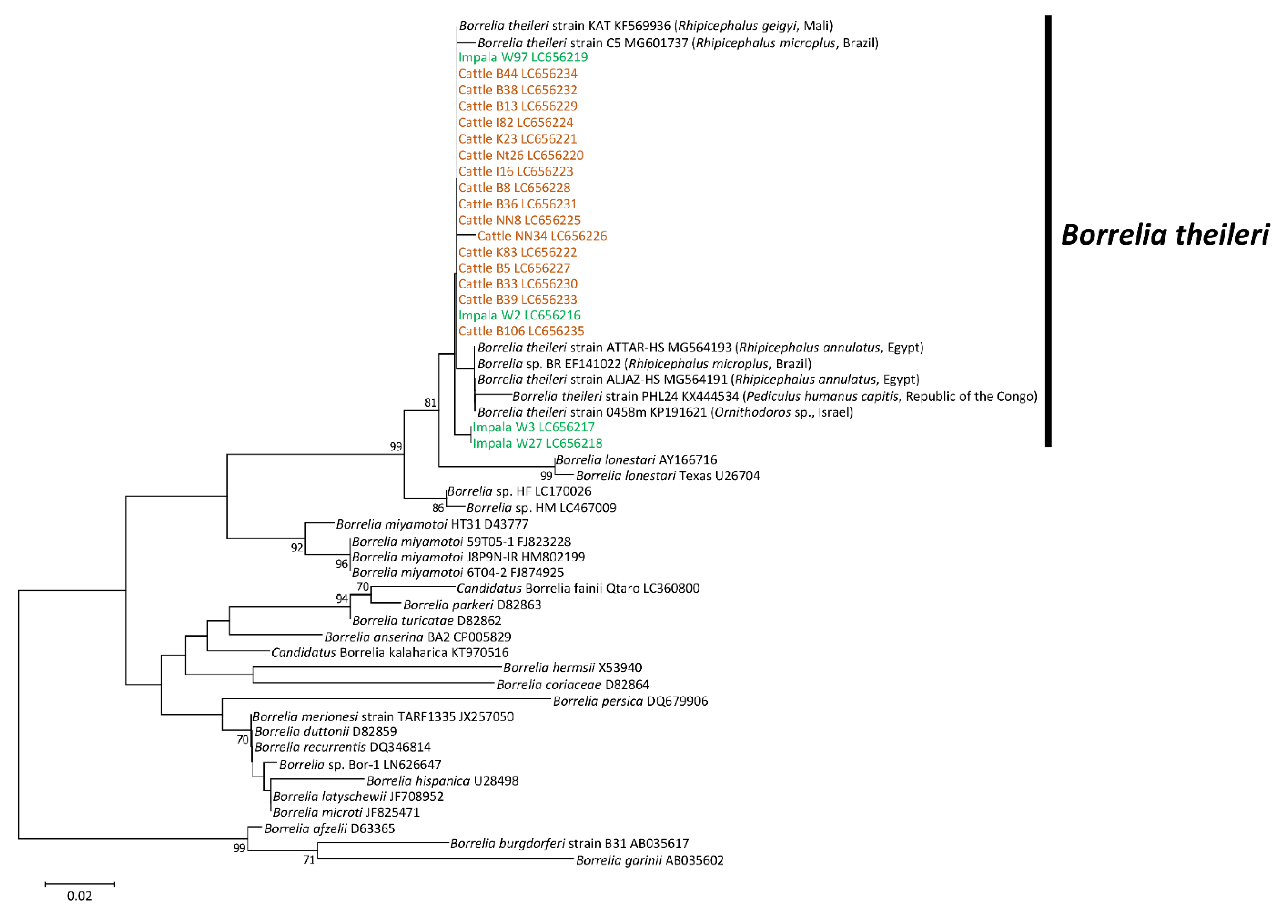
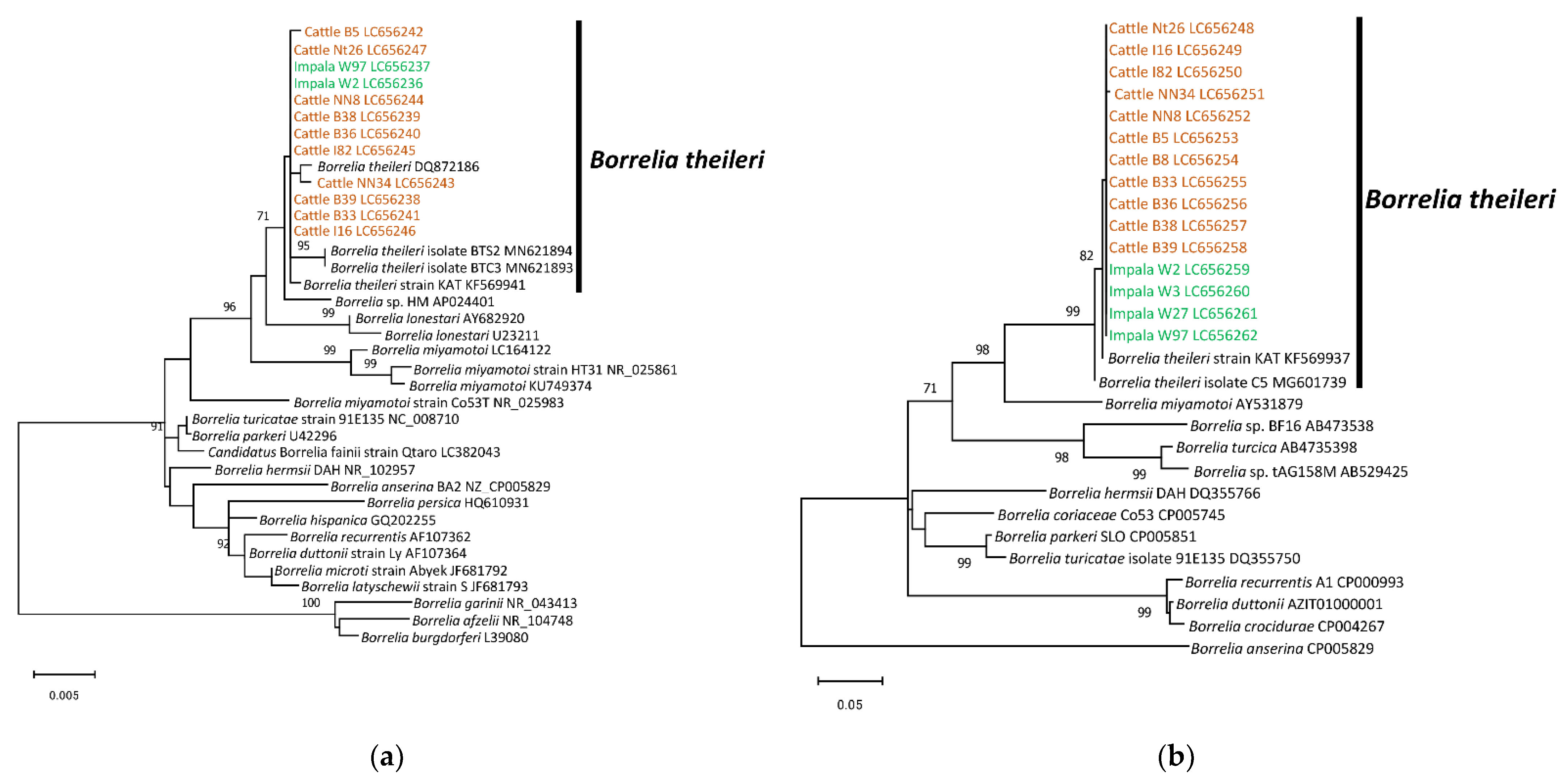
3.3. Phylogenetic Analysis

Phylogenetic trees were constructed to obtain information on the genetic association of our detected Borrelia spp. with other Borrelia species in the database. Based on the phylogenetic inference of the flaB gene, our detected Borrelia spp. from impalas and cattle were located within the clade of Borrelia theileri (Figure 1).
Similarly, the detected Borrelia spp. from impalas and cattle were positioned within the clade of Borrelia theileri in the phylogenetic trees based on the almost full-length 16S rDNA and the partial sequence of hpt (Figure 2).

3.4. Statistical Analysis

The mean PCV values of Borrelia infected and non-infected cattle were 32.1 (standard deviation (SD): 6.16, 95% confidence interval (CI): 29.22–34.98) and 31.9 (SD: 5.81, 95% CI: 31.40–32.46), respectively. There was no significant difference in PCV between infected and non-infected cattle (p = 0.74).

4. Discussion

In this study, we investigated the presence of Borrelia spirochetes in four different wild animal species and domestic cattle in the Kafue ecosystem in Zambia using molecular methods and successfully identified Borrelia theileri in impalas and cattle. To the best of our knowledge, this study provides the first evidence of Borrelia spirochetes in impalas in Africa and the first report of Borrelia theileri in cattle in Zambia.
Borrelia theileri is the causative agent of bovine borreliosis, first reported in cattle in South Africa in 1903 [21]. Zambia is a landlocked country located in south central Africa and shares its borders with Zimbabwe, Namibia, Botswana, Mozambique, Malawi, Tanzania, Democratic Republic of Congo, and Angola. Among these surrounding countries, only Botswana has reported cases of bovine borreliosis [41]. However, the previous report did not have any molecular information on the detected Borrelia theileri. The present study provides molecular evidence for a new geographical record of Borrelia theileri in Zambia. Furthermore, considering that cross-border trade is common in cattle and that wildlife migration occurs between Zambia and its neighbors, Borrelia theileri might have spread to the surrounding countries through infected animals.
A previous study in North Cameroon, using PCR, found that 17.9% (225/1260) of cattle were infected with Borrelia spp. [42]. However, out of 225 positive cattle, only 42 were confirmed to be infected with Borrelia theileri by sequence analysis. Thus, the infection rate of Borrelia theileri was 3.3% (42/1260) in the cattle population in North Cameroon. The present study also showed a similar infection rate (4.1%) in cattle to a previous study in North Cameroon. In addition, the present study revealed that 5.1% of the impalas in the Kafue ecosystem were infected with Borrelia theileri. However, there have been no studies on the infection rate of Borrelia theileri in large and middle-sized wildlife. Nevertheless, previous studies in Japan showed that 10.6% (25/235) and 25.9% (165/638) of wild sika deer were infected with a Borrelia sp., similar to Borrelia lonestari, which is a closely related species to Borrelia theileri [27,43].
Zambia is an endemic region of the East Coast Fever (ECF), a fatal disease in cattle caused by the blood parasite Theileria parva [44] and Redwater or bovine babesiosis caused by Babesia bigemina and Babesia bovis [45]. Another severe cattle disease, African animal trypanosomiasis, caused by Trypanosoma spp., is also endemic to Zambia [42]. Animals infected with these diseases have common clinical symptoms, such as fever, anemia, jaundice, hemoglobinuria, and swollen lymph nodes [44,45,46], and these diseases often result in death. Therefore, these diseases are of great veterinary and economic importance, as they have a considerable effect on livestock production [44]. In contrast, bovine borreliosis shows mild but similar symptoms, such as fever, lethargy, and anemia [42]. In this study, Borrelia infection was not related to anemia, statistically. Nevertheless, these symptoms can affect cattle performance and reduce their production. Furthermore, the livelihoods of an estimated 700 million rural African poor are supported or maintained by livestock ownership [47]. In addition to its intrinsic value, livestock provide a flow of food and income over time, helping to increase farm productivity; for most rural communities, livestock production is their only available livelihood [47]. Therefore, recognizing the existence of Borrelia theileri in Zambia was helpful in the clinical differentiation between bovine borreliosis and other bovine diseases for better treatment and control measures, which would ensure that farmers bring healthier and more valuable products to the market.
Human activities and associated land use changes have caused an increase in proximity between domestic and wild animals in many places, resulting in the mutual exchange of pathogens [20,48]. As is often the case near national parks in Zambia, domestic and wild animals often share a grazing place. In this study, we detected Borrelia theileri, which had sequences identical to those of cattle and impalas. Thus, the same strain of Borrelia theileri might be circulating between cattle and impalas in the Kafue ecosystem. Furthermore, Espinaze et al. (2018) revealed that large and middle-sized animals were highly connected by the tick species they shared, facilitating cross-infection with ticks and the transmission of tick-borne pathogens, including Borrelia theileri [49]. In particular, domestic animals can play an important role in accelerating the spread of ticks and tick-borne pathogens in Southern African animal communities [49], which means that these ticks and tick-borne pathogens could be introduced into the Kafue ecosystem. Therefore, although the pathogenicity of Borrelia theileri in wild animals is not known, limiting contact between cattle and wild animals might be a good conservation measure for reducing tick-borne diseases in the Kafue ecosystem. Furthermore, investigations of vector tick species for Borrelia theileri in the Kafue ecosystem are required for understanding their natural life cycle in the ecosystem.
In this study, we revealed the presence of Borrelia theileri, a causative agent of bovine borreliosis in wild impalas and domestic cattle in the Kafue ecosystem in Zambia. Our findings indicate the circulation of spirochetes between impalas and cattle. Furthermore, bovine borreliosis has veterinary and economic implications in livestock, even though the clinical presentation is not as severe as in other bovine diseases. As the study was conducted with samples collected only from the Kafue ecosystem, further investigations are required to clarify comprehensive Borrelia theileri status in the country by expanding the sampling area and including additional wild animal and livestock species, especially ungulates.

Supplementary Materials

The following are available online at https://0-www-mdpi-com.brum.beds.ac.uk/article/10.3390/microorganisms9112405/s1, Table S1: Accession numbers of the DNA sequences obtained in this study.

Author Contributions

Conceptualization, Y.Q. and H.K.; methodology, Y.Q. and A.O.; formal analysis, Y.Q. and A.C.C.L.; investigation, Y.Q.; resources, D.S., Y.N., L.C.M., N.K., K.H., J.Y. and B.N.; writing—original draft preparation, Y.Q.; writing—review and editing, all authors; supervision, Y.Q. and H.K.; funding acquisition, Y.Q., R.N., H.S. and H.K. All authors have read and agreed to the published version of the manuscript.

Funding

This research was funded by the Ministry of Education, Culture, Sports, Science and Technology, Japan (MEXT)/Japan Society for the Promotion of Science (JSPS), KAKENHI grant numbers 19K15992 and 20KK0151, and by the Japan Agency for Medical Research and Development (AMED), the Japan Program for Infectious Diseases Research and Infrastructure grant numbers JP21wm0125008 and JP21wm0225016. The funders had no role in the study design, data collection and analysis, decision to publish, or preparation of the manuscript.

Institutional Review Board Statement

In this study, DNA samples from wildlife and cattle, which were prepared in the previous studies, were used. Collecting the blood from animals in the previous studies was approved from the Excellence in Research Ethics and Science Converge in Zambia (Ref. No. 2019-Jul-010 for wildlife and Ref. No. 2019-Feb-081 for cattle).

Informed Consent Statement

Not applicable.

Data Availability Statement

All relevant data are provided in the manuscript.

Acknowledgments

We would like to thank the staff members and students at the International Institute for Zoonosis Control, Hokkaido University, Japan, staff of the Department of National Parks and Wildlife, Ministry of Tourism and Arts, Zambia, and technicians at the School of Veterinary Medicine, University of Zambia for technical assistance.

Conflicts of Interest

The authors declare no conflict of interest.

References

  1. Takano, A.; Goka, K.; Une, Y.; Shimada, Y.; Fujita, H.; Shiino, T.; Watanabe, H.; Kawabata, H. Isolation and characterization of a novel Borrelia group of tick-borne borreliae from imported reptiles and their associated ticks. Environ. Microbiol. 2010, 12, 134–146. [Google Scholar] [CrossRef] [PubMed]
  2. Franke, J.; Hildebrandt, A.; Dorn, W. Exploring gaps in our knowledge on Lyme borreliosis spirochaetes—Updates on complex heterogeneity, ecology, and pathogenicity. Ticks Tick Borne Dis. 2013, 4, 11–25. [Google Scholar] [CrossRef] [PubMed]
  3. Trevisan, G.; Cinco, M.; Trevisini, S.; Di Meo, N.; Ruscio, M.; Forgione, P.; Bonin, S. Borreliae Part 2: Borrelia Relapsing Fever Group and Unclassified Borrelia. Biology 2021, 10, 1117. [Google Scholar] [CrossRef]
  4. Trape, J.-F.; Diatta, G.; Arnathau, C.; Bitam, I.; Sarih, M.H.; Belghyti, D.; Bouattour, A.; Elguero, E.; Vial, L.; Mané, Y.; et al. The Epidemiology and Geographic Distribution of Relapsing Fever Borreliosis in West and North Africa, with a Review of the Ornithodoros erraticus Complex (Acari: Ixodida). PLoS ONE 2013, 8, e78473. [Google Scholar] [CrossRef]
  5. Smith, R.D.; Brener, J.; Osorno, M.; Ristic, M. Pathobiology of Borrelia theileri in the tropical cattle tick, Boophilus microplus. J. Invertebr. Pathol. 1978, 32, 182–190. [Google Scholar] [CrossRef]
  6. Fukunaga, M.; Takahashi, Y.; Tsuruta, Y.; Matsushita, O.; Ralph, D.; McClelland, M.; Nakao, M. Genetic and Phenotypic Analysis of Borrelia miyamotoi sp. nov., Isolated from the Ixodid Tick Ixodes persulcatus, the Vector for Lyme Disease in Japan. Int. J. Syst. Evol. Microbiol. 1995, 45, 804–810. [Google Scholar] [CrossRef] [Green Version]
  7. Barbour, A.G.; Maupin, G.O.; Teltow, G.J.; Carter, C.J.; Piesman, J. Identification of an uncultivable Borrelia species in the hard tick Amblyomma americanum: Possible agent of a Lyme disease-like illness. J. Infect. Dis. 1996, 173, 403–409. [Google Scholar] [CrossRef] [Green Version]
  8. Scoles, G.A.; Papero, M.; Beati, L.; Fish, D. A relapsing fever group spirochete transmitted by Ixodes scapularis ticks. Vector Borne Zoonotic Dis. 2001, 1, 21–34. [Google Scholar] [CrossRef]
  9. Brouqui, P. Arthropod-Borne Diseases Associated with Political and Social Disorder. Annu. Rev. Entomol. 2011, 56, 357–374. [Google Scholar] [CrossRef]
  10. McNeil, E.; Hinshaw, W.R.; Kissling, R.E. A study of Borrelia anserina infection (spirocheteosis) in Turkeys. J. Bacteriol. 1949, 57, 191–206. [Google Scholar] [CrossRef] [Green Version]
  11. Margos, G.; Fingerle, V.; Cutler, S.; Gofton, A.; Stevenson, B.; Estrada-Peña, A. Controversies in bacterial taxonomy: The example of the genus Borrelia. Ticks Tick Borne Dis. 2020, 11, 101335. [Google Scholar] [CrossRef] [PubMed]
  12. James, A.M.; Liveris, D.; Wormser, G.P.; Schwartz, I.; Montecalvo, M.A.; Johnson, B.J. Borrelia lonestari Infection after a Bite by an Amblyomma Americanum Tick. J. Infect. Dis. 2001, 183, 1810–1814. [Google Scholar] [CrossRef] [PubMed] [Green Version]
  13. Stromdahl, E.Y.; Williamson, P.C.; Kollars, T.M., Jr.; Evans, S.R.; Barry, R.K.; Vince, M.A.; Dobbs, N.A. Evidence of Borrelia lonestari DNA in Amblyomma americanum (Acari: Ixodidae) removed from humans. J. Clin. Microbiol. 2003, 41, 5557–5562. [Google Scholar] [CrossRef] [Green Version]
  14. Feder, H.M., Jr.; Hoss, D.M.; Zemel, L.; Telford, S.R., 3rd; Dias, F.; Wormser, G.P. Southern Tick-Associated Rash Illness (STARI) in the North: STARI following a tick bite in Long Island, New York. Clin. Infect. Dis. 2011, 53, e142–e146. [Google Scholar] [CrossRef] [Green Version]
  15. Burkot, T.R.; Mullen, G.R.; Anderson, R.; Schneider, B.S.; Happ, C.M.; Zeidner, N.S. Borrelia lonestari DNA in Adult Amblyomma americanum Ticks, Alabama. Emerg. Infect. Dis. 2001, 7, 471–473. [Google Scholar] [CrossRef]
  16. Moore, V.A.t.; Varela, A.S.; Yabsley, M.J.; Davidson, W.R.; Little, S.E. Detection of Borrelia lonestari, putative agent of southern tick-associated rash illness, in white-tailed deer (Odocoileus virginianus) from the southeastern United States. J. Clin. Microbiol. 2003, 41, 424–427. [Google Scholar] [CrossRef] [PubMed] [Green Version]
  17. Moyer, P.L.; Varela, A.S.; Luttrell, M.P.; Moore, V.A.t.; Stallknecht, D.E.; Little, S.E. White-tailed deer (Odocoileus virginianus) develop spirochetemia following experimental infection with Borrelia lonestari. Vet. Microbiol. 2006, 115, 229–236. [Google Scholar] [CrossRef]
  18. Platonov, A.E.; Karan, L.S.; Kolyasnikova, N.M.; Makhneva, N.A.; Toporkova, M.G.; Maleev, V.V.; Fish, D.; Krause, P.J. Humans Infected with Relapsing Fever Spirochete Borrelia miyamotoi, Russia. Emerg. Infect. Dis. 2011, 17, 1816–1823. [Google Scholar] [CrossRef]
  19. Krause, P.J.; Fish, D.; Narasimhan, S.; Barbour, A.G. Borrelia miyamotoi infection in nature and in humans. Clin. Microbiol. Infect. 2015, 21, 631–639. [Google Scholar] [CrossRef] [Green Version]
  20. Han, S.; Hickling, G.J.; Tsao, J.I. High Prevalence of Borrelia miyamotoi among Adult Blacklegged Ticks from White-Tailed Deer. Emerg. Infect. Dis. 2016, 22, 316–318. [Google Scholar] [CrossRef]
  21. Laveran, A. Sur la spirillose des bovidés. CR Acad. Sci. Paris 1903, 136, 939–941. [Google Scholar]
  22. Theiler, A. Spirillosis of cattle. J. Comp. Path 1904, 17, 47–55. [Google Scholar] [CrossRef]
  23. Callow, L.L. Observations on tick-transmitted spirochaetes of cattle in Australia and South Africa. Br. Vet. J. 1967, 123, 492–497. [Google Scholar] [CrossRef]
  24. Uilenberg, G.; Hinaidy, H.K.; Perie, N.M.; Feenstra, T. Borrelia infections of ruminants in Europe. Vet. Q. 1988, 10, 63–67. [Google Scholar] [CrossRef] [PubMed]
  25. Cutler, S.; Abdissa, A.; Adamu, H.; Tolosa, T.; Gashaw, A. Borrelia in Ethiopian ticks. Ticks Tick Borne Dis. 2012, 3, 14–17. [Google Scholar] [CrossRef]
  26. Amanzougaghene, N.; Akiana, J.; Mongo Ndombe, G.; Davoust, B.; Nsana, N.S.; Parra, H.-J.; Fenollar, F.; Raoult, D.; Mediannikov, O. Head Lice of Pygmies Reveal the Presence of Relapsing Fever Borreliae in the Republic of Congo. PLoS Negl. Trop. Dis. 2016, 10, e0005142. [Google Scholar] [CrossRef]
  27. Kumagai, Y.; Sato, K.; Taylor, K.R.; Zamoto-Niikura, A.; Imaoka, K.; Morikawa, S.; Ohnishi, M.; Kawabata, H. A relapsing fever group Borrelia sp. is widely distributed among wild deer in Japan. Ticks Tick Borne Dis. 2018, 9, 465–470. [Google Scholar] [CrossRef]
  28. Kirchgessner, M.S.; Freer, H.; Whipps, C.M.; Wagner, B. Detection of Borrelia burgdorferi outer surface protein antibodies in wild white-tailed deer (Odocoileus virginianus) in New York and Pennsylvania, USA. Vet. Immunol. Immunopathol. 2013, 153, 165–169. [Google Scholar] [CrossRef] [PubMed]
  29. Stöbel, K.; Schönberg, A.; Staak, C. A new non-species dependent ELISA for detection of antibodies to Borrelia burgdorferi s. l. in zoo animals. Int. J. Med. Microbiol. 2002, 291, 88–99. [Google Scholar] [CrossRef]
  30. Stoebel, K.; Schoenberg, A.; Streich, W.J. The seroepidemiology of Lyme borreliosis in zoo animals in Germany. Epidemiol. Infect. 2003, 131, 975–983. [Google Scholar] [CrossRef]
  31. Širmarová, J.; Tichá, L.; Golovchenko, M.; Salát, J.; Grubhoffer, L.; Rudenko, N.; Nowotny, N.; Růžek, D. Seroprevalence of Borrelia burgdorferi sensu lato and tick-borne encephalitis virus in zoo animal species in the Czech Republic. Ticks Tick Borne Dis. 2014, 5, 523–527. [Google Scholar] [CrossRef]
  32. Diatta, G.; Duplantier, J.M.; Granjon, L.; Ba, K.; Chauvancy, G.; Ndiaye, M.; Trape, J.F. Borrelia infection in small mammals in West Africa and its relationship with tick occurrence inside burrows. Acta Trop. 2015, 152, 131–140. [Google Scholar] [CrossRef]
  33. Qiu, Y.; Nakao, R.; Hang’ombe, B.M.; Sato, K.; Kajihara, M.; Kanchela, S.; Changula, K.; Eto, Y.; Ndebe, J.; Sasaki, M.; et al. Human Borreliosis Caused by a New World Relapsing Fever Borrelia-like Organism in the Old World. Clin. Infect. Dis. 2019, 69, 107–112. [Google Scholar] [CrossRef] [PubMed]
  34. Squarre, D.; Nakamura, Y.; Hayashida, K.; Kawai, N.; Chambaro, H.; Namangala, B.; Sugimoto, C.; Yamagishi, J. Investigation of the piroplasm diversity circulating in wildlife and cattle of the greater Kafue ecosystem, Zambia. Parasit. Vectors 2020, 13, 599. [Google Scholar] [CrossRef] [PubMed]
  35. Nakamura, Y.; Hayashida, K.; Delesalle, V.; Qiu, Y.; Omori, R.; Simuunza, M.; Sugimoto, C.; Namangala, B.; Yamagishi, J. Genetic Diversity of African Trypanosomes in Tsetse Flies and Cattle from the Kafue Ecosystem. Front. Vet. Sci. 2021, 8, 599815. [Google Scholar] [CrossRef] [PubMed]
  36. Parola, P.; Ryelandt, J.; Mangold, A.J.; Mediannikov, O.; Guglielmone, A.A.; Raoult, D. Relapsing fever Borrelia in Ornithodoros ticks from Bolivia. Ann. Trop. Med. Parasitol. 2011, 105, 407–411. [Google Scholar] [CrossRef] [PubMed] [Green Version]
  37. Takano, A.; Fujita, H.; Kadosaka, T.; Konnai, S.; Tajima, T.; Watanabe, H.; Ohnishi, M.; Kawabata, H. Characterization of reptile-associated Borrelia sp. in the vector tick, Amblyomma geoemydae, and its association with Lyme disease and relapsing fever Borrelia spp. Environ. Microbiol. Rep. 2011, 3, 632–637. [Google Scholar] [CrossRef] [PubMed]
  38. McCoy, B.N.; Maiga, O.; Schwan, T.G. Detection of Borrelia theileri in Rhipicephalus geigyi from Mali. Ticks Tick Borne Dis. 2014, 5, 401–403. [Google Scholar] [CrossRef] [Green Version]
  39. Roux, V.; Raoult, D. Body Lice as Tools for Diagnosis and Surveillance of Reemerging Diseases. J. Clin. Microbiol. 1999, 37, 596–599. [Google Scholar] [CrossRef] [Green Version]
  40. Kumar, S.; Stecher, G.; Li, M.; Knyaz, C.; Tamura, K. MEGA X: Molecular Evolutionary Genetics Analysis across Computing Platforms. Mol. Biol. Evol. 2018, 35, 1547–1549. [Google Scholar] [CrossRef]
  41. Sharma, S.P.; Amanfu, W.; Losho, T.C. Bovine borreliosis in Botswana. Onderstepoort. J. Vet. Res. 2000, 67, 221–223. [Google Scholar]
  42. Abanda, B.; Paguem, A.; Abdoulmoumini, M.; Kingsley, M.T.; Renz, A.; Eisenbarth, A. Molecular identification and prevalence of tick-borne pathogens in zebu and taurine cattle in North Cameroon. Parasit. Vectors 2019, 12, 448. [Google Scholar] [CrossRef] [PubMed]
  43. Lee, K.; Takano, A.; Taylor, K.; Sashika, M.; Shimozuru, M.; Konnai, S.; Kawabata, H.; Tsubota, T. A relapsing fever group Borrelia sp. similar to Borrelia lonestari found among wild sika deer (Cervus nippon yesoensis) and Haemaphysalis spp. ticks in Hokkaido, Japan. Ticks Tick Borne Dis. 2014, 5, 841–847. [Google Scholar] [CrossRef] [PubMed]
  44. Nambota, A.; Samui, K.; Sugimoto, C.; Kakuta, T.; Onuma, M. Theileriosis in Zambia: Etiology, epidemiology and control measures. Jpn. J. Vet. Res. 1994, 42, 1–18. [Google Scholar]
  45. Simuunza, M.; Weir, W.; Courcier, E.; Tait, A.; Shiels, B. Epidemiological analysis of tick-borne diseases in Zambia. Vet. Parasitol. 2011, 175, 331–342. [Google Scholar] [CrossRef]
  46. Laohasinnarong, D.; Goto, Y.; Asada, M.; Nakao, R.; Hayashida, K.; Kajino, K.; Kawazu, S.-I.; Sugimoto, C.; Inoue, N.; Namangala, B. Studies of trypanosomiasis in the Luangwa valley, north-eastern Zambia. Parasit. Vectors 2015, 8, 1–8. [Google Scholar] [CrossRef] [Green Version]
  47. De Leeuw, P.N.; McDermott, J.J.; Lebbie, S.H.B. Monitoring of livestock health and production in sub-Saharan Africa. Prev. Vet. Med. 1995, 25, 195–212. [Google Scholar] [CrossRef]
  48. Hassell, J.M.; Begon, M.; Ward, M.J.; Fèvre, E.M. Urbanization and Disease Emergence: Dynamics at the Wildlife–Livestock–Human Interface. Trends Ecol. Evol. 2017, 32, 55–67. [Google Scholar] [CrossRef] [Green Version]
  49. Espinaze, M.P.A.; Hellard, E.; Horak, I.G.; Cumming, G.S. Domestic mammals facilitate tick-borne pathogen transmission networks in South African wildlife. Biol. Conserv. 2018, 221, 228–236. [Google Scholar] [CrossRef] [Green Version]
Figure 1. Phylogenetic inference of Borrelia spp. based on partial sequence of flaB. The accession numbers for nucleotide sequences are shown after the species names. The analysis was performed using the neighbor-joining method. Bootstrap values > 70% based on 1000 replications are presented on the interior branch nodes.
Microorganisms 09 02405 g001
Figure 2. Phylogenetic trees of Borrelia spp. based on the sequences of 16S rDNA and hpt: (a) 16S rDNA; (b) hpt. The accession numbers for nucleotide sequences are shown after the species names. The analyses were performed using the maximum likelihood method. Bootstrap values > 70% based on 1000 replications are presented on the interior branch nodes.
Microorganisms 09 02405 g002
Table 1. Primers used in the conventional PCRs.
Target GenePrimer NameSequence (5′–3′)Annealing TemperatureExpected SizeReference
Flagellin (P41)BflaPAD 1GATCARGCWCAAYATAACCAWATGCA50 °C453 bp[37]
BflaPDU 1AGATTCAAGTCTGTTTTGGAAAGC
BflaPBU 2GCTGAAGAGCTTGGAATGCAACC50 °C347 bp[37]
BflaPCR 2TGATCAGTTATCATTCTAATAGCA
hypoxanthine-guanine
phosphoribosyltransferase
hptdegFGCAGAYATTACAAGAGARATGG55 °C433 bp[38]
hptdegRCYTCRTCACCCCATTGAGTTCC
16S ribosomal DNABF1GCTGGCAGTGCGTCTTAAGC55 °C1371 bp[39]
BR1GCTTCGGGTATCCTCAACTC
1 Primer for first PCR. 2 Primer for nested PCR.
Table 2. Results of each PCR assay.
SpeciesReal-Time PCRflaB16S rDNAhpt
Impala5/97 (5.1%)4/52/54/5
Hartebeest0/37 (0%)NANANA
Lion0/4 (0%)NANANA
Wild dog0/2 (0%)NANANA
Cattle20/488 (4.1%)16/2010/2011/20
NA: Not applicable.
Publisher’s Note: MDPI stays neutral with regard to jurisdictional claims in published maps and institutional affiliations.

