1. Introduction
Cardenolides, a chemical class within the cardiac glycosides, have a five-membered lactone group in the β position at C17 [
1]. The mechanisms of these compounds are known to inhibit Na
+/K
+-ATPase, activate the cation pump, and increase in intracellular calcium concentration through cellular output of Na
+ and intake of K
+ [
2]. Because of these biological actions, cardenolides have been used in the treatment of heart failure [
3]. In addition, many researchers have suggested that cardenolides may inhibit the growth of cancer cells, and have described them as anticancer agents with fewer side effects [
4,
5].
Cardiac glycosides were isolated from several plant families of Ranunculaceae, Scrophulariazea, Apocynaceae, and Liliaceae, along with pregnane glycosides [
6]. In Korea, the
Adonis family is mainly comprised of three species,
A. amurensis,
A. pseudoamurensis, and
A. multiflora based on RAPD analysis [
7,
8]. Previous phytochemical studies conducted on the roots of
A. amurensis, the most well-known
Adonis species, have identified several cardenolides: corchoroside A, covallatoxin, cymarin, cymarol, digitoxigenin 3-
O-β-
d-cymaroside, k-strophanthin, and k-strophanthin-β [
9]. However, little has been reported concerning the biological and phytochemical properties of
A. multiflora, except a brief report [
10]. We have confirmed the presence of cardenolide spots in the TLC of ethanolic extracts from whole plants of
A. multiflora based on the UV absorption pattern and the colors produced by spraying with a 10% H
2SO
4 solution and heating. Over the course of investigating cardenolides in whole plants of
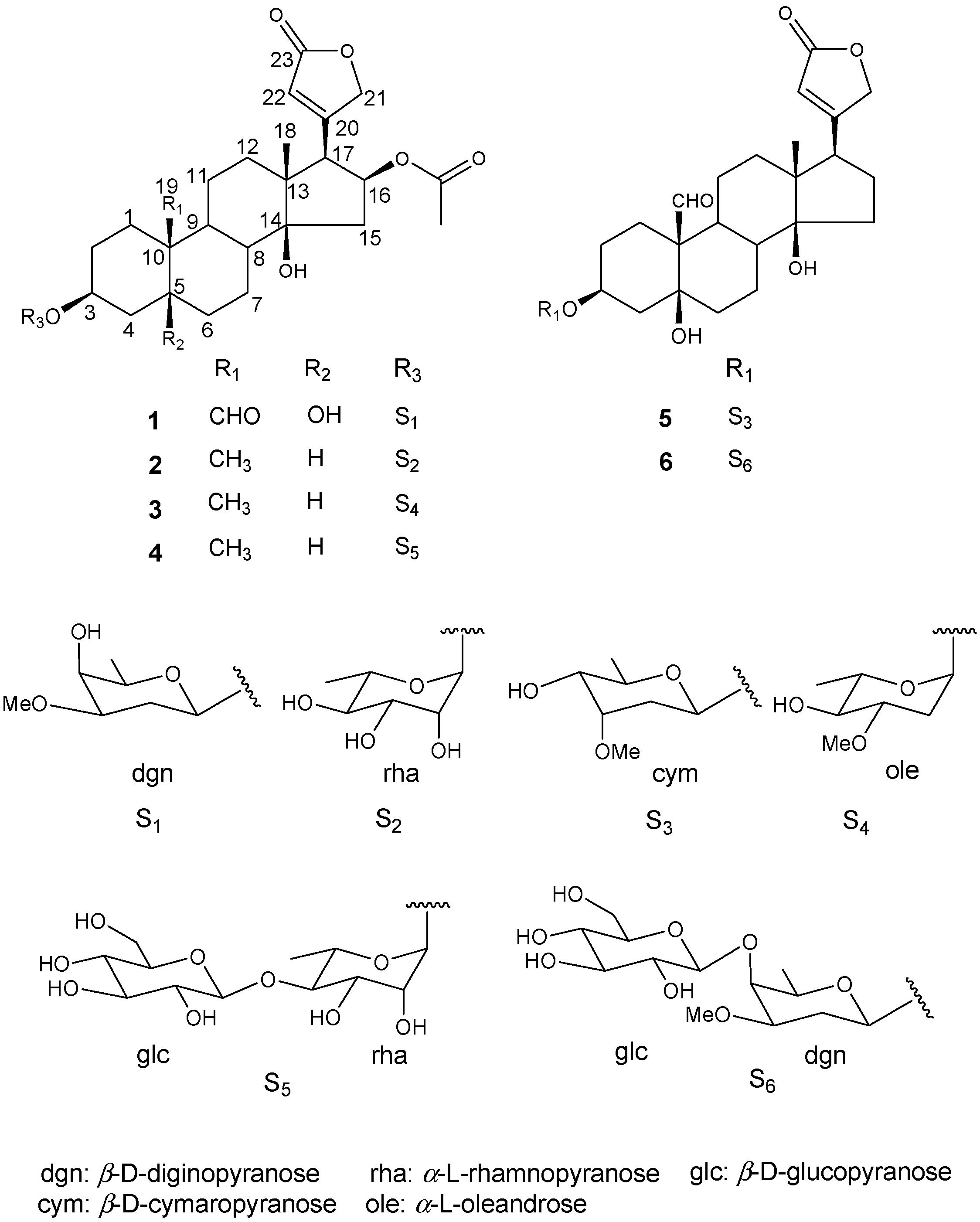
A. multiflora Nishikawa & Koki Ito, two new cardenolides
1 and
6 were identified and structurally determined, along with four known ones
2–
5 (
Figure 1). The cardenolides were then evaluated for cytotoxity against six human cancer cell lines (HCT-116, HepG2, HeLa, SK-OV-3, SK-BR-3, and SK-MEL-5).
Figure 1.
Compounds 1–6 isolated from the whole plants of Adonis multiflora.
Figure 1.
Compounds 1–6 isolated from the whole plants of Adonis multiflora.
2. Results and Discussion
The EtOH extracts were partitioned into CH
2Cl
2, EtOAc, BuOH, and H
2O fractions. Repeated SiO
2 and ODS column chromatography of the CH
2Cl
2 and BuOH fractions resulted in the identification of two new cardenolides, named adonioside A (
1) and adonioside B (
6), along with four known cardenolides
2–
5. The known compounds were identified as tupichinolide (
2), oleandrine (
3), cryptostigmin II (
4), and cymarin (
5) on the basis of spectroscopic analysis and the identities were confirmed by comparing their measured spectroscopic data with those reported in the literature [
11,
12,
13,
14].
Compound
1 was isolated as a white powder and showed IR absorbance bands representing OH (3384 cm
−1), CHO (1737 cm
−1), and C=C (1639 cm
−1) groups. The molecular weight was determined to be 606 from the molecular ion peak
m/z 605 [M − H]
− in the negative FAB-MS spectrum, and a molecular formula of C
32H
46O
11 was determined from the high-resolved molecular ion peak ([M − H]
−,
m/
z 605.2971, calc. for C
32H
45O
11, 605.2962) in the negative HR-FAB-MS. The
1H-NMR spectrum (
Table 1) exhibited the characteristics of an α,β-unsaturated-γ-lactone ring, with signals at δ(H) 5.95 (dd,
J = 1.6, 1.6 Hz, H-22), 4.94 (dd,
J = 18.4, 1.6 Hz, H-21
a), and 4.83 (dd,
J = 18.4, 1.6 Hz, H-21
b) as well as a tertiary methyl signal at δ(H) 0.92 (s, H-18), a formyl signal at δ(H) 9.92 (s, H-19), two O-bearing CH signals at δ(H) 4.19 (br.s, H-3) and 5.41 (ddd,
J = 9.6, 8.4, 8.4 Hz, H-16), and an AcO signal at δ(H) 1.95 (s, H-AcO), which suggested the presence of a cardenolide moiety with two oxygenated methines and an AcO group. In addition, a hemiacetal signal at δ(H) 4.50 (dd,
J = 9.6, 2.4 Hz, H-1′), three O-bearing CH signals at δ(H) 3.31~3.67 (H-3′~5′), an O-bearing CH
3 signal at δ(H) 3.36 (s, H-CH
3O), a CH
2 signal at δ(H) 2.05 (m, H-2′
a) and 1.66 (m, H-2′
b), and a CH
3 signal at δ(H) 1.33 (d,
J = 6.8 Hz, H-6′), indicated that
1 was a cardiac monoglycoside with a β-diginopyranoside.
The
13C-NMR spectrum showed 32 C-atoms signals (
Table 1). The aglycone with α,β-unsaturated-γ-lactone ring signals observed at δ(C) 173.9 (C-23), 167.3 (C-20), 121.5 (C-22), and 75.5 (C-21), a formyl signal at δ(C) 208.1 (C-19), two O-bearing quaternary signals at δ(C) 73.5 (C-5) and 83.9 (C-14), AcO signals at δ(C) 170.4 (C-OAc) and 21.0 (C-OAc), two O-bearing CH signals at δ(C) 74.1 (C-3) and 73.7 (C-16), and a tertiary CH
3 signal at δ(C) 15.8 (C-18) indicated that the aglycone was a cardenolide with four hydroxyls, one formyl, and one AcO group. The monosaccharide carbon signals, including a hemiacetal signal at δ(C) 98.9 (C-1′), three O-bearing CH signals at δ(C) 77.6 (C-3′), 70.7 (C-5′), 66.9 C-4′), a CH
3O signal at δ(C) 55.8 (C-CH
3O), a CH
2 signal at δ(C) 31.5 (C-2′), and a CH
3 signal at δ(C) 16.7 (C-6′), allowed us to conclude that the sugar was β-diginopyranose.
Acid hydrolysis of
1 and purification of the hydrolysate using column chromatography resulted in a sugar compound, which was identified to be a diginopyranose by direct comparison between its
Rf values on the SiO
2 TLC (0.47 with CHCl
3/MeOH 9:1, and 0.19 with CH
2Cl
2/EtOH 9:1) and those of an authentic sample. The specific rotation value of the obtained sugar (
= +56.8,
c = 0.11, H
2O), and the large
J value of the anomeric signal at δ(H) 4.50 (dd,
J = 9.6, 2.4 Hz, H-1′) revealed the sugar to be β-
d-diginopyranose. The location of β-
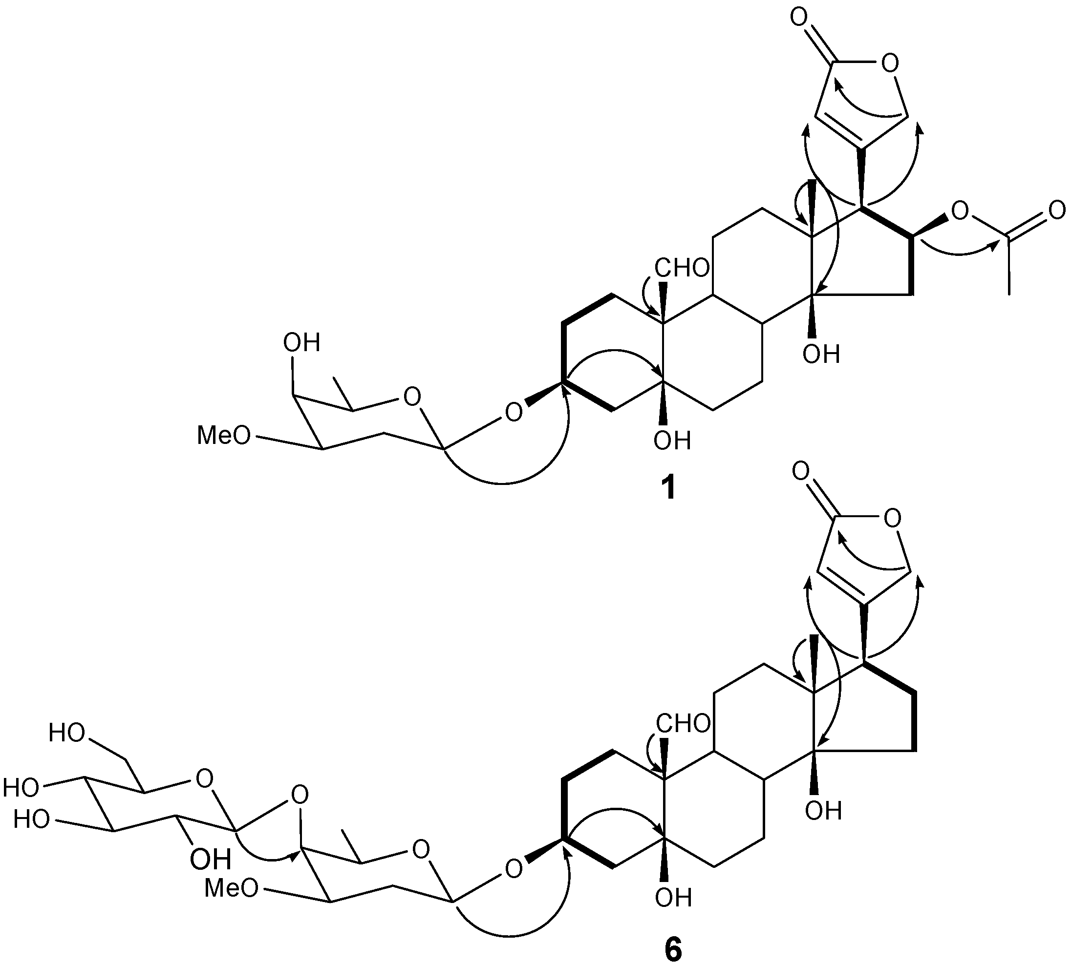
d-diginopyranose, methyl, formyl, hydroxyls, and AcO groups of
1 were determined from the connectivity between the oxygenated methine proton δ(H) 4.50 (1H, d,
J = 9.6, 2.4 Hz, H-1′) and O-bearing CH carbon δ(C) 74.1 (C-3), tertiary methyl proton δ(H) 0.92 (s, H-18) and quaternary carbon δ(C) 49.8 (C-13), formyl proton δ(H) 9.92 (s, H-19) and quaternary carbon δ(C) 54.3 (C-10), tertiary methyl proton δ(H) 0.92 (s, H-18) and O-bearing quaternary carbon δ(C) 83.9 (C-14) and O-bearing methine proton δ(H) 5.41 (ddd,
J = 9.6, 8.4, 8.4 Hz, H-16) and AcO carbon δ(C) 170.4 (C-OAc) in the HMBC spectrum, respectively. The location of the lactone group was deduced from the connectivity between the methylene protons H-15 δ(H) 2.59 (dd,
J = 15.6, 9.6 Hz, H-15), O-bearing methine proton δ(H) 5.41 (ddd,
J = 9.6, 8.4, 8.4 Hz, H-16) and the methine proton δ(H) 3.15 (d,
J = 8.4 Hz, H-17) in the COSY spectrum (
Figure 2). Taken together, compound
1 was determined to be a 16-β-acetoxystrophanthidin 3-
O-β-
d-digonopyranoside, a new cardenolide named adonioside A.
Compound
6 was also isolated as a white powder and showed IR absorbance bands of OH (3387 cm
−1), CHO (1742 cm
−1), and C=C (1647 cm
−1) groups. The molecular weight was determined to be 710 due to the pseudomolecular ion peak
m/z 733 [M + Na]
+ in the positive FAB-MS spectrum, and the molecular formula of C
36H
54O
14 was determined by the high-resolution pseudomolecular ion peak ([
M + Na]
+,
m/
z 733.3511, calc. for C
36H
54O
14Na, 733.3411) in the positive HR-FAB-MS. The
1H-NMR spectrum (
Table 1) displayed a formyl signal at δ(H) 10.33 (s, H-19), an olefin CH signal at δ(H) 6.10 (s, H-22), O-bearing CH
2 signals at δ(H) 5.25 (d,
J = 18.4 Hz, H-21
a) and 4.99 (d,
J = 18.4 Hz, H-21
b), O-bearing CH signal at δ(H) 4.64 (br.s, H-3), and a tertiary CH
3 at δ(H) 0.98 (s, H-18) indicating that
6 has a cardenolide skeleton. Also, two hemiacetal signals at δ(H) 5.07 (d,
J = 7.6 Hz, H-1′) and 4.69 (br.d,
J = 9.2 Hz, H-1′′) were observed, and their large
J values confirmed that the anomer hydroxyls were in β form. Two hexoses were determined to be β-diginopyranosyl-(1→4)-β-diginopyranose through comparisons between
13C-NMR data and those reported in previously published literature [
15]. Acid hydrolysis of
6 and comparison of the specific rotation values of two isolated sugars [
6a:
= +55.5 (
c = 0.12, H
2O),
6b:
= +49.3 (
c = 0.12, H
2O)] led to the identification of two sugars,
d-diginopyranose (
= +59.6) and
d-glucopyranose (
= +52.5) [
16,
17]. The locations of functional groups were determined by gCOSY and gHMBC experiments (
Figure 2). Thus, compound
6 was identified as strophanthidin 3-
O-β-
d-diginopyranosyl-(1→4)-β-
d-glucopyronoside, a new cardenolide named adonioside B.
Table 1.
1H- and 13C-NMR Data (400 and 100 MHz, resp.) of compounds 1 and 6.
Table 1.
1H- and 13C-NMR Data (400 and 100 MHz, resp.) of compounds 1 and 6.
| Position | 1 (CD3OD) | 6 (C5D5N) |
|---|
| | δ(H) | δ(C) | δ(H) | δ(C) |
| 1 | 2.05 (m) | 23.6 | 2.52 (m) | 18.6 |
| | 1.13 (m) | | 1.83 (m) | |
| 2 | 2.04 (m) | 25.1 | 2.06 (m) | 25.5 |
| | 1.42 (m) | | 1.64 (m) | |
| 3 | 4.19 (br.s) | 74.1 | 4.64 (br.s) | 74.5 |
| 4 | 1.94 (m) | 35.2 | 2.27 (m) | 37.0 |
| | 1.62 (m) | | 1.81 (m) | |
| 5 | | 73.5 | | 73.8 |
| 6 | 1.93 (m) | 35.9 | 2.17 (m) | 36.0 |
| | 1.57 (m) | | 1.76 (m) | |
| 7 | 1.51 (m) | 21.4 | 1.52 (m) | 24.8 |
| | 1.43 (m) | | 1.32 (m) | |
| 8 | 1.96 (m) | 41.4 | 2.26 (m) | 41.9 |
| 9 | 1.41 (m) | 39.0 | 1.64 (m) | 39.5 |
| 10 | | 54.3 | | 55.2 |
| 11 | 2.26 (br.dd, J = 14.8, 3.6) | 18.1 | 2.43 (m) | 22.6 |
| | 1.65 (m) | | 1.40 (m) | |
| 12 | 1.55 (m) | 39.1 | 1.39 (m) | 39.6 |
| | 1.23 (m) | | 1.28 (m) | |
| 13 | | 49.8 | | 49.8 |
| 14 | | 83.9 | | 84.4 |
| 15 | 2.59 (dd, J = 15.6, 9.6) | 40.0 | 2.02 (m) | 32.2 |
| | 1.74 (m) | | 1.79 (m) | |
| 16 | 5.41(ddd, J = 9.6, 5.8, 2.8) | 73.7 | 2.07 (m) | 27.2 |
| | | | 1.96 (m) | |
| 17 | 3.15 (d, 8.4) | 55.6 | 2.76 (m) | 51.1 |
| 18 | 0.92 (s) | 15.8 | 0.98 (s) | 16.0 |
| 19 | 9.92 (s) | 208.1 | 10.33 (s) | 208.4 |
| 20 | | 167.3 | | 175.6 |
| 21 | 4.94 (dd, J = 18.4, 1.6) | 75.5 | 5.25 (d, J = 18.4) | 73.7 |
| | 4.83 (dd, J = 18.4, 1.6) | | 4.99 (d, J = 18.4) | |
| 22 | 5.95 (dd, J = 1.6, 1.6) | 121.5 | 6.10 (s) | 117.8 |
| 23 | | 173.9 | | 174.4 |
| AcO | 1.93 (s) | 170.4, 21.0 | | |
| | Dgn (a) | | Dgn | D-Dig |
| 1′ | 4.50 (dd, J = 9.6, 2.4) | 98.9 | 5.07 (br.d, J = 9.6) | 99.5 |
| 2′ | 2.05 (m) | 31.5 | 2.15 (m) | 32.6 |
| | 1.66 (m) | | | |
| 3′ | 3.31 (ddd, J = 12.8, 5.2, 2.0) | 77.6 | 3.34 (br.dd, J = 12.0, 3.6) | 79.7 |
| 4′ | 3.67 (br.s) | 66.9 | 4.10 (br.s) | 73.9 |
| 5′ | 3.43 (br.q, J = 6.8) | 70.7 | 3.48 (br.q, J = 6.4) | 71.1 |
| 6′ | 1.33 (d, J = 6.8) | 16.7 | 1.49 (d, J = 6.4) | 17.8 |
| MeO | 3.36 (s) | 55.8 | 3.28 (s) | 56.1 |
| | | | Glc (b) | |
| 1′′ | | | 4.69 (d, J = 7.6) | 104.9 |
| 2′′ | | | 3.91 (dd, J = 8.8, 7.6) | 75.9 |
| 3′′ | | | 4.16 (dd, J = 8.8, 8.8) | 78.5 |
| 4′′ | | | 4.10 (dd, J = 8.8, 8.8) | 71.9 |
| 5′′ | | | 3.89 (m) | 78.3 |
| 6′′ | | | 4.51 (dd, J = 11.2, 1.6) | 63.1 |
| | | | 4.30 (dd, J = 11.2, 6.0) | |
Figure 2.
1H-1H-COSY (−) and gHMBC (H→C) key correlations of compounds 1 and 6.
Figure 2.
1H-1H-COSY (−) and gHMBC (H→C) key correlations of compounds 1 and 6.
All of the isolated cardenolides from
A. multiflora were evaluated for cytotoxicity against six human cancer cell lines (HCT-116, HepG2, HeLa, SK-OV-3, SK-BR-3, and SK-MEL-5). As shown in
Table 2, compounds
1,
2,
5, and
6 showed significant inhibition activity against HCT-116, SK-OV-3, and SK-MEL-5 cell lines with IC
50 values ranging from 0.06 ± 0.02 to 7.44 ± 1.98 µM. Compound
3 showed cytotoxic effects against the HeLa cell line with an IC
50 value of 8.85 ± 0.39 µM. Compound
4 showed cytotoxicity against the SK-MEL-5 cell line with an IC
50 value of 1.99 ± 0.28 µM.
Table 2.
Cytotoxic activity of compounds 1–6 against human cancer cell lines (IC50 [µM] (a)).
Table 2.
Cytotoxic activity of compounds 1–6 against human cancer cell lines (IC50 [µM] (a)).
| Compound | Cell lines (IC50) μM |
|---|
| HCT-116 | HepG2 | HeLa | SK-OV-3 | SK-MEL-5 | SK-BR-3 |
|---|
| 1 | 4.10 ± 0.38 | 14.65 ± 0.47 | 38.54 ± 1.08 | 2.34 ± 0.10 | 3.40 ± 0.67 | 38.35 ± 1.49 |
| 2 | 0.41 ± 0.13 | 17.99 ± 0.61 | 9.38 ± 0.15 | 0.06 ± 0.02 | 0.28 ± 0.06 | 2.58 ± 0.23 |
| 3 | 34.99 ± 1.39 | 30.12 ± 1.60 | 8.85 ± 0.39 | 25.38 ± 0.51 | 34.17 ± 1.78 | 80.38 ± 1.13 |
| 4 | 24.32 ± 1.26 | 26.61 ± 0.70 | 23.27 ± 1.73 | 41.02 ± 0.13 | 1.99 ± 0.28 | 23.94 ± 1.47 |
| 5 | 1.64 ± 0.13 | 2.87 ± 0.77 | 25.38 ± 0.15 | 0.76 ± 0.15 | 0.73 ± 0.14 | 5.10 ± 0.87 |
| 6 | 7.44 ± 1.98 | 13.71 ± 0.75 | 44.71 ± 0.89 | 4.63 ± 0.47 | 4.98 ± 0.56 | 21.30 ± 1.50 |
| Doxorubicin | 9.20 ±0.90 | 27.30 ± 0.50 | 3.20 ± 0.30 | 0.58 ± 0.08 | 4.80 ± 0.23 | 0.71 ± 0.05 |
3. Experimental Section
3.1. General
Column chromatography (CC): SiO2 (Kieselgel 60, Merck, Darmastdt, Germany) and ODS (LiChroprep RP-18, Merck) resins. TLC: Kieselgel 60 F254 and RP-18 F254S (Merck) plates; visualization with UV lamp Spectroline Model ENF-240 C/F (Spectronics Corporation, Westbury, NY, USA) and spraying 10% H2SO4 soln. in MeOH and heating. Optical rotations: JASCO P-1010 digital polarimeter (Jasco, Tokyo, Japan). IR spectra: Perkin Elmer Spectrum One FT-IR spectrometer (Perkin Elmer, Beaconsfield, UK). FAB-MS: JEOL JMSAX-700 mass spectrometer (Jeol, Tokyo, Japan). NMR spectra: Varian Unity Inova AS-400 FT-NMR spectrometer (Varian, Palo Alto, CA, USA).
3.2. Plant Materials
A. multiflora Nishikawa & Koki Ito was supplied from the BMI Corporation (Uiwang, Korea) in January 2014, and was identified by professor Dae-Keun Kim, College of Pharmacy, Woosuk University, Jeonju, Korea. A voucher specimen (KHU2014-0117) has been reserved at the Laboratory of Natural Products Chemistry, Kyung Hee University, Yongin, Korea.
3.3. Extraction and Isolation
The whole plants of A. multiflora (1.5 kg) were extracted with 70% aqueous EtOH (30 L) at room temperature for 24 h. The concentrated EtOH extracts (106 g) were suspended in H2O (3 L) and then successively extracted with CH2Cl2 (AAC; 2.6 g), AcOEt (AAE; 0.7 g), BuOH (AAB; 12 g), and H2O (AAW; 89.2 g). The AAC (2.6 g) was subjected to CC [SiO2 (φ 4 × 11 cm); CH2Cl2/MeOH 18:1, 15:1, 7:1, 1.6 L of each] yielding 16 fractions, AAC-1–AAC-16. Fr. AAC-3 (200 mg, elution volume/total volume (Ve/Vt) 0.03–0.06) was subjected to CC [ODS (φ 3 × 7 cm); MeOH/H2O 3:1, 2.4 L], yielding 14 fractions, AAC-3-1–AAC-3-14. Fr. AAC-3-1 (52 mg, Ve/Vt 0.00–0.09) was subjected to CC [SiO2 (φ 1.5 × 15 cm); Hexane/AcOEt 1:12, 0.5 L], yielding six fractions, AAC-3-1-1–AAC-3-1-6 along with a purified compound 1 [AAC-3-1-2; 12 mg; Ve/Vt 0.46–0.52; TLC (ODS F254S; MeOH/H2O 5:2): Rf 0.60]. Fr. AAC-4 (130 mg, Ve/Vt 0.06–0.08) was subjected to CC [ODS (φ 3 × 7 cm); MeOH/H2O 4:5, 1.3 L], yielding 14 fractions, AAC-4-1–AAC-4-12 along with a purified compound 5 [AAC-4-9; 40 mg; Ve/Vt 0.63–0.81; TLC (ODS F254S; MeOH/H2O 3:2): Rf 0.45]. Fr. AAC-7 (200 mg, Ve/Vt 0.17–0.22) was subjected to CC [ODS (φ 3 × 5 cm); MeOH/H2O 3:1, 1.6 l], yielding nine fractions, AAC-7-1–AAC-7-9 along with a purified compound 2 [AAC-7-2; 12 mg; Ve/Vt 0.05–0.07; TLC (ODS F254S; MeOH/H2O 4:1): Rf 0.45]. Fr. AAC-14 (121 mg, Ve/Vt 0.62–0.66) was subjected to CC [ODS (φ 3 × 6 cm); MeOH/H2O 1:2, 2.7 L], yielding 11 fractions, AAC-14-1–AAC-14-11 along with a purified compound 3 [AAC-14-2; 8 mg; Ve/Vt 0.04–0.19; TLC (ODS F254S; MeOH/H2O 2:1): Rf 0.60], and compound 4 [AAC-14-6; 12 mg; Ve/Vt 0.53–0.69; TLC (ODS F254S; MeOH/H2O 2:1): Rf 0.50]. The AAB (12 g) was subjected to CC [SiO2 (φ 7.5 × 16 cm); CH2Cl2/MeOH/H2O 13:3:1, 9:3:1, 7:3:1, 65:35:10, 7 L of each] yielding 15 fractions, AAB-1–AAC-15. Fr. AAC-5 (300 mg, Ve/Vt 0.06–0.10) was subjected to CC [ODS (φ 2.5 × 5 cm); MeOH/H2O 2:3, 1 L], yielding nine fractions, AAB-5-1–AAC-5-9 along with a purified compound 6 [AAB-5-7; 28 mg; Ve/Vt 0.38–0.71; TLC (ODS F254S; MeOH/H2O 6:5): Rf 0.30].
3.4. Spectroscopic Data
Adonioside A (
1)
. White powder.
= +23.9 (
c = 0.5, MeOH). IR (CaF
2): 3384, 2923, 1737, 1639, 1167, 1077 cm
−1.
1H- and
13C-NMR:
Table 1. negative HR-FAB-MS: 605.2971 ([M − H]
−, C
32H
45O
11; calc. 605.2962).
Adonioside B (
6). White powder.
= −94.4 (
c = 0.7, pyridine). IR (CaF
2): 3387, 2933, 1742, 1647, 1178, 1097 cm
−1.
1H- and
13C-NMR:
Table 1. positive HR-FAB-MS: 733.3511 ([M + Na]
+, C
36H
54O
14Na; calc. 733.3414).
3.5. Acid Hydrolysis of 1 and 6
Compound
1 (10 mg) and compound
6 (20 mg) were refluxed in 2 N HCl (0.3 mL) at 80 °C for 5 h, followed by neutralization with Ag
2CO
3 in excess and filtered through filter paper. The filtrate of
1 was subjected to CC [SiO
2 (φ 1 × 10 cm); CHCl
3/MeOH 12:1] to give fractions of sugar (
1a) and aglycone, and that of
6 was subjected to CC [SiO
2 (φ 1 × 10 cm); CHCl
3/MeOH 12:1, 1:1] to give fractions of sugars
6a, and
6b and aglycone. The monosaccharides
1a,
6a, and
6b in each sugar fraction were identified to be diginose, diginose, and glucose, respectively, by TLC comparison with authentic sugars. The
Rf values of diginose was 0.37 with CHCl
3/MeOH 9:1 and 0.47 with CH
2Cl
2/EtOH 9:1, and that of glucose was 0.30 with CHCl
3/MeOH/ H
2O 7:3:0.5 [
18,
19].
3.6. Determination of Absolute Configuration of 1a, 6a, and 6b
The sugar fractions,
1a (1 mg),
6a (1.2 mg), and
6b (1.2 mg), were measured for optical rotation values and compared with those reported in literature. Diginose,
1a and
6a, were determined to be
d-form [
1a:
= +56.8 (
c = 0.11, H
2O),
6a:
= +55.5 (
c = 0.12, H
2O);
d-diginose:
= +59.6]. Glucose
6b was determined to be
d-form [
6b: (
= +49.3 (
c = 0.12, H
2O);
d-glucose:
= +52.5] [
16,
17].
3.7. Cell Culture
Human hepatoma (HepG2), human cervix adenocarcinoma (HeLa), human ovarian adenocarcinoma (SK-OV-3), human breast adenocarcinoma (SK-BR-3), human colon carcinoma (HCT-116), human melanoma (SK-MEL-5) cells were obtained from the Korean Cell Line Bank (KCLB, Seoul, Korea). HepG2 and HeLa cells were maintained in Dulbecco’s modified Eagle’s medium (DMEM) supplemented with 10% (v/v) heat-inactivated fetal bovine serum (FBS) and 1% (v/v) penicillin-streptomycin in a humidified incubator with 5% CO2 at 37 °C. SK-OV-3, SK-BR-3, HCT-116, and SK-MEL-5 cells were maintained in RPMI1640 medium containing 10% (v/v) heat-inactivated FBS and 1% (v/v) penicillin-streptomycin in a humidified incubator with 5% CO2 at 37 °C. All cell culture media and reagents were purchased form Thermo Scientific Hyclone (Logan, UT, USA).
3.8. Cytotoxicity Assay
The cytotoxicity of cardenolides from A. multiflora was measured by a MTT colorimetric assay. Compounds were dissolved with dimethylsulfoxide (DMSO). The cells were seeded onto 96-well microplates at a density of 1 x 10⁴ cells per well in 100 μL of medium each. After incubation at 37 °C in a humidified incubator for 24 h, the cells were treated with various concentrations (1, 0.1, 0.5, 1, 5, 10, 50, 100 μM) of each compound in serum-free medium for 24 h. After incubation, 50 μL of MTT (5 mg/mL in PBS) was added to each well of the plate. The cells were incubated at 37 °C for 2 h. After removal from the medium, the cells were treated with 100 μL DMSO for 5 min and optical density measured using a microplate reader (BIO-TEK Inc., Winooski, VT, USA) at 550 nm. Cell viability was calculated as a percentage of viable cells in the compound-treated group vs. the control group by the following equation: Cell viability (%) = [OD (Compound) − OD (Blank)/OD (Control)-OD (Blank)] × 100.
3.9. Statistical Analysis
All experiments were performed with triplicate samples and repeated at least three times. The data are presented as means ± SD and statistical comparisons between groups were performed using 1-way ANOVA followed by Student’s t-test.


 ,
, 


