Article

Chemical Composition and in Vitro Antimicrobial, Cytotoxic, and Central Nervous System Activities of the Essential Oils of Citrus medica L. cv. ‘Liscia’ and C. medica cv. ‘Rugosa’ Cultivated in Southern Italy

1
Department of Pharmacy, University of Salerno, Via Giovanni Paolo II, 132, 84084 Fisciano (Salerno), Italy
2
Istituto di Scienze dell’Alimentazione, Consiglio Nazionale delle Ricerche (ISA-CNR), Via Roma 64, 83100 Avellino, Italy
3
Post-Doctoral by National Counsel of Technological and Scientific Development (CNPq/Brazil), 70000-000 Brasília, Brazil
*
Author to whom correspondence should be addressed.
Submission received: 19 July 2016 / Revised: 6 September 2016 / Accepted: 12 September 2016 / Published: 18 September 2016
(This article belongs to the Collection Recent Advances in Flavors and Fragrances)

Abstract

:
Citrus medica cv. ‘liscia’ and C. medica cv. ‘rugosa’ are two taxa of citron, belonging to the biodiversity of South Italy, in particular of Amalfi Coast, in the Campania region. The chemical composition of the essential oils (EOs) from fruit peels of both C. medica cultivars was studied by gas chromatography (GC) and gas chromatography-mass spectrometry (GC-MS) analyses. In all, 100 compounds were identified, 82 for C. medica cv. ‘liscia’, accounting for 91.4% of the total oil, and 88 for C. medica cv. ‘rugosa’, accounting for 92.0% of the total oil. Monoterpene hydrocarbons are the main constituents in both oils of C. medica cv. ‘liscia’ (79.1%) and C. medica cv. ‘rugosa’ (80.2%). In both oils, limonene (67.2%–62.8%) and camphene (8.5%–10.9%) are the main constituents. The antimicrobial activity of the EOs was assayed against some bacterial strains: Bacillus cereus (DSM 4313), Bacillus cereus (DSM 4384), Staphylococcus aureus (DSM 25693), Pseudomonas aeruginosa (ATCC 50071), and Escherichia coli (DSM 8579). Low concentrations of C. medica cv. ‘rugosa’ EO showed an inhibitory effect on P. aeruginosa and higher concentrations inhibited more B. cereus (4384) and E. coli than S. aureus. The cytotoxicity of the EO was evaluated against SH-SY5Y cell line. The influence of the EO on the expression of adenylate cyclase 1 (ADCY1) was also studied. The antimicrobial activity registered confirm their traditional uses as food preserving agents and led us to hypothesize the possible use of these oils as antimicrobials. The alterations in ADCY1 expression suggested a role for limonene in effects on the central nervous system.

1. Introduction

Citron, native to Southeast Asia, was imported to the Mediterranean around 300 B.C. Probably, it arrived in Italy through the Hebrews who introduced the cultivation of citron on the Calabrian coasts, Amalfi Coast, and Garda Lake [1,2].
Two local cultivars of Citrus medica L. are grown on the Amalfi Coast: C. medica cv. ‘liscia’, known by the vernacular name of ‘cedro’, and C. medica cv. ‘rugosa’, known as ‘ponsino’. These two cultivars contributed to the agricultural biodiversity of this area, as well as other Citrus species. However, their diffusion is decreasing, due to the technical difficulties for their cultivation and to the competition of lemon cultivations. The taxonomy of Citrus species is complex. In fact, recent genetic analyses have shown that only three species belong to the genus Citrus: C. maxima (Burm.) Merr., C. medica L., and C. reticulata Blanco [3]. Moreover, the Citrus species are able to crossbreed, producing fruits with a wide range of morphological and organoleptic characteristics.
Today, the fruits of both cultivars are used locally only for fresh alimentary consumption. In past times, both citrons have also been employed in traditional medicine as an anti-infective, an anti-inflammatory, and to treat digestive disorders.
These traditional uses agree with the folkloric uses of citron. In fact, fruits and leaves are used in different countries in the treatment of allergic inflammation, for treating colds, as a decongestant, an expectorant, and a carminative or, in the case of pathologies of the intestinal tract and rectum, as well as a stomachic, an antispasmodic, a diuretic and a digestive [3,4]. The citron essential oils (EOs) are used for flavoring, for perfuming, in fruit beverages, in soft drinks, in cosmetics, and in household products [4].
Different studies reported evidence that Citrus consumption is associated with a reduced cancer incidence [5]. Menichini and coworkers [6] reported the chemical profile and the photo-induced cytotoxic activity of Citrus bergamia Risso and Poit. and Citrus medica cv. ‘Diamante’. Both oils exhibited a selective inhibition of the A375 tumoral cell line. Russo and coworkers [7] studied the cytotoxic effect of the Bergamot EO on SH-SY5Y neuroblastoma cells and its components, limonene and linalyl acetate, were able to induce cell death.
Moreover, some EOs and their components are known for their activity on the central nervous system, interacting with N-methyl-d-aspartate (NMDA) receptor complex [8,9], and showing an inhibitory effect of the acetylcholine release and on the channel open time in the mouse neuromuscular junction [10].
The aims of this paper were to study the chemical composition of the EOs obtained from the peel of the fruits of the two cultivars grown in Amalfi Coast, to evaluate their potential antimicrobial activity, their cytotoxicity and the possible effects on central nervous system.

2. Results

2.1. Essential Oil Yield and Composition

Hydrodistillation of the peel from fruits of C. medica cv. ‘liscia’ and C. medica cv. ‘rugosa’ gave yellow EOs characterized by a typical citrusy and floral odor, with yields of 0.9% and 0.75%, respectively. Table 1 shows the chemical composition of the two oils; the compounds are listed according to their elution order on an HP-5 MS capillary column. All 100 compounds were identified, 82 for C. medica cv. ‘liscia’, accounting for 91.4% of the total oil, and 88 for C. medica cv. ‘rugosa’ accounting for 92.0% of the total oil. Monoterpene hydrocarbons are the main constituents in both oils, 79.1% for cv. ‘liscia’ and 80.2% for cv. ‘rugosa’. In both oils, limonene (67.2%–62.8%), camphene (8.5%–10.9%), and β-pinene (1.4%–1.7%) were other main components. In the oil from C. medica cv. ‘liscia’ other components in a lesser amount are geranyl acetate (0.9%), and α-trans-bergamotene (0.5%); in the oil from cv. ‘rugosa’ geraniol (0.7%), geranial (0.7%), neral (0.5%), isopulegol (0.7%), and α-bisabolol (0.5%) are present.

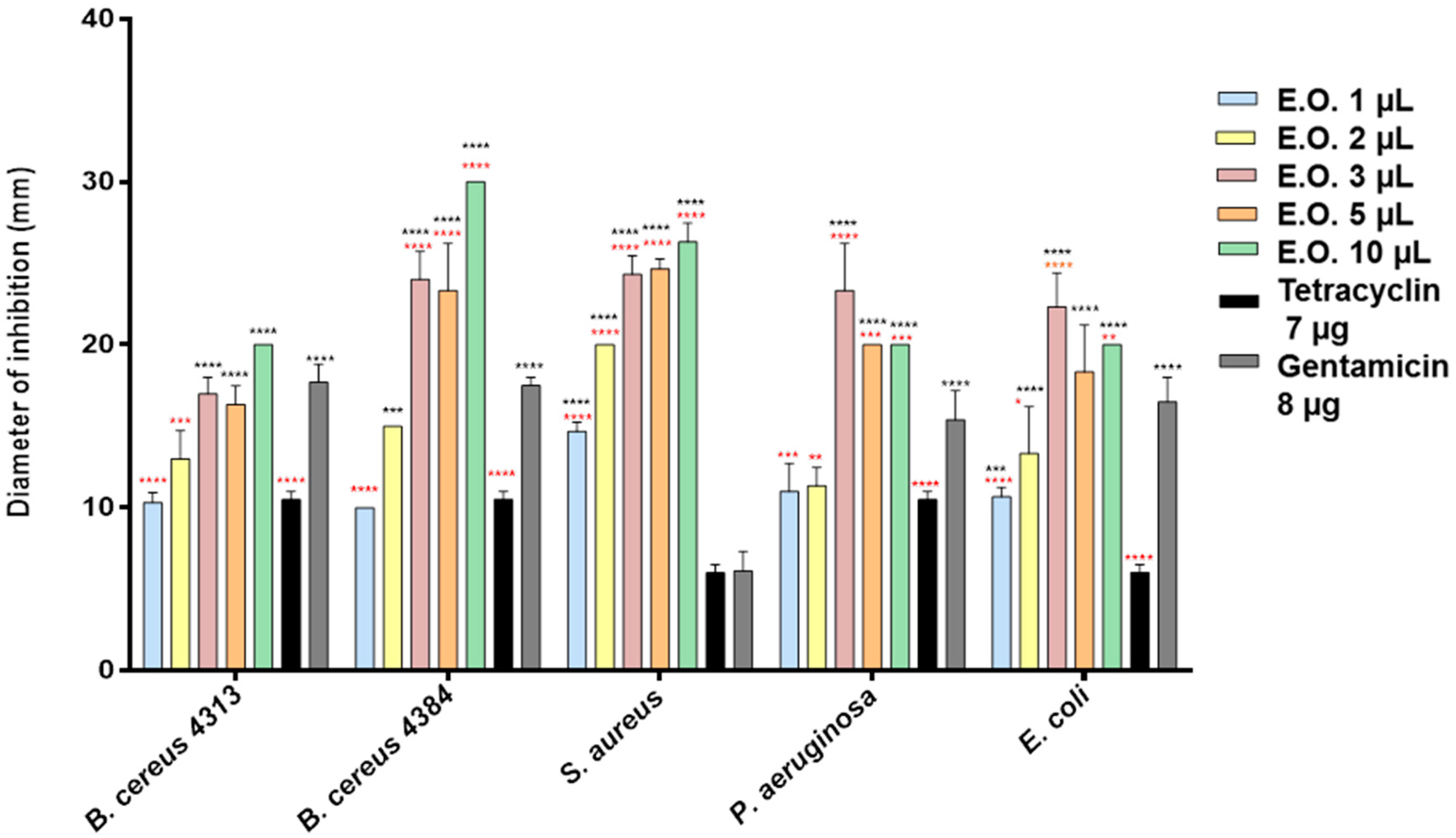
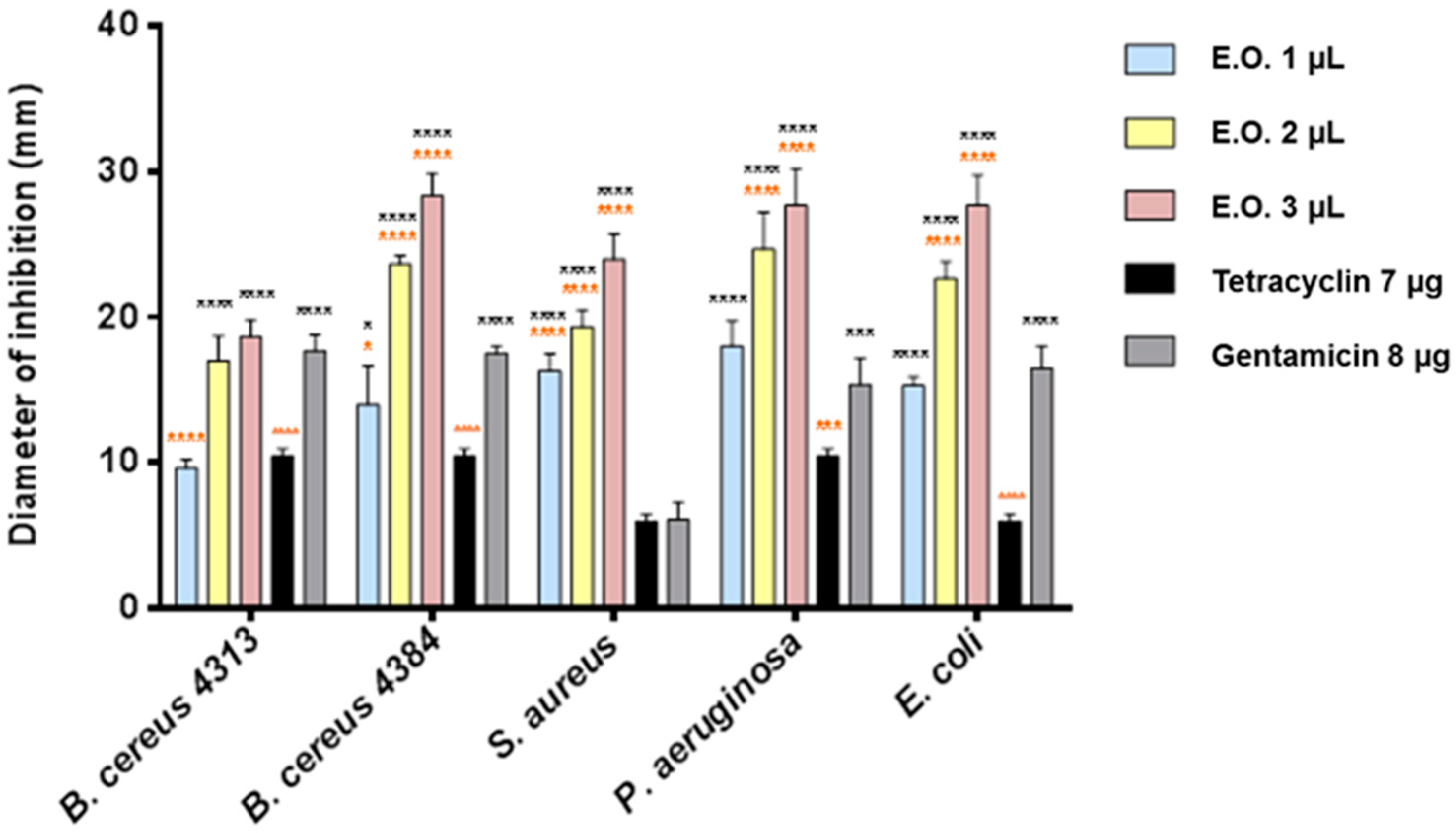
2.2. Antibacterial Activity

Using the inhibition halo technique, we evaluated the antimicrobial activity of the EOs of both C. medica cultivars. Among the Gram-positive bacteria, Bacillus cereus 4384 and Staphylococcus aureus were the more sensitive bacterial strains to C. medica cv. ‘liscia’ EO (Figure 1).
Low concentrations of C. medica cv. ‘rugosa’ and cv. ‘liscia’ EOs showed strong effect on all tester strains (inhibition halos ranging between 8.5 and 10 mm, Figure 1 and Figure 2). The effect was higher also respect to the antibiotics used as a control. B. cereus 4384 resulted in the most sensitive strain; S. aureus was the most sensitive at the lowest amount of Citrus medica cv. ‘liscia’. In any case, C. medica cv. ‘rugosa’ EO showed a greater inhibitory power than C. medica cv. ‘liscia’ EO on all bacterial strains and already at low concentrations (Figure 2). Three microliters of the two EOs seemed to give the most effective antimicrobial activity. At this concentration, the behavior exhibited by the pathogen strains was different in terms of sensitivity. Certainly, C. medica cv. ‘rugosa’ EO was much more effective than the EO obtained from C. medica cv. ‘liscia’. P. aeruginosa and E. coli were the most sensitive strains to this last EO, with inhibition halos not inferior to 27 mm. A strong activity was also exhibited by C. medica cv. ‘rugosa’ EO against S. aureus. C. medica cv. ‘liscia’ EO was effective, although in not such a marketable mode against the strains, with inhibition halos never superior to 22 mm. The two strains of B. cereus used in the experiments (4313 and 4384) reflected, in some way, the different behavior exhibited by the microorganisms used as tester. Both strains were sensitive to the two EOs. B. cereus 4313 showed the same behavior, giving similar inhibition halo; on the contrary, B. cereus 4384 showed a different behavior, resulting more sensitive to the C. medica cv. ‘rugosa’ EO respect to the EO obtained from C. medica cv. ‘liscia’ (Δ = 19%). Minimum inhibitory concentration (MIC) values reported a remarkable action of the two EOs. C. medica cv. ‘liscia’ EO (MIC = 0.1 μL/mL, Table 2) was more effective than that of C. medica cv. ‘rugosa’ (MIC ranging between 0.1 and 0.8 μL/mL), except than against B. cereus 4384 (MIC = 0.1 μL/mL) and much more effective against the antibiotic gentamycin. In all cases, the results of the MIC assay indicated that the antimicrobial activity of the two EOs was much more effective than their two main components, limonene and camphene.

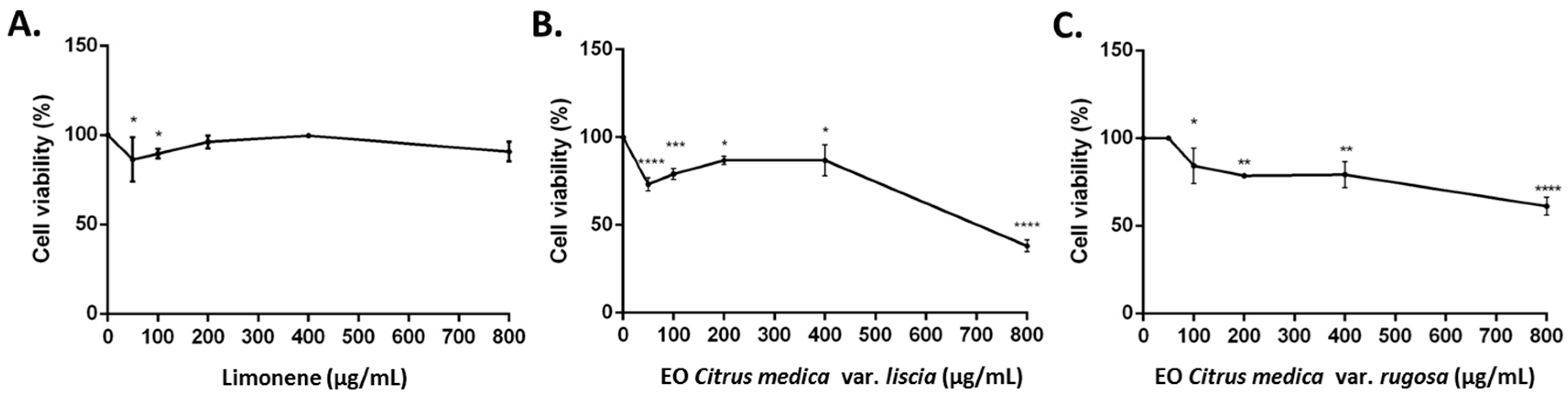
2.3. Cytotoxicity of Limonene, C. medica cv. ‘liscia’ and C. medica cv. ‘rugosa’ Essential Oils

Limonene, C. medica cv. ‘liscia’, and C. medica cv. ‘rugosa’ EOs were evaluated for their ability to inhibit the growth of human neuroblastoma cell line (SH-SY5Y). The EOs and limonene revealed different cytotoxic activities. Limonene and C. medica cv. ‘rugosa’ EOs showed an IC50 > 2000 μg/mL, instead C. medica cv. ‘rugosa’ EO showed an IC50 of 718.2 μg/mL.
Treatment of SH-SY5Y neuroblastoma cells with 800 μg/mL of limonene for 24 h resulted in a low cytotoxic activity. However, treatment with 800 μg/mL of C. medica cv. ‘liscia’ EOs resulted in a stronger cytotoxicity than C. medica cv. ‘rugosa’ EO with 38% cell death (Figure 3).

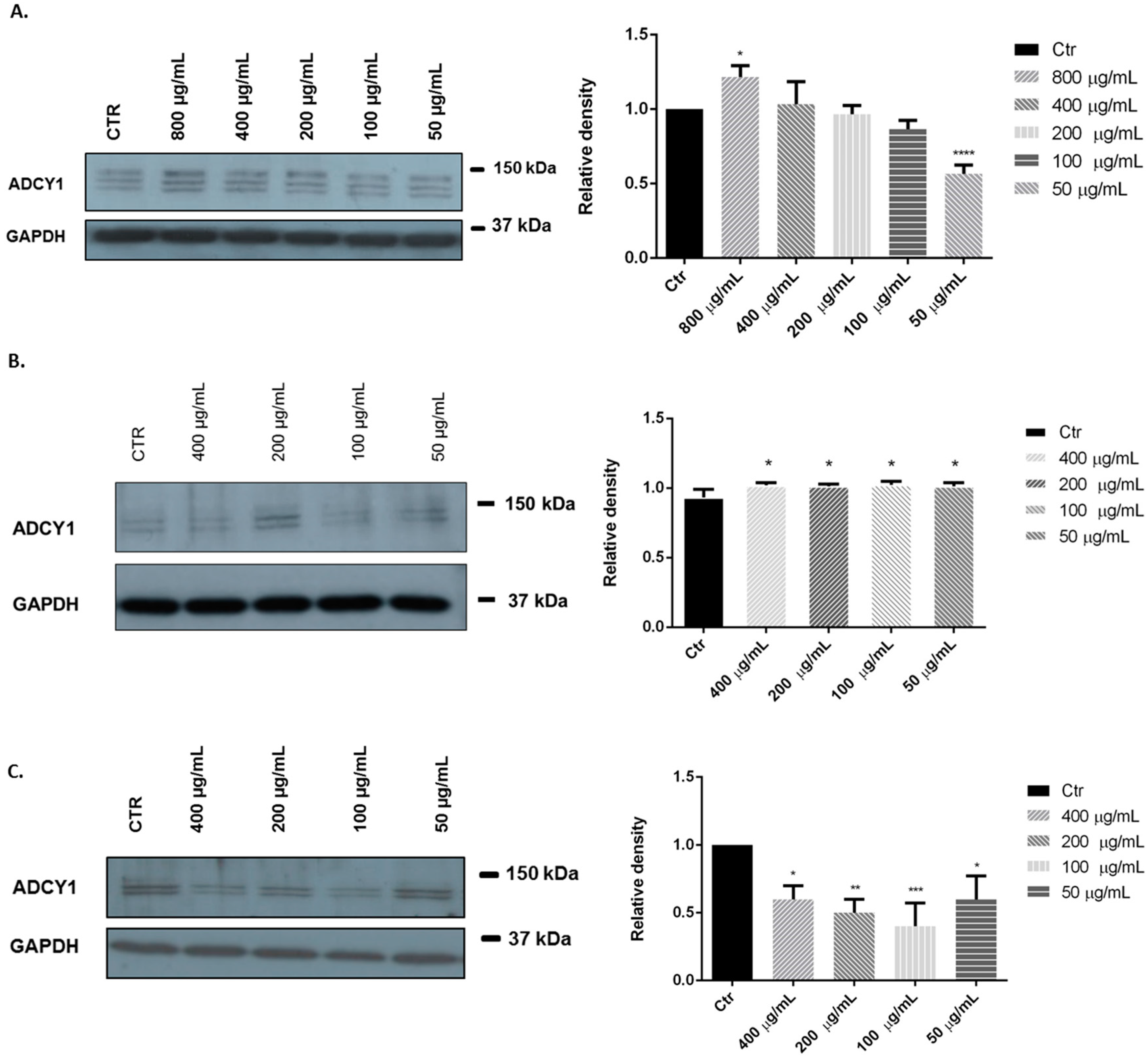
2.4. ADCY1: Western Blotting Analysis

We investigated the effects of limonene, C. medica cv. ‘liscia’ and C. medica cv. ‘rugosa’ EOs in SH-SY5Y human neuroblastoma cells. Representative Western blots and quantitative densitometric analysis for adenylate cyclase 1 (ADCY1) protein expression in SH-SY5Y following exposure to different concentrations of limonene and EOs are shown in Figure 4. Treatments of SH-SY5Y neuroblastoma cells with 800 and 50 μg/mL of limonene for 24 h significantly influenced ADCY1 expression in different way: high concentrations appear to increase ADCY1 expression, instead low concentrations reduced the ADCY1 expression (Figure 4A). However, treatments with 400, 200, 100, 50 μg/mL of C. medica cv. ‘liscia’ and C. medica cv. ‘rugosa’ EOs appear to influence significantly ADCY1 expression with an over expression and a down expression of ADCY1, respectively (Figure 4B,C).

3. Discussion

There were no relevant quali-quantitative differences in the composition of the two EOs, neither with regard to the percentages of the classes of compounds nor to the main components. In both oils, limonene and camphene are the main components, even if the composition of the EO of C. medica cv. ‘rugosa’ is more complex, but many of its constituents are present in very low percentages or even in traces.
The comparison with the available literature concerning the chemical composition of the EO of citron from other countries, showed substantial differences. Limonene percentages found (67.2% and 62.8%) are significantly higher than the values recorded in different parts of the world or even in Italy: South Korea, 52.44% [11]; China, 33.84% [12]; Santa Maria del Cedro (Italy), 59% [1]; and Iran, 56.6% [13]. Other notable differences relate to the content in γ-terpinene exceeding 20% and of α- and β-pinene, reported between 7.73% [12] and 16.3% [13]. Camphene range is 8.5%–10.9% (cv. ‘liscia’ and cv. ‘rugosa’, respectively) in samples studies in this research, but this compound is practically absent or in traces in the oils reported in literature.
The antimicrobial activity of the two EOs was evaluated against different microorganisms: Bacillus cereus (DSM 4313 and DSM 4384), an agent of different foodborne illnesses which can be found in raw and processed foods; Staphylococcus aureus which plays a significant role in invasive skin disease, and actually representing one of the most significant emerging pathogens; an enterotoxigenic strain of Escherichia coli, generally involved in episodes of food poisoning; and Pseudomonas aeruginosa, concerned to different urinary, pulmonary, skin, ear and eye infections.
The EO from cv. ’rugosa’ was tested at 1, 2, and 3 mg/mL, because it was more active in antimicrobial tests than the EO from cv. ‘liscia’, tested at 1, 2, 3, 5, and 10 mg/mL.
The modest inhibitory activity exhibited by the two EOs against the Gram-negative strains could be due to the different structures of the cell walls of Gram-positive and Gram-negative bacteria [14]. Our results supported previous studies [15], which reported a different sensitivity shown by the two strains of B. cereus against EO of Hypericum perfoliatum.
Some authors reported the antimicrobial activity of C. medica against different pathogens. Sah and coworkers [16] evaluated the antimicrobial activity of fruit juice and ethanol extract of different plant portion of Citrus medica which resulted to be less effective than the EOs of Citrus medica cv. liscia and rugosa evaluated in our experiments. Belletti and coworkers evaluated the effects of citron EO on microbial spoilage and growth and survival of pathogenic microorganisms during storage of a fruit based salad, reporting the capability of these substances to prolong the microbial shelf life of these products [17]. Therefore, the values of MIC observed testing limonene and camphene, the two most abundant components present in the two EOs, seemed to corroborate the hypothesis–almost for all pathogens tested–that the activity is due to a synergy among different components, more than to a specific component. In fact, MIC of limonene was always 1 μL, ten-fold higher than that of the two EOs; camphene, generally recognized as good antimicrobial agent [18], was less effective (MIC > 6 μL) except than against E. coli (MIC = 0.084 μL). Considering such value and comparing it with the MIC of the two EOs of Citrus medica, we could hypothesize a strong influence of camphene on the antimicrobial. On the other hand, these two monoterpenes generally do not exhibit a strong antimicrobial activity [19] per se. Limonene was indicated for its inhibitory activity against the Gram-negative pathogens, P. aeruginosa and E. coli, but at higher concentrations [20,21,22]. Limonene exists in two enantiomeric forms, with different biological activity. Aggarwal and coworkers have demonstrated that (S)-(−)-limonene had less antimicrobial activity than (R)-(+)-limonene. In fact, the first failed to inhibit many bacterial strains and the majority of the fungal strains, except Microsporum gypseum. In contrast, (R)-(+)-limonene was highly active against a wide spectrum of microorganisms [23].
The antimicrobial activity of the two EOs might be due to a global synergy among different singular components, more than to the presence of a specific component.
It is remarkable the stronger antimicrobial activity exhibited by the two EOs. This is still more noteworthy, considering their MIC values. The increasing resistance of microorganisms to conventional chemicals and drugs has prompted scientists to search for novel sources of biocides with broad-spectrum activities. In this concern, the activity of the two EOs studied comforts us towards an ever-greater increase of studies and research aimed at the discovery, exploitation, and potential use of natural substances capable of effectively countering the action of pathogenic microorganisms.
The cytotoxic activity of the two EOs and limonene were evaluated in human neuroblastoma cell line (SH-SY5Y). The IC50 values were >700 μg/mL, indicating that the substances were not cytotoxic as judged by the criterion set by the National Cancer Institute that stated that extracts with IC50 < 20 μg/mL were considered to be cytotoxic against the treated cells [24].
However, comparing the IC50 values, our findings indicated that C. medica cv. ‘liscia’ EO is more cytotoxic than C. medica cv. ‘rugosa’ EO and limonene. The cytotoxicity can probably be attributed to a synergistic activity of limonene and other minor components. Monajemi and coworkers [13] reported that the EO of C. limon with large amounts of limonene (98.4%) was less cytotoxic than C. medica with low content of limonene (56.6%) on MCF-7 and HeLa cell lines. Our results agree with previous studies reporting that low concentrations of limonene were ineffective in cell death in SH-SY5Y cells [7,25].
Many plant species with EOs are used as sedatives, hypnotics, and tranquilizers and they may be useful in treating disorders of the central nervous system [26]. Different studies reported a well-established role for adenylyl cyclase in the regulation of synaptic plasticity, memory, and other multiple brain processes. Moreover, cross-talk between the cyclic adenosine-3′,5′-monophosphate (cAMP) signal transduction system and other signaling pathways, such as the extracellular signal-regulated kinase/mitogen-activated protein (ERK/MAP) regulatory system, has been described [27,28]. In this perspective, we evaluated the possible influence of limonene and C. medica cv. ‘liscia’ and C. medica cv. ‘rugosa’ EOs on ADCY1 expression in SH-SY5Y cells, a human-derived neuroblastoma cell line often used as an in vitro neuronal model [29]. Adenylyl cyclase is an enzyme that regulates the physiological effects of numerous drugs and hormones through the production of cAMP. Clinical and epidemiologic research provided suggestive evidence regarding the association between adenylyl cyclase activity and major depression [30,31]. Moreover, platelet adenylyl cyclase activity might serve as a biological marker for major depression and the therapeutic effect of antidepressants [31,32,33,34].
Our results showed that the higher concentration of limonene (800 μg/mL) appears to increase ADCY1 expression in SH-SY5Y cell and, consequently, the intracellular production of cAMP. Our results agree with results of Watt and coworkers [35] that showed that some volatile substances stimulate cAMP-response element (CRE)-mediated transcription in olfactory sensory neurons through Ca2+ activation of the ERK/MAPK/p90rsk (Ribosomal Protein S6 Kinases, 90-kDa) pathway.
C. medica cv. ‘liscia’ EO showed similar effects with all concentrations tested, probably because its main component is limonene (67.2%). Limonene is reported to increase cytosolic cAMP concentration and induce activation of protein kinase A and phosphorylation of cAMP-response element-binding protein in Chinese hamster ovary cells transfected with the human adenosine A2A receptor gene [36]. On the contrary, treatment with C. medica cv. ‘rugosa’ EO showed an inhibition on ADCY1 expression in SH-SY5Y cell, possibly due to the complex phytochemical composition of this EO. These results showed that the use of both EOs could increase intracellular production of cAMP and have an excitatory effect on the Central Nervous System [26].

4. Materials and Methods

4.1. Plant Material

Fruits of Citrus medica cv. ‘liscia’ and C. medica cv. ‘rugosa’ were collected in February 2016 from biological cultivations in the Coast of Amalfi. The plants were identified by Prof. V. De Feo. Voucher specimens (labeled as De Feo/134/2016 for cv. ‘liscia’ and De Feo/133/2016 for cv. ‘rugosa’, respectively) are stored in the Herbarium of the Medical Botany Chair at the University of Salerno.

4.2. Isolation of the Essential Oils

One hundred grams of fruit peels of each cultivar were ground in a Waring blender and then subjected to hydrodistillation for 3 h according to the standard procedure described in the European Pharmacopoeia [37]. The oils were solubilized in n-hexane, filtered over anhydrous sodium sulphate and stored under N2 at 4 °C in the dark, until tested and analyzed.

4.3. GC-FID Analysis

Analytical gas chromatography (GC) was carried out on a Perkin-Elmer Sigma-115 gas chromatograph (Perkin Elmer, Waltham, MA, USA) equipped with a flame ionization detector (FID) and a data handling processor. The separation was achieved using a HP-5 MS fused-silica capillary column (30 m × 0.25 mm i.d., 0.25 μm film thickness, Agilent, Roma, Italy). Column temperature: 40 °C, with 5 min initial hold, and then to 270 °C at 2 °C/min, 270 °C (20 min); injection mode, splitless (1 μL of a 1:1000 n-hexane solution). Injector and detector temperatures were 250 °C and 290 °C, respectively. Analysis was also run by using a fused silica HP Innowax polyethylene glycol capillary column (50 m × 0.20 mm i.d., 0.25 μm film thickness, Agilent). In both cases, helium was used as the carrier gas (1.0 mL/min).

4.4. GC/MS Analysis

Analysis was performed on an Agilent 6850 Ser. II apparatus (Agilent), fitted with a fused silica DB-5 capillary column (30 m × 0.25 mm i.d., 0.33 μm film thickness, Agilent), coupled to an Agilent Mass Selective Detector MSD 5973; ionization energy voltage 70 eV; electron multiplier voltage energy 2000 V. Mass spectra (MS) were scanned in the range 40–500 amu, scan time 5 scans/s. Gas chromatographic conditions were as reported in the previous paragraph; transfer line temperature, 295 °C.

4.5. Identification of the Essential Oil Components

Most constituents were identified by GC by comparison of their Kovats retention indices (Ri) (determined relative to the retention times (tR) of n-alkanes (C10–C35)), with either those of the literature [38,39,40,41] and mass spectra on both columns or those of authentic compounds available in our laboratories by means of NIST 02 and Wiley 275 libraries [42]. The components’ relative concentrations were obtained by peak area normalization. No response factors were calculated.

4.6. Antimicrobial Activity

To study antimicrobial activity a filter paper disc method was used [43]. The bacteria used in this study included Bacillus cereus DSM 4313, Bacillus cereus 4384, Staphylococcus aureus DSM 25693, Pseudomonas aeruginosa ATCC 50071, and Escherichia coli DSM 8579. Bacteria were purchased from the Deutsche SammLung von Mikroorganismen und Zellkulturen GmbH (DSMZ, Deutsche Sammlung von Mikroorganismen und Zellkulturen, Braunschweig, Germany).
A 100 mg sample of each EO was re-suspended in dimethyl sulfoxide (DMSO) to produce the same concentration of 10 mg/mL, then diluted to be subjected to biological analyses. The strains were incubated in nutrient broth (Oxoid, Milano, Italy) at 37 °C for 18 h. The optical densities of all cultures were adjusted to match a 0.5 McFarland standard of 1 × 10⁸ colony-forming units (cfu)/mL. Sterile filter paper discs (5 mm) were impregnated with 1 and 10 μL of the EOs and placed in Petri dishes. Plates were left for 30 min at room temperature under sterile conditions and then incubated at 37 °C for 24 h, and the inhibition halo around the disc was measured. A disc treated with DMSO alone served as the negative control. Tetracycline (7 μg/disc; Sigma) and gentamycin (8 μg/disc; Sigma) were used as reference drugs. The experiments were performed in triplicate and averaged.

4.7. Minimum Inhibitory Concentration

For the MIC of the EOs and their main components, limonene and camphene, a modified version of the resazurin microtiter-plate assay of Sarker and coworkers was used [44]. Briefly, different volumes of the two EOs, prepared as above described, and of the pure compounds limonene (dissolved in DMSO, 1:10 v/v), and camphene (dissolved in a mix hexane/DMSO 1:10 v/v), were used and pipetted into the first row of the 96 well plates. To all wells, different volumes of Muller-Hinton broth was added. Two-fold serial dilutions were performed such that each well had 50 μL of the test material in serially descending concentrations. 30 μL of 3.3× strength isosensitised broth and 10 μL of resazurin indicator solution (previously prepared by dissolving 270 mg tablet in 40 mL of sterile distilled water) were added in each well, to reach a final volume/well of 240 μL. Finally, 10 μL of bacterial suspension was added to each well to achieve a concentration of approx. 5 × 10⁵ cfu/mL. Each plate was wrapped loosely with cling film to ensure that bacteria did not become dehydrated. In each plate a set of controls were added: a column with a ciprofloxacin (1 mg/mL in DMSO, Sigma) as positive control; a column with all solutions with the exception of the test compound; a column with all solutions with the exception of the bacterial solution adding 10 μL of nutrient broth instead and a column with DMSO solution as a negative control. The plates were prepared in triplicate, and incubated at 37 °C for 24 h. The growth was indicated by color changes (visually assessed) from blue/dark purple to pink (or colorless). The lowest concentration at which color change occurred indicated the MIC value.

4.8. Cell Cultures

Human neuroblastoma (SH-SY5Y) cancer cells were cultured in Roswell Park Memorial Institute Medium (RPMI) medium supplemented with 1% l-glutamine, 10% heat-inactivated fetal bovine serum (FBS), 1% penicillin/streptomycin (all from Sigma-Aldrich, St. Louis, MO, USA) at 37 °C in an atmosphere of 95% O2 and 5% CO2.

4.9. MTT Bioassay

Human neuroblastoma cancer cells (SH-SY5Y) were plated (5 × 10³) in 96-well culture plates in 150 μL of culture medium and incubated at 37 °C in humidified 5% CO2. The day after, a 150 μL aliquot of serial dilutions of limonene and EOs (800–50 μg/mL) was added to the cells and incubated for 24 h. DMSO alone was used as control. Cell viability was assessed through 3-(4,5-dimethylthiazol-2-yl)-2,5-diphenyl tetrazolium bromide (MTT) assay. Briefly, 30 μL of MTT (5 mg/mL) was added and the cells incubated for additional 3 h. Thereafter, cells were lysed and the dark blue crystals solubilized with 30 μL of a solution containing 50% v/v N,N-dimethylformamide and 20% w/v SDS with an adjusted pH of 4.5. The optical density (OD) of each well was measured with a microplate spectrophotometer (Thermo Scientific Multiskan GO, Monza, Italy) equipped with a 520 nm filter. Cell viability in response to treatment was calculated as a percentage of control cells treated with DMSO at the final concentration (0.1%): viable cells = (100 × OD treated cells)/OD control cells [45].

4.10. Extraction Proteins and Western Blotting

Cells were treated with different concentrations (800–100 μg/mL) of limonene and EOs. After 24 h, cells were collected and lysed using LaemmLi buffer to extract total proteins. For Western blot analysis, an aliquot of total protein was run on 8% SDS-PAGE gel and transferred to nitrocellulose. Nitrocellulose blots were blocked with 10% nonfat dry milk in Tris buffer saline 0.1% Tween-20 over night at 4 °C and incubated with primary anti-ADCY1 (Santa Cruz Biotechnology, Santa Cruz, CA, USA) for 3 h at room temperature. Immunoreactivity was detected by sequential incubation with horseradish peroxidase-conjugated secondary antibody (Amersham Biosciences, Pittsburgh, PA, USA) and enhanced chemiluminescence reagents (ImmunoCruz, Santa Cruz Biotechnology) [46].

4.11. Statistical Analysis

All experiments were carried out in triplicate. Data of each experiment were statistically analyzed using GraphPad Prism 6.0 software (GraphPad Software Inc., San Diego, CA, USA) followed by comparison of means (two-way ANOVA) using Dunnett’s multiple comparisons test, at the significance level of p < 0.05.

5. Conclusions

We characterized the chemical composition of the EOs of two C. medica cultivars, characteristics of Amalfi Coast. These oils present a new composition, with high presence of limonene, which seems a ’fingerprint’ of these oils. The EOs showed higher antimicrobial activity at the tested doses. The EOs showed higher antimicrobial activity than antibiotics used as control. The presence of different components present in the two oils, more than the high content of limonene and camphene might be responsible for this EOs activity. This finding led us to hypothesize the possible use of these oils as antimicrobials and confirm their traditional uses as food preserving agents.
The alterations in ADCY1 expression suggested a major role for limonene in effects on the central nervous system. The precise mechanism of the biochemical effects of Citrus medica EOs is unclear; however, ADCY1 pathway might be involved. Moreover, the chemical and biological study of these EOs contribute to the knowledge and promotion of two cultivars belonging to the biodiversity of South Italy.

Author Contributions

Vincenzo De Feo and Filomena Nazzaro conceived and designed the experiments; Luigi Aliberti, Lucia Caputo and Lucéia Fátima Souza performed the experiments; Luigi Aliberti and Laura De Martino analyzed the data; Vincenzo De Feo wrote the paper.

Conflicts of Interest

The authors declare no conflict of interest.

Abbreviations

The following abbreviations were used in this manuscript:
ADCY1Adenylate Cyclase 1
cAMPCyclic adenosine-3′,5′-monophosphate
EOEssential oil
ERKExtracellular signal-regulated kinase
MAPMitogen-activated protein
MTT3-(4,5-dimethylthiazol-2-yl)-2,5-diphenyl tetrazolium bromide
p90rskRibosomal Protein S6 Kinases, 90-kDa
RPMIRoswell Park Memorial Institute Medium
ODOptical Density

References

  1. Gabriele, B.; Fazio, A.; Dugo, P.; Costa, R.; Mondello, L. Essential oil composition of Citrus medica var. liscia L. cv. Diamante (Diamante citron) determined after using different extraction methods. J. Sep. Sci. 2009, 32, 99–108. [Google Scholar] [CrossRef] [PubMed]
  2. Savo, V.; Caneva, G.; Guarrera, P.M.; Reedy, D. Folk phytotherapy of the Amalfi Coast (Campania, Southern Italy). J. Ethnopharmacol. 2011, 135, 376–392. [Google Scholar] [CrossRef] [PubMed]
  3. Uzun, A.; Yesiloglu, T. Genetic Diversity in Citrus. In Genetic Diversity in Plants; Caliskan, M., Ed.; InTech: Rijeka, Croatia, 2012; pp. 213–230. [Google Scholar]
  4. Yeung, H.C. Handbook of Chinese Herbs and Formulas; Institute of Chinese Medicine: Los Angeles, CA, USA, 1985. [Google Scholar]
  5. Li, W.Q.; Kuriyama, S.; Li, Q.; Nagai, M.; Hozawa, A.; Nishino, Y.; Tsuji, I. Citrus consumption and cancer incidence: The Ohsaki cohort study. Int. J. Cancer 2010, 127, 1913–1922. [Google Scholar] [CrossRef] [PubMed]
  6. Menichini, F.; Tundis, R.; Loizzo, M.R.; Bonesi, M.; Provenzano, E.; Cindio, B.D.; Menichini, F. In vitro photo-induced cytotoxic activity of Citrus bergamia and C. medica L. cv. Diamante peel essential oils and identified active coumarins. Pharm. Biol. 2010, 48, 1059–1065. [Google Scholar] [CrossRef] [PubMed]
  7. Russo, R.; Cassiano, M.G.V.; Ciociaro, A.; Adornetto, A.; Varano, G.P.; Chiappini, C.; Corasaniti, M.T. Role of d-limonene in autophagy induced by bergamot essential oil in SH-SY5Y neuroblastoma cells. PLoS ONE 2014, 9, e113682. [Google Scholar] [CrossRef] [PubMed]
  8. Souto-Maior, F.M.; Lélis de Carvalho, F.; Soares Lima de Morais, L.S.; Mendonça Netto, S.; Pergentino de Sousa, D.; Nóbrega de Almeida, R. Anxiolytic-like effects of inhaled linalool oxide in experimental mouse anxiety models. Pharmacol. Biochem. Behav. 2011, 100, 259–263. [Google Scholar] [CrossRef] [PubMed]
  9. Coelho, V.R.; Gianesini, J.; von Borowski, R.; Mazzardo-Martins, L.; Martins, D.F.; Picada, J.N.; Santos, A.R.; Brum, L.F.; Pereira, P. (−)-Linalool, a naturally occurring monoterpene compound, impairs memory acquisition in the object recognition task, inhibitory avoidance test and habituation to a novel environment in rats. Phytomedicine 2011, 18, 896–901. [Google Scholar] [CrossRef] [PubMed]
  10. Re, L.; Barocci, S.; Sonnino, S.; Mencarelli, A.; Vivani, C.; Paolucci, G.; Scarpantonio, A.; Rinaldi, L.; Mosca, E. Linalool modifies the nicotinic receptor–ion channel kinetics at the mouse neuromuscular junction. Pharmacol. Res. 2000, 42, 177–182. [Google Scholar] [CrossRef] [PubMed]
  11. Kim, N.K.; Ko, Y.J.; Yang, H.M.; Ham, Y.M.; Roh, S.W.; Jeon, Y.J.; Ahn, G.; Kang, M.C.; Yoon, W.J.; Kim, D.; et al. Anti-inflammatory effect of essential oil and its constituents from fingered citron (Citrus medica L. var. sarcodactylis) through blocking JNK, ERK and NF-kB signaling pathways in LPS-activated RAW 264.7 cells. Food Chem. Toxicol. 2013, 57, 126–131. [Google Scholar] [CrossRef] [PubMed]
  12. Wu, Z.; Li, H.; Yang, Y.; Zhan, Y.; Tu, D. Variation in the components and antioxidant activity of Citrus medica L. var. sarcodactylis essential oils at different stages of maturity. Ind. Crops Prod. 2013, 46, 311–316. [Google Scholar] [CrossRef]
  13. Monajemi, R.; Oryan, S.; Haeri-Roohani, A.; Ghannadi, A.; Jafarian, A. Cytotoxic effects of essential oils of some Iranian Citrus peels. Iran. J. Pharm. Res. 2005, 3, 183–187. [Google Scholar]
  14. Nazzaro, F.; Fratianni, F.; de Martino, L.; Coppola, R.; de Feo, V. Effect of Essential Oils on Pathogenic Bacteria. Pharmaceuticals 2013, 6, 1451–1474. [Google Scholar] [CrossRef] [PubMed]
  15. Del Monte, D.; de Martino, L.; Marandino, A.; Fratianni, F.; Nazzaro, F.; de Feo, V. Phenolic content, antimicrobial and antioxidant activities of Hypericum perfoliatum L. Ind. Crops Prod. 2015, 74, 342–347. [Google Scholar] [CrossRef]
  16. Sah, A.N.; Vijay, J.; Anand, B.M. Antimicrobial activity of six different parts of the plant Citrus medica Linn. Pharmacogn. J. 2011, 3, 80–83. [Google Scholar] [CrossRef]
  17. Belletti, N.; Lanciotti, R.; Patrignani, F.; Gardini, F. Antimicrobial Efficacy of Citron Essential Oil on Spoilage and Pathogenic Microorganisms in Fruit-Based Salads. J. Food Sci. 2008, 73, 331–338. [Google Scholar] [CrossRef] [PubMed]
  18. Kazemi, M. Chemical composition and antimicrobial activity of essential oil of Matricaria chamomilla. Bull. Environ. Pharmacol. Life Sci. 2014, 3, 148–153. [Google Scholar]
  19. Dorman, H.J.D.; Deans, S.G. Antimicrobial agents from plants: Antibacterial activity of plant volatile oils. J. Appl. Microbiol. 2000, 88, 308–316. [Google Scholar] [CrossRef] [PubMed]
  20. Dai, J.; Zhu, L.; Yang, L.; Qiu, J. Chemical composition, antioxidant and antimicrobial activities of essential oil from Wedelia prostrata. EXCLI J. 2013, 12, 479–490. [Google Scholar] [PubMed]
  21. Vimal, M.; Vijaya, P.P.; Mumtaj, P.; Farhath, M.S.S. Antibacterial activity of selected compounds of essential oils from indigenous plants. J. Chem. Pharm. Res. 2013, 5, 248–253. [Google Scholar]
  22. Bakkali, F.; Averbeck, S.; Averbeck, D.; Idaomar, M. Biological effects of essential oils: A review. Food Chem. Toxicol. 2008, 46, 446–475. [Google Scholar] [CrossRef] [PubMed]
  23. Aggarwal, K.K.; Khanuja, S.P.S.; Ahmad, A.; Santha Kumar, T.R.; Gupta, V.K.; Kumar, S. Antimicrobial activity profiles of the two enantiomers of limonene and carvone isolated from the oils of Mentha spicata and Anethum sowa. Flavour Fragr. J. 2002, 17, 59–63. [Google Scholar] [CrossRef]
  24. Geran, R.I.; Greenberg, N.H.; Macdonald, M.M.; Schumacher, A.M.; Abbott, B.J. Protocols for screening chemical agents and natural products against animal tumours and other biological systems. Cancer Chemother. Rep. 1972, 3, 59–61. [Google Scholar]
  25. Corasaniti, M.T.; Maiuolo, J.; Maida, S.; Fratto, V.; Navarra, M; Russo, R.; Bagetta, G. Cell signaling pathways in the mechanisms of neuroprotection afforded by bergamot essential oil against NMDA-induced cell death in vitro. Br. J. Pharmacol. 2007, 151, 518–529. [Google Scholar] [CrossRef] [PubMed]
  26. Pimenta, F.C.F.; Correia, N.D.A.; Albuquerque, K.L.G.D.; de Sousa, D.P.; da Rosa, M.R.D.; Pimenta, M.B.F.; de Almeida, R.N. Naturally occurring anxiolytic substances from aromatic plants of genus citrus. J. Med. Plants Res. 2012, 6, 342–347. [Google Scholar] [CrossRef]
  27. Impey, S.; Obrietan, K.; Storm, D.R. Making new connections: Role of ERK/MAP kinase signaling in neuronal plasticity. Neuron 1999, 23, 11–14. [Google Scholar] [CrossRef]
  28. Davis, M.I.; Ronesi, J.; Lovinger, D.M. A predominant role for inhibition of the adenylate cyclase/protein kinase A pathway in ERK activation by cannabinoid receptor 1 in N1E-115 neuroblastoma cells. J. Biol. Chem. 2003, 49, 48973–48980. [Google Scholar] [CrossRef] [PubMed]
  29. Uğuz, A.C.; Ahmi, Ö.; Mustafa, N. Curcumin inhibits apoptosis by regulating intracellular calcium release, reactive oxygen species and mitochondrial depolarization levels in SH-SY5Y neuronal cells. J. Recept Signal Transduct. Res. 2016, 36, 395–401. [Google Scholar] [CrossRef] [PubMed]
  30. Cowburn, R.F.; Marcusson, J.O.; Eriksson, A.; Wiehager, B.; O’Neill, C. Adenylyl cyclase activity and G-protein subunit levels in postmortem frontal cortex of suicide victims. Brain Res. 1994, 633, 297–304. [Google Scholar] [CrossRef]
  31. Reiach, J.S.; Li, P.P.; Warsh, J.J.; Kish, S.J.; Young, L.T. Reduced adenylyl cyclase immunolabeling and activity in postmortem temporal cortex of depressed suicide victims. J. Affect. Disord. 1999, 56, 141–151. [Google Scholar] [CrossRef]
  32. Donati, R.J.; Rasenick, M.M. G protein signaling and the molecular basis of antidepressant action. Life Sci. 2003, 73, 1–17. [Google Scholar] [CrossRef]
  33. Hines, L.M.; Tabakoff, B. Platelet adenylyl cyclase activity: A biological marker for major depression and recent drug use. Biol. Psychiatry 2005, 58, 955–962. [Google Scholar] [CrossRef] [PubMed]
  34. Abdel-Razaq, W.; Bates, T.E.; Kendall, D.A. The effects of antidepressants on cyclic AMP-response element-driven gene transcription in a model cell system. Biochem. Pharmacol. 2007, 73, 1995–2003. [Google Scholar] [CrossRef] [PubMed]
  35. Watt, W.C.; Storm, D.R. Odorants stimulate the ERK/mitogen-activated protein kinase pathway and activate cAMP-response element-mediated transcription in olfactory sensory neurons. J. Biol. Chem. 2001, 276, 2047–2052. [Google Scholar] [CrossRef] [PubMed]
  36. Park, H.M.; Lee, J.H.; Yaoyao, J.; Jun, H.J.; Lee, S.J. Limonene, a natural cyclic terpene, is an agonistic ligand for adenosine A 2A receptors. Biochem. Biophys. Res. Commun. 2001, 404, 345–348. [Google Scholar] [CrossRef] [PubMed]
  37. Council of Europe. European Pharmacopoeia, 5th ed.; Council of Europe: Strasbourg Cedex, France, 2004; Volume I, p. 217. [Google Scholar]
  38. Jennings, W.; Shibamoto, T. Qualitative Analysis of Flavour and Fragrance Volatiles by Glass Capillary Gas Chromatography; Academic Press: New York, NY, USA, 1980. [Google Scholar]
  39. Davies, N.W. Gas chromatographic retention indices of monoterpenes and sesquiterpenes on methyl silicone and Carbowax 20M phases. J. Chromatogr. 1990, 503, 1–24. [Google Scholar] [CrossRef]
  40. Goodner, K.L. Practical retention index models of OV-101, DB-1, DB-5, and DB-Wax for flavor and fragrance compounds. LWT-Food Sci. Technol. 2008, 41, 951–958. [Google Scholar] [CrossRef]
  41. Adams, R.P. Identification of Essential Oil Components by Gas Chromatography/Mass Spectroscopy, 4th ed.; Allured Publishing Corporation: Carol Stream, IL, USA, 2007. [Google Scholar]
  42. Wiley Registry of Mass Spectral Data, with NIST Spectral Data CD Rom, 7th ed.; John Wiley & Sons: New York, NY, USA, 1998.
  43. De Martino, L.; de Feo, V.; Nazzaro, F. Chemical Composition and in Vitro Antimicrobial and Mutagenic Activities of Seven Lamiaceae Essential Oils. Molecules 2009, 14, 4213–4230. [Google Scholar] [CrossRef] [PubMed]
  44. Sarker, S.D.; Nahar, L.; Kumarasamy, Y. Microtitre plate-based antibacterial assay incorporating resazurin as an indicator of cell growth, and its application in the in vitro antibacterial screening of phytochemicals. Methods 2007, 42, 321–324. [Google Scholar] [CrossRef] [PubMed]
  45. Picerno, P.; Autore, G.; Marzocco, S.; Meloni, M.; Sanogo, R.; Aquino, R.P. Anti-inflammatory activity of verminoside from Kigelia africana and evaluation of cutaneous irritation in cell cultures and reconstituted human epidermis. J. Nat. Prod. 2005, 68, 1610–1614. [Google Scholar] [CrossRef] [PubMed]
  46. Petrella, A.; Ercolino, S.F.; Festa, M.; Gentilella, A.; Tosco, A.; Conzen, S.D.; Parente, L. Dexamethasone inhibits TRAIL-induced apoptosis of thyroid cancer cells via Bcl-xL induction. Eur. J. Cancer 2006, 42, 3287–3293. [Google Scholar] [CrossRef] [PubMed]
Figure 1. Antibacterial activity of essential oil (EO) of Citrus medica cv. ‘liscia’ and of the reference compounds, gentamicin and tetracycline. Results are the mean of three experiments. Dunnett’s test vs. gentamicin 8 μg: **** p < 0.0001; *** p < 0.001; ** p < 0.01; * p < 0.05; Dunnett’s test vs. tetracycline 7 μg: **** p < 0.0001; *** p < 0.001.
Molecules 21 01244 g001
Figure 2. Antibacterial activity of EO of Citrus medica cv. ‘rugosa’ and of the reference compounds, gentamicin and tetracycline. Results are the mean of three experiments. Dunnett’s test vs. gentamicin 8 μg: **** p < 0.0001; *** p < 0.001; * p < 0.05; Dunnett’s test vs. tetracycline 7 μg: **** p < 0.0001; *** p < 0.001; * p < 0.05.
Molecules 21 01244 g002
Figure 3. Percentage of cell viability after 3-(4,5-dimethylthiazol-2-yl)-2,5-diphenyl tetrazolium bromide (MTT) assay. Cells were treated with different concentrations (50–800 μg/mL) of limonene (A); C. medica cv. ‘liscia’ (B) and C. medica cv. ‘rugosa’ EOs, for 24 h and solvent (DMSO, 0.1%) alone. Data are the mean ± SD of three experiments * p < 0.05, ** p < 0.01, *** p < 0.001, **** p < 0.0001 vs. DMSO.
Molecules 21 01244 g003
Figure 4. Relative expression levels of the ADCY1 in SH-SY5Y treated with limonene (A); C. medica cv. ‘liscia’ (B); and C. medica cv. ‘rugosa (C) EOs. Each panel shows a representative Western blot and densitometric analysis of bands in the control and treated groups. Values are the mean ± SD in each group (n = 3). * p < 0.05, ** p < 0.01, *** p < 0.001, **** p < 0.0001, compared to control (ANOVA followed by Dunnett’s multiple comparison test).
Molecules 21 01244 g004
Table 1. Chemical composition of the essential oils (EOs) isolated from the peels of C. medica cv. ‘liscia’ (CL) and C. medica cv. ‘rugosa’ (CR) grown in Amalfi Coast.
No.CompoundLRI aLRI bCLCRIdentification c
1α-Thujene915930-0.11, 2
2α-Pinene9219390.81.21, 2
3α-Fenchene9349520.10.11, 2
4Camphene9649548.510.91, 2, 3
5β-Pinene9809791.41.71, 2
6α-Phellandrene99110020.50.61, 2
7δ-2-Carene100410020.10.31, 2
8p-Cymene10121024-1.01, 2
9Limonene1022102967.262.81, 2
10(Z)-β-Ocimene10281037Tr0.11, 2
11(E)-β-Ocimene103810500.10.31, 2, 3
12γ-Terpinene104710590.30.71, 2
13Linalool oxide furanoid106410720.3Tr1, 2
14trans-Linalool oxide 10860.1-1, 2
15Terpinolene107710880.10.31, 2
16Linalool109110960.31.31, 2
17α-Pinene oxide10951099Tr0.11, 2
181,3,8-p-Menthatriene11001110-Tr1, 2
19Perillene11031103TrTr1, 2
20trans-Thujone11061114Tr0.11, 2, 3
21Dehydro sabina ketone111111200.10.11, 2
22allo-Ocimene11191132Tr0.11, 2
23cis-p-Mentha-2,8-dien-1-ol11261137-Tr1, 2
24cis-Limonene oxide11331136Tr0.51, 2
25trans-Limonene oxide11401142-Tr1, 2, 3
26Isopulegol11441149Tr0.11, 2
27neo allo-Ocimene11521144-Tr 1, 2
28Citronellal11551153Tr0.21, 2
29neo iso-Isopulegol116711710.80.71, 2
30Isoborneol11631160-Tr1, 2, 3
31α-Terpineol118011880.70.61, 2
32Hexyl butanoate11831192-Tr1, 2
33Dihydrocarveol11851193TrTr1, 2
34Methyl chavicol11901196-Tr1, 2
35trans-4-Caranone119511960.30.11, 2, 3
36Decenal11981196-Tr1, 2
372-Decanol120211990.30.11, 2
38cis-4-Caranone120912000.10.31, 2
39endo-Fenchyl acetate121912200.90.41, 2
40Thymol methyl-ether12231235-Tr1, 2
41Neral123112380.10.51, 2
42Geraniol124612520.90.71, 2, 3
43Geranial126112670.10.71, 2
44n-Decanol126312690.3-1, 2
45trans-Carvone oxide127612760.10.11, 2, 3
46Thymol12831290-0.41, 2, 3
47p-Cymene-7-ol12921290-Tr1, 2
48Undecen-10-en-1-al129612990.1Tr1, 2
49n-Nonanyl acetate130113120.1Tr1, 2
50Citronellic acid13141313TrTr1, 2
51δ-Elemene132613380.40.21, 2, 3
52α-Terpinyl acetate13391349Tr0.11, 2, 3
53Citronellyl acetate134313520.10.11, 2, 3
54Eugenol13481359-Tr1, 2
55Neryl acetate135413610.70.61, 2, 3
56α-Ylangene13641375TrTr1, 2, 3
57α-Copaene13681376-Tr1, 2, 3
58Geranyl acetate137313810.90,51, 2, 3
59β-Patchoulene138013810.10.11, 2
60Methyl eugenol139614030.10.11, 2, 3
61Italicene139914050.1Tr1, 2
62Sesquithujiene140314050.1Tr1, 2
63Longifolene140714070.50.61, 2, 3
64β-Duprezianene141714220.10.11, 2
65γ-Elemene142214360.10.11, 2, 3
66α-trans-Bergamotene142414340.50.41, 2
67α-Guaiene14321439TrTr1, 2, 3
68Aromadendrene144114410.10.11, 2, 3
69(Z)-β-Farnesene144514420.10.11, 2, 3
70(E)-β-Farnesene14491456TrTr1, 2
71cis-Cadin-1(6),4-diene14571463-Tr 1, 2
729-epi-(E)-Caryophyllene14691466Tr0.11, 2, 3
73β-Acoradiene14731470TrTr1, 2
74γ-Gurjenene14781477Tr-1, 2
75α-Amorphene148214840.1Tr1, 2, 3
76Aristolochene14861488TrTr1, 2
77β-Selinene149014900.10.11, 2
78α-Selinene149614981.00.61, 2
79α-Cuprenene150215050.1Tr1, 2
80δ-Amorphene15111512-0.11, 2
81δ-Cadinene152315230.1-1, 2
82(Z)-Nerolidol15261532Tr-1, 2
83γ-Cuprenene15301533 Tr Tr1, 2
84(E)-Nerolidol155215630.3Tr1, 2
85Caryophyllene oxide15721583-0.11, 2, 3
86Globulol15801590TrTr1, 2
87β-Oplopenone15971607TrTr1, 2
88Guaiol159916000.1Tr1, 2
891-epi-Cubenol16181628TrTr1, 2
90Eremoligenol16291631Tr-1, 2
91α-Muurolol16311646Tr-1, 2, 3
92epi-α-Muurolol164416420.10.11, 2
93Pogostol164716530.3Tr1, 2
94Cedranol165816730.10,11, 2
95α-Bisabolol167416850.1-1, 2, 3
96Eudesm-7(11)-en-4-ol16821700Tr0.51, 2
97Z-α-trans-Bergamotol168816900.1-1, 2
98Nootkatol170317150.3-1, 2
99(2Z,6E)-Farnesol17111723Tr-1, 2
100Oplopanone17171740Tr-1, 2
Monoterpene hydrocarbons 79.180.2
Oxygenated monoterpenes 4.86.9
Sesquiterpene hydrocarbons 4.23.2
Oxygenated sesquiterpenes 2.51.6
Non terpenes 0.80.1
Total 91.492.0
a Linear retention index on a HP-5MS column; b Linear retention index on a HP Innowax column; c Identification method: 1 = linear retention index; 2 = identification based on the comparison of mass spectra; 3 = Co-Gas chromatography retention time identical to authentic compounds; -: not detected; Tr = trace (<0.1%).
Table 2. Minimal inhibitory concentration (MIC, μL) of the EOs of Citrus medica cv. ‘liscia’ and cv. ‘rugosa’ and of their main components, limonene and camphene.
MicroorganismMIC (μL)
C. medica cv. ‘Liscia’C. medica cv. ‘Rugosa’LimoneneCamphene
Bacillus cereus 43130.1 μL0.5 μL1 μL>6 μL
Bacillus cereus 43840.1 μL0.1 μL1 μL>8 μL
Escherichia coli0.1 μL0.2 μL1μL0.084 μL
Pseudomonas aeruginosa0.1 μL0.8 μL1 μL>10 μL
Staphylococcus aureus0.1 μL0.8 μL1 μL>10 μL

