Article

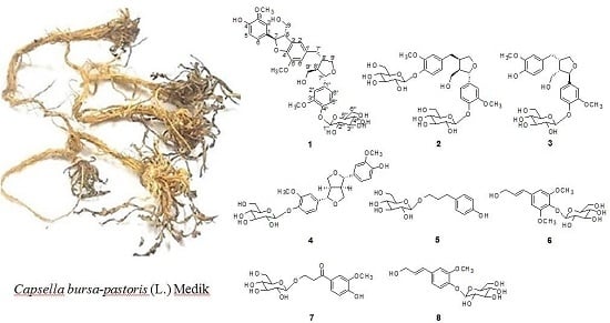
Phenolic Glycosides from Capsella bursa-pastoris (L.) Medik and Their Anti-Inflammatory Activity

1
Natural Products Laboratory, School of Pharmacy, Sungkyunkwan University, Suwon 16419, Korea
2
Gachon Institute of Pharmaceutical Science, Gachon University, 191 Hambakmoero, Yeonsu-gu, Incheon 21936, Korea
3
College of Pharmacy, Gachon University, 191 Hambakmoero, Yeonsu-gu, Incheon 21936, Korea
*
Author to whom correspondence should be addressed.
Submission received: 16 May 2017 / Revised: 12 June 2017 / Accepted: 18 June 2017 / Published: 20 June 2017
(This article belongs to the Collection Bioactive Compounds)

Abstract

:
A new sesquilignan glycoside 1, together with seven known phenolic glycosides 28 were isolated from the aerial parts of Capsella bursa-pastoris. The chemical structure of the new compound 1 was elucidated by extensive nuclear magnetic resonance (NMR) data (1H- and 13C-NMR, 1H-1H correlation spectroscopy (1H-1H COSY), heteronuclear single-quantum correlation (HSQC), heteronuclear multiple bond correlation (HMBC), and nuclear overhauser effect spectroscopy (NOESY)) and HR-FABMS analysis. The anti-inflammatory effects of 18 were evaluated in lipopolysaccharide (LPS)-stimulated murine microglia BV-2 cells. Compounds 4 and 7 exhibited moderate inhibitory effects on nitric oxide production in LPS-activated BV-2 cells, with IC50 values of 17.80 and 27.91 µM, respectively.

Graphical Abstract

1. Introduction

Capsella bursa-pastoris (L.) Medik (Cruciferae), commonly known as ‘shepherd’s purse’ is an annual plant which is widely distributed throughout the world [1]. The roots of this plant have been used in Korean traditional medicine for the treatment of edema and hypertension [2]. Previous phytochemical investigations of this plant led to reports of several flavonoids, terpenoids, and phenolic compounds with antimicrobial, antibacterial, antitumor, and liver catalase activities [3,4,5,6,7,8]. Some of these compounds of C. bursa-pastoris, such as flavonoids and sulforaphane, have been reported to exhibit good antibacterial and anti-inflammatory activity [4,9]. In the course of our continuing search for bioactive constituents in Korean medicinal plants, we found that the n-BuOH fraction of the MeOH extract of C. bursa-pastoris inhibited NO production in a lipopolysaccharide (LPS)-activated microglia BV-2 cell line. The n-BuOH fraction was subjected to repeated column chromatography with Diaion HP-20, silica gel, and semipreparative HPLC separation to yield a new sesquilignan glycoside 1, and seven known compounds 28 (Figure 1). The structure of the new compound 1 was determined by spectroscopic methods, including 1D and 2D NMR (1H- and 13C-NMR, 1H-1H COSY, HSQC, HMBC, and NOESY), and HR-FABMS analysis. The known compounds 28 were identified by comparison of their spectroscopic data and specific optical rotation with reported data. The isolated compounds 18 were tested for their inhibitory effects on NO production in an LPS-activated microglia BV-2 cell line.

2. Results and Discussion

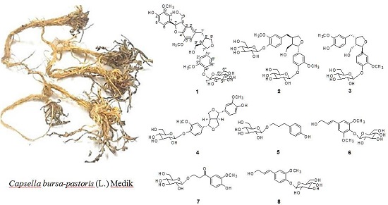
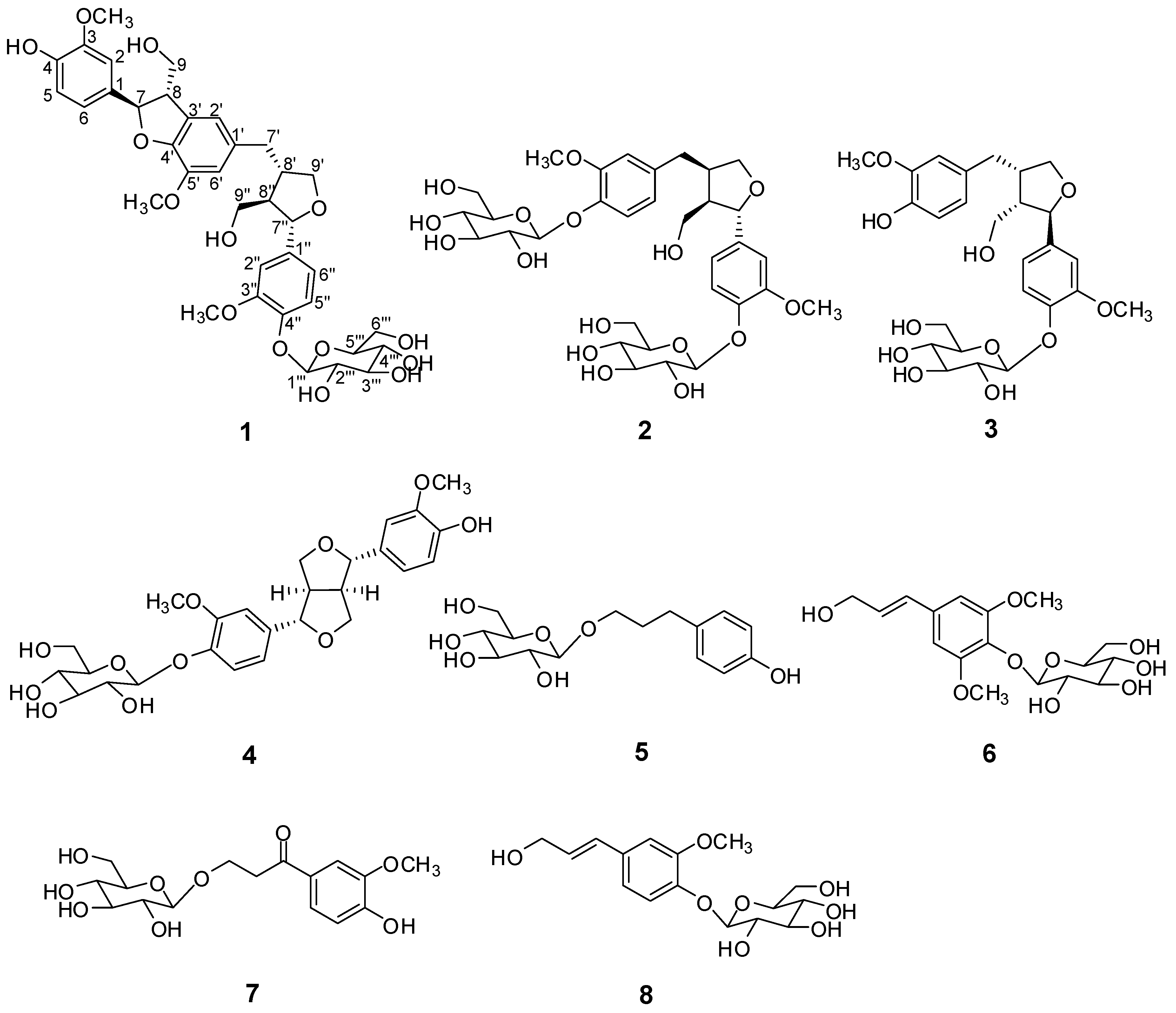
2.1. Structure Elucidation

Purification of the n-BuOH layer by column chromatography afforded a new sesquilignan glycoside 1, together with three known lignin glycosides 24 and four known phenolic glycosides 58. The known compounds were identified as 7S, 8R, 8′R-(−)-lariciresinol-4,4′-bis-O-glucopyranoside (2) [10], lariciresinol4′-O-β-d-glucoside (3) [11], (+)-pinoresinol-β-d-glucoside (4) [12], salidroside (5) [13], 3-(4-β-d-glucopyranosyloxy-3,5-dimethoxy)-phenyl-2E-propanol (6) [14], β-hydroxy-propiovanillone 3-O-β-d-glucopyranoside (7) [15], and coniferin (8) [16] by comparing their 1H-,13C-NMR and MS spectral data with values in the literature.
Compound 1 was obtained as a brownish gum. The molecular formula was established as C36H44O14 using HR-FABMS, which showed a positive ion [M + Na]+ at m/z: 723.2626 (calcd. for C36H44O14Na, 723.2629, so the molecular formula was deduced to be C36H44O14. The 1H-NMR spectrum of 1 revealed the presence of two 1,3,4-trisubstituted aromatic rings (δH 7.16 (d, J = 8.3 Hz, H-5′′), 7.15 (d, J = 8.2 Hz, H-5), 7.05 (d, J = 1.7 Hz, H-2), 7.01 (d, J = 1.7 Hz, H-2′′), 6.95 (dd, J = 8.2, 1.7 Hz, H-6), and 6.90 (dd, J = 8.2, 1.7 Hz, H-6″)), a 1,3,4,5-tetrasubstitued aromatic ring (δH 6.76 (s, H-6′), 6.78 (s, H-2′)), two oxygenated methines (δH 5.58 (d, J = 5.9 Hz, H-7) and 4.85 (m, H-7′′)), two oxygenated methylenes (δH 3.85 (m, H-9b), 3.78 (m, H-9a), and 3.88 (m, H-9′′a), 3.68 (dd, J = 10.9, 6.7 Hz, H-9b′′)), a methylene 2.96 ((dd, J = 13.5, 5.0 Hz, H-7′a), and 2.57 (dd, J = 13.3, 11.0 Hz, H-7′b)), three methines (δH 3.48 (m, H-8), 2.75 (sep, J = 6.5, 5.5 Hz, H-8′), and 2.38 (quin, J = 6.9 Hz, H-8″)), three methoxy groups (δH 3.88 (s, 3-OCH3), 3.87 (s, 5′-OCH3) and 3.84 (s, 3′′-OCH3)), and a glucopyanosyl unit (δH 4.91 (d, J = 7.3 Hz, H-1′′′), 3.69 (m, H-6′′′), 3.51 (m, H-2′′′), 3.41 (overlap, H-4′′′, 5′′′) and 3.39 (m, H-3′′′). The 13C-NMR spectrum displayed 36 carbon signals, including 18 aromatic carbons δC 151.1 (C-3″), 147.9 (C-4′, 3′), 147.8 (C-3), 147.5 (C-4, 4″), 145.5 (C-5′), 139.7 (C-1″), 135.7 (C-1), 134.9 (C-1′), 119.7 (C-6″), 119.5 (C-6), 118.4 (C-6′), 118.2 (C-5″), 118.1 (C-5), 114.7 (C-2′), 111.5 (C-2″) and 111.3 (C-2), three oxygenated carbons δC 88.6 (C-7), 83.9 (C-7″), and 73.5 (C-9′), three methylene carbons δC 65.1 (C-9), 60.5 (C-9″), 33.9 (C-7′) and three methine carbons δC 55.7 (C-8), 54.2 (C-8″), 44.0 (C-8′), and, three methoxy carbons (δC 56.9 and 56.8 (×2)), and glucose carbons δc 103.1 (C-1′′′), 78.5 (C-3′′′), 78.4 (C-5′′′), 75.1 (C-2′′′), 71.6 (C-4′′′), and 62.6 (C-6′′′) (Table 1).
The 1H- and 13C-NMR spectra (Table 1) were very similar to those of abiesol A [17], except for the presence of a glucose unit in 1. The positions of three methoxy groups were confirmed as 3-OCH3H 3.88)/C-3 (δC 147.8), 5′-OCH3C 3.87)/C-5′ (δC 145.5), and 3″-OCH3H 3.84)/C-3″ (δC 151.1). HMBC correlation (H-1′′′ to C-4″) indicated that a D-glucose moiety was linked to C-4″ (Figure 2a), and identified the β form by the coupling constant (J = 7.3 Hz) [18]. The stereochemistry of 1 was assigned on the basis of examination of the CD spectrum in combination with the NOESY experiment. The absolute configurations of C-7 and C-8 were confirmed as 7R and 8S from the positive Cotton effect at 223 nm and the negative effect at 245 and 291 nm in the CD spectrum [19]. The absolute configurations of C-8′/C-7″/C-8″ were identified as 8′S, 7″S and 8R from the negative Cotton effect at 231 nm and 280 nm, respectively (Figure S8, Supplementary Materials) [19]. HMBC correlations and NOESY cross-peaks (Figure 2) reconfirmed the suggested structure of 1. The enzymatic hydrolysis of 1 afforded d-glucose, which was identified by the sign of the specific rotation [ α ] D 25 +48.2 (c = 0.03, H2O) and by co-TLC (CHCl3:MeOH:H2O = 2:1:0.1; Rf = 0.21) [20] and the aglycone 1a, which was identified by 1H-NMR and MS data [17]. Thus, the structure of 1 was determined as (7R, 8S, 8′S, 7″S, 8″R)-abiesol A 4″-O-β-d-glucopyranoside, and it was named capselloside.

2.2. Anti-Inflammatory Activity

Nitric oxide (NO) is one of the pro-inflammatory mediators and also a signaling molecule responsible for the induction of inflammation and pain through the pronounced induction of pro-inflammatory cytokines [21]. It is believed to be produced excessively in the inflammatory conditions so it has been considered as a major biomarker for the screening of the anti-inflammatory activity of compound [22]. Compounds that can inhibit the NO production possess an anti-inflammatory activity against LPS-induced neuroinflammation [23].
The anti-inflammatory activities of the isolates 18 were evaluated by measuring the levels of nitric oxide (NO) produced in lipopolysaccharide (LPS)-activated microglia BV-2 cells [24]. Compound 4 significantly inhibited LPS-stimulated NO production, with an IC50 value of 17.80 μM and compound 7 showed moderate NO production, with an IC50 value of 27.91 μM without cell toxicity (Table 2), which represented more potent activity than the positive control, L-NMMA (20.76 μM).

3. Materials and Methods

3.1. General Experimental Procedures

Optical rotations were measured on a P-1020 polarimeter in MeOH (Jasco, Easton, MD, USA). Infrared (IR) spectra were recorded on a IFS-66/S FT-IR spectrometer (Bruker, Karlsruhe, Germany). Circular dichroism (CD) spectra were measured on a Jasco J-715 spectropolarimeter using methanol as a solvent. Ultraviolet (UV) spectra were recorded using a UV-1601 UV-Visible spectrophotometer (Shimadzu, Tokyo, Japan). HR-FABMS spectra were obtained on a JMS700 mass spectrometer (JEOL, Peabody, MA, USA). TLC was performed using precoated silica gel F254 plates and RP-18 F254s plates (Merck, Darmstadt, Germany). NMR spectra, including COSY, HMQC, and HMBC experiments were recorded on an AVANCE III 700 NMR spectrometer at 700 MHz (1H) and 175 MHz (13C). (Bruker, Billerica, MA, USA). Preparative high performance liquid chromatography (HPLC) was conducted using a 306 pump (Gilson, Middleton, WI, USA) equipped with a Shodex refractive index detector (Shodex, New York, NY, USA) and a Phenomenex-Luna-10u-silica-(2) column (250 mm × 10.00 mm i.d.) or a YMC J’sphere ODS-M80 column (250 mm × 10.00 mm i.d.). Low-pressure liquid chromatography (LPLC) was carried out over a Li-Chroprep Lobar-A RP-18 column (240 mm × 10 mm i.d.; Merck) with an FMI QSY-0 pump (Teledyne Isco, Lincoln, NE, USA). Silica gel 60 and RP-C18 silica gel (230–400 mesh, Merck) were used for column chromatography. The packing material for macroporous adsorbent column chromatography was a Diaion HP-20 column (Sigma, St. Louis, MO, USA). Spots were detected by thin layer chromatography (TLC) under UV light or by heating after spraying with anisaldehyde-sulfuric acid.

3.2. Plant Materials

Whole plants of C. bursa-pastoris (3 kg) were purchased from Anmyeon-Island, Chungcheongnam-do, Korea in March 2015. A voucher specimen (SKKU-NPL 1410) has been deposited in the herbarium of the School of Pharmacy, Sungkyunkwan University, Suwon, Korea.

3.3. Extraction and Isolation

Whole plants of C. bursa-pastoris (3 kg) were extracted three times at room temperature with 80% aqueous MeOH (10 L × 1 day) and then filtered. The filtrate was evaporated under vacuum to afford a crude MeOH extract (280 g), which was suspended in distilled H2O and successively partitioned with n-hexane, CHCl3, EtOAc, and n-BuOH, yielding 33, 5, 4, and 26 g respectively. The BuOH-soluble layer (26 g) was separated on a Diaion HP-20 column eluted with a gradient solvent system of 100% H2O and 100% MeOH, yielding subfractions A (100% H2O) and B (100% MeOH). Subfraction B (7 g) was separated over a silica gel column (230–400 mesh, 300 g) with CHCl3–MeOH–H2O (5:1:0.1) to afford six subfractions (B1–B6). Subfraction B2 (168 mg) was chromatographed over a Lobar-A RP-18 column (25% MeCN) to give four subfractions (B21–B24). Subfraction B22 (54 mg) was purified by semi-preparative reversed-phase HPLC using a solvent system of 32% MeOH (flow rate; 2 mL/min) to isolate compound 4 (7 mg). Subfraction B24 (41 mg) was purified by semi-preparative reversed-phase HPLC using a solvent system of 45% MeOH (flow rate; 2 mL/min) to isolate compound 3 (6 mg). Subfraction B3 (1 g) was separated over a RP-C18 column (230–400 mesh, 150 g) with a solvent system of 10% MeOH to obtain eleven subfractions (B301–B311). Compounds 5 (7 mg), 8 (30 mg), 6 (9 mg), 7 (5 mg) and, 9 (13 mg) were acquired by purification of fractions B303 (17 mg), B304 (66 mg), B305 (21 mg), B306 (14 mg), and B311 (16 mg), respectively, using semi-preparative normal-phase HPLC with CHCl3–MeOH–H2O (5:1:0.1; flow rate; 2 mL/min). Subfraction B5 (1 g) was separated over a RP-C18 column (230–400 mesh, 150 g) with a solvent system of 10% MeOH to obtain ten subfractions (B501–B510). Compounds 1 (3 mg), and 2 (14 mg) were acquired by purification of fractions B503 (17 mg) and B505 (21 mg), respectively, using semi-preparative normal-phase HPLC with a solvent system of CHCl3–MeOH–H2O (2:1:0.1; flow rate; 2 mL/min).

3.4. Characterization

Capselloside (1): Brownish gum; [ α ] D 25 −34.0 (c = 0.05, MeOH); UV (MeOH) λmax (log ε) 280 (1.4), 228 (3.3), 214 (3.1) nm; IR (KBr) νmax 3261, 2946, 2816, 1638, 1489, 1261 cm−1; CD (MeOH) λmax (Δ ε) 291 (−), 280 (−) 257 (−) 230 (−) 223 (+); 1H (700 MHz) and 13C (175 MHz) NMR data, see Table 1; HR-FABMS (positive-ion mode) m/z: 723.2626 [M + Na]+ (calcd. for C36H44O14Na, 723.2629).

3.5. Enzymatic Hydrolysis

Compound 1 (1.0 mg) in H2O (3.0 mL) was hydrolyzed with crude β-glucosidase (10.0 mg, from almonds; Sigma-Aldrich) at 37 °C for 48 h. The reaction mixture was extracted with CHCl3 (3 × 3 mL). The CHCl3 layer (0.6 mg) was chromatographed over a silica gel Waters Sep-Pak Vac 12 cc eluted with CHCl3–MeOH (30:1) to give the aglycone 1a (0.4 mg), which was identified by 1H‑NMR and MS data. The sugar (d-glucose) from the aqueous phase of the hydrolysate of 1 was identified by silica gel TLC (CHCl3–MeOH–H2O 2:1:0.1; Rf = 0.21) comparison with a d-glucose standard. The specific rotation of the sugar obtained from 1 was [ α ] D 25 +48.2 (c = 0.03, H2O).

3.6. Measurement of Nitric Oxide Production and Cell Viability in LPS-Activated BV-2 Cells

The inhibitory effect of the test compounds on LPS-stimulated NO production was studied using BV-2 cells. BV-2 cells were seeded on a 96-well plate (4 × 10⁴ cells/well) and treated with or without different concentrations of the compounds. The cells were stimulated with LPS (100 µg/mL) and incubated for 24 h. The concentration of nitrite (NO2), a soluble oxidation product of NO, in the culture medium was measured using Griess reagent (0.1% N-1-naphthylethylenediamine dihydrochloride and 1% sulfanilamide in 5% phosphoric acid). Fifty microliters of supernatant was mixed with an equal volume of Griess reagent. Absorbance was measured after 10min using a microplate reader (Emax, Molecular Devices, Sunnyvale, CA, USA) at a wavelength of 570 nm. L-NMMA, a well-known nitric oxide synthase (NOS) inhibitor, was used as a positive control. Graded sodium nitrite solution was used as a standard to calculate nitrite concentration. Cell viability was evaluated by observing the ability of viable cells to reduce the yellow-colored MTT to purple formazan, using an MTT assay.

4. Conclusions

We have isolated and identified from C. bursa-pastoris a new sesquilignan glycoside 1, which we have named capselloside, together with seven known compounds 28 and evaluated their anti-inflammatory effects. Nitric oxide synthesis inhibition potency of phenolic glycosides suggests their anti-inflammatory activity against LPS-induced neuroinflammatory model in vitro. (+)-Pinoresinol-β-d-glucoside (4) showed the most potent anti-inflammatory effects and β-hydroxypropiovanillone 3-O-β-d-glucopyranoside (7) showed moderate NO production in an LPS-activated microglia BV-2 cell line. Compound 4 showed better potency than that of the commercial NOS inhibitor to inhibit LPS-induced NO production and neuroinflammation. In conclusion, our results collectively support the notion that phenolic glycosides from Capsella bursa-pastoris (L.) Medik may be potentially responsible for the observed in vitro anti-inflammatory activity in a LPS-induced neuroinflammatory model.

Supplementary Materials

The NMR, HR-FABMS, IR, UV, and CD spectra of 1 are available as supplementary materials available online.

Acknowledgments

We are thankful to the Korea Basic Science Institute (KBSI) for the measurements of NMR and MS spectra. This research was supported by the Basic Science Research Program through the National Research Foundation of Korea (NRF) funded by the Ministry of Education, Science and Technology (2012R1A5A2A28671860). We are thankful to the Korea Basic Science Institute (KBSI) for the MS spectra measurements.

Author Contributions

J.M.C. and K.R.L. conceived and designed the experiments. J.M.C., W.S.S., and T.H.L. performed the extraction and isolation. J.M.C., W.S.S. and K.R.L. structure elucidation of the compounds, and analyzed the data. L.S. and S.Y.K. carried out the bioassay experiments. J.M.C. wrote the paper.

Conflicts of Interest

The authors declare no conflicts of interest.

References

  1. Lee, Y.N. Flora of Korea; Kim, S., Ed.; Kyohaksa: Seoul, Korea, 1998; p. 259. [Google Scholar]
  2. Kuroda, K.; Kaku, T. Pharmacological and chemical studies on the alcohol extract of Capsella bursa-pastoris. Life Sci. 1969, 8, 151–155. [Google Scholar] [CrossRef]
  3. Song, N.; Xu, W.; Guan, H.; Liu, X.; Wang, Y.; Nie, X. Several flavonoids from Capsella bursa-pastoris (L.) Medic. Asian J. Tradit. Med. 2007, 2, 218–222. [Google Scholar]
  4. Grosso, C.; Vinholes, J.; Silva, L.R.; Pinho, P.G.d.; Gonçalves, R.F.; Valentão, P.; Jäger, A.K.; Andrade, P.B. Chemical composition and biological screening of Capsella bursa-pastoris. Rev. Bras. Farmacogn. 2011, 21, 635–643. [Google Scholar] [CrossRef]
  5. Selenu, M.; Carrus, F.; Bonsignore, L. Phytochemical study on capsella bursa pastoris l. Boll. Chim. Farm. 2005, 144, 66–78. [Google Scholar]
  6. Park, C.J.; Park, C.B.; Hong, S.S.; Lee, H.S.; Lee, S.Y.; Kim, S.C. Characterization and cdna cloning of two glycine-and histidine-rich antimicrobial peptides from the roots of shepherd’s purse, Capsella bursa-pastoris. Plant Mol. Biol. 2000, 44, 187–197. [Google Scholar] [CrossRef] [PubMed]
  7. Kuroda, K.; Akao, M. Antitumor and anti-intoxication activities of fumaric acid in cultured cells. Gann 1981, 72, 777–782. [Google Scholar] [PubMed]
  8. Kuroda, K.; Mitsutaro, A. Effect of Capsella bursa-pastoris on liver catalase activity in rats fed 3′-methyl-4-(dimethylamino) azobenzene. Gann 1975, 66, 461–462. [Google Scholar] [PubMed]
  9. Hur, J.; Yoo, M.; Shin, D.B.; Lee, S. Inhibition of nitric oxide production corresponds to the sulforaphane content in Korean sheperd’s purse (Capsella bursa-pastoris) and related species in BV-2 cell. Food Sci. Biotechnol. 2013, 22, 1085–1089. [Google Scholar] [CrossRef]
  10. El Gamal, A.; Takeya, K.; Itokawa, H.; Halim, A.; Amer, M.; Saad, H.E. Lignan bis-glucosides from Galium sinaicum. Phytochemistry 1997, 45, 597–600. [Google Scholar] [CrossRef]
  11. Karioti, A.; Protopappa, A.; Megoulas, N.; Skaltsa, H. Identification of tyrosinase inhibitors from Marrubium velutinum and Marrubium cylleneum. Bioorg. Med. Chem. 2007, 15, 2708–2714. [Google Scholar] [CrossRef] [PubMed]
  12. Kim, D.K.; Lim, J.P.; Kim, J.W.; Park, H.W.; Eun, J.S. Antitumor and antiinflammatory constituents from Celtis sinensis. Arch. Pharm. Res. 2005, 28, 39–43. [Google Scholar] [CrossRef] [PubMed]
  13. Akita, H.; Kawahara, E.; Kishida, M.; Kato, K. Synthesis of naturally occurring β-D-glucopyranoside based on enzymatic β-glycosidation. J. Mol. Catal. B: Enzym. 2006, 40, 8–15. [Google Scholar] [CrossRef]
  14. Della Greca, M.; Ferrara, M.; Fiorentino, A.; Monaco, P.; Previtera, L. Antialgal compounds from Zantedeschia aethiopica. Phytochemistry 1998, 49, 1299–1304. [Google Scholar] [CrossRef]
  15. Kim, J.S.; Kwon, Y.S.; Sa, Y.J.; Kim, M.J. Isolation and identification of sea buckthorn (Hippophae rhamnoides) phenolics with antioxidant activity and α-glucosidase inhibitory effect. J. Agric. Food Chem. 2010, 59, 138–144. [Google Scholar] [CrossRef] [PubMed]
  16. Han, M.H.; Yang, X.W.; Zhang, M.; Zhong, G.Y. Phytochemical study of the rhizome of Pinellia ternata and quantification of phenylpropanoids in commercial Pinellia tuber by RP-LC. Chromatographia 2006, 64, 647–653. [Google Scholar] [CrossRef]
  17. Ozawa, S.; Sasaya, T. Extractives of Todomatsu Abies sachalinensis Masters, 7: New phenylpropane trimers from the wood of Abies sachalinensis. Jpn. Wood Res. Soc. 1991, 37, 69–75. [Google Scholar]
  18. Perkins, S.J.; Johnson, L.N.; Phillips, D.C.; Dwek, R.A. High resolution 1H-and 13N-NMR spectra of d-gluco-pyranose, 2-acetamido-2-deoxy-d-glucopyranose, and related compounds in aqueous media. Carbohydr. Res. 1977, 59, 19–34. [Google Scholar] [CrossRef]
  19. Yang, Y.N.; Huang, X.Y.; Feng, Z.M.; Jiang, J.S.; Zhang, P.C. Hepatoprotective activity of twelve novel 7′-hydroxy lignan glucosides from Arctii fructus. J. Agric. Food Chem. 2014, 62, 9095–9102. [Google Scholar] [CrossRef] [PubMed]
  20. Kim, K.H.; Choi, J.W.; Choi, S.U.; Lee, K.R. Terpene glycosides and cytotoxic constituents from the seeds of Amomum xanthioides. Planta Med. 2010, 76, 461–464. [Google Scholar] [CrossRef] [PubMed]
  21. Abramson, S.B. Nitric oxide in inflammation and pain associated with osteoarthritis. Arthritis Res. Ther. 2008, 10, S2. [Google Scholar] [CrossRef] [PubMed]
  22. Sharma, J.; Al-Omran, A.; Parvathy, S. Role of nitric oxide in inflammatory diseases. Inflammopharmacology 2007, 15, 252–259. [Google Scholar] [CrossRef] [PubMed]
  23. Subedi, L.; Gaire, B.P.; Do, M.H.; Lee, T.H.; Kim, S.Y. Anti-neuroinflammatory and neuroprotective effects of the Lindera neesiana fruit in vitro. Phytomedicine 2016, 23, 872–881. [Google Scholar] [CrossRef] [PubMed]
  24. Ko, W.; Sohn, J.H.; Kim, Y.C.; Oh, H. Viridicatol from the marine-derived fungal strain Penicillium sp. SF-5295 exerts anti-inflammatory effects through inhibiting NF-ΚB signaling pathway on lipopolysaccharide-induced RAW264. 7 and BV2 cells. Nat. Prod. Sci. 2015, 21, 240–247. [Google Scholar] [CrossRef]
Sample Availability: Samples of the compounds are available from the authors.
Figure 1. Chemical structures of compounds 18.
Molecules 22 01023 g001
Figure 2. Key 1H-1H COSY, HMBC (a) and NOESY (b) correlations of 1.
Molecules 22 01023 g002
Table 1. 1H (700 MHz) and 13C (175 MHz) NMR data of 1 in methanol-d4 (δ in ppm) a.
PositionδC, TypeδH (J in Hz)
1135.7, C
2111.3, CH7.05, d (1.7)
3147.8, C
4147.5, C
5118.1, CH7.15, d (8.2)
6119.5, CH6.95, dd (8.2, 1.7)
788.6, CH5.58, d (5.91)
855.7, CH3.48, m
965.1, CH2
a 3.78, m
b 3.85, m
1′134.9, C
2′114.7, CH6.78, s
3′147.9, C
4′147.9, C
5′145.5, C
6′118.4, CH6.76, s
7′33.9, CH2
a 2.96, dd (13.5, 5.0)
b 2.57, dd (13.3, 11.0)
8′44.0, CH2.75, sep (6.5, 5.5)
9′73.5, CH2
a 3.78, m
b 4.05, dd (8.3, 6.6)
1″139.7, C
2″111.5, CH7.01, d (1.7)
3″151.1, C
4″147.5, C
5″118.2, CH7.16, d (8.3)
6″119.7, CH6.90, dd (8.2, 1.7)
7″83.9, CH4.85, overlap
8″54.2, CH2.38, quin (6.9)
9″60.5, CH2
a 3.88, m
b 3.68, dd (10.9, 6.7)
Glc-1′′′103.1, CH4.91, d (7.3)
2′′′75.1, CH3.51, m
3′′′78.5, CH3.39, m
4′′′71.6, CH3.41, m
5′′′78.4, CH3.41, m
6′′′62.6, CH3.69, overlap
OCH3 (3″)56.93.84, s
OCH3 (3)56.83.88, s
OCH3 (5′)56.83.87, s
a The assignments were based on HSQC and HMBC experiments.
Table 2. Effects of compounds 18 on NO production in LPS-activated BV-2 cells.
CompoundIC50 a (µM)Cell Viability b (%)
175.13103.04 ± 7.08
248.80116.68 ± 3.69
330.70109.17 ± 8.61
417.80117.36 ± 10.23
531.14116.09 ± 10.67
662.21110.02 ± 9.52
727.91119.20 ± 6.23
849.21111.55 ± 10.40
c L-NMMA20.76112.89 ± 4.90
a The IC50 value of each compound was defined as the concentration (µM) that caused 50% inhibition of NO production in LPS-activated BV-2 cells; b Cell viability after treatment with 20 µM of each compound was determined by the MTT assay and is expressed as a percentage (%). The results are averages of three independent experiments, and the data are expressed as mean ± SD; c L-NMMA was used as a positive control.

