Article

A Novel Brominated Alkaloid Securidine A, Isolated from the Marine Bryozoan Securiflustra securifrons

1
MARBIO, UiT–The Arctic University of Norway, Breivika, Tromsø N-9037, Norway
2
Department of Chemistry, UiT–The Arctic University of Norway, Breivika, Tromsø N-9037, Norway
*
Authors to whom correspondence should be addressed.
These authors contributed equally to this work.
Submission received: 20 June 2017 / Revised: 12 July 2017 / Accepted: 17 July 2017 / Published: 23 July 2017
(This article belongs to the Section Natural Products Chemistry)

Abstract

:
A novel brominated alkaloid, Securidine A, was isolated from the cold water marine bryozoan Securiflustra securifrons. Securidine A was isolated using semi-preparative HPLC, and the structure was elucidated by spectroscopic methods. The isolated Securidine A was tested for cytotoxic, antibacterial, and anti-diabetic activities as well as for its potential for inhibition of biofilm formation. No significant biological activity was observed in the applied bioassays, thus expanded bioactivity profiling is required, in order to reveal any potential applications for Securidine A.

1. Introduction

Marine organisms are considered to be a rich source for natural products, and currently almost 50,000 marine natural products are reported in the Dictionary of Marine Natural Products [1]. A statistical analysis of marine natural products discovered between 1985 and 2008 estimates that approximately 75% of the compounds are isolated from sessile and soft-bodied invertebrates including sponges, ascidians, cnidarians, mollusks and bryozoans [2,3]. In order to protect themselves from predators and to compete for space and food, many invertebrates have evolved the ability to produce secondary metabolites as a chemical defense, which increase their ability to survive, grow, and reproduce [4,5]. Bryozoans, or moss animals, are aquatic invertebrates that belong to the phylum Bryozoa (Polyzoa). They are sedentary and filter feeders. These colonial organisms consist of tiny intercommunicating individuals called zooids [6]. Bryozoans are widely distributed in tropical and temperate marine waters [7], and are common fouling organisms on marine substrates [6]. The phylum consists of 3 classes, 4 orders, 187 families, and 808 genera, and approximately 8000 species are described, most of them are observed in marine habitats [8,9]. Marine bryozoans are known to produce a significant number of biologically active secondary metabolites, including macrolides, alkaloids, sterols, and heteroatom-containing compounds [3,10] that possess antitumor [11], antibacterial [12,13], and muscle relaxant activities [14]. The well-known bioactive compound bryostatin 1 is a macrolide isolated from the marine bryozoan Bugula neritina, and it is proven to be an antineoplastic agent [15]. Secondary metabolites isolated from marine bryozoans have been extensively reviewed [6,8,16,17].
In the present study, we isolated the novel mono brominated tyrosine derivative Securidine A (1) from the marine bryozoan Securiflustra securifrons. S. securifrons (Pallas, 1766) is a temperate and subtidal species abundant in the North Sea [18,19], and the encrusting basal portion of the colonies are attached to hard substrates such as stones and shells. In previous reports, seven halogenated indole-imidazole alkaloids, Securamines A–G and Securines A and B (obtained by dissolving Securamines A and B in DMSO-d6), have been isolated from S. securifrons, but no biological activities were recorded for any of these compounds. The halogenated Securamines A–G (Figure 1) differ from each other by the presence and absence of a bromine substitution or a number of bromine substitutions in the benzene ring. [20,21]. These compounds are structurally similar to chartellines A–C and chartellamides A and B, isolated from another marine bryozoan Chartella papyracea. Both species belong to the Flustridae family (Order: Cheilostomata). Many of the Cheilostome bryozoans including S. securifrons, C. papyracea, and Flustra foliacea are producing indole alkaloids and brominated alkaloids [12,20,21,22,23,24,25,26,27,28,29,30,31,32]. However, the isolated new brominated alkaloid, Securidine A (1) is a novel structure (Figure 1), which is not structurally similar to any other listed compounds of S. securifrons.
The current paper describes the isolation of new compound Securidine A (1), its structural characterization and potential biological activities were also investigated.

2. Results and Discussion

The marine bryozoan S. securifrons was collected in April 2014 in the North Sea. Securidine A (1) was isolated from the aqueous extract of S. securifrons through stepwise fractionation. The lyophilized animal material was ground and extracted with ultrapure water. The freeze-dried aqueous extract was pre-fractionated using column chromatography with a Biotage SP24 HPFC flash purification system. The extract was eluted with a gradient of H2O/CH3OH/CH3-CO-CH3 and the fractions were assayed for cytotoxicity against melanoma (A2058) and colon adenocarcinoma (HT29) cell lines using an MTS based anticancer assay. Flash fractions four and five showed cytotoxic activity. UPLC-HR-MS analysis of these fractions, followed by isotopic pattern examination of the eluting compounds, revealed the presence of a mono brominated compound. For the isolation, the aqueous extract was injected onto semi-preparative RP-HPLC C18 column and Securidine A (1) was eluted with a 10 minutes gradient, changing from 10% to 40% CH3CN over 10 minutes. For the second purification step, a Phenyl-Hexyl prep HPLC column was used with a 10 min gradient, changing from 10% to 34% CH3CN.
The HR-MS analysis suggested that Securidine A (1) contained a bromine. The molecular formula was determined to be C14H21BrN4O2, corresponding to the protonated molecular peak at m/z 357.0858 ([M + H]+, calculated m/z 357.0926), suggesting six degrees of unsaturation. A database search indicated that the isolated compound was novel, and its structure was solved based on a number of 1D and 2D NMR experiments.
Interpretation of the available 1D 1H (presat, 1DNOE and excitation sculpting), 1D 13C, HMBC, ME-HSQC, H2BC, COSY, and ROESY (Supplementary materials Figures S1 to S6) led to an unambiguous structural elucidation of 1. Examination and comparison of the 1D 13C, HSQC, and HMBC showed that 1 had 14 carbons consisting of one methyl group, five methylenes, three methines, and five quaternary carbon atoms. Two 1H resonances, H-10 and H-15, were identified as NH groups based on the absence of any 1JCH, broadened line widths and deshielded chemical shifts. Three methines indicated the presence of a disubstituted phenyl ring attached to the main chain. The methyl group protons (δH 3.84) showed HMBC correlation to the quaternary C-2 (δC 156.2) as well as ROESY correlation to the H-3 (δH 6.96) methine proton. The relatively deshielded chemical shift value of the methyl protons (δH 3.84) was assigned as the protons of a methoxy group. The high chemical shift value of C-2 (δC 156.2) further confirmed this. COSY and ROESY correlations between H-3 (δH 6.96) and H-4 (δH 7.22) as well as a shared coupling constant of 8.6 between the two, places C-3 and C-4 next to each other. Additionally, the H-3 methine showed HMBC correlation to a halogenated quaternary carbon (C-5, δC 112.2), the quaternary C-7, (δC 130.5) as well as to the final methine carbon (C-6, δC 134.5). The distinction between C-7 and C-5 was made based on their chemical shift values and a 2JCH from C-5 to H-6, which is commonly observed from halogenated carbons but not from carbons in phenyl rings further away from heteroatoms. The H-8 (δH 3.41) methylene showed ROESY and HMBC correlations to the protons and carbons in position 4 (δH 7.22, δC 130.2) and 6 (δH 7.47, δC 134.5), placing it in ortho position to C-4 and C-6 in the phenyl ring. The phenyl group was thus methoxy-substituted in the para position and brominated in the meta position, while and the main chain of the molecule was attached to C-7. The H-8 (δH 3.41) protons further showed a HMBC correlation to a deshielded quaternary carbon (C-9, δC 173.7), characteristic for a carbonyl carbon. HMBC correlations were observed to H-10 (δH 8.20) from C-9 and to H-10 from C-11 (δC 39.8). COSY and ROESY correlations were observed between the methylene groups in the 11 (δH 3.20, δC 39.8) and 12 positions (δH 1.56, δC 27.5). The same correlations were observed between the methylene groups in position 13 (δH 1.55, δC 27.0) and 14 (δH 3.16, δC 42.0). The methylene groups in the 12 and 13 position were near isochronous, but could be distinguished by their respective couplings to H-11 and H-14 in H2BC and COSY. H-14 further correlated with the quaternary C-16 (δC 158.8) in HMBC spectra. H-13 (δH 1.55) and H-14 (δH 3.16) both showed ROESY correlations to H-15 (δH 7.57). This, in addition to a COSY correlation between H-14 and H-15, places a NH-group in the 15 position. Thus, the remaining part of the chemical formula was N2H3, leaving the only possible structural solution to be the attachment of an NH- and an NH2- group to C-16. The deshielded shift value of C-16 (δC 158.8) was in good agreement with this conclusion (Table 1). Selected 2D correlations utilized in determining the structure of 1 are shown in Figure 2. The compound was named Securidine A.

Bioactivity

The pure Securidine A (1) was evaluated for its biological activity in a selection of bioassays including anti-cancer and anti-bacterial activity, inhibition of protein tyrosine phosphatase 1B (PTP-1B) for anti-diabetic activity, as well as for inhibition of biofilm formation.
Securidine A (1) was assessed for its cytotoxicity against three cancer cell lines (A2058 melanoma, HT29 colon adenocarcinoma and MCF7 breast carcinoma) by the MTS method, since flash fractions of the extract from S. securifrons showed anticancer activity against A2058 melanoma and HT29 colon adenocarcinoma cancer cell lines in the MabCent screening program [33]. However, the isolated compound, Securidine A (1), did not show any significant activity against the cancer cell lines at 50 µM (17.87 µg/mL) or 100 µM (37.73 µg/mL). Securidine A (1) was also tested for its antibacterial activity, and no activity was seen against Staphyloccoccus aureus, Escherichia coli, Enterococcus faecalis, Pseudomonas aeruginosa, or Streptococcus agalactiae.
Securidine A (1) is a brominated tyrosine derivative. It is structurally related to two other brominated tyrosine derivatives, Pulmonarin B (2) and Synoxazolidinone B (3) (Figure 3), both isolated from the sub-Arctic ascidian Synoicum pulmonaria [34,35]. Pulmonarin B (2) contains a quaternary ammonium group in the side-chain instead of the guanidine group of Securidine A (1) and Pulmonarin B (2) did not have any antibiotic or cytotoxic activity. Both Securidine A (1) and Synoxazolidinone B (3) have a guanidine group in the side chain, but Synoxazolidinone B (3) has a central 4-oxazolidinone core, linking a di-brominated tyrosine derivative to an arginine [36]. Synoxazolidinone B (3) is active against the Gram-positive bacteria, methicillin resistant S. aureus. Macherla et al. suggested that a 4-oxazolidinone core is a good pharmacophore for antibacterial activity [36]. The difference between Securidine A (1) and Synoxazolidinone B (3) in activity against bacteria might thus be explained by the lack of a central 4-oxazolidinone core in Securidine A (1). Moreover, most of the antimicrobial peptides or alkaloids are amphipathic molecules, including Synoxazolidinone B, which displays two cationic groups and two bulky lipophilic groups in order to fulfill the structural requirements of antibacterial activity [37,38,39]. A study of the pharmacophore of small antibacterial peptides demonstrated that one cationic group as well as a minimum of two bulky groups were required for activity against Gram-positive staphylococci [40].
The effect of Securidine A (1) against PTP-1B was also examined, since PTP-1B is regarded as a promising target for the treatment of diabetes [41]. However, no inhibition of PTP-1B was observed at 50 µM or 100 µM. Many potent antifouling compounds are brominated tyrosine derivatives that have cationic guanidine or guanidine-like groups [42]. Nevertheless, no inhibition of biofilm formation by Staphylococcus epidermidis was found at 100 µM.

3. Materials and Methods

3.1. Animal Material

The marine bryozoan Securiflustra securifrons (Palllas, 1766) was collected by an Agassiz trawl at 73 m depth from west Spitzbergen (71.0759° N, 24.4355° E) in April 2014. The fresh sample weighed 336.01 g and was frozen immediately at −23 °C. The specimen was identified by Norwegian marine biobank Marbank (Institute of Marine Research, Tromsø, Norway) and they have a taxonomical reference sample in their collection. The sample was stored at −23 °C until further processing.

3.2. Extraction

The lyophilized animal material was ground and extracted twice with ultrapure water (Milli-Q; Millipore, Billerica, MA, USA) at 4 °C for 24 h in darkness. After centrifugation (4000 rpm at 5 °C for 30 min), the supernatants were pooled and reduced under reduced pressure at 40 °C, yielding 24.78 g of aqueous extract. The remaining pellet was freeze-dried and extracted twice with 1:1 v/v of dichloromethane (Merck, Darmstadt, Germany) and methanol (MeOH; Sigma-Aldrich, Steinheim, Germany) at 4 °C. The combined organic extract was subsequently filtered through a Whatman No. 3 filter paper (Little Chalfont, UK) and concentrated under reduced pressure, affording 5.13 g of organic extract. Both extracts were stored at −23 °C until further use. Aqueous extract was used for isolation of Securidine A (1), in order to avoid including lower polarity molecules in the purification process.

3.3. Isolation and Purification of Compound 1

An aliquot of the lyophilized aqueous extract (1.5 g) was mixed with 90% MeOH (6–8 mL), cooled to 4 °C, and centrifuged at 4000 rpm and 4 °C, in order to precipitate salt. The supernatant was mixed with 3.0 g resin (Dianon ® HP-20, Supelco Analytical, Bellefonte, PA, USA) and the mixture was dried by SpeedVac. The dried resin loaded with the aqueous extract was deposited on top of a column pre-packed with 6.5 g HP20SS, and the extract eluted from the column using a gradient of H2O/CH3OH/CH3COCH3. The pre-fractionation was performed using a Biotage SP4 chromatography HPFC flash purification system (Biotage, Charlottesville, NA, USA), resulting in eight fractions. These eight fractions were assayed for cytotoxicity against A2058 melanoma and HT29 colon adenocarcinoma cell lines using the MTS based anticancer assay. Fractions four and five showed cytotoxic activity and these active fractions were de-replicated using HR-MS. Securidine A (1) was isolated directly from the aqueous extract by mass guided prep-HPLC.
Isolation of Securidine A (1) was performed by a Waters HPLC purification system. The system was composed of a 600 pump, a 2996 photodiode array UV detector, a 3100 mass detector and a 2767 sample manager (Milford, MA, USA). The system was controlled with Waters MassLynx version 4.1 and the FractionLynx application manager. The mass of Securidine A (1) (m/z 356.2520) was used as the collection trigger. The aqueous extract was injected onto a Waters Xterra RP18 (10 mm × 300 mm, 10 µm) HPLC column. Securidine A (1) was eluted with a 10 min with a gradient from 10% to 40% CH3CN (Chromasolv; Sigma-Aldrich) in ultrapure water, both containing 0.1% formic acid (Pro-analysis, Merck), and the flow rate was 6 mL/min. Further purification was done by Waters Phenyl-Hexyl Prep (10 mm × 250 mm, 5 µm) HPLC column with a gradient from 10% to 34% of CH3CN (Chromasolv; Sigma-Aldrich) in ultrapure water with a 10 min and the flow rate of 6 mL/min.

3.4. Structure Determination

UPLC-HR-MS analysis and NMR were used to confirm the structure of Securidine A (1). UPLC-HR-MS analysis was performed on an Acquity UPLC and a LCT premier Time-of-Fight MS using ESI+ ionization (Waters). MassLynx version 4.1 was used to process the MS data. The NMR experiments were acquired on a Bruker Avance III HD spectrometer operating at 599.90 MHz for protons, equipped with an inverse detected cryo-probe enhanced for 1H, 13C, and 2H. The NMR samples were prepared by dissolving Securidine A (1) in 500 µL methanol-d3 and transferred into a 5 mm disposable tube. Experiments were typically acquired using gradient selected adiabatic versions where applicable. All experiments were acquired using Top Spin 3.5 pl2 at 298 K.

3.5. Bioassays

3.5.1. Cytotoxicity

Cell culturing—Human melanoma A2058 (ATCC-CRL-1114 TM, Manassas, VA, USA) and human colon adenocarcinoma HT29 (ATCC HTB-38 TM) were grown in Dulbecco’s Modified Eagle Medium (DMEM) (Life technologies TM, city, UK) and Roswell Park Memorial Institute (RPMI)–1640 medium (Biochrome GmbH, Germany) respectively. Both media were supplemented with fetal bovine serum (10%), gentamycin (10 mg/ml), and l-alanyl-l-glutamine (200 mM). Human breast cancer MCF7 (ATCC HTB-22 TM) cells were grown in Eagle’s Minimum Essential Medium (E-MEM) (Biochrom, GmbH, Berlin, Germany) supplemented with fetal bovine serum (10%), gentamycin (10 mg/mL), non-essential amino acid without l-alanyl-l-glutamine, sodium pyruvate (100 mM), and sodium bicarbonate (7.5% w/v). The supplements were purchased from Biochem, GmbH, Berlin, Germany. All cells were incubated at 37 °C with 5% CO2 and 95% air. Cells were sub-cultured with fresh medium every three to four days. The test concentrations (50 µM and 100 µM) were prepared from the stock solution of pure compound (10 mg/mL in 100% DMSO) which is dissolved in supplemented RPMI-1640 medium.
Cell viability was tested against three adherent cancer cell lines, human melanoma A2058, human colon adenocarcinoma HT29, and human breast cancer MCF7 cell lines in vitro, using an MTS [3-(4,5-dimethylthiazol-2-yl)-5-(3-carboxymethoxyphenyl)-2-(4-sulfophenyl)-2H-tetrazolium inner salt] colorimetric method. The growing cells were suspended in 100 µL of supplemented medium seeded at a density of 2000 cells/well in a 96-well microtiter plates (Nunclon TM Delta Surface, Thermo Fisher Scientific, Denmark). The cells were incubated at 37 °C for 24 h. After removal of old medium, the cells were treated with 50 µM and 100 µM of Securidine A (1). After 72 h incubation, 10 µL of Cell titer 96 ® Aqueous One Solution Reagent (Promega, Madison, WI, USA) was added to each well and the plates were incubated for 1 h. The absorbance readings at 485 nm was measured using DTX 880 multimode detector (Beckman coulter, CA, USA). Cells with RPMI-1640 medium were used as negative control and cells treated with Triton® X-100 (Sigma-Aldrich, Steinheim, Germany) as positive control. Growth inhibition was measured by optical density (OD) and was calculated as: Cell survival (%) = (Average of parallels – positive control)/(negative control – positive control) × 100

3.5.2 Antibacterial Assay

Test strains and culture media—The antibacterial activity of Securidine A (1) was determined by using a minimum inhibitory concentration (MIC) assay against Staphylococcus aureus (ATCC 25923), Escherichia coli (ATCC 25922), Enterococcus faecalis (ATCC 29212), Pseudomonas aeruginosa (ATCC 27853), and Streptococcus agalactiae (ATCC 12386). Antibacterial test was performed in sterile Muller Hinton broth (Becton Dickinson, Sparks, MD, USA) and brain heart infusion broth (Becton Dickinson). The bacterial cultures were obtained from the stock (−80 °C) and were grown on blood agar (acquired from the University hospital, UiT, Tromsø, Norway) at 37 °C for 24 h and working bacterial stock culture were maintained at 4 °C. An overnight culture of each strain was prepared and 2 mL of overnight culture was inoculated in 25 mL of growth media and incubated at 37 °C on the shaker at 180 rpm, until the culture reaches the turbidity according to 0.5 McFarland standard (1.0 × 10⁸ colony forming unit (CFU)/mL). In this study, bacterial cultures were diluted in 1:100 and then 1:10 in growth media and the final concentration of bacterial cells in the wells were adjusted to 0.5–3.0 × 10⁵ (CFU)/mL of S. aureus, E. coli, E. faecalis, and Streptococcus agalactiae and 3.0–7.0 × 10⁴ CFU/mL of P. aeruginosa. Test concentrations (50 µM and 100 µM) were prepared from the stock solution of Securidine A (1) (10 mg/mL in 100% DMSO), and was dissolved in MilliQ water.
From the test concentrations, 50 µL was transferred into a 96-well microtiter plate (NunclonTM Delta Surface, Thermo Fisher Scientific, Denmark). Subsequently, 50 µL of final concentration of bacterial cells were added and incubated at 37 °C for 24 h. After incubation, the activity was measured as absorbance at 600 nm in a plate reader (1420 Victor3 TM multilabel counter, PerkinElmer®, Singapore) and WorkOut 2.0 software (Dazdaq Ltd, Brighton, UK) was used for plate reading. Bacterial suspension with MilliQ water was used as positive control and growth medium with MilliQ water as negative control. Gentamycin (Amresco, OH, USA) was used as the reference control of this assay. The inhibition was evaluated by the average of parallel OD value. The OD value ≤0.05 considered as active and ≥0.09 was considered inactive.

3.5.3 Protein Tyrosine Phosphatase 1B Assay

Reagents—(4-(2-hydroxyethyl)-1-piperazineethanesulfonic acid (HEPES; Sigma-Aldrich, Steinheim, Germany), sodium chloride (NaCl; Sigma-Aldrich), dithiothreitol (Sigma-Aldrich), ethylene diamine tetra-acetic acid disodium salt dehydrate (EDTA; Sigma-Aldrich), bovine serum albumin (Sigma Aldrich), human recombinant protein tyrosine phosphatase 1B enzyme (PTP1B; Merck-Calbiochem, Darmstadt, Germany), protein tyrosine phosphatase inhibitor IV (Merck-Calbiochem), 6,8-difluoro-4-methylumbelliferylphosphate (DiFMUP; VWR, Leuven, Belgium).
The test concentration (10 µM and 50 µM) was obtained from stock solution (10 mg/mL in 100% DMSO) of Securidine A (1) and was dissolved in the assay buffer.
PTP1B activity of Securidine A (1) was measured in the presence of human recombinant PTP1B enzyme, flurogenic substrate DiFMUP, and the buffer solution in black polystyrene 96-well microtiter plate (FluoroNuncTM MaxiSorp Surface, Capital Scientific, TX, USA). The final volume of the mixture was 100 µL in each well. Each well contained 25 µL of buffer solution (pH 7.2) (25 mM HEPES, 50 mM NaCl, 2 mM dithiothreitol, 2.5 mM EDTA and 0.01 mg/mL bovine serum albumin), 25 µL of Securidine A (1), and 50 µL of PTP1B enzyme (31.2 ng/mL). Then, the plate was incubated at 37 °C. After 30 min incubation, 25 µL of 10 µM fluorogenic substrate DiFMUP was added into wells and incubated again at 37 °C for 10 min. The inhibition of enzyme activity was measured as the intensity of fluorescence at 360 nm excitation and 465 nm emission wavelengths by using DTX 880 Multimode detector (Beckman coulter, CA, USA). The assay buffer was used as the positive control and protein tyrosine inhibitor IV (160 µM) was used as the negative control. The inhibition of enzyme activity was measured from the fluorescence and calculated with positive and negative control. The inhibition of enzyme activity was expressed in percentage and the activity lower than 30% was considered active. The inhibition of enzyme activity was calculated by (Average of triplicates – negative control)/(positive control – negative control) × 100.

3.5.4. Biofilm Assay

Test strains—The test strains Staphylococcus epidermidis (RP62A-42-77 ATCC-35984) and Staphylococcus haemolyticus (Clinical isolate 8-7A) from the stock were grown on blood agar (acquired from University hospital, UiT, Tromsø) at 37 °C for 24 h and these working bacterial stock were maintained at 4 °C.
The bacterial strain S. epidermidis was used to test the biofilm formation on the polystyrene 96-well microtiter plate (Nunclon TM Delta Surface, 734-2097, Thermo Fisher Scientific, Denmark). The overnight culture was grown in 5 mL of sterile tryptic soy broth (TSB; Merck, Darmstadt, Germany) in a shaking incubator at 37 °C and diluted in 1:100 in TSB medium with 1% of glucose (Sigma-Aldrich). Securidine A (1) from the stock (10 mg/mL) was dissolved in the MilliQ water, to prepare 100 µM test concentration. From the test concentration, 50 µL was transferred into the wells, subsequently the diluted test strain 50 µL was added and incubated at 37 °C for overnight. S. epidermidis with sterile MilliQ water was used as positive, S. haemolyticus with sterile MilliQ water was used as negative control and medium, with sterile MilliQ water used as blank.
The biofilm formation in the well was determined by using crystal violet (Merck) staining. The bacterial suspension was removed carefully on the cell paper and the plate was gently washed with tap water two or three times. After the biofilm fixation was done at 65 °C for 1 h, the cells were stained with 70 µL 0.1% of crystal violet. After 10 min incubation, the crystal violet was removed carefully on the cell paper and the plate was washed with tap water two or three times. The plate was dried at 65 °C for 1 h and 70 µL 70% ethanol (VWR) was added into the wells. Thereafter, the microtiter plate was placed on a shaker for 5–10 min. The inhibition of biofilm was assessed by using in the Victor3 TM multilabel counter at 600 nm and measuring the optical density (OD). If the inhibition of biofilm rate was 0.25 or lower the OD value was considered active.

4. Conclusions

The new brominated tyrosine derivative Securidine A (1) was isolated from the aqueous extract of the marine bryozoan Securiflustra securifrons. The structure was determined by interpretation of data from 1D and 2D NMR experiments and mass spectrometry analysis. In this study, Securidine A (1) did not show any significant biological activities in the applied bioassays. Further bioactivity profiling is required in order to identify any potential biological activities of the molecule.

Supplementary Materials

Supplementary data S1–S6 (1H, 13C, HSQC, HMBC, H2BC, COSY and ROESY NMR spectral data of isolated compound) associated with this article can be found online.

Acknowledgments

The authors would like to thank Marbank for marine bryozoan collection, identification, and extraction preparation. MabCent is a center at the Arctic University of Tromsø and is supported by the Research council of Norway, Grant No. 174885/130. We also would like to acknowledge the department engineer Marte Albrigtsen for her technical assistance with bioassay experiments. The publication charges for this article have been funded by a grant from the publication fund of UiT The Arctic University of Norway.

Author Contributions

K.Ø.H. isolated the compound; K.Ø.H. and J.I. elucidated the structure; P.M. tested the biological activity; K.Ø.H., J.H.A., and E.H. designed the study; P.M., K.Ø.H., and E.H. prepared the manuscript.

Conflicts of Interest

The authors declare no conflict of interest.

References

  1. Hansen, E.; Andersen, H.J. Screening for marine natural products with potential as chemotherapeutics for acute myeloid leukemia. Curr. Pharm. Biotechnol. 2016, 17, 71–77. [Google Scholar] [CrossRef] [PubMed]
  2. Hu, G.P.; Yuan, J.; Sun, L.; She, Z.G.; Wu, J.H.; Lan, X.J.; Zhu, X.; Lin, Y.C.; Chen, S.P. Statistical research on marine natural products based on data obtained between 1985 and 2008. Mar. Drugs 2011, 9, 514–525. [Google Scholar] [CrossRef] [PubMed]
  3. Sinko, J.; Rajchard, J.; Balounova, Z.; Fikotova, L. Biologically active substances from water invertebrates: A review. Vet. Med. Czech 2012, 57, 177–184. [Google Scholar]
  4. Avila, C.; Taboada, S.; Núñez-pons, L. Antarctic marine chemical ecology: What is next? Mar. Ecol. 2008, 29, 1–71. [Google Scholar] [CrossRef]
  5. Schumacher, M.; Kelkel, M.; Dicato, M.; Diederich, M. Gold from the sea: Marine compounds as inhibitors of the hallmarks of cancer. Biotechnol. Adv. 2011, 29, 531–547. [Google Scholar] [CrossRef] [PubMed]
  6. Christophersen, C. Secondary metabolites from marine bryozoans. A review. Acta. Chem. Scand. 1985, 39, 517–529. [Google Scholar] [CrossRef]
  7. Blackman, A.J.; Davies, N.W.; Ralph, C.E. Volatile and odorous compounds from the bryozoan Biflustra perfragilis. Biochem. Syst. Ecol. 1992, 20, 339–342. [Google Scholar] [CrossRef]
  8. Bock, P.E.; Gordon, D.P. Phylum bryozoa ehrenberg, 1831. In Animal Biodiversity: An Outline of Higher-Level Classification and Survey of Taxonomic Richness (Addenda 2013) Zootaxa; Zhang, Z.Q., Ed.; Magnolia Press: Auckland, New Zealand, 2013; Volume 3703, pp. 67–74. [Google Scholar]
  9. Sharp, J.H.; Winson, M.K.; Porter, J.S. Bryozoan metabolites: An ecological perspective. Nat. Prod. Rep. 2007, 24, 659–673. [Google Scholar] [CrossRef] [PubMed]
  10. Kiuru, P.; D’Auria, M.V.; Muller, C.D.; Tammela, P.; Vuorela, H.; Yli-Kauhaluoma, J. Exploring marine resources for bioactive compounds. Planta Med. 2014, 80, 1234–1246. [Google Scholar] [CrossRef] [PubMed]
  11. Wang, A.T.; Prinsep, M.R.; Martinus, R.D. Pterocellin A isolated from marine bryozoan Pterocella vesiculosa is cytotoxic to human HeLa cells via mitochondrial apoptotic processes. SpringerPlus 2016, 5, 1–11. [Google Scholar] [CrossRef] [PubMed] [Green Version]
  12. Wright, J.L. A new antibiotic from the marine bryozoan Flustra foliaceae. J. Nat. Prod. 1984, 47, 893–895. [Google Scholar] [CrossRef] [PubMed]
  13. Tadesse, M.; Tabudravu, J.N.; Jaspars, M.; Strom, M.B.; Hansen, E.; Andersen, J.H.; Kristiansen, P.E.; Haug, T. The antibacterial ent-eusynstyelamide B and eusynstyelamides D, E, and F from the Arctic bryozoan Tegella cf. spitzbergensis. J. Nat. Prod. 2011, 74, 837–841. [Google Scholar] [CrossRef] [PubMed]
  14. Sjöblom, T.; Bohlin, L.; Christophersen, C. Studies of Swedish marine organisms. II. Muscle-relaxant alkaloids from the marine bryozoan Flustra foliacea. Acta. Pharm. Suec. 1983, 20, 415–418. [Google Scholar] [PubMed]
  15. Leal, M.C.; Sheridan, C.; Osinga, R.; Dionisio, G.; Rocha, R.J.M.; Silva, B.; Rosa, R.; Calado, R. Marine microorganism-invertebrate assemblages: Perspectives to solve the “Supply Problem” in the initial steps of drug discovery. Mar. Drugs 2014, 12, 3929–3952. [Google Scholar] [CrossRef] [PubMed]
  16. Anthoni, U.; Nielsen, P.H.; Pereira, M.; Christophersen, C. Bryozoan secondary metabolites: A chemotaxonomical challenge. Comp. Biochem. Physiol. 1990, 96B, 431–437. [Google Scholar] [CrossRef]
  17. Blunt, J.W.; Copp, B.R.; Keyzers, R.A.; Munro, M.H.G.; Prinsep, M.R. Marine natural products. Nat. Prod. Rep. 2015, 32, 116–211. [Google Scholar] [CrossRef] [PubMed]
  18. Sonnewald, M.; Türkay, M. The megaepifauna of the Dogger Bank (North Sea): Species composition and faunal characteristics 1991–2008. Helgol. Mar. Res. 2012, 66, 63–75. [Google Scholar] [CrossRef]
  19. Ryland, J.S.; Hayward, P.J. Marine Flora and Fauna of the Northern United States Erect Bryozoa; National Marine Fisheries Service: Marmora, NJ, USA, 1991; pp. 1–47.
  20. Rahbaek, L.; Anthoni, U.; Christophersen, G.; Nielsen, P.H.; Petersen, B.O. Marine alkaloids. 18. Securamines and securines, halogenated indole-imidazole alkaloids from the marine bryozoan Securiflustra securifrons. J. Org. Chem. 1996, 61, 887–889. [Google Scholar] [CrossRef]
  21. Rahbæk, L.; Christophersen, C. Marine alkaloids. 19. Three new alkaloids, securamines E-G, from the marine bryozoan Securiflustra securifrons. J. Nat. Prod. 1997, 60, 175–177. [Google Scholar] [CrossRef]
  22. Chevolot, L.; Chevolot, A.M.; Gajhede, M.; Larsen, C.; Anthoni, U.; Christophersen, C. Marine alkaloids. 10. Chartelline A: A pentahalogenated alkaloid from the marine bryozoan Chartella papyracea. J. Am. Chem. Soc. 1985, 107, 4542–4543. [Google Scholar] [CrossRef]
  23. Anthoni, U.; Bock, K.; Chevolot, L.; Larsen, C.; Nielsen, P.H.; Christophersen, C. Chartellamide A and B, halogenated β-lactam indole-imidazole alkaloids from the marine bryozoan Chartella papyracea. J. Org. Chem. 1987, 52, 5638–5639. [Google Scholar] [CrossRef]
  24. Carle, J.S.; Christophersen, C. Bromo-substituted physostigmine alkaloids from a marine bryozoa Flustra foliacea. J. Am. Chem. Soc. 1979, 101, 4012–4013. [Google Scholar] [CrossRef]
  25. Carle, J.S.; Christophersen, C. Marine alkaloids. 2. Bromo alkaloids from a marine bryozoan Flustra foliacea. Isolation and structure elucidation. J. Org. Chem. 1980, 45, 1586–1589. [Google Scholar] [CrossRef]
  26. Carle, J.S.; Christophersen, C. Marine alkaloids. 3. Bromo-substituted alkaloids from the marine bryozoan Flustra foliacea, flustramine C and flustraminol A and B. J. Org. Chem. 1981, 46, 3440–3443. [Google Scholar] [CrossRef]
  27. Holst, P.; Anthoni, U.; Christophersen, C.; Nielsen, P. Marine alkaloids, 15. 2 alkaloids, flustramine-E and debromoflustramine-B, from the marine bryozoan Flustra foliacea. J. Nat. Prod. 1994, 57, 997–1000. [Google Scholar] [CrossRef] [PubMed]
  28. Peters, L.; König, G.M.; Wright, A.D.; Terlau, H. Four new bromotryptamine derivatives from the marine bryozoan Flustra foliacea. J. Nat. Prod. 2002, 65, 1633–1637. [Google Scholar] [CrossRef] [PubMed]
  29. Wulff, P.; Carlé, J.S.; Christophersen, C. Marine alkaloids—6. The first naturally occurring bromo-substituted quinoline from Flustra foliacea. Comp. Biochem. Physiol. Part B: Biochem. Mol. Biol. 1982, 71, 525–526. [Google Scholar] [CrossRef]
  30. Wulff, P.; Carl, J.S.; Christophersen, C. Marine alkaloids 5. Flustramide a and 6-bromo-nb-methyl-nb-formyltryptamine from the marine bryozoan Flustra foliacea. Comp. Biochem. Physiol. 1982, 71B, 523–524. [Google Scholar]
  31. Wulff, P.; Carlé, J.S.; Christophersen, C. Marine alkaloids. Part 4. A formamide, flustrabromine, from the marine bryozoan Flustra foliacea. J. Chem. Soc. JCS Perkin I 1981, 2895–2898. [Google Scholar] [CrossRef]
  32. Rochfort, S.J.; Moore, S.; Craft, C.; Martin, N.H.; Van Wagoner, R.M.; Wright, J.L.C. Further studies on the chemistry of the flustra alkaloids from the bryozoan Flustra foliacea. J. Nat. Prod. 2009, 72, 1773–1781. [Google Scholar] [CrossRef] [PubMed]
  33. Svenson, J. MabCent: Arctic marine bioprospecting in Norway. Fundam. Perspect. Nat. Prod. Res. 2013, 12, 567–578. [Google Scholar] [CrossRef] [PubMed] [Green Version]
  34. Tadesse, M.; Strøm, M.B.; Svenson, J.; Jaspars, M.; Milne, B.F.; Tørfoss, V.; Andersen, J.H.; Hansen, E.; Stensvåg, K.; Haug, T. Synoxazolidinones A and B: Novel bioactive alkaloids from the ascidian Synoicum pulmonaria. Org. lett. 2010, 12, 4752–4755. [Google Scholar] [CrossRef] [PubMed]
  35. Tadesse, M.; Svenson, J.; Sepčić, K.; Trembleau, L.; Engqvist, M.; Andersen, J.H. Isolation and synthesis of pulmonarin A and B, acetylcholinesterase inhibitors from the colonial ascidian Synoicum pulmonaria. J. Nat. Prod. 2014, 77, 364–369. [Google Scholar] [CrossRef] [PubMed]
  36. Macherla, V.; Liu, J.; Sunga, M.; White, D.; Grodberg, J.; Teisan, S.; Lam, K.; Potts, B.; Macherla, V. Lipoxazolidinones A, B, and C: Antibacterial 4-oxazolidinones from a marine actinomycete isolated from a Guam marine sediment. J. Nat. Prod. 2007, 70, 1454–1457. [Google Scholar] [CrossRef] [PubMed]
  37. Paulsen, V.S.; Blencke, H.-M.; Benincasa, M.; Haug, T.; Eksteen, J.J.; Styrvold, O.B.; Scocchi, M.; Stensvag, K. Structure-activity relationships of the antimicrobial peptide Arasin 1 - and mode of action studies of the N-terminal, proline-rich region. PLoS ONE 2013, 8, 1–11. [Google Scholar] [CrossRef] [PubMed] [Green Version]
  38. Igumnova, E.M.; Mishchenko, E.; Haug, T.; Blencke, H.-M.; Sollid, J.U.E.; Fredheim, E.G.A.; Lauksund, S.; Stensvåg, K.; Strøm, M.B. Synthesis and antimicrobial activity of small cationic amphipathic aminobenzamide marine natural product mimics and evaluation of relevance against clinical isolates including ESBL–CARBA producing multi-resistant bacteria. Bioorg. Med. Chem. 2016, 24, 5884–5894. [Google Scholar] [CrossRef] [PubMed]
  39. Solstad, R.; Li, C.; Isaksson, J.; Johansen, J.; Svenson, J.; Stensvag, K.; Haug, T.; Solstad, R. Novel antimicrobial peptides EeCentrocins 1, 2 and EeStrongylocin 2 from the edible sea urchin Echinus esculentus have 6-Br-Trp post-translational modifications: e0151820. PLoS ONE 2016, 11, 1–25. [Google Scholar] [CrossRef] [PubMed]
  40. Strøm, M.; Haug, B.; Skar, M.; Stensen, W.; Stiberg, T.; Svendsen, J.; Stroem, M. The pharmacophore of short cationic antibacterial peptides. J. Med. Chem. 2003, 46, 1567–1570. [Google Scholar] [CrossRef] [PubMed]
  41. Wang, L.J.; Jiang, B.; Wu, N.; Wang, S.Y.; Shi, D.Y. Natural and semisynthetic protein tyrosine phosphatase 1B (PTP1B) inhibitors as anti-diabetic agents. RSC Adv. 2015, 5, 48822–48834. [Google Scholar] [CrossRef]
  42. Trepos, R.; Cervin, G.; Hellio, C.; Pavia, H.; Stensen, W.; Stensvåg, K.; Svendsen, J.S.; Haug, T.; Svenson, J. Antifouling compounds from the sub-arctic ascidian Synoicum pulmonaria: Synoxazolidinones A and C, pulmonarins A and B, and synthetic analogues. J. Nat. Prod. 2014, 77, 2105–2113. [Google Scholar] [CrossRef] [PubMed]
Sample Availability: Samples of the compounds are available from the authors.
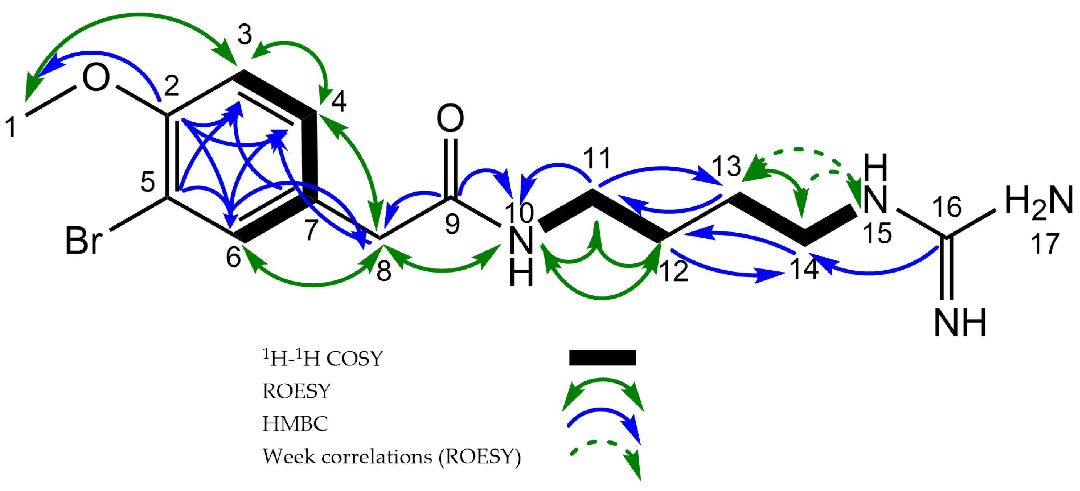
Figure 1. Structure of novel compound Securidine A (1), Securamine A–G (28), and Securines A and B (910).
Molecules 22 01236 g001
Figure 2. Key HMBC, ME-HSQC, H2BC, COSY, and ROESY correlations of Securidine A (1).
Molecules 22 01236 g002
Figure 3. The structurally related compounds Securidine A (1), Pulmonarin (2), and Synoxazolidinone B (3).
Molecules 22 01236 g003
Table 1. NMR spectroscopic data a (600 MHz, methanol-d3).
PositionδC, TypeδH (J in Hz)COSY1H, 13C-HMBC b1H, 13C H2BCROESY
156.6, CH33.84, s 2 3
2156.2, C
3113.1, CH6.96, d (8.6)42,5,741,4
4130.2, CH7.22, dd (8.6, 2.3)3,62,3,6,833,8
5112.2, C
6134.5, CH7.47, d (2.3)42,4,5,8 8
7130.5, C
842.4, CH23.41, s 4,6,9 4,6,10
9173.7, C
10 8.20, s119,11118,11,12 w
1139.8, CH23.20, q (6.4)10,129,(12/13) d1210,12
1227.5, CH21.56, m c11(11/14) d1110 w,11
1327.0, CH21.55, m c14(11/14) d1414,15 w
1442.0, CH23.16, m c13,1516,(12/13) d1313,15 w
15 7.57, s14 1413 w,14 w
16158.8, C
a 1H 1D, 13C, 1H, 1H-COSY, 1H,13C-HMBC (full and band selective) spectra are included in the supplementary data, Figures S1–S6 (Supplementary materials). b 1H 1D, 13C-HMBC correlations are from the proton(s) stated to the indicated carbon; c Overlapping and/or broadened peaks impeding complete multiplet analysis. Chemical shift extracted from 1H, 13C-HSQC, 1H, 13C-HMBC and/or 1H, 13C-H2BC; d ambiguous correlations in HMBC, C12 and C13 assignment based on H2BC correlations (Figure S4); w Weak correlations.

