
Paclitaxel-Loaded Magnetic Nanoparticles Based on Biotinylated N-Palmitoyl Chitosan: Synthesis, Characterization and Preliminary In Vitro Studies

 , , , and
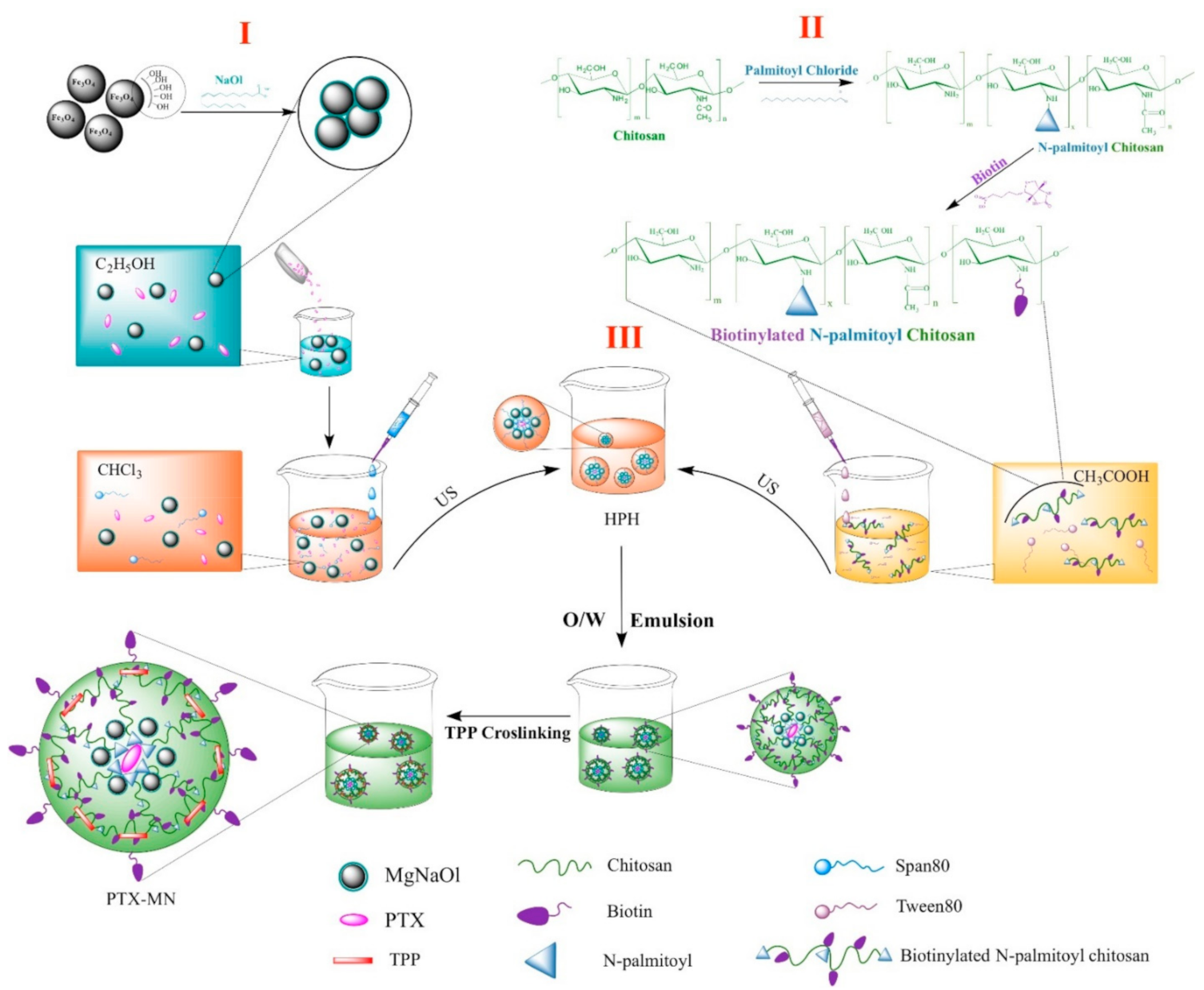
Abstract
:1. Introduction
2. Results and Discussions
2.1. Paclitaxel-Loaded Magnetic Biotinylated N-Palmitoyl Chitosan Nanoparticles Preparation
2.2. Nanoparticles Features
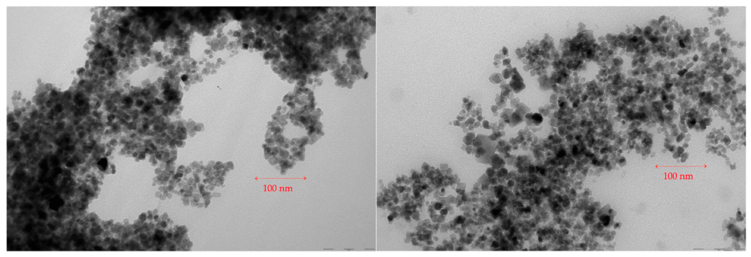
2.2.1. Size, Surface Charge and Morphology
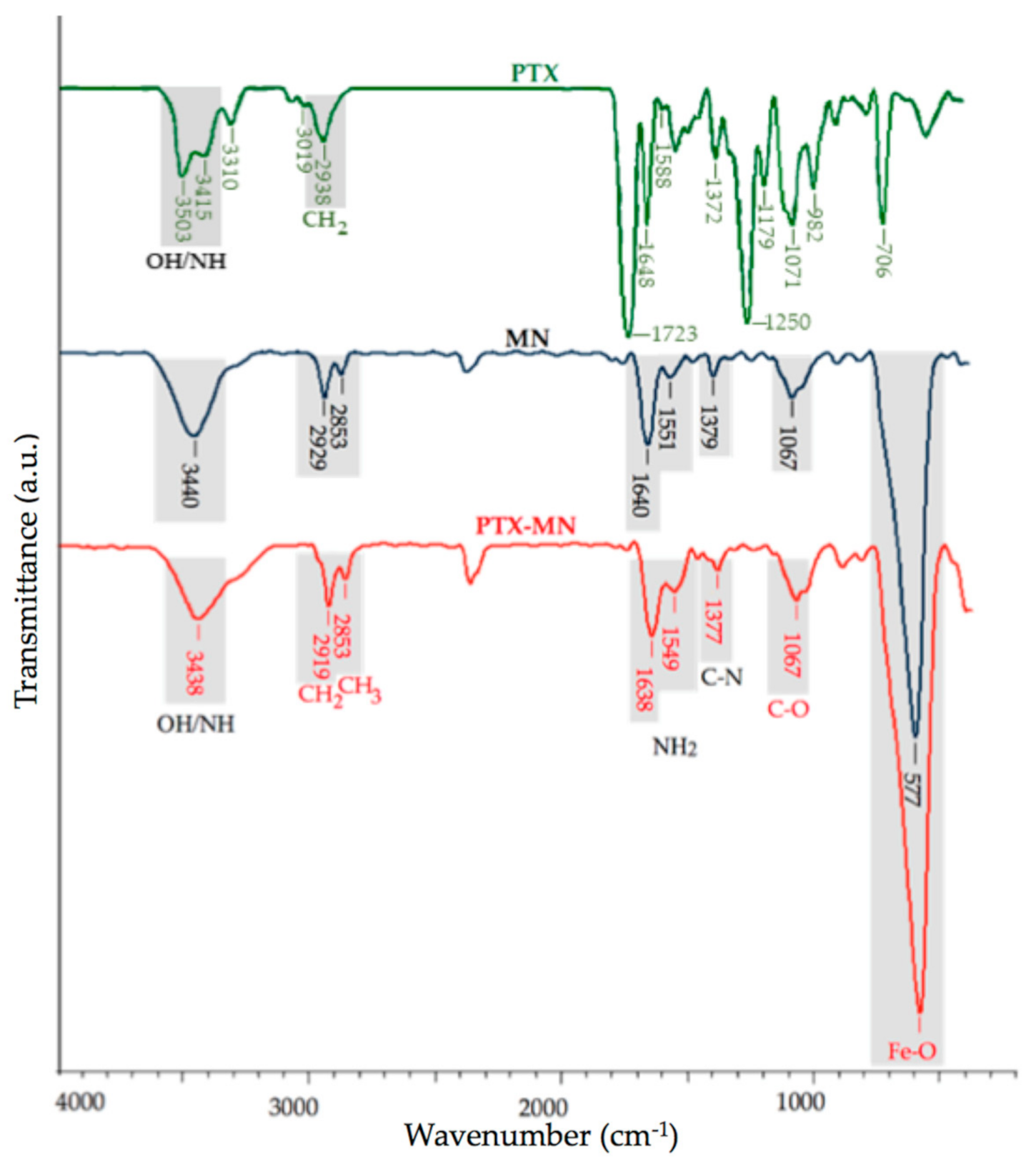
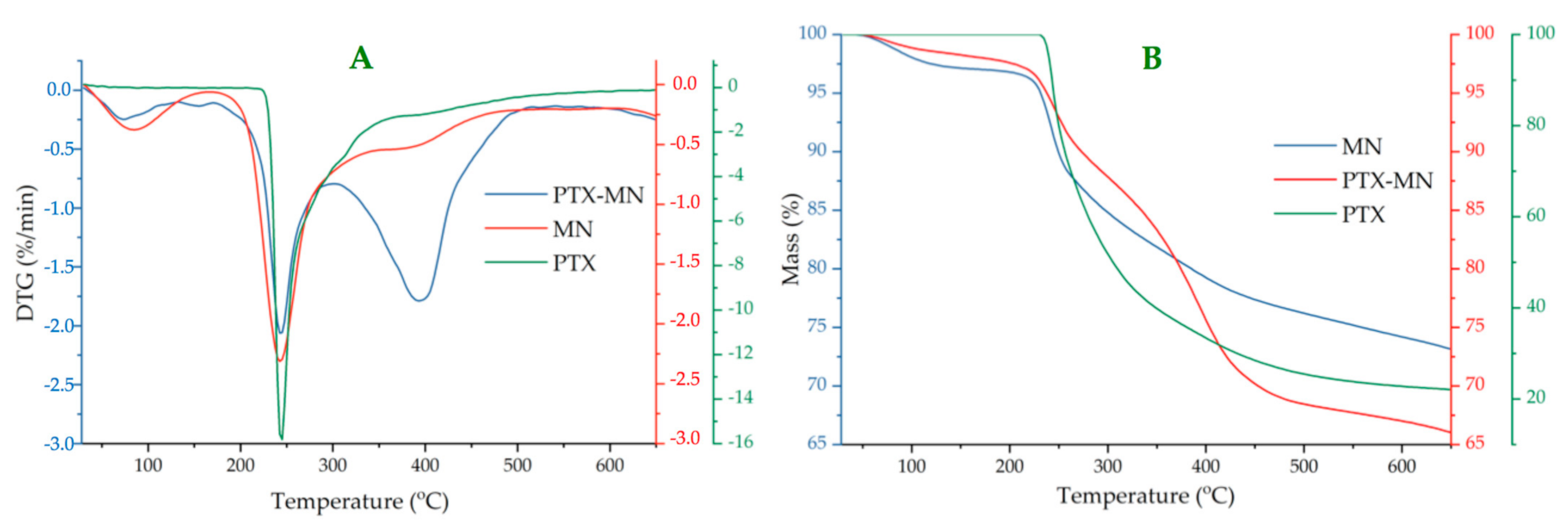
2.2.2. Composition and Thermal Behavior
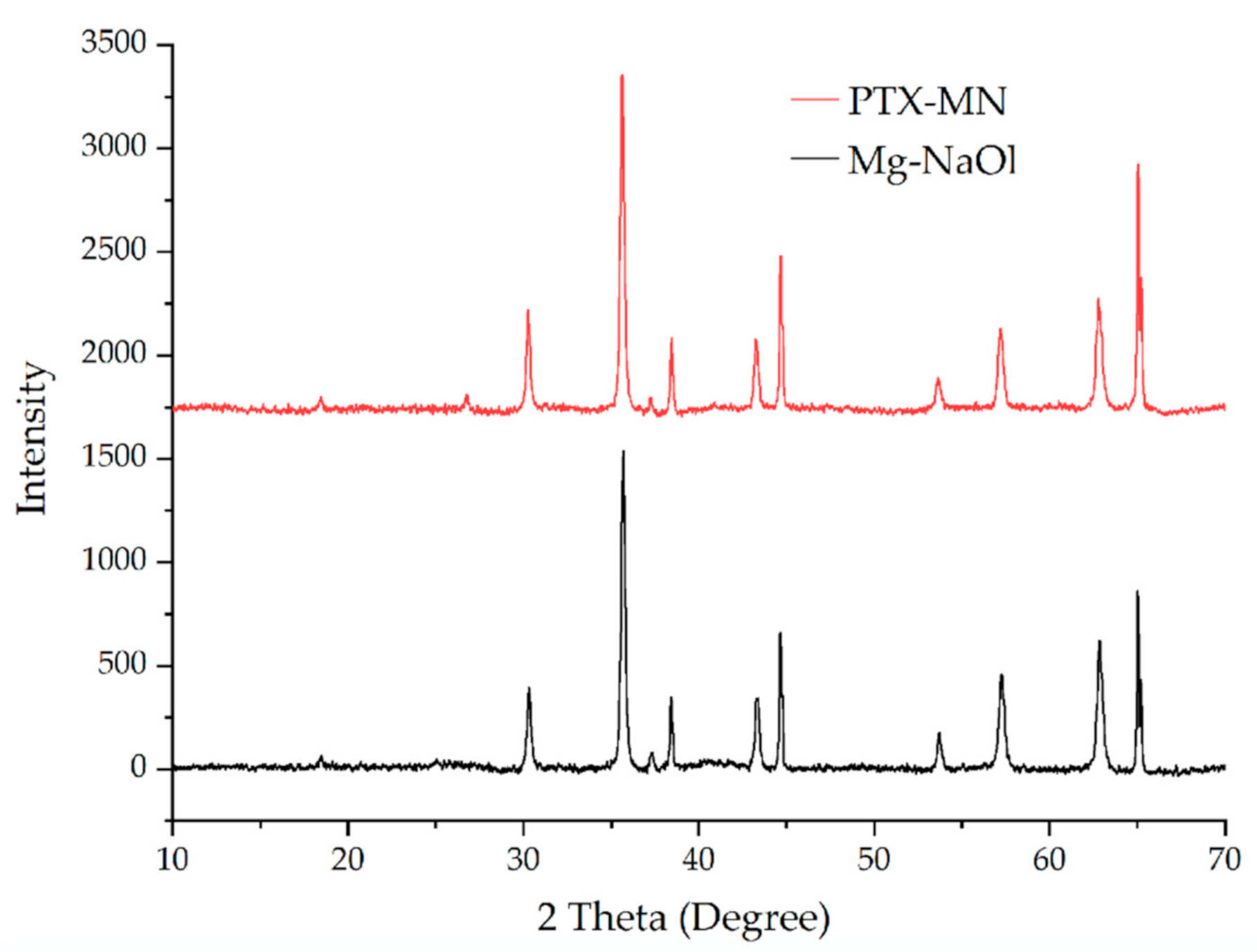
2.2.3. X-ray Diffraction Analysis
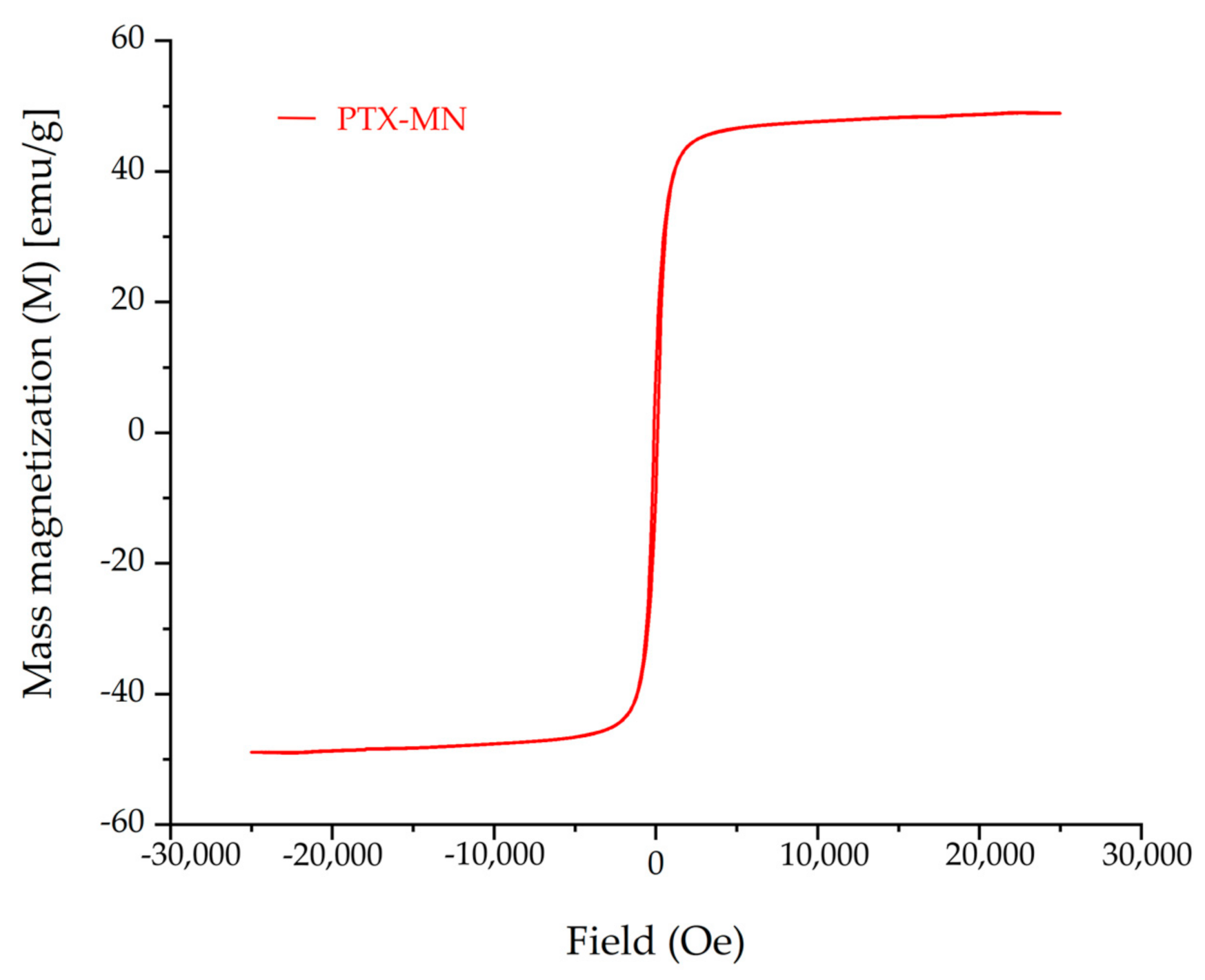
2.2.4. Magnetic Properties
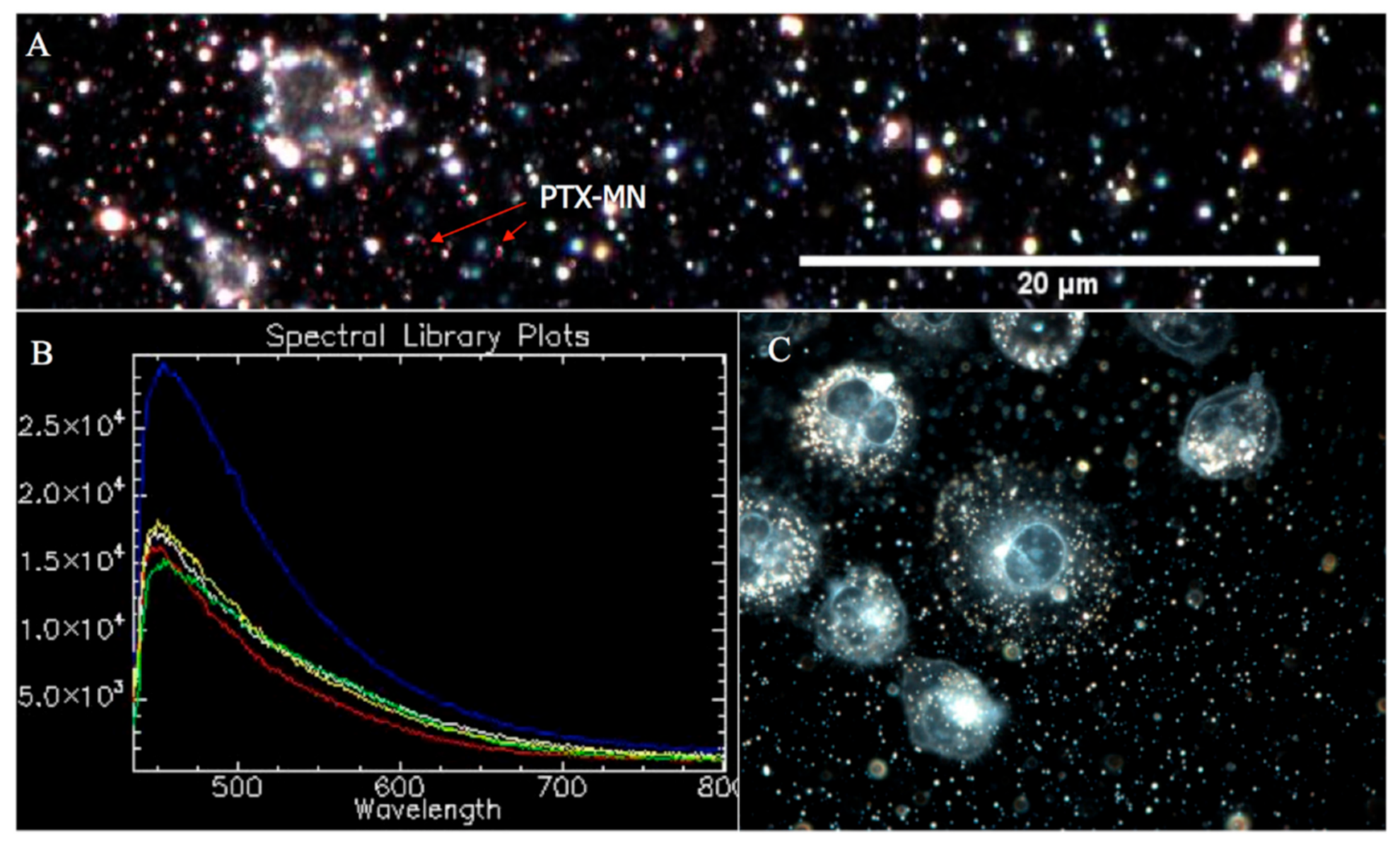
2.2.5. CytoViva Results
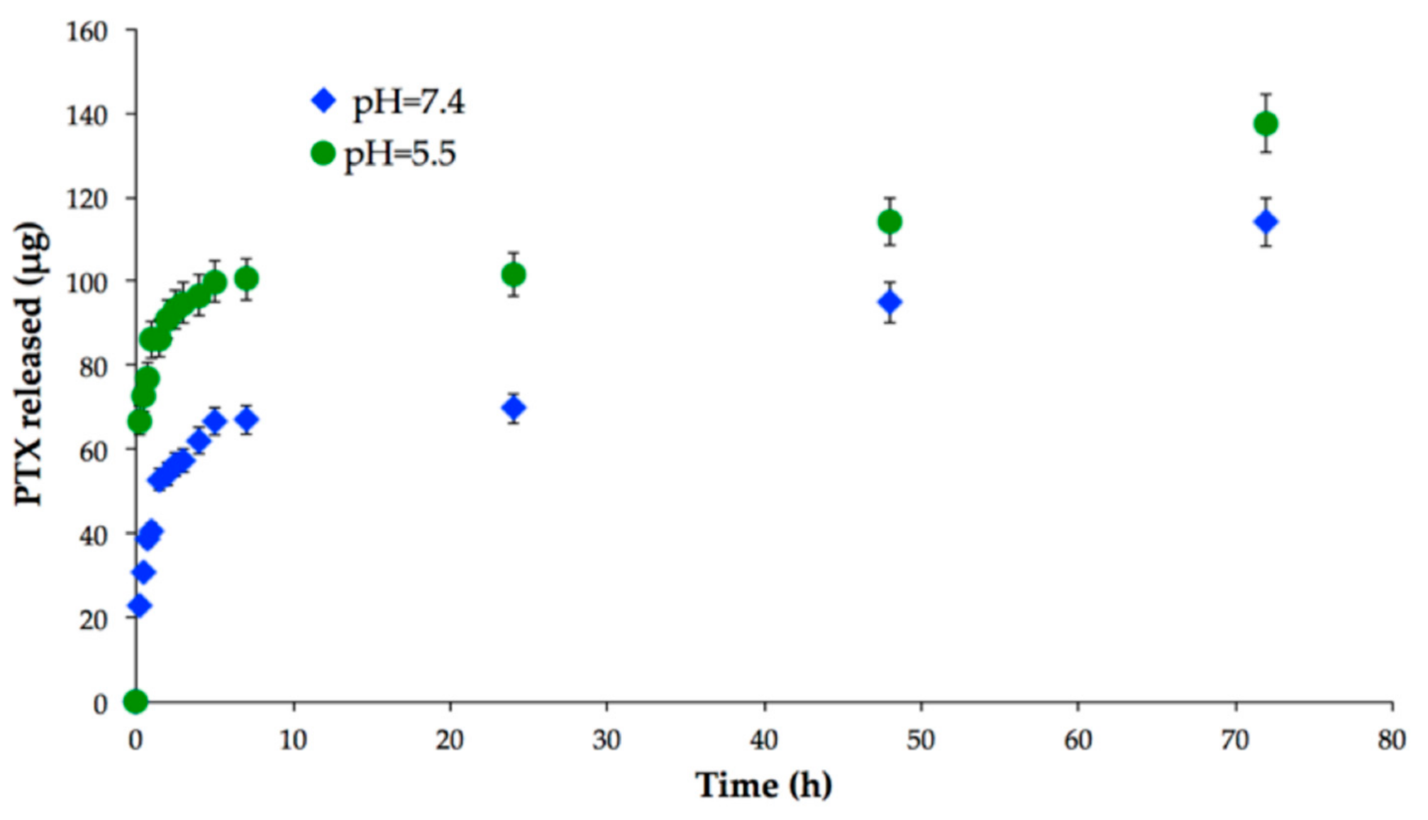
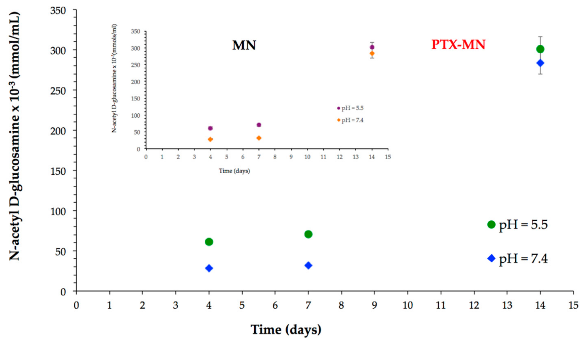
2.3. In Vitro Drug Release and Biodegradation Studies
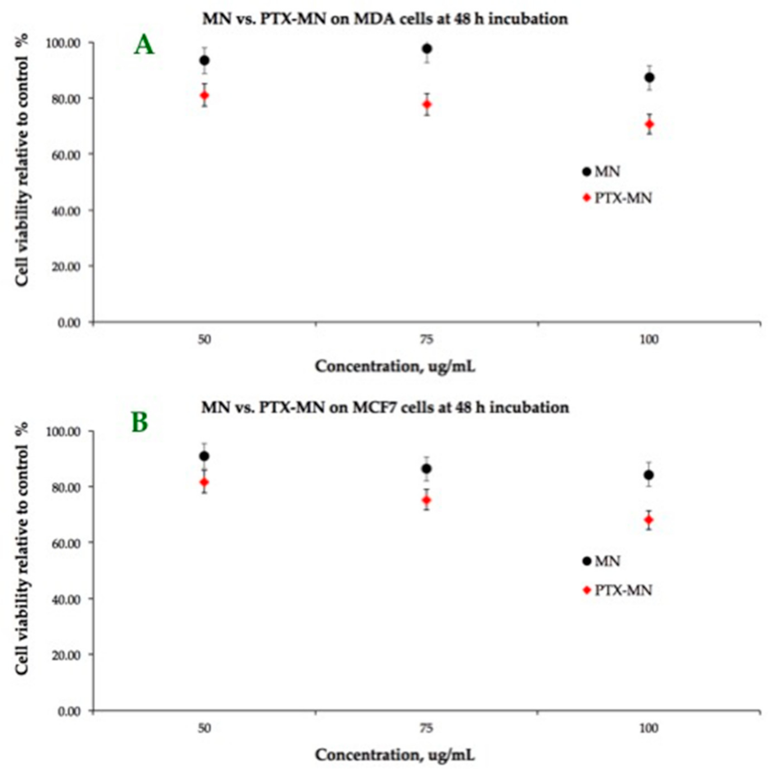
2.4. In Vitro Cell Investigations
3. Materials and Methods
3.1. Materials
3.2. Synthesis of Paclitaxel-Loaded Magnetic Nanoparticles Based on Biotinylated N-Palmitoyl Chitosan
3.3. Characterization of Composite Magnetic Nanoparticles
3.4. In Vitro Drug Release Studies
3.5. Biodegradability Studies
3.6. Cell Culture Assays
4. Conclusions
Author Contributions
Funding
Conflicts of Interest
Sample Availability
References
- Zhi, D.; Yang, T.; Yang, J.; Fu, S.; Zhang, S. Targeting Strategies for Superparamagnetic Iron Oxide Nanoparticles in Cancer Therapy. Acta Biomater. 2020, 102, 13–34. [Google Scholar] [CrossRef]
- Li, K.; Nejadnik, H.; Daldrup-Link, H.E. Next-Generation Superparamagnetic Iron Oxide Nanoparticles for Cancer Theranostics. Drug Discov. Today 2017, 22, 1421–1429. [Google Scholar] [CrossRef]
- Chomoucka, J.; Drbohlavova, J.; Huska, D.; Adam, V.; Kizek, R.; Hubalek, J. Magnetic Nanoparticles and Targeted Drug Delivering. Pharmacol. Res. 2010, 62, 144–149. [Google Scholar] [CrossRef]
- McCarthy, J.R.; Kelly, K.A.; Sun, E.Y.; Weissleder, R. Targeted Delivery of Multifunctional Magnetic Nanoparticles. Nanomedicine 2007, 2, 153–167. [Google Scholar] [CrossRef]
- Bulte, J.W.M.; Kraitchman, D.L. Iron Oxide MR Contrast Agents for Molecular and Cellular Imaging. NMR Biomed. 2004, 17, 484–499. [Google Scholar] [CrossRef] [PubMed]
- Rostami, E. Progresses in Targeted Drug Delivery Systems Using Chitosan Nanoparticles in Cancer Therapy: A Mini-Review. J. Drug Deliv. Sci. Technol. 2020, 58, 101813. [Google Scholar] [CrossRef]
- Bonferoni, M.C.; Gavini, E.; Rassu, G.; Maestri, M.; Giunchedi, P. Chitosan Nanoparticles for Therapy and Theranostics of Hepatocellular Carcinoma (HCC) and Liver-Targeting. Nanomaterials 2020, 10, 870. [Google Scholar] [CrossRef] [PubMed]
- Bano, S.; Afzal, M.; Waraich, M.M.; Alamgir, K.; Nazir, S. Paclitaxel Loaded Magnetic Nanocomposites with Folate Modified Chitosan/Carboxymethyl Surface; A Vehicle for Imaging and Targeted Drug Delivery. Int. J. Pharm. 2016, 513, 554–563. [Google Scholar] [CrossRef] [PubMed]
- Nag, M.; Gajbhiye, V.; Kesharwani, P.; Jain, N.K. Transferrin Functionalized Chitosan-PEG Nanoparticles for Targeted Delivery of Paclitaxel to Cancer Cells. Colloids Surf. B Biointerfaces 2016, 148, 363–370. [Google Scholar] [CrossRef]
- Poudel, I.; Ahiwale, R.; Pawar, A.; Mahadik, K.; Bothiraja, C. Development of Novel Biotinylated Chitosan-Decorated Docetaxel-Loaded Nanocochleates for Breast Cancer Targeting. Artif. Cells Nanomed. Biotechnol. 2018, 46, 229–240. [Google Scholar] [CrossRef] [Green Version]
- Cheng, M.; Ma, D.; Zhi, K.; Liu, B.; Zhu, W. Synthesis of Biotin-Modified Galactosylated Chitosan Nanoparticles and Their Characteristics in Vitro and in Vivo. Cell. Physiol. Biochem. 2018, 50, 569–584. [Google Scholar] [CrossRef]
- Ashrafizadeh, M.; Ahmadi, Z.; Mohamadi, N.; Zarrabi, A.; Abasi, S.; Dehghannoudeh, G.; Tamaddondoust, R.N.; Khanbabaei, H.; Mohammadinejad, R.; Thakur, V.K. Chitosan-Based Advanced Materials for Docetaxel and Paclitaxel Delivery: Recent Advances and Future Directions in Cancer Theranostics. Int. J. Biol. Macromol. 2020, 145, 282–300. [Google Scholar] [CrossRef] [PubMed]
- Said, H.M. Cell and Molecular Aspects of Human Intestinal Biotin Absorption. J. Nutr. 2009, 139, 158–162. [Google Scholar] [CrossRef] [PubMed] [Green Version]
- Dasgupta, A. Chapter 2—Biotin: Pharmacology, Pathophysiology, and Assessment of Biotin Status. In Biotin and Other Interferences in Immunoassays; Dasgupta, A., Ed.; Elsevier: Amsterdam, The Netherlands, 2019; pp. 17–35. ISBN 978-0-12-816429-7. [Google Scholar]
- Shi, J.-F.; Wu, P.; Jiang, Z.-H.; Wei, X.-Y. Synthesis and Tumor Cell Growth Inhibitory Activity of Biotinylated Annonaceous Acetogenins. Eur. J. Med. Chem. 2014, 71, 219–228. [Google Scholar] [CrossRef] [PubMed]
- Chen, S.; Zhao, X.; Chen, J.; Chen, J.; Kuznetsova, L.; Wong, S.S.; Ojima, I. Mechanism-Based Tumor-Targeting Drug Delivery System. Validation of Efficient Vitamin Receptor-Mediated Endocytosis and Drug Release. Bioconjugate Chem. 2010, 21, 979–987. [Google Scholar] [CrossRef] [PubMed] [Green Version]
- Nosrati, H.; Barzegari, P.; Danafar, H.; Manjili, H.K. Biotin-Functionalized Copolymeric PEG-PCL Micelles for in Vivo Tumour-Targeted Delivery of Artemisinin. Artif. Cells Nanomed. Biotechnol. 2019, 47, 104–114. [Google Scholar] [CrossRef] [Green Version]
- Wang, Y.; van Steenbergen, M.J.; Beztsinna, N.; Shi, Y.; Lammers, T.; van Nostrum, C.F.; Hennink, W.E. Biotin-Decorated All-HPMA Polymeric Micelles for Paclitaxel Delivery. J. Control. Release 2020, 328, 970–984. [Google Scholar] [CrossRef]
- Rompicharla, S.V.K.; Kumari, P.; Bhatt, H.; Ghosh, B.; Biswas, S. Biotin Functionalized PEGylated Poly (Amidoamine) Dendrimer Conjugate for Active Targeting of Paclitaxel in Cancer. Int. J. Pharm. 2019, 557, 329–341. [Google Scholar] [CrossRef]
- Balan, V.; Redinciuc, V.; Tudorachi, N.; Verestiuc, L. Biotinylated N-Palmitoyl Chitosan for Design of Drug Loaded Self-Assembled Nanocarriers. Eur. Polym. J. 2016, 81, 284–294. [Google Scholar] [CrossRef]
- Balan, V.; Moise, C.I.; Verestiuc, L. Synthesis and Characterization of Self-assembled Submicron Particles Based on Biotinylated N-palmitoyl Chitosan. In 4th International Conference on Nanotechnologies and Biomedical Engineering; Tiginyanu, I., Sontea, V., Railean, S., Eds.; Springer International Publishing: Cham, Switzerland, 2020; Volume 77, pp. 325–329. ISBN 978-3-030-31865-9. [Google Scholar]
- Chowdhury, P.; Nagesh, P.K.B.; Hatami, E.; Wagh, S.; Dan, N.; Tripathi, M.K.; Khan, S.; Hafeez, B.B.; Meibohm, B.; Chauhan, S.C.; et al. Tannic Acid-Inspired Paclitaxel Nanoparticles for Enhanced Anticancer Effects in Breast Cancer Cells. J. Colloid Interface Sci. 2019, 535, 133–148. [Google Scholar] [CrossRef]
- Rajora, A.K.; Ravishankar, D.; Zhang, H.; Rosenholm, J.M. Recent Advances and Impact of Chemotherapeutic and Antiangiogenic Nanoformulations for Combination Cancer Therapy. Pharmaceutics 2020, 12, 592. [Google Scholar] [CrossRef] [PubMed]
- Dodi, G.; Hritcu, D.; Draganescu, D.; Popa, M.I. Iron Oxide Nanoparticles for Magnetically Assisted Patterned Coatings. J. Magn. Magn. Mater. 2015, 388, 49–58. [Google Scholar] [CrossRef]
- Vikesland, P.J.; Rebodos, R.L.; Bottero, J.Y.; Rose, J.; Masion, A. Aggregation and Sedimentation of Magnetite Nanoparticle Clusters. Environ. Sci. Nano 2016, 3, 567–577. [Google Scholar] [CrossRef] [Green Version]
- Kathiravan, G.; Rajasekar, A. Infra-Red Spectral Analysis of Taxol Produced by Different Species of Pestalotiopsis. J. Anal. Bioanal. Tech. 2014, 5. [Google Scholar] [CrossRef]
- Yang, D.; Van, S.; Liu, J.; Wang, J.; Jiang, X.; Wang, Y.; Yu, L. Physicochemical Properties and Biocompatibility of a Polymer-Paclitaxel Conjugate for Cancer Treatment. Int. J. Nanomed. 2011, 6, 2557–2566. [Google Scholar] [CrossRef] [Green Version]
- Hwang, H.-Y.; Kim, I.-S.; Kwon, I.C.; Kim, Y.-H. Tumor Targetability and Antitumor Effect of Docetaxel-Loaded Hydrophobically Modified Glycol Chitosan Nanoparticles. J. Control. Release 2008, 128, 23–31. [Google Scholar] [CrossRef]
- Kean, T.; Thanou, M. Biodegradation, Biodistribution and Toxicity of Chitosan. Adv. Drug Deliv. Rev. 2010, 62, 3–11. [Google Scholar] [CrossRef]
- Vizoso, F.; Plaza, E.; Vázquez, J.; Serra, C.; Lamelas, M.L.; González, L.O.; Merino, A.M.; Méndez, J. Lysozyme Expression by Breast Carcinomas, Correlation with Clinicopathologic Parameters, and Prognostic Significance. Ann. Surg. Oncol. 2001, 8, 667–674. [Google Scholar] [CrossRef] [PubMed]
- Ren, D.; Yi, H.; Wang, W.; Ma, X. The Enzymatic Degradation and Swelling Properties of Chitosan Matrices with Different Degrees of N-Acetylation. Carbohydr. Res. 2005, 340, 2403–2410. [Google Scholar] [CrossRef]


Publisher’s Note: MDPI stays neutral with regard to jurisdictional claims in published maps and institutional affiliations. |
© 2021 by the authors. Licensee MDPI, Basel, Switzerland. This article is an open access article distributed under the terms and conditions of the Creative Commons Attribution (CC BY) license (https://creativecommons.org/licenses/by/4.0/).
Share and Cite
Ursachi, V.C.; Dodi, G.; Rusu, A.G.; Mihai, C.T.; Verestiuc, L.; Balan, V. Paclitaxel-Loaded Magnetic Nanoparticles Based on Biotinylated N-Palmitoyl Chitosan: Synthesis, Characterization and Preliminary In Vitro Studies. Molecules 2021, 26, 3467. https://0-doi-org.brum.beds.ac.uk/10.3390/molecules26113467
Ursachi VC, Dodi G, Rusu AG, Mihai CT, Verestiuc L, Balan V. Paclitaxel-Loaded Magnetic Nanoparticles Based on Biotinylated N-Palmitoyl Chitosan: Synthesis, Characterization and Preliminary In Vitro Studies. Molecules. 2021; 26(11):3467. https://0-doi-org.brum.beds.ac.uk/10.3390/molecules26113467
Chicago/Turabian StyleUrsachi, Vlad Constantin, Gianina Dodi, Alina Gabriela Rusu, Cosmin Teodor Mihai, Liliana Verestiuc, and Vera Balan. 2021. "Paclitaxel-Loaded Magnetic Nanoparticles Based on Biotinylated N-Palmitoyl Chitosan: Synthesis, Characterization and Preliminary In Vitro Studies" Molecules 26, no. 11: 3467. https://0-doi-org.brum.beds.ac.uk/10.3390/molecules26113467