Article

Amidine- and Amidoxime-Substituted Heterocycles: Synthesis, Antiproliferative Evaluations and DNA Binding

by
Silvija Maračić
1,
Petra Grbčić
2,
Suresh Shammugam
3,
Marijana Radić Stojković
3,*,
Krešimir Pavelić
4,
Mirela Sedić
5,
Sandra Kraljević Pavelić
6 and
Silvana Raić-Malić
1,*
1
Department of Organic Chemistry, Faculty of Chemical Engineering and Technology, University of Zagreb, Marulićev trg 19, HR-10000 Zagreb, Croatia
2
Department of Biotechnology, University of Rijeka, Ulica Radmile Matejčić 2, HR-51000 Rijeka, Croatia
3
Division of Organic Chemistry and Biochemistry, Laboratory for Biomolecular Interactions and Spectroscopy, Ruđer Bošković Institute, Bijenička 54, HR-10000 Zagreb, Croatia
4
Faculty of Medicine, Juraj Dobrila University of Pula, HR-52100 Pula, Croatia
5
Centre for Applied Bioanthropology, Institute for Anthropological Research, Ljudevita Gaja 32, HR-10000 Zagreb, Croatia
6
Faculty of Health Studies, University of Rijeka, Ulica Viktora Cara Emina 5, HR-51000 Rijeka, Croatia
*
Authors to whom correspondence should be addressed.
Submission received: 27 October 2021 / Revised: 16 November 2021 / Accepted: 19 November 2021 / Published: 22 November 2021
(This article belongs to the Special Issue Ligand Binding to DNA and RNA)

Abstract

:
The novel 1,2,3-triazolyl-appended N- and O-heterocycles containing amidine 411 and amidoxime 1222 moiety were prepared and evaluated for their antiproliferative activities in vitro. Among the series of amidine-substituted heterocycles, aromatic diamidine 5 and coumarine amidine 11 had the most potent growth-inhibitory effect on cervical carcinoma (HeLa), hepatocellular carcinoma (HepG2) and colorectal adenocarcinoma (SW620), with IC50 values in the nM range. Although compound 5 was toxic to non-tumor HFF cells, compound 11 showed certain selectivity. From the amidoxime series, quinoline amidoximes 18 and 20 showed antiproliferative effects on lung adenocarcinoma (A549), HeLa and SW620 cells emphasizing compound 20 that exhibited no cytostatic effect on normal HFF fibroblasts. Results of CD titrations and thermal melting experiments indicated that compounds 5 and 10 most likely bind inside the minor groove of AT-DNA and intercalate into AU-RNA. Compounds 6, 9 and 11 bind to AT-DNA with mixed binding mode, most probably minor groove binding accompanied with aggregate binding along the DNA backbone.

1. Introduction

Nitrogen- and oxygen-containing heterocyclic compounds may be found in pharmaceuticals, natural products, dyes, organic materials, and particularly in biologically active molecules [1,2]. Natural and synthetic N- and O-heterocycles have been discovered as promising anti-cancer agents used in clinic or clinical evaluations, suggesting their prominent place in anti-cancer drugs development. Although the design of bioactive molecules is primarily focused on novel N-heterocycles, due to their ease of synthesis and possible mimicking of physiological molecules, O-heterocycles have provided equally potent molecules with a lesser risk of toxicity [2,3]. These compounds were found to act via various targets such as protein kinases, histone deacetylases, topoisomerase I and II, carbonic anhydrase, aromatase, sulfatase, vascular endothelial growth factor (VEGF), and epidermal growth factor (EGF) receptors, monocarboxylate transporters, DNA polynucleotide, tubulin/microtubule system [1,4,5,6,7,8,9,10,11,12,13,14].
Applying the molecular hybridization approach for the synthesis of new molecules where 1,2,3-triazole ring acts as pharmacophore or linker leads to libraries of compounds with significant anticancer activity [15]. Thus, indole scaffold, found to be widely distributed in natural products and bioactive molecules, was linked to 1,2,3-triazole and yielded new hybrids with potent anticancer activity [11]. For example, in order to obtain histone deacetylases (HDAC) inhibitors with lower side effects, indole-derived N-hydroxyarylamide I with the triazole ring as a linker was obtained and showed the positive effects of 5-methoxy group and the one-carbon-bridge between triazole and the indole ring on antiproliferative activity (Figure 1) [16].
Bis-1,2,3-triazole–indole hybrids were further synthesized and evaluated for their in vitro and in vivo anticancer activity, among which compound II caused DNA cleavage and displayed significant activity against breast cancer (MCF-7), cervical carcinoma (HeLa) and embryonic kidney (HEK 293) cell lines, as compared with cisplatin [17]. Indole–1,2,3-triazole–chalcone III acted via noncovalent intercalative mode of binding to DNA, causing antiproliferative activity on human cervical cancer (SiHa) and colorectal epithelial carcinoma (SW620) cancer cell lines with no cytotoxicity in human embryonic kidney cells (HEK293) at the similar concentration [18]. Another triazolyl-appended N-heterocycles that exhibited anticancer activities are quinoline-based 1,2,3-triazole hybrids. New quinoline–1,2,3-triazole–dihydroquinoline derivatives were synthesized [19] and displayed cytotoxicity against lung adenocarcinoma (A549) and breast cancer (MCF7) cells, emphasizing the compound IV (Figure 1), that showed antiproliferative activity against A549 cells comparable to that of the reference drug doxorubicin. 8-Hydroxyquinoline derivative V with sugar moiety linked via 1,2,3-triazole ring exhibited potent antiproliferative activity and high selectivity toward ovarian cancer (OVCAR-03) cells, with higher activity than the doxorubicin [20]. The 1,2,3-triazole-4-carboxamide VI was identified as a multitargeted receptor tyrosine kinase inhibitor with a more potent growth-inhibition effect on human colon adenocarcinoma (HT-29) cells than foretinib [21].
Hybridization of coumarin moiety with other anticancer pharmacophores was found to provide novel anticancer candidates with low toxicity, high specificity and excellent efficacy against drug-susceptible and drug-resistant cancers [3,13,22]. It was observed that substituent, length and position of alkyl spacer had a profound effect on the anticancer potency. 1,2,3-Triazole-containing novobiocin analogues with triazole ring at C-3 of coumarin were investigated [23] and showed that coumarin–1,2,3-triazole–indole VII (Figure 2) exhibited potent antiproliferative activity against two breast cancer cell lines (SKBr-3 and MCF-7), which was directly related to inhibition of the Hsp90-mediated protein folding process. Artemisinin–coumarin compound VIII accumulated in cancer-cell mitochondria efficiently and induced the generation of intracellular ROS, triggered cancer cell apoptosis, and consequently caused cancer cell death [24]. In addition, chalcone–coumarin hybrids displayed cytotoxicity against lymphoblastic leukemia (MOLT-3) cells without affecting normal cells [25]. Among these, compound IX showed strong activity against human liver cancer (HepG2) cells and no toxicity to Vero cells. Further investigations revealed that coumarin–1,2,3-triazole–benzimidazole derivative X induced cell death, mainly due to early apoptosis, and its inhibition of 5-lipoxygenase activity and perturbation of sphingolipid signaling by interfering with intracellular acid ceramidase activity [26]. Another coumarin–1,2,3-triazole hybrid XI containing amide showed moderate to excellent activity against human breast cancer (MDA-MB-231) cells, revealing general enhancement of proliferation-inhibiting activity under hypoxia, contrasted with normoxia [27]. 6-Hydroxycoumarin XII linked through triazole with ortho-substituted phenyl moiety showed to be selectively cytotoxic against lung cancer (A-549) cell line [28].
Heterocyclic diamidines have been extensively studied as DNA minor groove binders [29]. The research on amidines has been successful in demonstrating a correlation between the structure and biological activity with their ability to bind in minor grooves [29,30].
To improve bioavailability and oral absorption of amidines, the strategy of amidoxime prodrugs was successfully applied [31,32].
Based on the above-mentioned data and in continuation of our research on 1,2,3-triazolyl- appended heterocycles [26,33,34,35] and their amidino derivatives [8,36,37] as anticancer and antipathogenic agents, herein, we report the synthesis and antiproliferative evaluations of novel indole, quinoline and coumarin-based aromatic amidines 111 and amidoximes 1222. Since nucleic acids are a major target for a large number of anticancer drugs, one of our aims was to investigate the binding of small molecules to DNA and RNA [29]. Assessing the binding strength and mode of binding of small molecule-DNA/RNA interactions was performed using UV–Vis, fluorescence and circular dichroism (CD) spectroscopy.

2. Results and Discussion
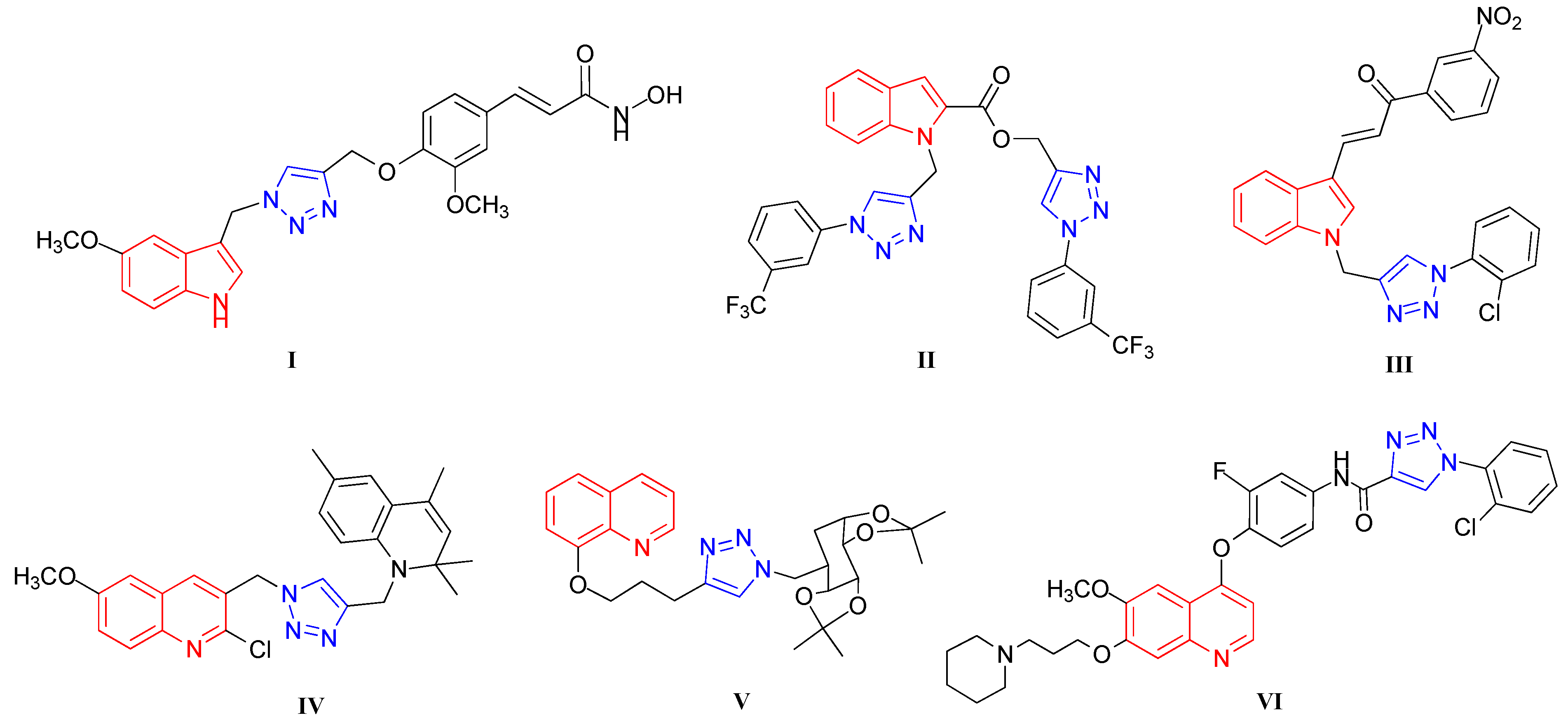
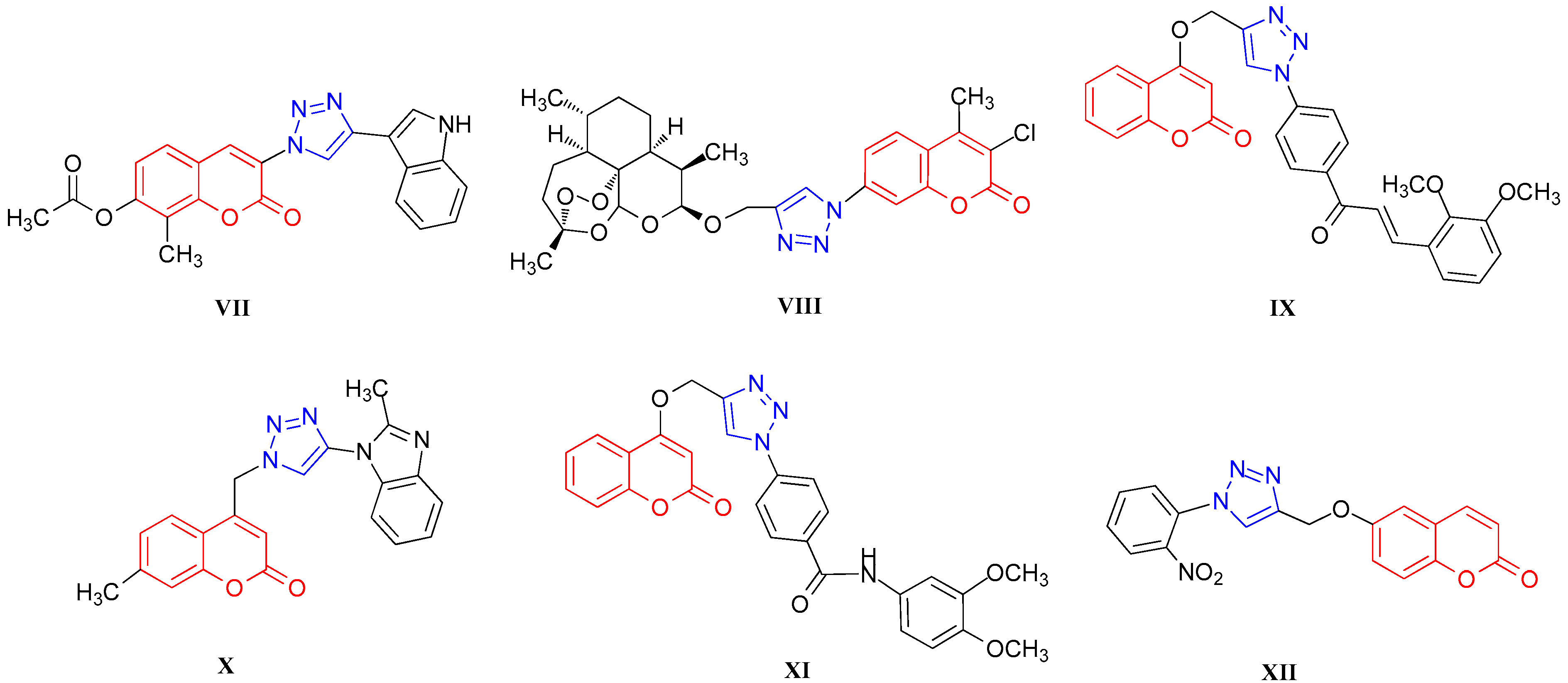
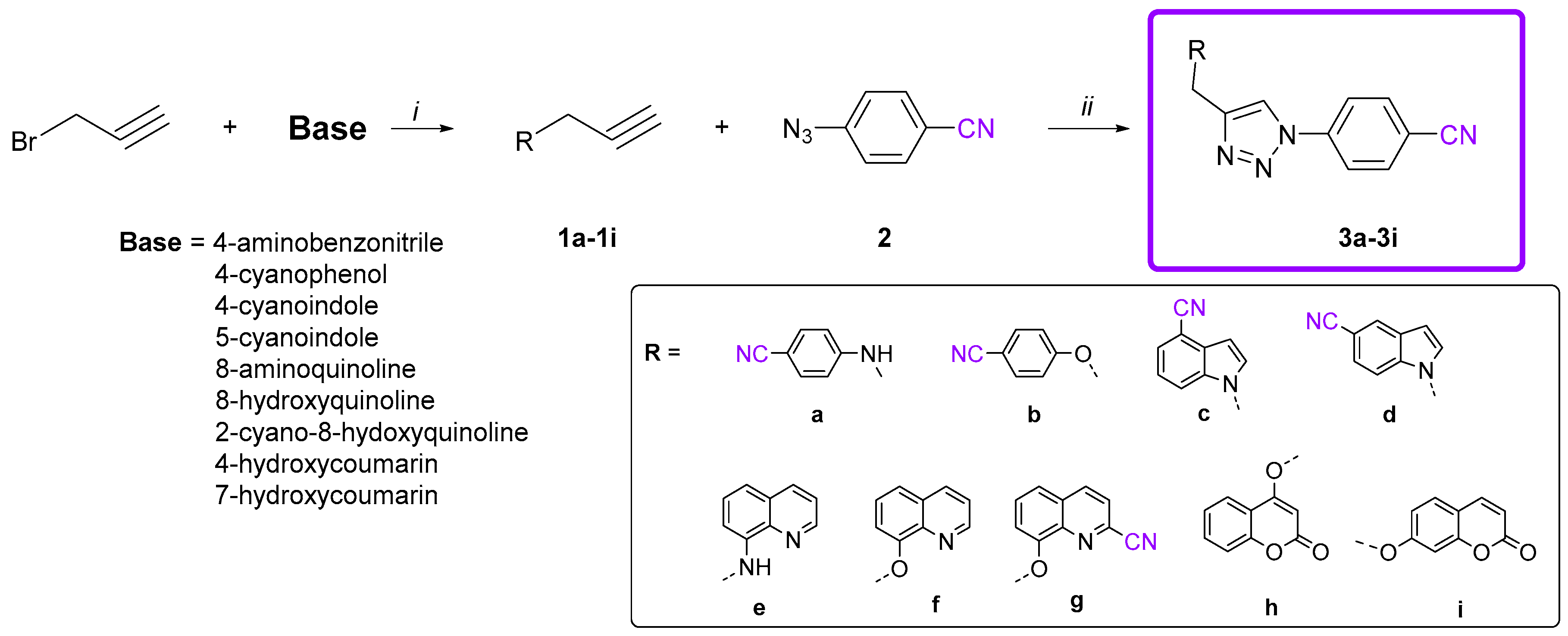
2.1. Chemistry

Novel hybrids of aromatic nitrile and heterocycle linked via 1,2,3-triazole scaffold 3a3i were synthesized by regioselective Cu(I)-catalyzed azide-alkyne 1,3-dipolar cycloaddition of 4-azidobenzonitrile and corresponding alkynes. Alkynyl derivatives of benzonitrile (1a and 1b), indole (1c and 1d), quinoline (1e1g) and coumarin (1h and 1i) were synthesized by alkylation with propargyl bromide in the presence of a base (Scheme 1), while azido derivative 2 was prepared by diazotation reaction according to the well-known procedure [38]. To study the influence of the type of linker between heterocycles on their antiproliferative activity, 1,2,3-triazolyl-appended aryl and quinoline analogues containing nitrogen- (3a and 3e) and oxygen-containing (3b, 3f and 3g) aliphatic linkers were prepared. Coumarin was attached to aryl-triazole unit at C-4 (3h) and C-7 (3i) positions.
Nitrile derivatives 3a3i were subsequently used as precursors for the synthesis of amidine- (411) and amidoxime-substituted (1222) heterocycles (Scheme 2 and Scheme 3). Amidines were synthesized according to the Pinner method [39]. Treatment of corresponding cyano derivative 3a3h with dry gaseous HCl in absolute ethanol following by the reaction of the resulting imidate salt with ethylenediamine afforded targeted 2-imidazolinyl substituted derivatives 411. Conversely, the reaction of nitriles 3a3i with hydroxylamine and triethylamine resulted in the desired amidoxime derivatives 1222. In the synthesis of amidoximes both mono- 12, 14, 16 and 19 and their bis-amidoxime analogues 13, 15, 17 and 20 were obtained (Scheme 2).

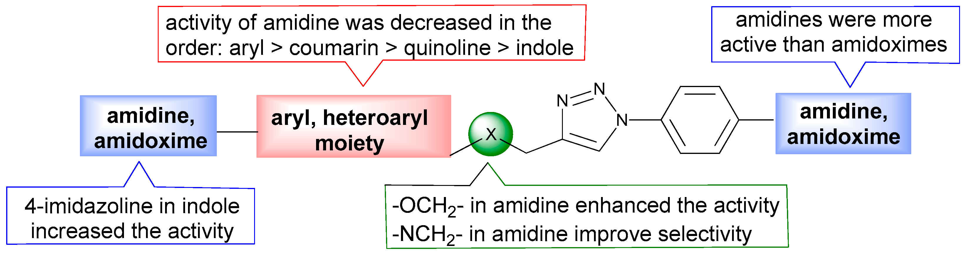
2.2. Biological Evaluation of Aromatic Amidines and Amidoximes

2.2.1. Evaluation of Antiproliferative Activity

Antiproliferative evaluations of amidine- and amidoxime-substituted heterocycles were performed on human tumor cell lines including cervical carcinoma (HeLa), colorectal adenocarcinoma, metastatic (SW620), lung adenocarcinoma (A549) and hepatocellular carcinoma (HepG2) and non-tumor HFF (human foreskin fibroblasts) cells (Table 1). 5-Fluorouracil (5-FU) was used as the reference drug.
From the amidine series, asymmetrical aryl bisamidine 5 linked via 4-methyleneoxy-1,2,3-triazolyl spacer showed strong antiproliferative activity on HeLa (IC50 = 0.80 μM), HepG2 (IC50 = 0.64 μM) and SW620 (IC50 = 0.22 μM) cell lines. Its analogue 4 bearing 4-aminomethylene-1,2,3-triazolyl spacer showed significantly reduced activity (Figure 3). Similarly, quinoline amidine 9 with 8-methyleneoxy-1,2,3-triazolyl unit exhibited 13-fold stronger inhibitory effects to tumor cells compared to its structural analogue 8. The only exception was the growth-inhibition effect on HEPG2 cells that was 2-fold increased by 8 relative to its congener 9. Conversely, aminomethylene units in amidines 4 and 8 contributed to their selectivity, showing less toxicity to normal HFF fibroblasts with selectivity index (SI) of 2 compared to amidines 5 and 9, with methyleneoxy spacer, which showed high cytotoxicity to normal fibroblasts. Antiproliferative evaluations of indole diamidines showed that imidazoline 4-substituted indole 6 was favorable for the activity, particularly on HEPG2 cells (IC50 = 2.37 μM). Conversely, placement of the imidazoline in the 5-position reduced the cytostatic activity of compound 7. Moreover, both 4- and 7-substituted coumarin amidines 10 and 11 exhibited strong cytostatic activity against all evaluated cell lines. However, 7-substituted coumarin 11 showed to be more selective compared to 10, particularly on HepG2 cells with SI of 11.
Generally, antiproliferative evaluations of amidoximes 1222 showed reduced antiproliferative activity compared to their corresponding amidine analogues 411. Only moderate inhibitory activities of aryl amidoxime 12, indole mono- 14 and diamidoximes 17 were observed on SW629 and HeLa cells. Coumarine amidoximes 21 and 22 were deprived of cytostatic activity, while quinoline amidoximes 18 and 20 displayed strong inhibitory effect on A549 (18, IC50 = 6.52 μM), HeLa (20, IC50 = 7.15 μM) and SW620 (20, IC50 = 7.24 μM) cells. Quinoline diamidoxime 20 was around 9-fold more active on HeLa and SW620 cells compared to its monoamidoxime 19. Both quinolines 19 and 20 with methyleneoxy spacer did not exhibit an inhibitory effect on non-tumor HFF cells and selectivity was more expressed by inhibition of compound 20 on HeLa and SW620 cells (SI > 14).
Comparison of the activity of evaluated compounds and reference drug (5-FU) showed that from amidine series, compounds 5, 10 and 11 had better antiproliferative effect on HeLa and HepG2 cells, while from amidoximes, only compound 20 had activity comparable to that of 5-FU.

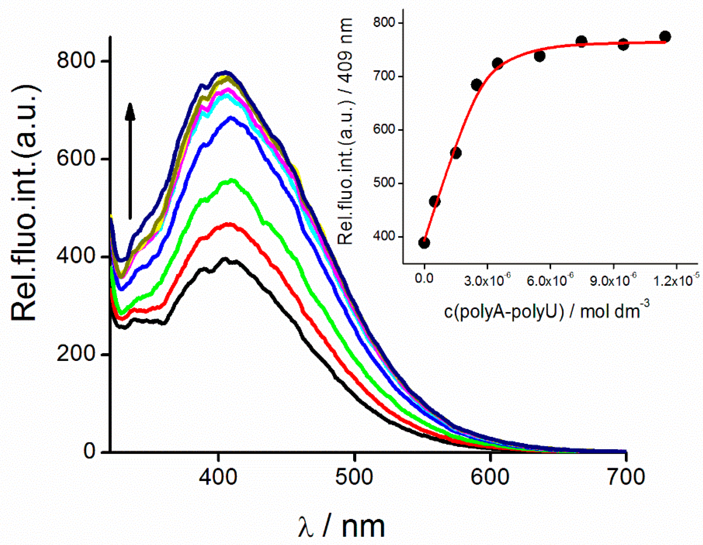
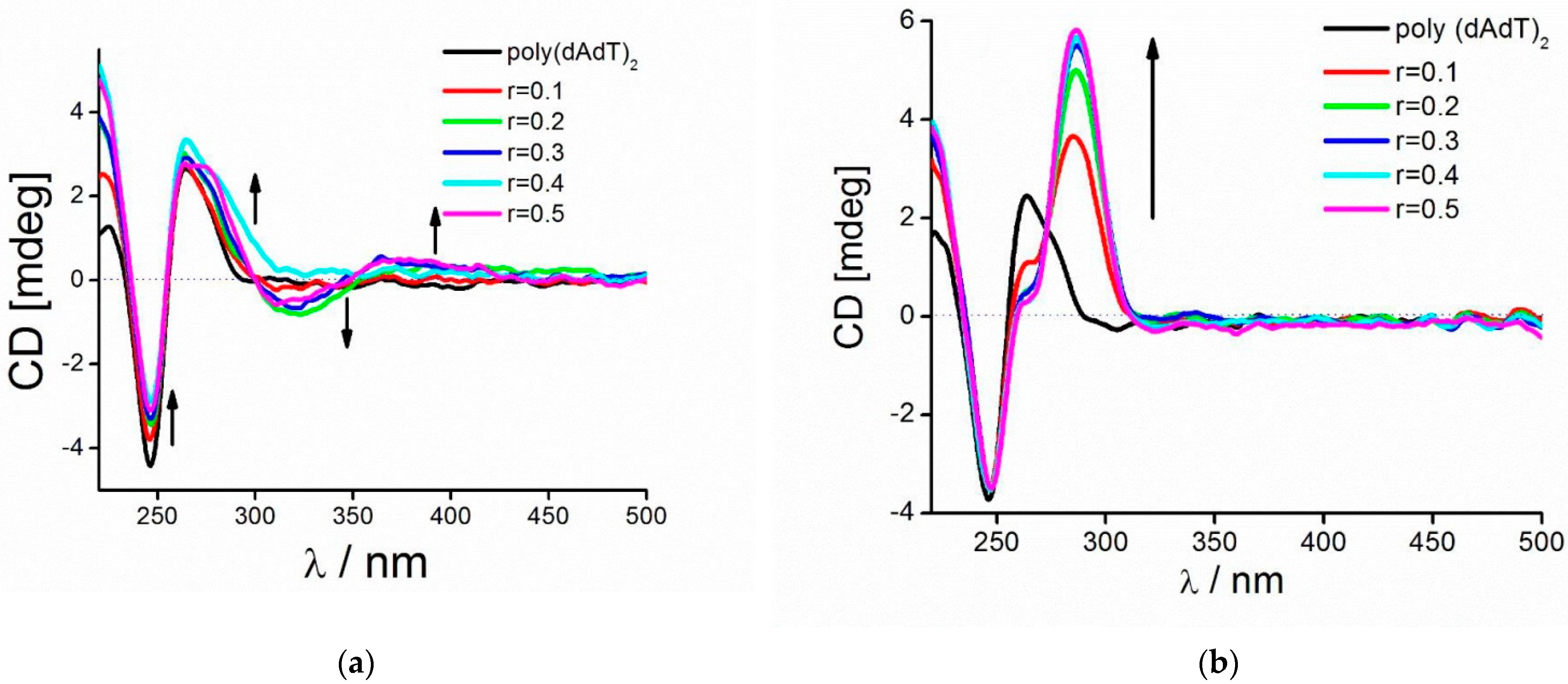
2.2.2. DNA Binding Study

Based upon the antiproliferative activity, several compounds from a series of novel amidine- and amidoxime-substituted heterocycles (5, 6, 811 and 20) were selected for the study with calf thymus DNA (ctDNA), alternating AT-DNA (poly (dAdT)2) and AU-RNA (poly A-poly U). Compounds 5, 6, 811 were soluble in water (c = 2 × 10⁻³ mol dm−3). Due to the small aggregation of 20 in water, a stock solution was also prepared in DMSO (c = 2 × 10⁻³ mol dm−3) to check the dependence of UV–Vis changes on concentration increase. The absorbencies of buffered aqueous solutions of studied compounds were proportional to their concentrations up to c = 2 × 10⁻⁵ mol dm−3 (Supplementary information (SI), Figures S1–S8) indicating that studied compounds do not aggregate by intermolecular stacking at experimental conditions used. Absorption maxima and corresponding molar extinction coefficients (ε) are given in Table S1 (SI).
The addition of ctDNA, poly A-poly U and poly (dAdT)2 yielded a fluorescence decrease of 6, 9, 11 and 20 whereas an emission increase was noticed in the case of 5, 8 and 10 with all studied polynucleotides (Table 2, Figure 4). The fluorescence changes of studied compounds were not dependent on the structure of polynucleotide added.
The binding constants Ks and ratios n (bound compound)/(DNA/RNA) obtained by processing of fluorometric titration data with Scatchard equation [40] are summarized in Table 2. Studied compounds showed similar affinity toward ds-DNA and ds-RNA. Still, some compounds such as 5, 6, 9 and 10 showed greater affinities towards p(dAdT)2 compared to other compounds. In addition, 5 displayed the best affinity towards AU-RNA than other compounds. Fluorescence changes of 8 and 10 with ctDNA and poly A-poly U were too small for accurate calculation of binding constants. Compounds with cyclic amidines at both ends of heterocyclic core (5, 6) or with cyclic amidine at one end and quinoline at the other (9) exhibited the preferential binding toward AT sequences. Interestingly, 9 exhibited greater affinities toward polynucleotides than 8, although the only difference was the linker between triazole and quinoline rings, O-alkyl vs. N-alkyl linker.
Compounds 911 and especially 5 and 6 showed moderate to big stabilization effect of AT-DNA (Table S2, Figure S41). In comparison to AT-DNA, stabilization of ctDNA by 5 and 6 was weaker as a consequence of the mixed base pair composition of ctDNA (58% AT, 42% GC). Other compounds showed either small or no stabilization effect of polynucleotides. Only a derivative possessing two imidazole moieties at the end of the heteroaromatic structure, 5 stabilized both DNA and RNA.
CD spectroscopy is a highly sensitive method for gaining insight into the conformational changes in the secondary structure of polynucleotides induced by small molecule binding [41]. In addition, it can provide information on the binding mode for small achiral molecules possessing UV–Vis spectra above 300 nm.
Generally, the addition of compounds caused either a small decrease or a small increase of CD intensity of DNA and RNA polynucleotides at their maximal values (ctDNA at 275 nm, AT-DNA and AU-RNA at 260 nm). Still, significantly induced CD (ICD) bands appeared in titrations of 5, 6 and 911 with AT-DNA (Figure 5, SI). Addition of AT-DNA to 6, 9 and 11 induced positive ICD signals in the region from 270–303 nm at ratios, r < 0.2 which could be ascribed to minor groove binding [42]. However, positive and negative signals positioned at 320 and 380 nm appeared at ratios of r higher than 0.2, suggesting additional binding mode—formation and binding of dimers or higher-order aggregates along the polynucleotide backbone. In titrations with AT-DNA, compounds 5 and 10 caused strong positive ICD bands centered around 276 and 287 nm, respectively which is consistent with minor groove binding (Figure 5, SI). In titrations with AU-RNA with 10 and 11, weak negative ICD signals visible around 300 nm are indicative of intercalative binding mode where the ligands are oriented “parallel” to the long axis of adjacent base pairs [42]. Compound 5 caused an appearance of similar ICD signals (strong positive ICD signals around 284 nm) with ctDNA as in titration with AT-DNA and additionally, positive and negative signals positioned at 320 and 390 nm were indicative of binding of aggregates along the polynucleotide backbone or most probably inside the groove.
In titrations of polynucleotides with 8 and 20, there were no significantly induced CD signals possibly due to the formation of aggregates such as in titration of 20 with ctDNA or a lack of any dominant orientation of 8 and 20 molecules with respect to the polynucleotide chiral axis.

3. Experimental Part

3.1. Materials and Methods

All solvents were purified following recommended drying agents and/or distilled over 3 Å molecular sieves. For monitoring the progress of a reaction and for comparison purposes, thin layer chromatography (TLC) was performed on pre-coated Merck (Darmstadt, Germany) silica gel 60F-254 plates using an appropriate solvent system and the spots were detected under UV light (254 nm). For column chromatography silica gel (Fluka, Buchs, Switzerland, 0.063–0.2 mm) was employed, glass column was slurry-packed under gravity. Melting points (uncorrected) were determined with Kofler micro hot-stage (Reichert, Wien, Austria). The IR spectra were recorded on PerkinElmer Spectrum One spectrophotometer (Boston, MA, USA) with KBr disks. Elemental composition analyses of all novel compounds were within the 0.4% of the calculated values. 1H and 13C NMR spectra were acquired on a Bruker 300 and 600 MHz NMR spectrometer (Bruker Biospin, Rheinstetten, Germany). All data were recorded in DMSO-d6 at 298 K. Chemical shifts were referenced to the residual solvent signal of DMSO-d6 at δ 2.50 ppm for 1H and δ 39.50 ppm for 13C. Individual resonances were assigned on the basis of their chemical shifts, signal intensities, multiplicity of resonances and H–H coupling constants. 1H and 13C NMR spectra of compounds 422 are provided in Supporting Information (Figures S43–S61).

3.2. Synthetic Procedures

Compounds 4-azidobenzonitrile (2) [39] and alkynyl derivatives of benzonitrile (1a and 1b) [43,44], quinoline (1e and 1f) [45,46] and coumarin (1h and 1i) [47] were synthesized according to the known procedure.

3.3. Preparation of Alkynyl Derivatives 1c, 1d and 1g

3.3.1. 4-Cyano-1-((prop-2-ynyl)amino)-1H-indole (1c)

4-Cyanoindole (500 mg; 3.52 mmol) was dissolved in DMF (10 mL); K2CO3 (1.2 equiv.) was added and reaction mixture was heated at 80 °C. After 30 min, propargyl bromide (1.2 equiv.) was added. Reaction mixture was stirred about 24 h. Solvent was removed under reduced pressure and residue purified by column chromatography with dichloromethane as eluens. Compound 1c was isolated as white powder (403 mg; 64%; m.p. = 137–140 °C). 1H NMR (300 MHz, DMSO-d6) (δ/ppm): 7.92 (d, J = 8.3 Hz, 1H, H-5), 7.72 (d, J = 3.2 Hz, 1H, H-2), 7.60 (dd, J = 7.4, 0.7 Hz, 1H, H-7), 7.39–7.31 (m, 1H, H-6), 6.63 (dd, J = 3.2, 0.8 Hz, 1H, H-3), 5.21 (d, J = 2.5 Hz, 2H, CH2), 3.46 (t, J = 2.5 Hz, 1H, CH). 13C NMR (151 MHz, DMSO-d6) (δ/ppm): 135.13 (C-7a), 131.70 (C-2), 129.10 (C-3a), 125.06 (C-5), 121.37 (C-6), 118.36 (CN), 115.59 (C-7), 101.67 C-4), 99.59 (C-3), 78.63 (C-2′), 76.04 (C-3′), 35.45 (CH2-1′).

3.3.2. 5-Cyano-1-((prop-2-ynyl)amino)-1H-indole (1d)

5-Cyanoindole (1.00 g; 7.03 mmol) was dissolved in DMF (20 mL), K2CO3 (1.2 equiv was added and reaction mixture was heated at 80 °C. After 30 min propargyl bromide (1.2 equiv.) was added. Reaction mixture was stirred about 24 h. Solvent was removed under reduced pressure and residue purified by column chromatography with dichloromethane as eluens. Compound 1d was isolated as pink powder (900 mg; 71%, m.p. = 107–109 °C). 1H NMR (300 MHz, DMSO-d6) (δ/ppm): 5.19 (d, J = 2.5 Hz, 2H, CH2), 3.44 (t, J = 2,4 Hz, 1H, CH), 7.54 (dd, J = 8.5, 1.4 Hz, 1H, H-6), 6.64 (d, J = 3.0 Hz, 1H, H-3), 7.62 (d, J = 3.2 Hz, 1H, H-2), 7.72 (d, J = 8.5, 1H, H-7), 8.12 (d, J = 0.8, 1H, H-4). 13C NMR (75 MHz, DMSO-d6) (δ/ppm): 137.02 (C-7a), 131.03 (C-2), 128.07 (C-3a), 126.15 (C-4), 124.11 (C-6), 120.48 (CN), 111.40 (C-7), 102.42 (C-3), 101.64 (C-5), 78.64 (C-2′), 76.01 (C-3′), 35.36 (CH2-1′).

3.3.3. 2-Cyano-8-(prop-2-ynyloxy)-quinoline (1g)

To a solution of 8-hydroxy-2-carbonitrile (500 mg; 2.94 mmol) in acetone (18 mL), K2CO3 (6 equiv.) was added and reaction mixture was heated under reflux. After 30 min, propargyl bromide (0.4 mL; 2.94 mmol) was added. Reaction mixture was stirred and heated under reflux for about 24 h. Solvent was removed under reduced pressure and residue purified by column chromatography with petroleum ether:ethyl acetate = 6:4 as eluens. Compound 1g was isolated as white powder (498 mg; 81%, m.p. = 144–147 °C). 1H NMR (600 MHz, DMSO-d6) (δ/ppm): 8.62 (d, J = 8.5 Hz, 1H, H-4), 8.05 (d, J = 8.4 Hz, 1H, H-3), 7.75 (t, J = 8.0 Hz, 1H, H-6), 7.69 (d, J = 7.5 Hz, 1H, H-5), 7.44 (d, J = 7.7 Hz, 1H, H-7), 5.10 (d, J = 2.3 Hz, 2H, CH2), 3.65 (t, J = 2.3 Hz, 1H, CH). 13C NMR (75 MHz, DMSO-d6) (δ/ppm): 152.62 (C-8), 139.53 (C-4a), 138.09 (C-4), 131.56 (C-8a), 130.00 (C-6), 129.77 (C-2), 124.32 (C-3), 120.28 (C-5), 117.72 (CN), 111.64 (C-7), 78.90 (C-3′), 78.68 (C-2′), 56.22 (C-1′).

3.4. General Procedure for Synthesis of Cyano-Substituted 1,2,3-Triazole Derivatives 3a3i

4-Azidobenzonitrile (2) (1 equiv.) and corresponding alkyne 1a1i (1 equiv.) were dissolved in methanol, followed by addition of Cu(OAc)2 (0.05 equiv.). Reaction mixture was stirred for 24 h at room temperature. Solvent was removed under reduced pressure and residue purified by column chromatography with dichloromethane as eluent.

3.4.1. 4-((1-(4-Cyanophenyl)-1H-1,2,3-triazole-4-yl)methylamino)benzonitrile (3a)

Compound 3a was prepared according to general procedure from 4-azidobenzonitrile (396 mg; 2.75 mmol) and compound 1a (428 mg; 2.75 mmol) in methanol (30 mL). After purification by column chromatography with dichloromethane:methanol = 100:1 as eluens, compound 3a was isolated as white powder (306.7 mg; 37%, m.p. = 247–248 °C). 1H NMR (300 MHz, DMSO-d6) (δ/ppm): 8.90 (s, 1H, CH-triazole), 8.12 (q, J = 9.0 Hz, 4H, Ph), 7.47 (d, J = 8.8 Hz, 2H, Ph), 7.25 (t, J = 5.6 Hz, 1H, NH), 6.75 (d, J = 8.8 Hz, 2H, Ph), 4,48 (d, J = 5.7 Hz, 2H, CH2). 13C NMR (75 MHz, DMSO-d6) (δ/ppm): 151.68 (Cq-Ph), 146.29 (Cq-Ph), 139.48 (Cq-triazole), 134.30 (C-Ph), 133.36 (C-Ph), 121.53 (CH-triazole), 120.46 (CN), 120.34 (C-Ph), 118.09 (CN), 112.19 (C-Ph), 110.97 (Cq-Ph), 96.36 (Cq-Ph), 37.64 (CH2).

3.4.2. 4-((1-(4-Cyanophenyl)-1H-1,2,3-triazol-4-yl)methyloxy)benzonitrile (3b)

Compound 3b was prepared according to general procedure from 4-azidobenzonitrile (250 mg; 1.74 mmol) and compound 1b (273 mg; 1.74 mmol) in methanol (20 mL). Compound 3b was isolated as white powder (418 mg; 80%, m.p. = 207–210 °C). 1H NMR (600 MHz, DMSO-d6) (δ/ppm): 9.13 (s, 1H, H-triazole), 8.14 (dd, J = 29.0, 8.8 Hz, 4H, Ph), 7.81 (d, J = 8.9 Hz, 2H, Ph), 7.26 (d, J = 8.9 Hz, 2H, Ph), 5.38 (s, 1H, CH2). 13C NMR (75 MHz, DMSO-d6) (δ/ppm): 161.36 (C-Ph), 143.66 (C-Ph), 139.44 (Cq-triazole), 134.36 (C-Ph), 134.31 (CH-triazole), 123.47 (C-Ph), 120.69 (C-Ph), 119.09 (CN), 118.11 (CN), 115.91 (C-Ph), 111.27 (C-Ph), 103.39 (C-Ph), 61.32 (CH2).

3.4.3. 4-Cyano-1-((1-(4-cyanophenyl)-1H-1,2,3-triazol-4-yl)methylamino)-1H-indole (3c)

Compound 3c was prepared according to general procedure from 4-azidobenzonitrile (240 mg; 1.67 mmol) and compound 1c (300 mg; 1.67 mmol) in methanol (20 mL). Compound 3c was isolated as white powder (308 mg; 57%, m.p. = 200–203 °C). 1H NMR (300 MHz, DMSO-d6) (δ/ppm): 8.95 (s, 1H, H-triazole), 8.13–8.05 (m, 4H, H-5), 8.02 (d, J = 8.3 Hz, 1H, H-5), 7.80 (d, J = 3.2 Hz, 1H, H-2), 7.57 (d, J = 7.3 Hz, 1H, H-7), 7.36–7.27 (m, 1H, H-6), 6.65 (d, J = 2.7 Hz, 1H, H-3), 5.70 (s, 2H, CH2). 13C NMR (75 MHz, DMSO-d6) (δ/ppm): 144.65 (C-Ph), 139.36 (Cq-triazole), 135.31(C-7a), 134.23 (C-Ph), 132.14 (C-2), 129.08 (C-3a), 124.90 (C-5), 122.03 (CH-triazole), 121.28 (C-6), 120.54 (C-Ph), 118.46 (CN), 118.04 (CN), 115.73 (C-7), 111.10 (C-Ph), 101.54 (C-Ph), 99.53 (C-3), 40.93 (CH2).

3.4.4. 5-Cyano-1-((1-(4-cyanophenyl)-1H-1,2,3-triazol-4-yl)methylamino)-1H-indole (3d)

Compound 3d was prepared according to general procedure from 4-azidobenzonitrile (250 mg; 1.74 mmol) and compound 1d (313 mg; 1.74 mmol) in methanol (20 mL). Compound 3d was isolated as white powder (513 mg; 91%, m.p. = 215–217 °C). 1H NMR (300 MHz, DMSO-d6) (δ/ppm): 8.96 (s, 1H, H-triazole), 8.22–8.04 (m, 5H, Ph, H-4), 7.83 (d, J = 8.6 Hz, 1H, H-7), 7.70 (d, J = 3.2 Hz, 1H, H-2), 7.51 (dd, J = 8.6, 1.5 Hz, 1H, H-6), 6.65 (d, J = 3.2 Hz, 1H, H-3), 5.67 (s, 2H, CH2). 13C NMR (75 MHz, DMSO-d6) (δ/ppm): 144.58 (C-Ph), 139.36 (Cq-triazole), 137.20 (C-7a), 134.23 (C-Ph), 131.44 (C-2), 128.03 (C-3a), 126.05 (C-4), 124.00 (C-6), 122.05 (CH-triazole), 120.54 (C-Ph), 118.03 (CN), 111.51 (C-7), 111.11 (C-Ph), 102.37 (C-3), 101.43 (C-5), 40.83 (CH2).

3.4.5. 8-((1-(4-Cyanophenyl)-1H-1,2,3-triazol-4-yl)methylamino)quinoline (3e)

Compound 3e was prepared according to general procedure from 4-azidobenzonitrile (209 mg; 1.45 mmol) and compound 1e (316 mg; 1.74 mmol) in methanol (20 mL). After purification by column chromatography with dichloromethane:methanol = 100:1 as eluens, compound 3e was isolated as yellow crystalline powder (269 mg; 60%, m.p. = 196–198 °C). 1H NMR (300 MHz, DMSO-d6) (δ/ppm): 8.90 (s, 1H, H-triazole), 8.76 (dd, J = 4.2, 1.6 Hz, 1H, H-2), 8.22 (dd, J = 8.3, 1.6 Hz, 1H, H-4), 8.10 (dd, J = 24.2, 8.9 Hz, 4H), 7.51 (dd, J = 8.3, 4.2 Hz, 1H, H-3), 7.36 (t, J = 7.9 Hz, 1H, H-6), 7.10 (d, J = 8.2 Hz, 1H, H-7), 7.05 (t, J = 6.0 Hz, 1H, NH), 6.80 (d, J = 7.6 Hz, 1H, H-5), 4.68 (d, J = 6.0 Hz, 2H, CH2). 13C NMR (151 MHz, DMSO-d6) (δ/ppm): 147.05 (C-2), 143.89 (C-8), 139.55 (Cq-triazole), 137.58 (C-4a), 135.95 (C-4), 134.22 (C-Ph), 128.23 (C-8a), 127.67 (C-6), 121.75 (C-3), 121.30 (CH-triazole), 120.37 (C-Ph), 118.09 (CN), 113.91 (C-5), 110.87 (C-Ph), 104.89 (C-7), 38.28 (CH2).

3.4.6. 8-((1-(4-Cyanophenyl)-1H-1,2,3-triazol-4-yl)methyloxy)quinoline (3f)

Compound 3f was prepared according to general procedure from 4-azidobenzonitrile (353 mg; 2.45 mmol) and compound 1f (538 mg; 2.94 mmol) in methanol (15 mL). Compound 3f was isolated as white powder (655 mg; 82%, m.p. = 158–162 °C). 1H NMR (300 MHz, DMSO-d6) (δ/ppm): 9.19 (s, 1H, H-triazole), 8.84 (dd, J = 4.1, 1.7 Hz, 1H, H-2), 8.33 (dd, J = 8.3, 1.7 Hz, 1H, H-7), 8.16 (dd, J = 23.9, 8.9 Hz, 4H, Ph), 7.65–7.39 (m, 4H, H-3, H-5, H-6, H-7), 5,47 (s, 2H, CH2). 13C NMR (75 MHz, DMSO-d6) (δ/ppm): 153.71 (C-8), 149.04 (C-2), 144.26 (C-Ph), 139.73 (C-4a), 139.47 (Cq-triazole), 135.82 (C-4), 134.31 (C-Ph), 129.09 (C-8a), 126.73 (C-6), 123.47 (CH-triazole), 121.90 (C-3), 120.59 (C-Ph), 120.30 (C-5), 118.08 (CN), 111.16 (C-Ph), 110.31 (C-7), 61.67 (CH2).

3.4.7. 2-Cyano-8-((1-(4-cyanophenyl)-1H-1,2,3-triazol-4-yl)methyloxy)quinoline (3g)

Compound 3g was prepared according to general procedure from 4-azidobenzonitrile (187 mg; 1.30 mmol) and compound 1g (270 mg; 1.30 mmol) in methanol (20 mL). Compound 3g was isolated as white powder (260 mg; 57%, m.p. = 263–265 °C). 1H NMR (300 MHz, DMSO-d6) (δ/ppm): 9,21 (s, 1H, H-triazole), 8.62 (d, J = 8.5 Hz, 1H, H-4), 8.15 (dd, J = 21.8, 8.9 Hz, 4H, Ph), 8.05 (d, J = 8.5 Hz, 1H, H-3), 7.60–7.80 (m, 3H, H-5, H-5, H-7), 5,54 (s, 2H, CH2). 13C NMR (75 MHz, DMSO-d6) (δ/ppm): 153.47 (C-8), 143.74 (C-Ph), 139.56 (C-4a), 139.45 (Cq-triazole), 138.07 (C-4), 134.27 (C-Ph), 131.43 (C-8a), 130.21 (C-6), 129.84 (C-2), 124.33 (C-3), 123.73 (CH-triazole), 120.66 (C-Ph), 120.07 (C-5), 118.06 (CN), 117.74 (CN), 111.49 (C-7), 111.18 (Ph), 61.66 (CH2).

3.4.8. 4-((1-(4-Cyanophenyl)-1H-1,2,3-triazol-4-yl)methyloxy)coumarin (3h)

Compound 3h was prepared according to general procedure from 4-azidobenzonitrile (158 mg; 1.10 mmol) and compound 1h (220 mg; 1.10 mmol) in methanol (20 mL). Compound 3h was isolated as white powder (245 mg; 71%, m.p. = 274–276 °C). 1H NMR (600 MHz, DMSO-d6) (δ/ppm): 9.23 (s, 1H, H-triazole), 8.17 (dd, J = 43.7, 8.4 Hz, 4H, Ph), 7.84 (d, J = 7.8 Hz, 1H, H-5), 7.67 (t, J = 7.7 Hz, 1H, H-7), 7.42 (d, J = 8.3 Hz, 1H, H-8), 7.36 (t, J = 7.6 Hz, 1H, H-6), 6,21 (s, 1H, H-3), 5.55 (s, 2H, CH2). 13C NMR (75 MHz, DMSO-d6) (δ/ppm): 164.28 (C-4), 161.50 (C-2), 152.77 (C-8a), 142.80 (C-Ph), 139.40 (Cq-triazole), 134.32 (C-Ph), 132.88 (C-7), 124.22 (C-6), 123.58 (CH-triazole), 123.02 (C-5), 120.70 (C-Ph), 118.07 (CN), 116.47 (C-8), 115.01 (C-4a), 111.29 (C-Ph), 91.53 (C-3), 62.71 (CH2).

3.4.9. 7-((1-(4-Cyanophenyl)-1H-1,2,3-triazol-4-yl)methyloxy)coumarin (3i)

Compound 3i was prepared according to general procedure from 4-azidobenzonitrile (187 mg; 1.30 mmol) and compound 1i (260 mg; 1.30 mmol) in methanol (20 mL). Compound 3i was isolated as white powder (187 mg; 41.8%, m.p. = 228–229 °C). 1H NMR (300 MHz, DMSO-d6) (δ/ppm): 9.16 (s, 1H, H-triazole), 8.14 (td, J = 8.9, 2.0 Hz, 4H, Ph), 8.01 (d, J = 9.5 Hz, 1H, H-4), 7.66 (d, J = 8.7 Hz, 1H, H-5), 7.20 (d, J = 2.3 Hz, 1H, H-8), 7.06 (dd, J = 8.6, 2.4 Hz, 1H, H-6), 6.32 (d, J = 9.5 Hz, 1H, H-3), 5.39 (s, 2H, CH2). 13C NMR (151 MHz, DMSO-d6) (δ/ppm): 160.97 (C-2), 160.26 (C-7), 155.31 (C-8a), 144.30 (C-4), 143.67 (C-Ph), 139.43 (Cq-triazole), 134.34 (C-Ph), 129.60 (C-5), 123.47 (CH-triazole), 120.63 (C-Ph), 118.10 (CN), 112.92 (C-3), 112.83 (C-6), 112.76 (C-Ph), 111.22 (C-4a), 101.64 (C-8), 61.52 (CH2).

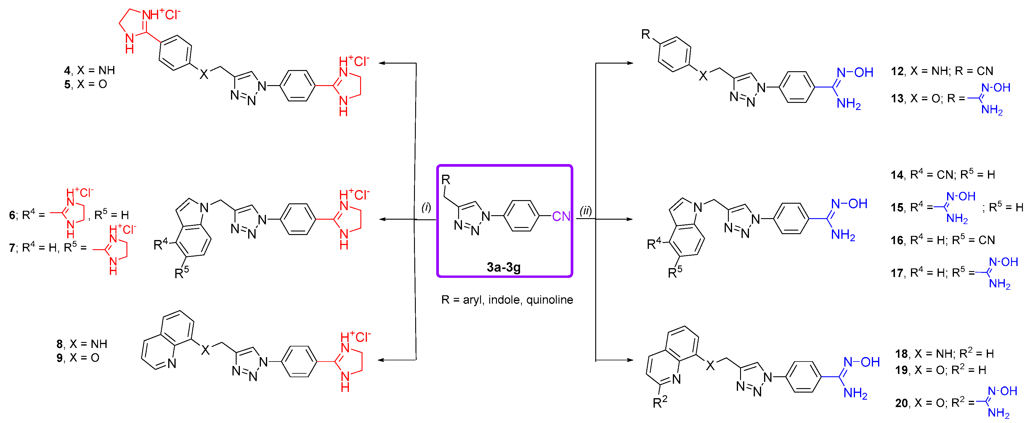
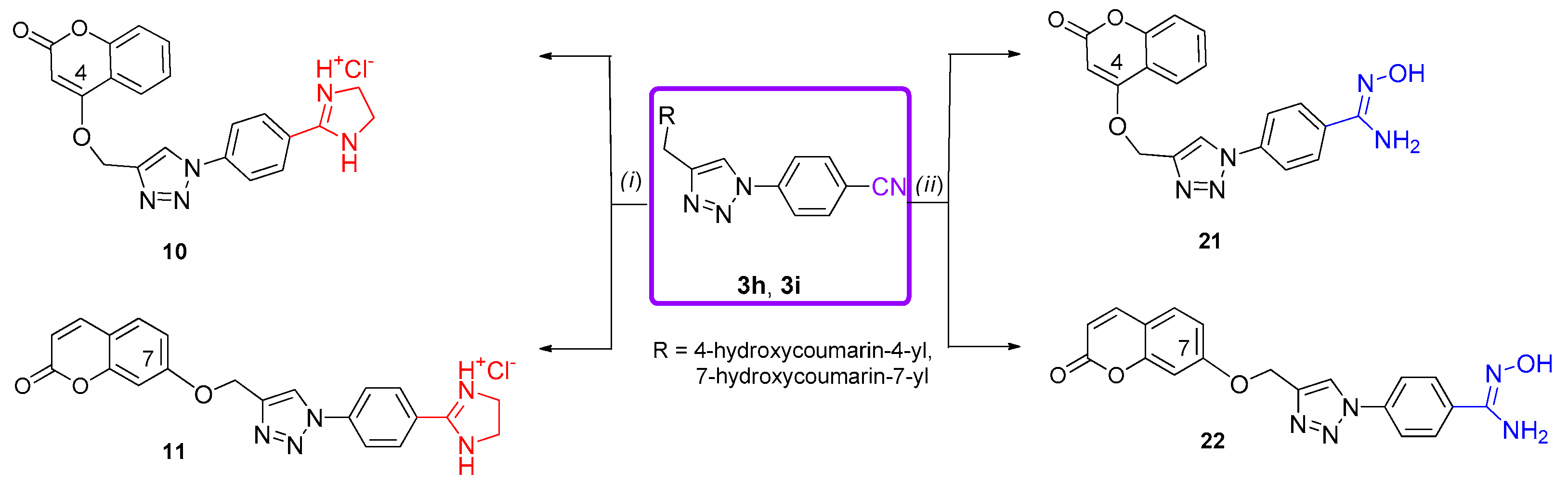
3.5. General Procedure for Synthesis of Imidazolinyl-Substituted 1,2,3-Triazole Derivatives 411

Suspension of corresponding cyano derivative 3a3i in absolute EtOH was cooled in an ice-salt bath and was saturated with HCl gas. The flask was sealed, and the mixture was stirred at room temperature until the nitrile band disappeared (monitored by the IR analysis at 2200 cm−1). The reaction mixture was diluted with dry diethylether, and the crude imidate was filtered off and suspended in absolute ethanol. Ethylenediamine was added and the mixture was stirred at reflux for 24 h. The reaction mixture was diluted with dry diethylether, and the crude base was filtered off. Suspension of base in absolute EtOH was cooled in an ice-salt bath and saturated with HCl gas, sealed and stirred for 24 h. Mixture was diluted with dry diethyl ether and the crude product was then filtered off, washed with diethylether to give powdered product as hydrochloride salt.

3.5.1. 1-(4-(4,5-Dihydro-1H-imidazol-2-yl)-phenyl)-4-((4-(4,5-dihydro-1H-imidazol-2-yl)phenyl)aminomethyl)-1H-1,2,3-triazole dihydrochloride (4)

Compound 4 was prepared according to general procedure using 3a (50 mg; 0.17 mmol) in absolute ethanol (15 mL). After 4 days, iminoester was filtered and suspended in absolute ethanol (10 mL) followed by the addition of EDA (0.07 mL, 1.02 mmol). Compound 4 was isolated as yellow powder (37 mg; 47%; m.p. > 250 °C). 1H NMR (300 MHz, DMSO-d6) (δ/ppm): 10.98 (s, 2H, NH), 10.12 (s, 2H, NH), 8.99 (s, 1H, H-triazole), 8.18–8.32 (m, 4H, Ph), 7.81 (d, J = 8.7 Hz, 2H, Ph), 7.57 (bs, 1H, NH), 6.83 (d, J = 8.8 Hz, 2H, Ph), 4.54 (d, J = 3,4 Hz, 2H, CH2), 4.02 (s, 4H, CH2), 3.89 (s, 4H, CH2). 13C NMR (75 MHz, DMSO-d6) (δ/ppm): 164.16 (C-imidazolyl), 163.86 (C-imidazolyl), 153.29 (C-1′), 153.46 (C-Ph), 146.13 (C-4), 140.33 (Cq-triazole), 130.71 (C-2,6), 130.57 (C-3′,5′), 121.81 (C-1), 121.61 (CH-triazole), 119.94 (C-3′,5′), 112.87 (C-2′,6′), 107.80 (C-4′), 44.38 (2 × CH2), 43.74 (2 × CH2), 37.60 (CH2). Anal. calcd. for C21H22N8 × 2 HCl × 1.5 H2O (Mr = 486.40): C 51.86, H 5.60, N 23.04; found: C 51.79, H 5.68, N 23.09%.

3.5.2. 1-(4-(4,5-Dihydro-1H-imidazol-2-yl)-phenyl]-4-((4-(4,5-dihydro-1H-imidazol-2-yl) phenyloxy)methyl]-1H-1,2,3-triazole dihydrochloride (5)

Compound 5 was prepared according to general procedure using 3b (100 mg; 0.33 mmol) in absolute ethanol (20 mL). After 10 days, iminoester was filtered and suspended in absolute ethanol (20 mL), followed by the addition of EDA (0.13 mL, 1.98 mmol). Compound 5 was isolated as white powder (34 mg; 22%); m.p. > 250 °C). 1H NMR (600 MHz, DMSO-d6) (δ/ppm): 11.06 (bs, 2H, NH-imidazolyl), 10.68 (bs, 2H, NH-imidazolyl), 9.23 (s, 1H, H-triazole), 8.24–8.36 (m, 4H, Ph), 8.11 (d, J = 9.0 Hz, 2H, Ph), 7.35 (d, J = 9.0 Hz, 2H, Ph), 5.43 (s, 2H, CH2), 4.03 (s, 4H, CH2), 3.96 (s, 4H, CH2). 13C NMR (151 MHz, DMSO-d6) (δ/ppm): 164.01 (C-imidazolyl), 163.69 (C-imidazolyl), 162.56 (C-1′), 143.58 (C-4), 140.26 (Cq-triazole), 130.93 (C-2,6), 130.72 (C-3′,6′), 123.51 (CH-triazole), 122.07 (C-1), 120.25 (C-3,5), 115.42 (C-2′,6′), 114.54 (C-4′), 61.36 (CH2), 44.41 (2 × CH2), 44.11 (2 × CH2). Anal. calcd. for C21H21N7O × 2 HCl × 3 H2O (Mr = 514.41): C 49.03, H 5.68, N 19.06; found: C 49.10, H 5.66, N 18.98%.

3.5.3. 1-((1-(4-(Dihydro-1H-imidazol-2-yl)phenyl)-1H-1,2,3-triazol-4-yl)methyl)-4-(dihydro-1H-imidazol-2-yl)-1H-indole dihydrochloride (6)

Compound 6 was prepared according to general procedure using 3c (150 mg; 0.46 mmol) in absolute ethanol (30 mL). After 4 days, iminoester was filtered and suspended in absolute ethanol (15 mL), followed by the addition of EDA (0.19 mL, 2.76 mmol). Compound 6 was isolated as brown powder (57 mg; 26%; m.p. > 250 °C). 1H NMR (300 MHz, DMSO-d6) (δ/ppm): 10.92 (s, 2H, NH-imidazolyl), 10.30 (s, 2H, NH-imidazolyl), 9.06 (s, 1H, H-triazole), 8.23 (m, 4H, Ph), 8.10 (d, J = 8.3 Hz, 1H, H-5), 7.84 (d, J = 3.3 Hz, 1H, H-2), 7.59 (d, J = 6.8 Hz, 1H, H-7), 7.36–7.43 (m, 1H, H-6), 6.79 (d, J = 2.6 Hz, 1H, H-3), 5.73 (s, 2H, CH2), 4.04 (s, 4H, 2 × CH2), 4.02 (s, 4H, 2 × CH2). 13C NMR (75 MHz, DMSO-d6) (δ/ppm): 165.66 (C-imidazolyl), 163.63 (C-imidazolyl), 144.51 (C-1′), 140.20 (Cq-triazole), 135.98 (C-7a), 131.87 (C-2), 130.71 (C-3′,5′), 125.68 (C-3a), 122.15 (CH-triazole), 121.97 (C-4), 121.60 (C-5), 120.89 (C-6), 120.13 (C-2′,6′), 116.39 (C-7), 113.76 (C-4′), 100.54 (C-3), 44.36 (CH2), 40.81 (CH2). Anal. calcd. for C23H22N8 × 2 HCl × 0.5 H2O (Mr = 492.41): C 56.10, H 5.12, N 22.76; found: C 56.98, H 5.14, N 22.81%.

3.5.4. 1-((1-(4-(Dihydro-1H-imidazol-2-yl)phenyl)-1H-1,2,3-triazol-4-yl)methyl)-5-(dihydro-1H-imidazol-2-yl)-1H-indole dihydrochloride (7)

Compound 7 was prepared according to general procedure using 3d (150 mg; 0.46 mmol) in absolute ethanol (30 mL). After 4 days, iminoester was filtered and suspended in absolute ethanol (15 mL), followed by the addition of EDA (0.19 mL, 2.76 mmol). Compound 7 was isolated as red powder (160 mg; 72%; m.p. = 228–231 °C). 1H NMR (300 MHz, DMSO-d6) (δ/ppm): 11.08 (s, 2H, NH-imidazolyl), 10.60 (s, 2H, NH-imidazolyl), 9.09 (s, 1H, H-triazole), 8.40 (s, 1H, H-4), 8.14–8.34 (m, 4H, Ph), 7.81–7.94 (m, 2H, H-2, H-7), 7.74 (d, J = 3.2 Hz, 1H, H-6), 6.71 (d, J = 3.1 Hz, 1H, H-3), 5.70 (s, 2H, CH2), 3.87–4.16 (m, 8H, 4xCH2). 13C NMR (151 MHz, DMSO-d6) (δ/ppm): 165.60 (C-imidazolyl), 163.62 (C-imidazolyl), 144.52 (C-1′), 140.20 (Cq-triazole), 138.37 (C-7a), 131.59 (C-2), 130.67 (C-3′,5′), 127.84 (C-3a), 122.63 (C-4), 122.14 (CH-triazole), 121.95 (C-4), 121.17 (C-6), 120.11 (C-2′,6′), 112.88 (C-5), 111.01 (C-7), 102.94 (C-3), 44.35 (2 × CH2), 44.06 (2 × CH2), 40.84 (CH2). Anal. calcd. for C23H22N8 × 2 HCl × 2.2 H2O (Mr = 523.03): C 52.82, H 5.47, N 21.46; found: C 52.62, H 5.45, N 21.53%.

3.5.5. 8-(((1-(4-(4,5-Dihydro-1H-imidazol-2-yl)phenyl)-1H-1,2,3-triazol-4-yl)methyl)amino)-quinoline hydrochloride (8)

Compound 8 was prepared according to general procedure using 3e (190 mg; 0.58 mmol) in absolute ethanol (30 mL). After 10 days iminoester was filtered and suspended in absolute ethanol (15 mL), followed by the addition of EDA (0.12 mL, 1.74 mmol). Compound 8 was isolated as white powder (48 mg; 21%; m.p. = 192–196 °C). 1H NMR (300 MHz, DMSO-d6) (δ/ppm): 10.93 (s, 2H, NH-imidazolyl), 8.96 (s, 1H, H-triazole), 8.82 (dd, J = 4.3, 1.5 Hz, 1H, H-2), 8.37 (d, J = 8.3 Hz, 1H, H-4), 8.18–8.30 (m, 4H, Ph), 7.60 (dd, J = 8.3, 4.3 Hz, 1H, H-3), 7.42 (t, J = 7.9 Hz, 1H, H-6), 7.19 (d, J = 8.0 Hz, 1H, H-7), 6.89 (d, J = 7.6 Hz, 1H, H-5), 4.71 (s, 2H, CH2), 4.01 (s, 4H, CH2). 13C NMR (75 MHz, DMSO-d6) (δ/ppm): 163.77 (C-imidazolyl), 146.90 (C-8), 146.42 (C-2), 143.14 (C-4a), 140.44 (Cq-triazole), 137.59 (C-4), 130.59 (C-3′,5′), 128.52 (C-8a), 128.12 (C-6), 121.79 (C-3), 121.72 (C-4′), 121.37 (CH-triazole), 120.00 (C-2′,6′), 114.25 (C-7), 106.00 (C-5), 44.41 (2 × CH2), 36.49 (CH2). Anal. calcd. for C21H19N7 × HCl × 2.5 H2O (Mr = 450.93): C 55.94, H 5.59, N 21.74; found: C 55.75, H 5.60, N 21.78%.

3.5.6. 8-((1-(4-(4,5-Dihydro-1H-imidazol-2-yl)phenyl)-1H-1,2,3-triazol-4-yl)methyloxy)-quinoline hydrochloride (9)

Compound 9 was prepared according to general procedure using 3f (175 mg; 0.53 mmol) in absolute ethanol (30 mL). After 6 days iminoester was filtered and suspended in absolute ethanol (15 mL), followed by the addition of EDA (0.11 mL, 1.59 mmol). Compound 9 was isolated as white powder (50 mg; 23%; m.p. = 229–232 °C). 1H NMR (300 MHz, DMSO-d6) (δ/ppm): 10.93 (s, 2H, NH-imidazolyl), 9.56 (s, 1H, H-triazole), 9.08 (d, J = 4.3 Hz, 1H, H-2), 8.91 (d, J = 7.3 Hz, 1H, H-4), 8.24–8.37 (m, 4H, Ph), 7.93 (bs, 1H, H-3), 7.76–7.86 (m, 3H, H-5, H-6, H-7), 5.64 (s, 2H, CH2), 4.04 (s, 4H, CH2). 13C NMR (151 MHz, DMSO-d6) (δ/ppm): 163.98 (C-imidazolyl), 148.54 (C-2), 144.02 (C-1′), 140.40 (Cq-triazole), 134.45 (C-4), 130.53 (C-3′,5′), 129.19 (C-8a), 127.20 (C-6), 123.48 (CH-triazole), 122.07 (C-3), 122.01 (C-4′), 120.40 (C-5), 120.30 (C-2′,6′), 110.84 (C-7), 61.87 (CH2), 44.50 (2 × CH2). Anal. calcd. for C21H18N6O × HCl × 3.8 H2O (Mr = 475.33): C 53.06, H 5.64, N 17.68; found: C 52.95, H 5.66, N 17.65%.

3.5.7. 4-((1-(4-(4,5-Dihydro-1H-imidazol-2-yl)phenyl)-1H-1,2,3-triazol-4-yl)methyloxy)-coumarin hydrochloride (10)

Compound 10 was prepared according to general procedure using 3h (100 mg; 0.29 mmol) in absolute ethanol (20 mL). After 10 days iminoester was filtered and suspended in absolute ethanol (10 mL), followed by the addition of EDA (0.06 mL, 0.87 mmol). Compound 10 was isolated as white powder (29 mg; 24%; m.p.> 250 °C). 1H NMR (300 MHz, DMSO-d6) (δ/ppm): 10.89 (s, 2H, NH-imidazolyl), 9.27 (s, 1H, H-triazole), 8.19–8.35 (m, 4H, Ph), 7.85 (d, J = 7.9 Hz, 1H, H-5), 7.68 (t, J = 7.1 Hz, 1H, H-7), 7.43 (d, J = 8.3 Hz, 1H, H-8), 7.36 (t, J = 7.6 Hz, 1H, H-6), 6.22 (s, 1H, H-3), 5.57 (s, 2H, CH2), 4.04 (s, 4H, 2xCH2). 13C NMR (75 MHz, DMSO-d6) (δ/ppm): 164.27 (C-4), 163.84 (C-imidazolyl), 161.49 (C-2), 152.78 (C-8a), 142.84 (C-1′), 140.29 (Cq-triazole), 132.89 (C-7), 130.57 (C-3′,5′), 124.23 (C-6), 123.52 (CH-triazole), 123.00 (C-5), 122.10 (C-4′), 120.39 (C-2′,6′), 116.48 (C-8), 115.01 (C-4a), 91.55 (C-3), 62.72 (CH2), 44.46 (2 × CH2). Anal. calcd. for C21H17N5O3 × HCl × 3.5 H2O (Mr = 486.91): C 51.80, H 5.18, N 14.38; found: C 51.71, H 5.16, N 14.40%.

3.5.8. 7-((1-(4-(4,5-Dihydro-1H-imidazol-2-yl)phenyl)-1H-1,2,3-triazol-4-yl)methyloxy)-coumarin hydrochloride (11)

Compound 11 was prepared according to general procedure using 3i (200 mg; 0.58 mmol) in absolute ethanol (30 mL). After 6 days iminoester was filtered and suspended in absolute ethanol (15 mL), followed by the addition of EDA (0.12 mL, 1.74 mmol). Compound 11 was isolated as white powder (72 mg; 29%; m.p. > 250 °C). 1H NMR (300 MHz, DMSO-d6) (δ/ppm): 10.98 (s, 2H, NH-imidazolyl), 9.19 (s, 1H, H-triazole), 8.27 (s, 4H, Ph), 8.02 (d, J = 9.5 Hz, 1H, H-4), 7.67 (d, J = 8.7 Hz, 1H, H-5), 7.20 (d, J = 2.2 Hz, 1H, H-8), 7.07 (dd, J = 8.6, 2.4 Hz, 1H, H-6), 6.32 (d, J = 9.5 Hz, 1H, H-3), 5.41 (s, 2H, CH2), 4.03 (s, 4H, CH2). 13C NMR (75 MHz, DMSO-d6) (δ/ppm): 163.91 (C-imidazolyl), 160.97 (C-2), 160.26 (C-7), 155.31 (C-8a), 144.30 (C-4), 143.74 (C-1′), 140.32 (Cq-triazole), 130.54 (C-3′,5′), 129.63 (C-5), 123.40 (CH-triazole), 122.12 (C-4′), 120.36 (C-2′,6′), 112.92 (C-3), 112.84 (C-6), 112.78 (C-4a), 101.66 (C-8), 61.55 (CH2), 44.53 (2 × CH2). Anal. calcd. for C21H17N5O3 × HCl × 1.3 H2O (Mr = 447.28): C 56.39, H 4.64, N 15.66; found: C 56.58, H 4.63, N 15.69%.

3.6. General Procedure for Synthesis of Amidoxime-Substituted 1,2,3-Triazole Derivatives 1222

Corresponding cyano derivative 3a3i was suspended in solvent mixture methanol:dimethylformamide = 2:1, and suspension was heated at 100 °C. Triethylamine (Et3N) (3 equiv. for bis cyano derivatives 3a3d and 3g, and 6 equiv. for cyano derivatives 3e, 3f, 3h and 3i) and NH2OH⸱HCl (3 equiv. for bis cyano derivatives 3a3d and 3g, and 6 equiv. for cyano derivatives 3e, 3f, 3h and 3i) were added and reaction mixture was stirred for 6 h. After cooling, mixture was diluted with water and precipitate was filtered off. If necessary, precipitate was purified by column chromatography with dichloromethane:methanol = 10:1 as eluent.

3.6.1. 4-(((1-(4-((Z)-N′-Hydroxycarbamimidoyl)phenyl)-1H-1,2,3-triazol-4-yl)methyl)amino)benzonitrile (12)

Compound 12 was prepared according to general procedure using 3a (50 mg; 0.17 mmol), Et3N (0.14 mL) and NH2OH⸱HCl (71 mg) in solvent mixture methanol:DMF = 2:1 (6 mL). After purification by column chromatography compound 12 was isolated as white powder (19 mg; 35%; m.p. = 222–225 °C). 1H NMR (300 MHz, DMSO-d6) (δ/ppm): 9.79 (s, 1H, OH), 8.77 (s, 1H, H-triazole), 7.76–7.95 (m, 4H, Ph), 7.47 (d, J = 8.7 Hz, 2H, Ph), 7.23 (t, J = 5.6 Hz, 1H, NH), 6.76 (d, J = 8.8 Hz, 2H, Ph), 5.93 (s, 2H, NH2), 4.46 (d, J = 5.6 Hz, 2H, CH2). 13C NMR (75 MHz, DMSO-d6) (δ/ppm): 151.73 (C-1′), 149.83 (C(NH2)=NOH), 145.74 (C-4), 136.68 (Cq-triazole), 133.35 (C-3′,5′), 129.21 (C-1), 126.71 (C-2,6), 121.21 (CH-triazole), 120.49 (CN), 119.47 (C-3,5), 112.18 (C-2′,6′), 96.27 (C-4′), 37.71 (CH2). Anal. calcd. for C17H15N7O (Mr = 333.36): C 61.25, H 4.54, N 29.41; found: C 61.42, H 4.52, N 29.47%.

3.6.2. (Z)-N′-Hydroxy-4-(4-((4-((Z)-N′-hydroxycarbamimidoyl)phenoxy)methyl)-1H-1,2,3-triazol-1-yl)benzimidamide (13)

Compound 13 was prepared according to general procedure using 3b (80 mg; 0.27 mmol), Et3N (0.22 mL) and NH2OH⸱HCl (113 mg) in solvent mixture methanol:DMF = 2:1 (6 mL). After purification by column chromatography compound 13 was isolated as white powder (80 mg; 82%; m.p. = 218–221 °C). 1H NMR (300 MHz, DMSO-d6) (δ/ppm): 9.75 (s, 1H, OH), 9.43 (s, 1H, OH), 8.95 (s, 1H, H-triazole), 7.80–8.03 (m, 4H, Ph), 7.63 (d, J = 8.8 Hz, 2H, Ph), 7.08 (d, J = 8.8 Hz, 2H, Ph), 5.89 (s, 2H, NH2), 5.70 (s, 2H, NH2), 5.28 (s, 2H, CH2). 13C NMR (75 MHz, DMSO-d6) (δ/ppm): 158.51 (C-Ph), 150.55 (C(NH2)=NOH), 149.84 (C(NH2)=NOH), 143.83 (C-Ph), 136.62 (Cq-triazole), 133.52 (C-Ph), 126.76 (C-Ph), 126.73 (C-Ph), 126.15 (C-Ph), 122.82 (CH-triazole), 119.69 (C-Ph), 114.27 (C-Ph), 61.02 (CH2). Anal. calcd. for C17H17N7O3 (Mr = 367.37): C 55.58, H 4.66, N 26.69; found: C 55.41, H 4.66, N 26.63%.

3.6.3. 1-((1-(4-((Z)-N′-Hydroxycarbamimidoyl)phenyl)-1H-1,2,3-triazol-4-yl)methyl)-4-cyanoindole (14) and (Z)-N′-hydroxy-1-((1-(4-((Z)-N′-hydroxycarbamimidoyl)phenyl)-1H-1,2,3-triazol-4-yl)methyl)-1H-indole-4-carboximidamide (15)

Compounds 14 and 15 were prepared according to general procedure using 3c (150 mg; 0.46 mmol), Et3N (0.38 mL) and NH2OH⸱HCl (192 mg) in solvent mixture methanol:DMF = 2:1 (12 mL). Purification by column chromatography yielded compound 14 as white powder (36 mg; 22%; m.p. = 229–232 °C) and compound 15 as white powder (74 mg; 41%; m.p. = 120–123 °C).
14: 1H NMR (300 MHz, DMSO-d6) (δ/ppm): 9.79 (s, 1H, OH), 8.83 (s, 1H, H-triazole), 8.04 (d, J = 8.3 Hz, 1H, H-5), 7.86 (s, 4H, Ph), 7.81 (d, J = 3.2 Hz, 1H, H-2), 7.57 (d, J = 7.3 Hz, 1H, H-7), 7.25–7.37 (m, 1H, H-6), 6.64 (d, J = 3.1 Hz, 1H, H-3), 5.93 (s, 2H, NH2), 5.68 (s, 2H, CH2). 13C NMR (151 MHz, DMSO-d6) (δ/ppm): 149.93 (C(NH2)=NOH), 144.26 (C-Ph), 136.61 (Cq-triazole), 135.39 (C-7a), 133.56 (C-3a), 132.20 (C-2), 129.15 (C-Ph), 126.77 (C-Ph), 124.97 (C-5), 121.76 (CH-triazole), 121.36 (C-6), 119.75 (C-Ph), 118.56 (CN), 115.81 (C-7), 101.59 (C-4), 99.56 (C-3), 41.04 (CH2). Anal. calcd. for C19H15N7O (Mr = 357.38): C 63.86, H 4.23, N 27.44; found: C 63.89, H 4.24, N 27.42%.
15: 1H NMR (600 MHz, DMSO-d6) (δ/ppm): 9.78 (s, 1H, OH), 9.57 (s, 1H, OH), 8.82 (s, 1H, H-triazole), 7.86 (s, 4H, Ph), 7.67 (d, J = 8.2 Hz, 1H, H-5), 7.49 (d, J = 3,2 Hz, 1H, H-2), 7.30 (dd, J = 7.3, 0.5 Hz, 1H, H-7), 7.09–7.21 (m, 1H, H-6), 6.88 (dd, J = 3.0, 0.5 Hz, 1H, H-3), 5.92 (s, 2H, NH2), 5.68 (s, 2H, NH2), 5.56 (s, 2H, CH2). 13C NMR (151 MHz, DMSO-d6) (δ/ppm): 152.02 (C(NH2)=NOH), 149.80 (C(NH2)=NOH), 144.59 (C-Ph), 136.55 (Cq-triazole), 135.89 (C-7a), 133.42 (C-3a), 128.70 (C-2), 126.65 (C-Ph), 125.61 (C-Ph), 125.55 (C-4), 121.53 (CH-triazole), 120.69 (C-6), 119.59 (C-Ph), 117.65 (C-7), 110.78 (C-5), 102.63 (C-3), 40.71 (CH2). Anal. calcd. for C19H18N8O2 (Mr = 390.41): C 58.45, H 4.65, N 28.70; found: C 58.40, H 4.65, N 28.72%.

3.6.4. 1-((1-(4-((Z)-N′-Hydroxycarbamimidoyl)phenyl)-1H-1,2,3-triazol-4-yl)methyl)-5-cyanoindole (16) and (Z)-N′-hydroxy-1-((1-(4-((Z)-N′-hydroxycarbamimidoyl)phenyl)-1H-1,2,3-triazol-4-yl)methyl)-indole-5-carboximidamide (17)

Compounds 16 and 17 were prepared according to general procedure using 3d (125 mg; 0.39 mmol), Et3N (0.32 mL) and NH2OH⸱HCl (163 mg) in solvent mixture methanol:DMF = 2:1 (12 mL). Purification by column chromatography yielded compound 16 as white powder (26 mg; 18%; m.p. = 232–234 °C) and compound 17 as white powder (67 mg; 44%; m.p. = 152–155 °C).
16: 1H NMR (300 MHz, DMSO-d6) (δ/ppm): 9.79 (s, 1H, OH), 8.84 (s, 1H, H-triazole), 8.11 (d, J = 1.0 Hz, 1H, H-4), 7.80–7.88 (m, 5H, Ph, H-7), 7.71 (d, J = 3.2 Hz, 1H, H-2), 7.51 (dd, J = 8.6, 1.5 Hz, 1H, H-6), 6,65 (d, J = 3.2 Hz, 1H, H-3), 5,92 (s, 2H, NH2), 5,65 (s, 2H, CH2). 13C NMR (75 MHz, DMSO-d6) (δ/ppm): 150.03 (C(NH2)=NOH), 144.28 (C-Ph), 137.32 (Cq-triazole), 136.67 (C-7a), 133.61 (C-Ph), 131.57 (C-2), 128.18 (C-3a), 126.86 (C-Ph), 126.21 (C-4), 124.14 (C-6), 121.85 (CH-triazole), 120.72 (CN), 119.83 (C-Ph), 111.65 (C-7), 102.51 (C-3), 101.50 (C-5), 40.98 (CH2). Anal. calcd. for C19H15N7O (Mr = 357.38): C 63.86, H 4.23, N 27.44; found: C 63.79, H 4.24, N 27.51%.
17: 1H NMR (600 MHz, DMSO-d6) (δ/ppm): 9.78 (s, 1H, OH), 9.39 (s, 1H, OH), 8.83 (s, 1H, H-triazole), 7.71–7.95 (m, 5H), 7.60 (d, J = 8.7 Hz, 1H, 2H), 7.43–7.54 (m, 2H, H-4, H-6), 6.38–6.55 (m, 1H, H-3), 5.91 (s, 2H, NH2), 5.72 (s, 2H, NH2), 5.56 (s, 2H, CH2). 13C NMR (75 MHz, DMSO-d6) (δ/ppm): 151.91 (C(NH2)=NOH), 149.81 (C(NH2)=NOH), 144.59 (C-Ph), 136.57 (Cq-triazole), 135.90 (C-7a), 133.45 (C-Ph), 129.31 (C-2), 127.77 (C-3a), 126.68 (C-Ph), 124.78 (C-5), 121.58 (CH-triazole), 119.61 (C-Ph), 119.26 (C-4), 117.77 (C-6), 109.68 (C-7), 101.71 (C-3), 40.78 (CH2). Anal. calcd. for C19H18N8O2 (Mr = 390.41): C 58.45, H 4.65, N 28.70; found: C 58.51, H 4.66, N 28.61%.

3.6.5. 8-(((1-(4-((Z)-N′-Hydroxycarbamimidoyl)phenyl)-1H-1,2,3-triazol-4-yl)methyl)amino)quinoline (18)

Compound 18 was prepared according to general procedure using 3e (80 mg; 0.25 mmol), Et3N (0.10 mL) and NH2OH⸱HCl (51 mg) in solvent mixture methanol:DMF = 2:1 (9 mL). After purification by column chromatography, compound 18 was isolated as white powder (35 mg; 40%; m.p. = 181–183 °C). 1H NMR (300 MHz, DMSO-d6) (δ/ppm): 9.78 (s, 1H, OH), 8.77 (s, 1H, H-triazole), 8.22 (dd, J = 8.3, 1.6 Hz, 1H, H-2), 7.80–7.97 (m, 5H, Ph, H-4), 7.51 (dd, J = 8.3, 4.2 Hz, 1H, H-3), 7.37 (t, J = 7.9 Hz, 1H, H-6), 7.11 (d, J = 8.1 Hz, 1H, H-5), 7.02 (t, J = 5.8 Hz, 1H, NH), 6.82 (d, J = 7.0 Hz, 1H, H-7), 5.92 (s, 2H, NH2), 4.67 (d, J = 6.0 Hz, 2H, CH2). 13C NMR (75 MHz, DMSO-d6) (δ/ppm): 150.00 (C(NH2)=NOH), 147.19 (C-2), 146.62 (C-8), 144.01 (C-1′), 137.66 (Cq-triazole), 136.84 (C-4a), 136.07 (C-4), 133.36 (C-1′), 128.33 (C-8a), 127.80 (C-6), 126.80 (C-3′,5′), 121.88 (C-3), 121.09 (CH-triazole), 119.63 (C-2′,6′), 114.00 (C-5), 105.02 (C-7), 35.90 (CH2). Anal. calcd. for C19H17N7O (Mr = 359.39): C 63.50, H 4.77, N 27.28; found: C 63.45, H 4.76, N 27.27%.

3.6.6. 8-((1-(4-((Z)-N′-Hydroxycarbamimidoyl)phenyl)-1H-1,2,3-triazol-4-yl)methoxy)quinoline (19)

Compound 19 was prepared according to general procedure using 3f (90 mg; 0.28 mmol), Et3N (0.12 mL) and NH2OH⸱HCl (58 mg) in solvent mixture methanol: DMF = 2:1 (9 mL). Compound 19 was isolated as white powder (78 mg; 79%; m.p. = 230–232 °C). 1H NMR (600 MHz, DMSO-d6) (δ/ppm): 9.81 (s, 1H, OH), 9.06 (s, 1H, H-triazole), 8.84 (dd, J = 4.1, 1.7 Hz, 1H, H-2), 8.33 (dd, J = 8.3, 1.6 Hz, 1H, H-4), 7.88–7.99 (m, 4H, Ph), 7.50–7.59 (m, 3H, H-3, H-5, H-6), 7.46 (dd, J = 6.3, 2.7 Hz, 1H, H-7), 5.95 (s, 2H, NH2), 5.46 (s, 2H, CH2). 13C NMR (151 MHz, DMSO-d6) (δ/ppm): 153.78 (C-8), 149.84 (C(NH2)=NOH), 149.02 (C-2), 143.80 (C-Ph), 139.74 (Cq-triazole), 136.65 (C-4a), 135.81 (C-4), 133.54 (C-Ph), 129.21 (C-6), 129.08 (C-8a), 126.74 (C-Ph), 123.13 (CH-triazole), 121.88 (C-3), 120.21 (C-5), 119.71 (C-Ph), 110.24 (C-7), 61.74 (CH2). Anal. calcd. for C19H16N6O2 (Mr = 360.38): C 63.33, H 4.48, N 23.32; found: C 63.21, H 4.49, N 23.33%.

3.6.7. (Z)-N′-Hydroxy-8-((1-(4-((Z)-N′-hydroxycarbamimidoyl)phenyl)-1H-1,2,3-triazol-4-yl)methoxy)quinoline-2-carboximidamide (20)

Compound 20 was prepared according to general procedure using 3g (80 mg; 0.23 mmol), Et3N (0.19 mL) and NH2OH⸱HCl (95 mg) in solvent mixture methanol: DMF = 2:1 (9 mL). Compound 20 was isolated as white powder (84 mg; 87%; m.p. = 238–241 °C). 1H NMR (600 MHz, DMSO-d6) (δ/ppm): 10.17 (s, 1H, OH), 9.81 (s, 1H, OH), 9.07 (s, 1H, H-triazol), 8.31 (d, J = 8.7 Hz, 1H), 7.99 (d, J = 8.6 Hz, 1H), 7.89–7.98 (m, 4H, Ph), 7.48–7.60 (m, 3H), 5.80–6.23 (m, 4H, 2 × NH2), 5,53 (s, 2H, CH2). 13C NMR (75 MHz, DMSO-d6) (δ/ppm): 153.58 (C-8), 149.85 (C(NH2)=NOH), 149.57 (C(NH2)=NOH), 148.58 (C-2), 144.07 (C-Ph), 138.30 (C-triazol), 136.65 (C-4a), 136.27 (C-4), 133.49 (C-Ph), 129.16 (C-8a), 127.18 (C-6), 126.74 (C-Ph), 122.90 (CH-triazol), 120.30 (C-3), 119.67 (C-Ph), 117.71 (C-5), 111.82 (C-7), 62.39 (CH2). Anal. calcd. for C20H18N8O3 (Mr = 418.42): C 57.41, H 4.34, N 26.78; found: C 57.50, H 4.34, N 26.74%.

3.6.8. 4-((1-(4-((Z)-N′-Hydroxycarbamimidoyl)phenyl)-1H-1,2,3-triazol-4-yl)methoxy)coumarin (21)

Compound 21 was prepared according to general procedure using 3h (100 mg; 0.29 mmol), Et3N (0.12 mL) and NH2OH⸱HCl (60 mg) in solvent mixture methanol:DMF = 2:1 (9 mL). Compound 21 was isolated as white powder (75 mg; 69%; m.p. = 235–237 °C). 1H NMR (300 MHz, DMSO-d6) (δ/ppm): 9.81 (s, 1H, OH), 9.11 (s, 1H, H-triazole), 7.94 (q, J = 8.9 Hz, 4H, Ph), 7,85 (d, J = 7,8 Hz, 1H, H-5), 7,61–7,72 (m, 1H, H-7), 7,42 (d, J = 8,2 Hz, 1H, H-8), 7.35 (t, J = 7.6 Hz, 1H, H-6), 6.21 (s, 1H, H-3), 5.96 (s, 2H, NH2), 5.53 (s, 2H, CH2). 13C NMR (151 MHz, DMSO-d6) (δ/ppm): 164.32 (C-4), 161.50 (C-2), 152.76 (C-8a), 149.79 (C(NH2)=NOH), 142.33 (C-triazole), 136.54 (C-Ph), 133.63 (C-Ph), 132.84 (C-7), 126.71 (C-Ph), 124.210 (C-6), 123.26 (CH-triazole), 123.04 (C-5), 119.78 (C-Ph), 116.43 (C-8), 115.02 (C-4a), 91.46 (C-3), 62.81 (CH2). Anal. calcd. for C19H15N5O4 (Mr = 377.36): C 60.48, H 4.01, N 18.56; found: C 60.39, H 4.01, N 18.59%.

3.6.9. 7-((1-(4-((Z)-N′-Hydroxycarbamimidoyl)phenyl)-1H-1,2,3-triazol-4-yl)methoxy)coumarin (22)

Compound 22 was prepared according to general procedure using 3i (100 mg; 0.29 mmol), Et3N (0.12 mL) and NH2OH⸱HCl (60 mg) in solvent mixture methanol: DMF = 2:1 (9 mL). Compound 22 was isolated as white powder (68 mg; 62%; m.p. = 232–235 °C). 1H NMR (300 MHz, DMSO-d6) (δ/ppm): 9.81 (s, 1H, OH), 9.03 (s, 1H, H-triazole), 8.01 (d, J = 9.5 Hz, 1H, H-4), 7.91 (q, J = 9.0 Hz, 4H, Ph), 7.67 (d, J = 8.7 Hz, 1H, H-5), 7.21 (d, J = 2.4 Hz, 1H, H-8), 7.07 (dd, J = 8.6, 2.4 Hz, 1H, H-6), 6.32 (d, J = 9.5 Hz, 1H, H-3), 5.95 (s, 2H, NH2), 5.37 (s, 3H, CH2). 13C NMR (151 MHz, DMSO-d6) (δ/ppm): 161.04 (C-2), 160.25 (C-7), 155.31 (C-8a), 149.86 (C(NH2)=NOH), 144.29 (C-4), 143.24 (C-Ph), 136.60 (Cq-triazole), 133.59 (C-Ph), 129.58 (C-5), 126.76 (C-Ph), 123.09 (CH-triazole), 119.77 (C-Ph), 112.92 (C-3), 112.78 (C-6), 112.72 (C-4a), 101.66 (C-8), 61.62 (CH2). Anal. calcd. for C19H15N5O4 (Mr = 377.36): C 60.48, H 4.01, N 18.56; found: C 60.54, H 4.01, N 18.50%.

3.7. Cell Culturing

The cell lines HeLa (cervical carcinoma), SW620 (colorectal adenocarcinoma, metastatic), A549 (lung adenocarcinoma), HepG2 (hepatocellular carcinoma) and HFF (human foreskin fibroblasts) were cultured as monolayers and maintained in Dulbecco’s modified Eagle medium (DMEM), supplemented with 10% fetal bovine serum (FBS), 2mM L-glutamine, 100 U/mL penicillin and 100 μg/mL streptomycin in a humidified atmosphere with 5% CO2 at 37 °C.

3.8. Proliferation Assays

Cells were seeded onto 96-well microtiter plates at a seeding density of 3000 cells/well for carcinoma cell lines, and 5000 cells/well for normal human fibroblasts. The next day, cells were treated with test agents at five different concentrations (0.01–100 µM) and further incubated for 72 h. DMSO (solvent) was tested for potential cytotoxic effect but it did not exceed 0.1%. Following 72 h incubation, the MTT assay was performed and measured absorbances were transformed into percentage of cell growth as described previously [48]. Results were obtained from three independent experiments. IC50 values were calculated using linear regression analysis and results were statistically analyzed by ANOVA, Tukey post hoc test (p < 0.05).

3.9. DNA Binding Study

Compound solutions were used for measurements in aqueous buffer (pH = 7, sodium cacodylate buffer, I = 0.05 mol dm−3). Polynucleotides were purchased as noted: poly A-poly U, poly (dAdT)2 and calf thymus (ct)DNA (Sigma-Aldrich). Polynucleotides were dissolved in Na-cacodylate buffer, I = 0.05 mol dm−3, pH = 7. The calf thymus ctDNA was additionally sonicated and filtered through a 0.45 mm filter [49]. Polynucleotide concentration was determined spectroscopically [50,51] as the concentration of phosphates.

3.9.1. UV–Vis Measurements

UV–Vis spectra were recorded on a Varian Cary 100 Bio spectrophotometer using 1cm path quartz cuvettes. Calibration experiments were performed at 25 °C and pH = 7 (I = 0.05 mol dm−3, sodium cacodylate buffer). Thermal melting curves for DNA and their complexes with studied compounds were determined as previously described by following the absorption change at 260 nm as a function of temperature. The absorbance of the ligands was subtracted from every curve and the absorbance scale was normalized. Tm values are the midpoints of the transition curves determined from the maximum of the first derivative and checked graphically by the tangent method. The ΔTm values were calculated by subtracting the Tm of the free nucleic acid from the Tm of the complex. Every ΔTm value reported here was the average of at least two measurements. The error in ΔTm is ±0.5 °C.

3.9.2. Fluorometric Measurements

Fluorescence spectra were recorded on a Varian Cary Eclipse spectrophotometer at 25 °C using appropriate 1cm path quartz cuvettes. Fluorometric experiments were performed at pH = 7 (I = 0.05 mol dm−3, sodium cacodylate buffer) by adding portions of polynucleotide solution into the solution of the studied compound. In fluorometric experiments, an excitation wavelength of λexc ≥ 300 nm was used to avoid the inner filter effect caused due to increasing absorbance of the polynucleotide. Emissions were determined in the range λem = 300–600 nm. Values for Ks obtained by processing titration data using the Scatchard equation [40], in most titrations have satisfactory correlation coefficients (≥0.98).

3.9.3. CD Measurements

CD spectra were recorded on a JASCO J815 spectrophotometer in 1cm path quartz cuvettes. CD parameters: range = 500–220 nm, data pitch = 2, standard sensitivity, scanning speed = 200 nm/min, accumulation = 3–5. CD experiments were performed at 25 °C and pH = 7 (I = 0.05 mol dm−3, sodium cacodylate buffer) by adding portions of compound stock solution into the polynucleotide solution.

4. Conclusions

The novel 1,2,3-triazolyl-appended indole, quinoline and coumarin heterocycles containing aryl imidazoline 411 and amidoxime 1222 moiety substituted at the various position of heterocycle or connected via nitrogen- and oxygen-containing linkers were synthesized with the aim to assess the influence of performed structural modification on antiproliferative activity.
Antiproliferative evaluations on hepatocellular carcinoma (HepG2), showed that asymmetrical aromatic diamidine 5 and coumarin amidine 11 were the most potent compounds, with IC50 values in the nM range. However, 5 was also highly cytotoxic to normal HFF fibroblasts, while 11 exhibited less toxicity to non-tumor cells with a selectivity index (SI) of 11. Quinoline amidine 8 with -NCH2- spacer exhibited marked and selective effects against HepG2. Its analogue 9 with -OCH2- unit significantly inhibited HFF fibroblasts. From the amidoxime series, only quinoline amidoximes 18 and 20 showed a strong antiproliferative effect on A549, HeLa and SW620. Bis-amidoxime moiety in 20 contributed to its better activity compared to mono-amidoxime analogue 19. Based on the results of CD titrations and thermal melting experiments, it can be concluded that 5 and 10 most likely bind inside the minor groove of AT-DNA and intercalate into AU-RNA. 6, 9 and 11 bind to AT-DNA with mixed binding mode, most probably minor groove binding accompanied with aggregate binding along the DNA backbone.

Supplementary Materials

The following are available online. UV–VIS spectra, fluorescence spectra, 1H and 13C NMR spectra.

Author Contributions

Conceptualization, S.R.-M. and M.R.S.; Methodology, S.M., P.G. and S.S.; Investigation, S.M., P.G. and S.S.; Writing—Original Draft Preparation, S.R.-M., S.M. and M.R.S.; Writing—Review and Editing, S.M., P.G., S.S., M.R.S., K.P., M.S., S.K.P. and S.R.-M.; Visualization, S.M. and S.R.-M.; Supervision, M.R.S., K.P., M.S., S.K.P. and S.R.-M.; Project Administration, S.R.-M. and M.R.S.; Funding Acquisition, S.R.-M. and M.R.S. All authors have read and agreed to the published version of the manuscript.

Funding

This research was funded by Croatian Science Foundation, grant numbers IP-2018-01-4682 and IP-2018-01-4694.

Data Availability Statement

Data is contained within the article and Supplementary Materials.

Conflicts of Interest

The authors declare no conflict of interest.

References

  1. Akhtar, J.; Khan, A.A.; Ali, Z.; Haider, R.; Shahar Yar, M. Structure-activity relationship (SAR) study and design strategies of nitrogen-containing heterocyclic moieties for their anticancer activities. Eur. J. Med. Chem. 2017, 125, 143–189. [Google Scholar] [CrossRef] [PubMed]
  2. Singh, P.K.; Silakari, O. The Current Status of O-Heterocycles: A Synthetic and Medicinal Overview. ChemMedChem 2018, 13, 1071–1087. [Google Scholar] [CrossRef] [PubMed]
  3. Zhang, L.; Xu, Z. Coumarin-containing hybrids and their anticancer activities. Eur. J. Med. Chem. 2019, 181, 111587. [Google Scholar] [CrossRef] [PubMed]
  4. Singla, P.; Luxami, V.; Paul, K. Benzimidazole-biologically attractive scaffold for protein kinase inhibitors. RSC Adv. 2014, 4, 12422–12440. [Google Scholar] [CrossRef]
  5. Carneiro, A.; Matos, M.J.; Uriarte, E.; Santana, L. Trending topics on coumarin and its derivatives in 2020. Molecules 2021, 26, 501. [Google Scholar] [CrossRef] [PubMed]
  6. Dhuguru, J.; Skouta, R. Role of Indole Scaffolds as Pharmacophores in the Development of Anti-Lung Cancer Agents. Molecules 2020, 25, 1615. [Google Scholar] [CrossRef] [Green Version]
  7. Meščić Macan, A.; Perin, N.; Jakopec, S.; Mioč, M.; Stojković, M.R.; Kralj, M.; Hranjec, M.; Raić-Malić, S. Synthesis, antiproliferative activity and DNA/RNA-binding properties of mono- and bis-(1,2,3-triazolyl)-appended benzimidazo[1,2-a]quinoline derivatives. Eur. J. Med. Chem. 2020, 185, 111845. [Google Scholar] [CrossRef] [PubMed]
  8. Bistrović, A.; Krstulović, L.; Harej, A.; Grbčić, P.; Sedić, M.; Koštrun, S.; Pavelić, S.K.; Bajić, M.; Raić-Malić, S. Design, synthesis and biological evaluation of novel benzimidazole amidines as potent multi-target inhibitors for the treatment of non-small cell lung cancer. Eur. J. Med. Chem. 2018, 143, 1616–1634. [Google Scholar] [CrossRef] [PubMed]
  9. Bistrović, A.; Harej, A.; Grbčić, P.; Sedić, M.; Pavelić, S.K.; Cetina, M.; Raić-Malić, S. Synthesis and anti-proliferative effects of mono-and bis-purinomimetics targeting Kinases. Int. J. Mol. Sci. 2017, 18, 2292. [Google Scholar] [CrossRef] [Green Version]
  10. Martorana, A.; la Monica, G.; Lauria, A. Quinoline-based molecules targeting c-Met, EGF, and VEGF receptors and the proteins involved in related carcinogenic pathways. Molecules 2020, 25, 4279. [Google Scholar] [CrossRef]
  11. Wan, Y.; Li, Y.; Yan, C.; Yan, M.; Tang, Z. Indole: A privileged scaffold for the design of anti-cancer agents. Eur. J. Med. Chem. 2019, 183, 111691. [Google Scholar] [CrossRef] [PubMed]
  12. Dadashpour, S.; Emami, S. Indole in the target-based design of anticancer agents: A versatile scaffold with diverse mechanisms. Eur. J. Med. Chem. 2018, 150, 9–29. [Google Scholar] [CrossRef] [PubMed]
  13. Al-Warhi, T.; Sabt, A.; Elkaeed, E.B.; Eldehna, W.M. Recent advancements of coumarin-based anticancer agents: An up-to-date review. Bioorg. Chem. 2020, 103, 104163. [Google Scholar] [CrossRef] [PubMed]
  14. Fotopoulos, I.; Hadjipavlou-Litina, D. Hybrids of Coumarin Derivatives as Potent and Multifunctional Bioactive Agents: A Review. Med. Chem. 2019, 16, 272–306. [Google Scholar] [CrossRef] [PubMed]
  15. Xu, Z.; Zhao, S.J.; Liu, Y. 1,2,3-Triazole-containing hybrids as potential anticancer agents: Current developments, action mechanisms and structure-activity relationships. Eur. J. Med. Chem. 2019, 183, 111700. [Google Scholar] [CrossRef] [PubMed]
  16. Cai, M.; Hu, J.; Tian, J.L.; Yan, H.; Zheng, C.G.; Hu, W. Le Novel hybrids from N-hydroxyarylamide and indole ring through click chemistry as histone deacetylase inhibitors with potent antitumor activities. Chin. Chem. Lett. 2015, 26, 675–680. [Google Scholar] [CrossRef]
  17. Narsimha, S.; Satheesh Kumar, N.; Kumara Swamy, B.; Vasudeva Reddy, N.; Althaf Hussain, S.K.; Srinivasa Rao, M. Indole-2-carboxylic acid derived mono and bis 1,4-disubstituted 1,2,3-triazoles: Synthesis, characterization and evaluation of anticancer, antibacterial, and DNA-cleavage activities. Bioorg. Med. Chem. Lett. 2016, 26, 1639–1644. [Google Scholar] [CrossRef]
  18. Aneja, B.; Arif, R.; Perwez, A.; Napoleon, J.V.; Hasan, P.; Rizvi, M.M.A.; Azam, A.; Rahisuddin; Abid, M. N-Substituted 1,2,3-Triazolyl-Appended Indole-Chalcone Hybrids as Potential DNA Intercalators Endowed with Antioxidant and Anticancer Properties. ChemistrySelect 2018, 3, 2638–2645. [Google Scholar] [CrossRef]
  19. Praveena, K.S.S.; Shivaji Ramarao, E.V.V.; Murthy, N.Y.S.; Akkenapally, S.; Ganesh Kumar, C.; Kapavarapu, R.; Pal, S. Design of new hybrid template by linking quinoline, triazole and dihydroquinoline pharmacophoric groups: A greener approach to novel polyazaheterocycles as cytotoxic agents. Bioorg. Med. Chem. Lett. 2015, 25, 1057–1063. [Google Scholar] [CrossRef]
  20. De O Freitas, L.B.; Borgati, T.F.; De Freitas, R.P.; Ruiz, A.L.T.G.; Marchetti, G.M.; De Carvalho, J.E.; Da Cunha, E.F.F.; Ramalho, T.C.; Alves, R.B. Synthesis and antiproliferative activity of 8-hydroxyquinoline derivatives containing a 1,2,3-triazole moiety. Eur. J. Med. Chem. 2014, 84, 595–604. [Google Scholar] [CrossRef] [PubMed]
  21. Liu, M.; Hou, Y.; Yin, W.; Zhou, S.; Qian, P.; Guo, Z.; Xu, L.; Zhao, Y. Discovery of a novel 6,7-disubstituted-4-(2-fluorophenoxy)quinolines bearing 1,2,3-triazole-4-carboxamide moiety as potent c-Met kinase inhibitors. Eur. J. Med. Chem. 2016, 119, 96–108. [Google Scholar] [CrossRef] [PubMed]
  22. Akkol, E.K.; Genç, Y.; Karpuz, B.; Sobarzo-Sánchez, E.; Capasso, R. Coumarins and coumarin-related compounds in pharmacotherapy of cancer. Cancers 2020, 12, 1959. [Google Scholar] [CrossRef]
  23. Peterson, L.B.; Blagg, B.S.J. Click chemistry to probe Hsp90: Synthesis and evaluation of a series of triazole-containing novobiocin analogues. Bioorg. Med. Chem. Lett. 2010, 20, 3957–3960. [Google Scholar] [CrossRef] [PubMed] [Green Version]
  24. Yu, H.; Hou, Z.; Tian, Y.; Mou, Y.; Guo, C. Design, synthesis, cytotoxicity and mechanism of novel dihydroartemisinin-coumarin hybrids as potential anti-cancer agents. Eur. J. Med. Chem. 2018, 151, 434–449. [Google Scholar] [CrossRef] [PubMed]
  25. Pingaew, R.; Saekee, A.; Mandi, P.; Nantasenamat, C.; Prachayasittikul, S.; Ruchirawat, S.; Prachayasittikul, V. Synthesis, biological evaluation and molecular docking of novel chalcone-coumarin hybrids as anticancer and antimalarial agents. Eur. J. Med. Chem. 2014, 85, 65–76. [Google Scholar] [CrossRef] [PubMed]
  26. Kraljević, T.G.; Harej, A.; Sedić, M.; Pavelić, S.K.; Stepanić, V.; Drenjančević, D.; Talapko, J.; Raić-Malić, S. Synthesis, in vitro anticancer and antibacterial activities and in silico studies of new 4-substituted 1,2,3-triazole–coumarin hybrids. Eur. J. Med. Chem. 2016, 124, 794–808. [Google Scholar] [CrossRef] [PubMed]
  27. An, R.; Hou, Z.; Li, J.T.; Yu, H.N.; Mou, Y.H.; Guo, C. Design, synthesis and biological evaluation of novel 4-substituted coumarin derivatives as antitumor agents. Molecules 2018, 23, 2281. [Google Scholar] [CrossRef] [PubMed] [Green Version]
  28. Shakeel-u-Rehman; Masood-ur-Rahman; Tripathi, V.K.; Singh, J.; Ara, T.; Koul, S.; Farooq, S.; Kaul, A. Synthesis and biological evaluation of novel isoxazoles and triazoles linked 6-hydroxycoumarin as potent cytotoxic agents. Bioorg. Med. Chem. Lett. 2014, 24, 4243–4246. [Google Scholar] [CrossRef] [PubMed]
  29. Demeunynck, M.; Bailly, C.; Wilson, W.D. Small Molecule DNA and RNA Binders: From Synthesis to Nucleic Acid Complexes; Wiley-VCH Verlag GmbH & Co. KGaA: Weinheim, Germany, 2002. [Google Scholar]
  30. Munde, M.; Kumar, A.; Peixoto, P.; Depauw, S.; Ismail, M.A.; Farahat, A.A.; Paul, A.; Say, M.V.; David-Cordonnier, M.H.; Boykin, D.W.; et al. The unusual monomer recognition of guanine-containing mixed sequence DNA by a dithiophene heterocyclic diamidine. Biochemistry 2014, 53, 1218–1227. [Google Scholar] [CrossRef] [PubMed]
  31. Reeh, C.; Wundt, J.; Clement, B. N,N′-dihydroxyamidines: A new prodrug principle to improve the oral bioavailability of amidines. J. Med. Chem. 2007, 50, 6730–6734. [Google Scholar] [CrossRef] [PubMed]
  32. Berger, O.; Ortial, S.; Wein, S.; Denoyelle, S.; Bressolle, F.; Durand, T.; Escale, R.; Vial, H.J.; Vo-Hoang, Y. Evaluation of amidoxime derivatives as prodrug candidates of potent bis-cationic antimalarials. Bioorg. Med. Chem. Lett. 2019, 29, 2203–2207. [Google Scholar] [CrossRef] [PubMed]
  33. Maračić, S.; Lapić, J.; Djaković, S.; Opačak-Bernardi, T.; Glavaš-Obrovac, L.; Vrček, V.; Raić-Malić, S. Quinoline and ferrocene conjugates: Synthesis, computational study and biological evaluations. Appl. Organomet. Chem. 2019, 33, e4628. [Google Scholar] [CrossRef] [Green Version]
  34. Bistrović, A.; Stipaničev, N.; Opačak-Bernardi, T.; Jukić, M.; Martinez, S.; Glavaš-Obrovac, L.; Raić-Malić, S. Synthesis of 4-aryl-1,2,3-triazolyl appended natural coumarin-related compounds with antiproliferative and radical scavenging activities and intracellular ROS production modification. New J. Chem. 2017, 41, 7531–7543. [Google Scholar] [CrossRef]
  35. Maračić, S.; Kraljević, T.G.; Paljetak, H.Č.; Perić, M.; Matijašić, M.; Verbanac, D.; Cetina, M.; Raić-Malić, S. 1,2,3-Triazole pharmacophore-based benzofused nitrogen/sulfur heterocycles with potential anti-Moraxella catarrhalis activity. Bioorg. Med. Chem. 2015, 23, 7448–7463. [Google Scholar] [CrossRef] [PubMed]
  36. Popov, A.B.; Krstulović, L.; Koštrun, S.; Jelić, D.; Bokulić, A.; Stojković, M.R.; Zonjić, I.; Taylor, M.C.; Kelly, J.M.; Bajić, M.; et al. Design, synthesis, antitrypanosomal activity, DNA/RNA binding and in vitro ADME profiling of novel imidazoline-substituted 2-arylbenzimidazoles. Eur. J. Med. Chem. 2020, 207, 112802. [Google Scholar] [CrossRef] [PubMed]
  37. Popov, A.B.; Stolić, I.; Krstulović, L.; Taylor, M.C.; Kelly, J.M.; Tomić, S.; Tumir, L.; Bajić, M.; Raić-Malić, S. Novel symmetric bis-benzimidazoles: Synthesis, DNA/RNA binding and antitrypanosomal activity. Eur. J. Med. Chem. 2019, 173, 63–75. [Google Scholar] [CrossRef]
  38. Barral, K.; Moorhouse, A.D.; Moses, J.E. Efficient Conversion of Aromatic Amines into Azides: A One-Pot Synthesis of Triazole Linkages. Org. Lett. 2007, 9, 1809–1811. [Google Scholar] [CrossRef]
  39. Hranjec, M.; Starčević, K.; Zamola, B.; Mutak, S.; Derek, M.; Karminski-Zamola, G. New amidino-benzimidazolyl derivatives of tylosin and desmycosin. J. Antibiot. 2002, 55, 308–314. [Google Scholar] [CrossRef] [Green Version]
  40. Scatchard, G. The attractions of proteins for small molecules and ions. Ann. N. Y. Acad. Sci. 1949, 51, 660–672. [Google Scholar] [CrossRef]
  41. Eriksson, M.; Nordén, B. Linear and circular dichroism of drug-nucleic acid complexes. Methods Enzymol. 2001, 340, 68–98. [Google Scholar]
  42. Šmidlehner, T.; Piantanida, I.; Pescitelli, G. Polarization spectroscopy methods in the determination of interactions of small molecules with nucleic acids—Tutorial. Beilstein J. Org. Chem. 2018, 14, 84–105. [Google Scholar] [CrossRef] [PubMed]
  43. Sakai, N.; Hori, H.; Ogiwara, Y. Copper(II)-Catalyzed [4+1] Annulation of Propargylamines with N,O-Acetals: Entry to the Synthesis of Polysubstituted Pyrrole Derivatives. Eur. J. Org. Chem. 2015, 2015, 1905–1909. [Google Scholar] [CrossRef]
  44. Ferroni, C.; Pepe, A.; Kim, Y.S.; Lee, S.; Guerrini, A.; Parenti, M.D.; Tesei, A.; Zamagni, A.; Cortesi, M.; Zaffaroni, N.; et al. 1,4-Substituted Triazoles as Nonsteroidal Anti-Androgens for Prostate Cancer Treatment. J. Med. Chem. 2017, 60, 3082–3093. [Google Scholar] [CrossRef] [PubMed]
  45. Wang, L.; Jin, J.; Zhao, L.; Shen, H.; Shen, C.; Zhang, P. Synthesis of C-glycosyl triazolyl quinoline-based fluorescent sensors for the detection of mercury ions. Carbohydr. Res. 2016, 433, 41–46. [Google Scholar] [CrossRef] [PubMed]
  46. Maity, D.; Govindaraju, T. A differentially selective sensor with fluorescence turn-on response to Zn2+ and dual-mode ratiometric response to Al3+ in aqueous media. Chem. Commun. (Camb.) 2012, 48, 1039–1041. [Google Scholar] [CrossRef] [PubMed]
  47. Yu, B.; Qi, P.-P.; Shi, X.-J.; Huang, R.; Guo, H.; Zheng, Y.-C.; Yu, D.-Q.; Liu, H.-M. Efficient synthesis of new antiproliferative steroidal hybrids using the molecular hybridization approach. Eur. J. Med. Chem. 2016, 117, 241–255. [Google Scholar] [CrossRef]
  48. Gazivoda, T.; Raić-Malić, S.; Krištafor, V.; Makuc, D.; Plavec, J.; Bratulić, S.; Kraljević-Pavelić, S.; Pavelić, K.; Naesens, L.; Andrei, G.; et al. Synthesis, cytostatic and anti-HIV evaluations of the new unsaturated acyclic C-5 pyrimidine nucleoside analogues. Bioorg. Med. Chem. 2008, 16, 5624–5634. [Google Scholar] [CrossRef]
  49. Chaires, J.B.; Dattagupta, N.; Crothers, D.M. Studies on Interaction of Anthracycline Antibiotics and Deoxyribonucleic Acid: Equilibrium Binding Studies on Interaction of Daunomycin with Deoxyribonucleic Acid. Biochemistry 1982, 21, 3933–3940. [Google Scholar] [CrossRef] [PubMed]
  50. Bresloff, J.L.; Crothers, D.M. Equilibrium Studies of Ethidium-Polynucleotide Interactions. Biochemistry 1981, 20, 3547–3553. [Google Scholar] [CrossRef]
  51. Chalikian, T.V.; Völker, J.; Plum, G.E.; Breslauer, K.J. A more unified picture for the thermodynamics of nucleic acid duplex melting: A characterization by calorimetric and volumetric techniques. Proc. Natl. Acad. Sci. USA 1999, 96, 7853–7858. [Google Scholar] [CrossRef] [PubMed] [Green Version]
Figure 1. Triazolyl-appended N-heterocycles indole and quinoline with anticancer activities.
Molecules 26 07060 g001
Figure 2. Coumarin-based 1,2,3-triazole hybrids with anticancer activities.
Molecules 26 07060 g002
Scheme 1. Synthesis of cyano-substituted 1,2,3-triazole derivatives. Reagents and conditions: (i) propargylation of hydroxyl group: propargyl bromide, acetone, K2CO3, reflux, 24 h; propargylation of amino group: propargyl bromide, DMF, K2CO3, 80 °C, 24 h; (ii) methanol, Cu(OAc)2, r.t., 24 h.
Molecules 26 07060 sch001
Scheme 2. Synthesis of amidine- (49) and amidoxime-substituted (1220) 1,2,3-triazolyl heterocycle derivatives. Reagents and conditions: (i) 1. HCl(g), abs. ethanol; 2. ethylenediamine (EDA), abs. ethanol, reflux, 24 h; 3. HCl(g), abs. ethanol; (ii) methanol:DMF = 2:1, Et3N, NH2OH⸱HCl, 100 °C, 6 h.
Molecules 26 07060 sch002
Scheme 3. Synthesis of amidino- (10 and 11) and amidoxime-substituted (12 and 22) 1,2,3-triazolyl coumarin derivatives. Reagents and conditions: (i) 1. HCl(g), abs. ethanol; 2. EDA, abs. ethanol, reflux, 24 h; 3. HCl(g), abs. ethanol; (ii) methanol:DMF = 2:1, Et3N, NH2OH⸱HCl, 100 °C, 6 h.
Molecules 26 07060 sch003
Figure 3. Structure-activity relationship of antiproliferative activity of the amidine- and amidoxime-substituted heterocycles.
Molecules 26 07060 g003
Figure 4. Changes in fluorescence spectrum of 5 (c = 2.0 × 10⁻⁶ mol dm−3, λexc = 300 nm) upon titration with poly A-poly U (c = 5.00 × 10⁻⁷–1.15 × 10⁻⁵ mol dm−3) with graph showing dependence of 5 absorbance at λmax = 409 nm on c(poly A-poly U), at pH = 7, sodium cacodylate buffer, I = 0.05 mol dm−3.
Molecules 26 07060 g004
Figure 5. CD titration of poly (dAdT)2 (c = 3.0 × 10⁻⁵ mol dm−3) with (a) 11 and (b) 5 at molar ratios r = compound/polynucleotide (pH = 7.0, buffer sodium cacodylate, I = 0.05 mol dm−3).
Molecules 26 07060 g005
Table 1. Antiproliferative activity of amidino- 411 and amidoxime-substituted 1222 1,2,3-triazole derivatives.
Molecules 26 07060 i001
4–22
CompdRR′IC50 a (µM)
A549HeLaHepG2SW620HFF
4 Molecules 26 07060 i002 Molecules 26 07060 i00346.6841.2348.8761.87NA
5 Molecules 26 07060 i004 Molecules 26 07060 i00515.670.800.640.22<0.01
6 Molecules 26 07060 i006 Molecules 26 07060 i00749.8713.542.3718.02<0.01
7 Molecules 26 07060 i008 Molecules 26 07060 i009>100>10069.0749.77NA
8 Molecules 26 07060 i010 Molecules 26 07060 i01129.1323.754.8435.5511.66
9 Molecules 26 07060 i012 Molecules 26 07060 i01325.2210.279.072.69<0.01
10 Molecules 26 07060 i014 Molecules 26 07060 i0157.861.903.551.75<0.01
11 Molecules 26 07060 i016 Molecules 26 07060 i0174.492.130.284.773.14
12 Molecules 26 07060 i018 Molecules 26 07060 i019>100>100>10065.14NA
13 Molecules 26 07060 i020 Molecules 26 07060 i021>100>100>100>100NA
14 Molecules 26 07060 i022 Molecules 26 07060 i023>10065.75>100>100NA
15 Molecules 26 07060 i024 Molecules 26 07060 i025>100>100>100>100NA
16 Molecules 26 07060 i026 Molecules 26 07060 i027>100>100>100>100NA
17 Molecules 26 07060 i028 Molecules 26 07060 i029>10060.66>100>100NA
18 Molecules 26 07060 i030 Molecules 26 07060 i0316.5225.55>100NA9.81
19 Molecules 26 07060 i032 Molecules 26 07060 i03376.6864.51>10072.71NA
20 Molecules 26 07060 i034 Molecules 26 07060 i03531.357.1551.317.24NA
21 Molecules 26 07060 i036 Molecules 26 07060 i037>10099.72>100>100NA
22 Molecules 26 07060 i038 Molecules 26 07060 i039>10087.3891.40>100NA
5-FU 2.808.818.900.08/
a IC50; 50% inhibitory concentration, or compound concentration required to inhibit tumor cell proliferation by 50%.
Table 2. Binding constants (logKs) a and ratios n b (bound compound/polynucleotide phosphate) calculated from the fluorescence titrations of 5, 6, 811 and 20 with ds-polynucleotides at pH = 7.0 (buffer sodium cacodylate, I = 0.05 mol dm−3).
ctDNAp(dAdT)2poly A-poly U
logKsnI/I0 clogKsnI/I0 clogKsnI/I0 c
55.70.12.06.5<0.12.27.00.72.0
65.00.70.86.60.10.94.20.60.1
8- d- d- d5.50.52.3- d- d- d
95.30.30.55.80.10.25.1<0.1<0.1
10- d- d- d6.70.81.3- d- d- d
114.5 d0.20.54.90.20.64.90.20.7
204.3 d0.2-4.7 d0.1-4.6 d0.1-
a,b Processing of titration data using Scatchard equation [40] gave values of ratio n (bound compound/polynucleotide); correlation coefficients were >0.98 for most of calculated Ka. c I0—starting fluorescence intensity of compound; I—fluorescence intensity of compound/polynucleotide complex calculated by Scatchard equation. d small fluorescence change disabled calculation of stability constant.
Publisher’s Note: MDPI stays neutral with regard to jurisdictional claims in published maps and institutional affiliations.

