
Design and Synthesis of Some New Furan-Based Derivatives and Evaluation of In Vitro Cytotoxic Activity

 ,
,  ,
,  , and
, and
Abstract
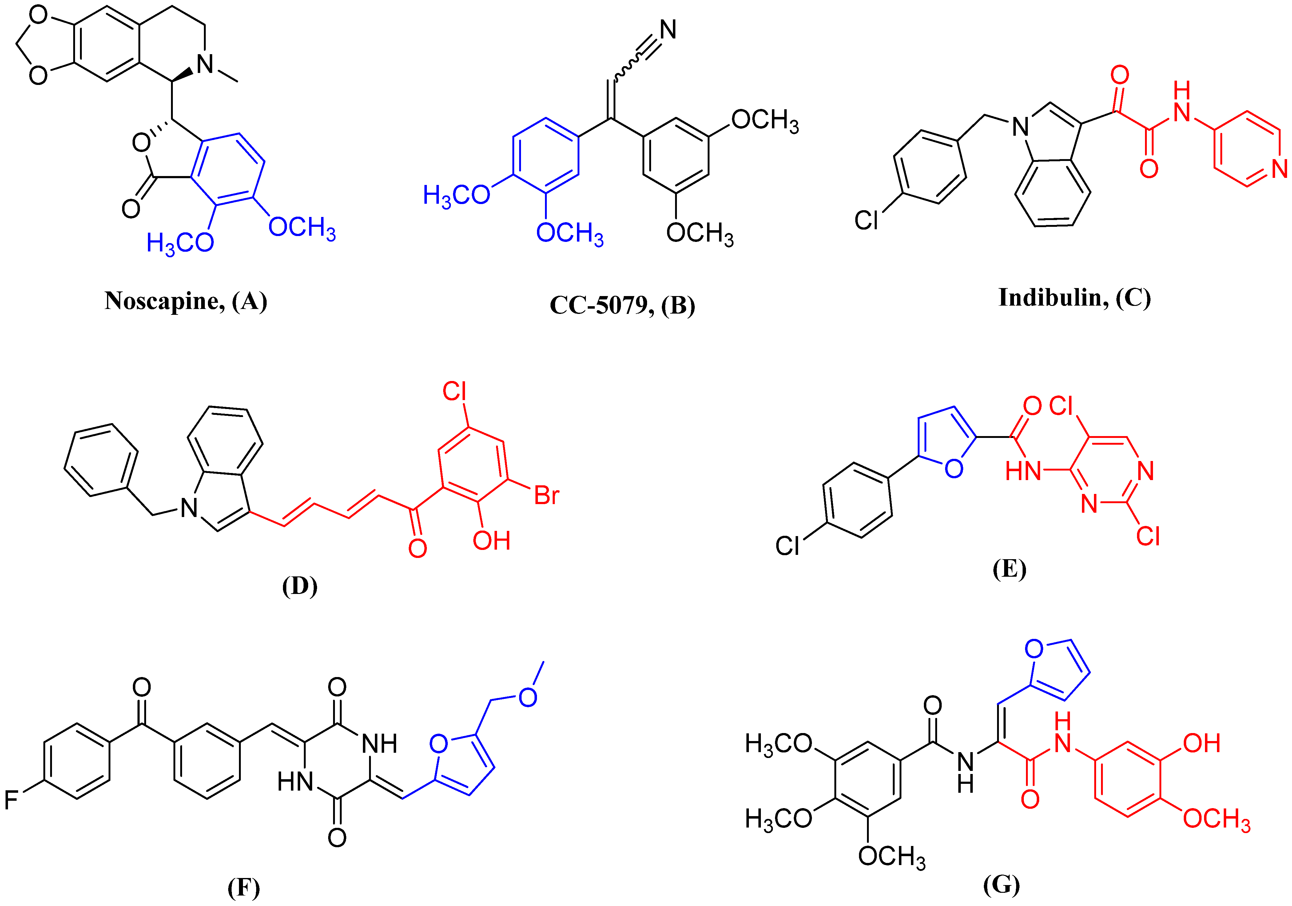
:1. Introduction
2. Results and Discussion
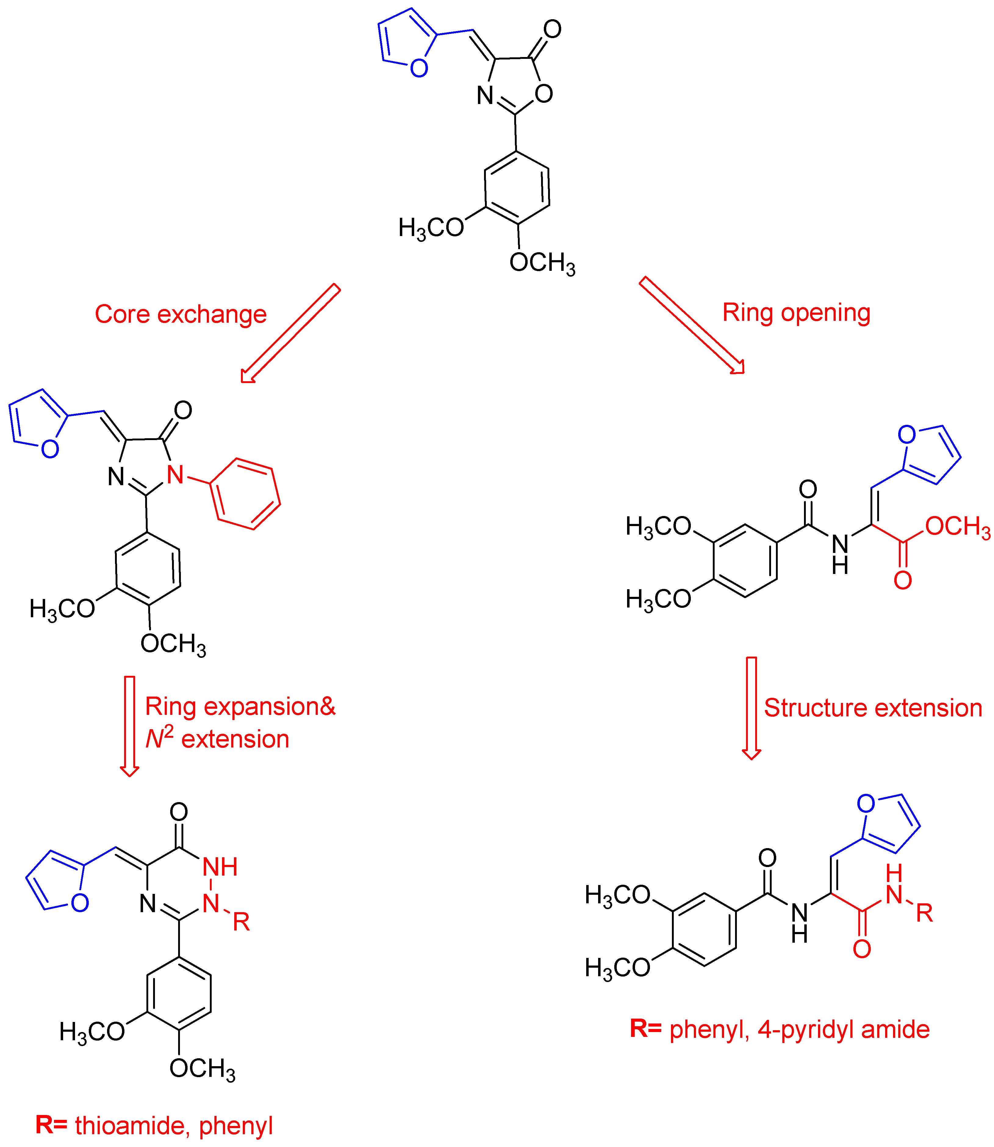
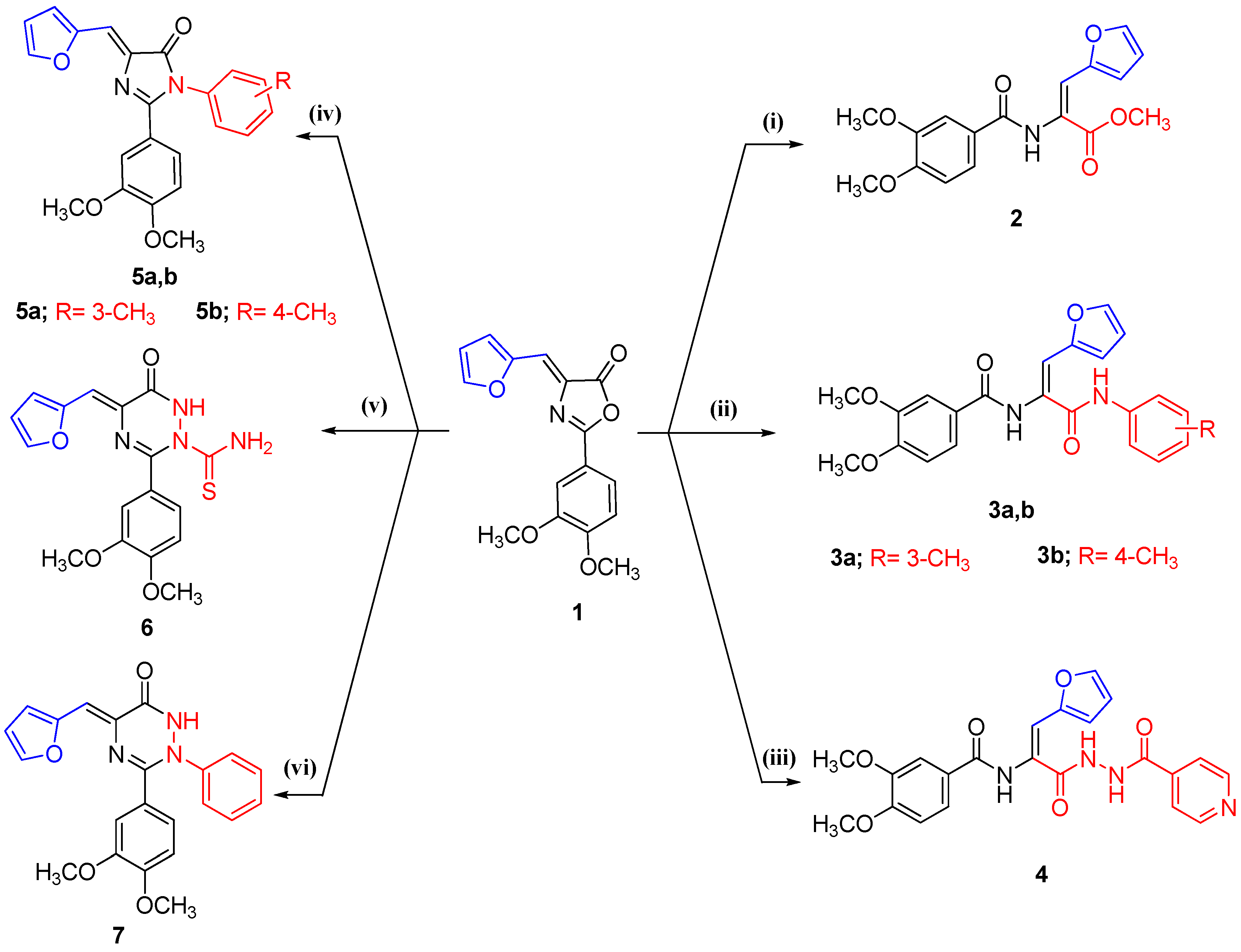
2.1. Chemistry
2.2. Biological Evaluation and Mechanistic Studies
2.2.1. Cytotoxic Activity against Breast Cancer Cell Lines (MCF-7)
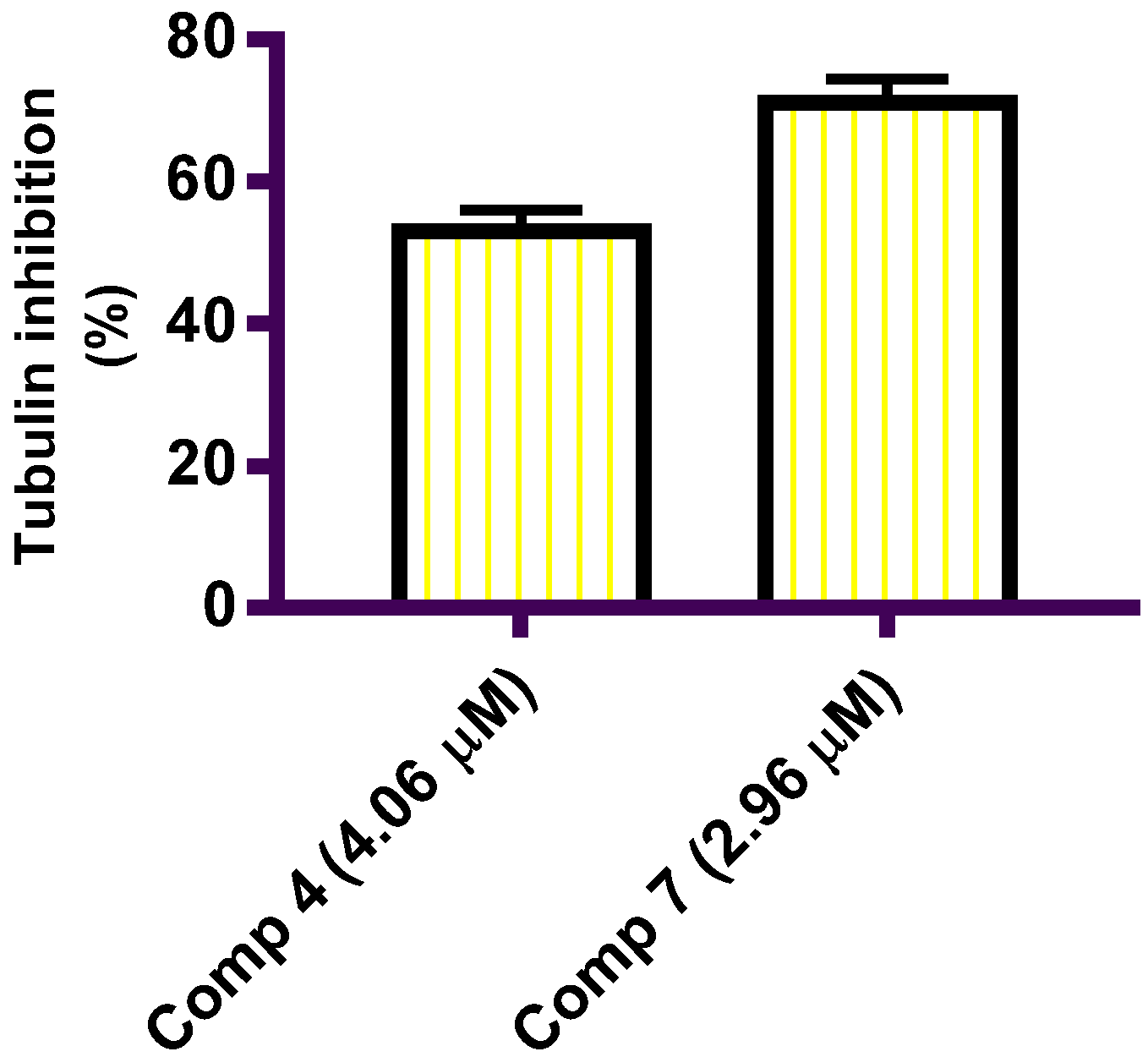
2.2.2. In Vitro β-Tubulin Polymerization Assay
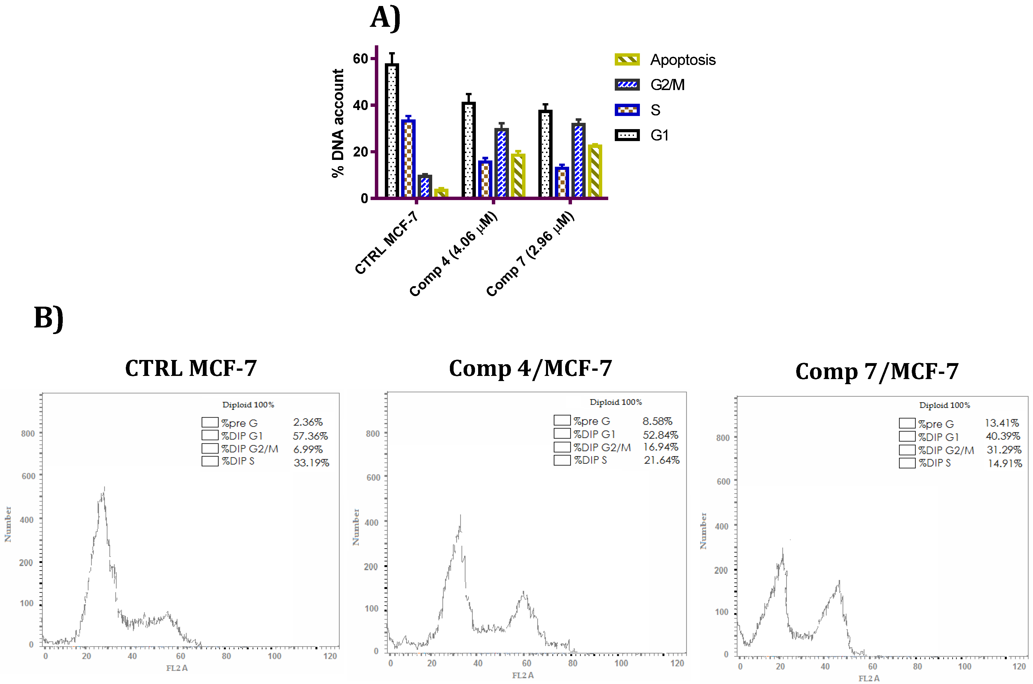
2.2.3. Cell Cycle Analysis
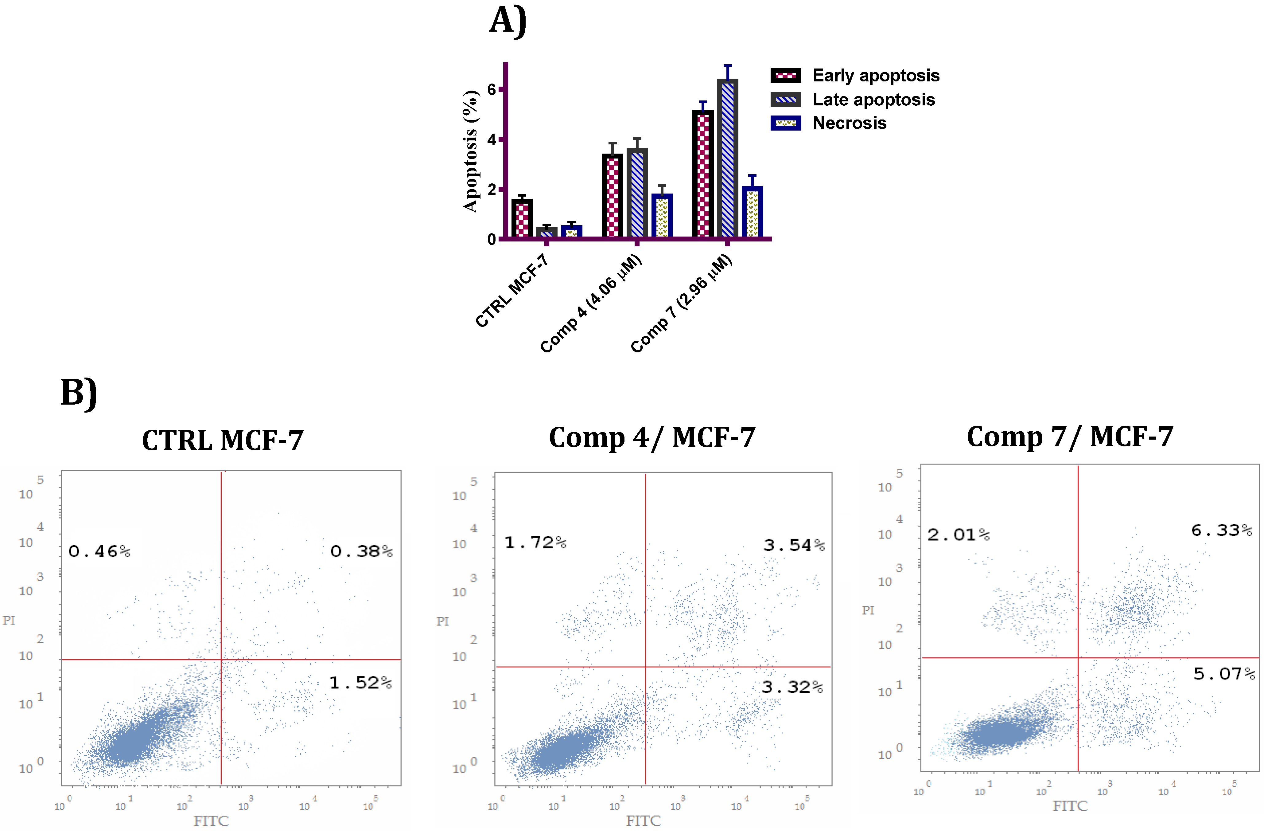
2.2.4. Annexin V-FITC/PI and Detection of Apoptosis
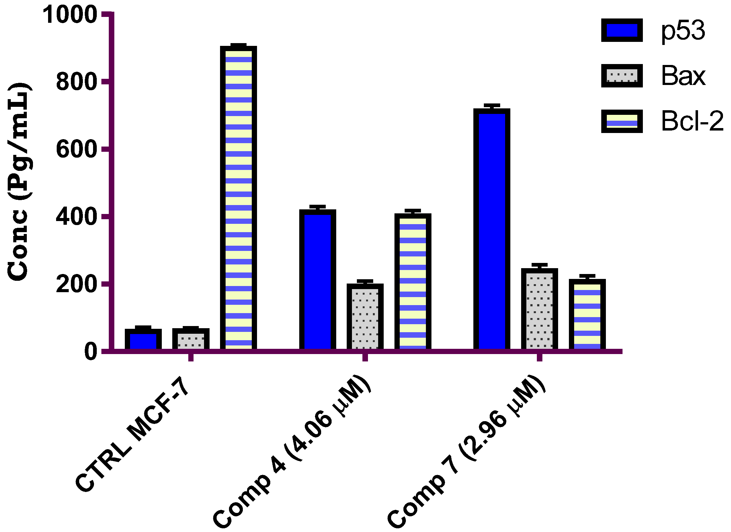
2.2.5. Effect of Compounds 4 and 7 on the Level of p53/Bax/Bcl-2
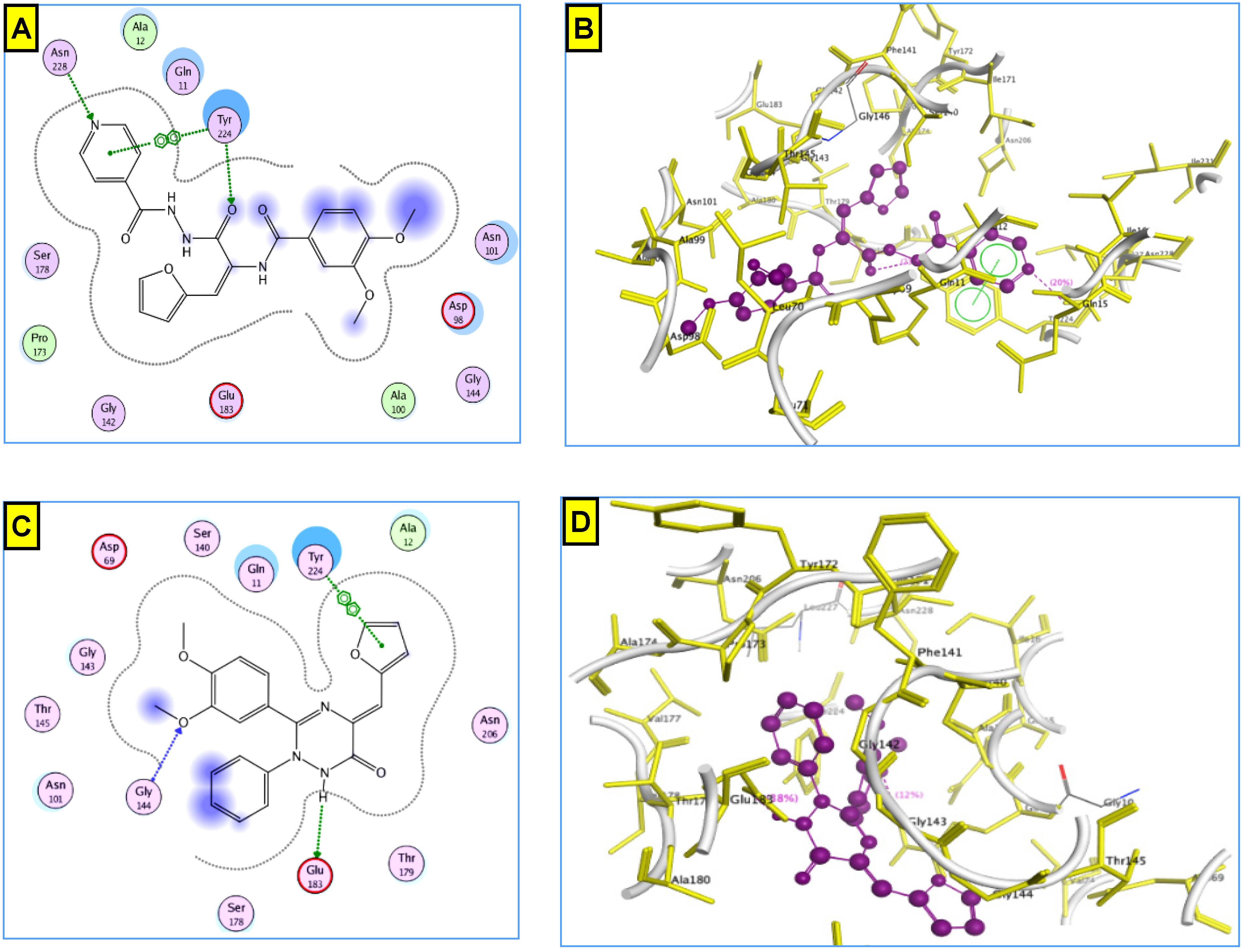
2.2.6. Molecular Docking Study
3. Experimental
3.1. Chemistry
3.1.1. General
3.1.2. General Procedure for the Synthesis of Methyl 2-(3,4-dimethoxybenzamido)-3-(furan-2-yl)acrylate (2)
3.1.3. General Procedure for the Synthesis of N-(1-(furan-2-yl)-3-oxo-3-(arylamino)prop-1-en-2-yl)-3,4-dimethoxybenzamides 3a,b
N-(1-(furan-2-yl)-3-oxo-3-(m-tolylamino)prop-1-en-2-yl)-3,4-dimethoxybenzamide (3a)
N-(1-(furan-2-yl)-3-oxo-3-(p-tolylamino)prop-1-en-2-yl)-3,4-dimethoxybenzamide (3b)
3.1.4. General Procedure for the Synthesis of N-(1-(furan-2-yl)-3-(2-isonicotinoylhydrazinyl)-3-oxoprop-1-en-2-yl)-3,4-dimethoxybenzamide (4)
3.1.5. General Procedure for the Synthesis of 1-Aryl-2-(3,4-dimethoxyphenyl)-4-(furan-2-ylmethylene)-1H-imidazol-5(4H)-ones 5a,b
2-(3,4-Dimethoxyphenyl)-4-(furan-2-ylmethylene)-1-m-tolyl-1H-imidazol-5(4H)-one (5a)
2-(3,4-Dimethoxyphenyl)-4-(furan-2-ylmethylene)-1-p-tolyl-1H-imidazol-5(4H)-one (5b)
3.1.6. General Procedure for the Synthesis of 3-(3,4-Dimethoxyphenyl)-5-(furan-2-ylmethylene)-6-oxo-5,6-dihydro-1,2,4-triazine-2(1H)-carbothioamide (6)
3.1.7. General Procedure for the Synthesis of 3-(3,4-Dimethoxyphenyl)-5-(furan-2-ylmethylene)-2-phenyl-2,5-dihydro-1,2,4-triazin-6(1H)-one (7)
3.2. Pharmacological Studies
3.2.1. Cytotoxic Activity against MCF-7 Cell Line
3.2.2. Tubulin Inhibitions Assay
3.2.3. Cell Cycle Analysis of Test Compounds
3.2.4. Annexin V FITC/PI Staining Assay
3.2.5. ELISA Measurements of p53, Bax and Bcl2
4. Conclusions
Supplementary Materials
Author Contributions
Funding
Institutional Review Board Statement
Informed Consent Statement
Data Availability Statement
Acknowledgments
Conflicts of Interest
References
- Adil, M.S.; Khulood, D.; Somanath, P.R. Targeting Akt-associated microRNAs for cancer therapeutics. Biochem. Pharmacol. 2021, 189, 114384–114392. [Google Scholar] [CrossRef] [PubMed]
- Yadav, A.R.; Mohite, S.K. Cancer-A silent killer: An overview. Asian J. Pharm. Res. 2020, 10, 213–216. [Google Scholar] [CrossRef]
- Pedron, C.N.; Andrade, G.P.; Sato, R.H.; Torres, M.D.T.; Cerchiaro, G.; Ribeiro, A.O.; Oliveira, V.X., Jr. Anticancer activity of VmCT1 analogs against MCF-7 cells. Chem. Biol. Drug Des. 2018, 91, 588–596. [Google Scholar] [CrossRef]
- Faramarzi, A.; Jahromi, M.G.; Ashourzadeh, S.; Jalilian, N. Metastatic and pathophysiological characteristics of breast cancer with emphasis on hereditary factors. Cent. Asian J. Med. Pharm. Sci. Innov. 2021, 1, 104–113. [Google Scholar]
- Trayes, K.P.; Cokenakes, S.E. Breast cancer treatment. Am. Fam. Phys. 2021, 104, 171–178. [Google Scholar]
- Liang, Y.; Zhang, H.; Song, X.; Yang, Q. Metastatic heterogeneity of breast cancer: Molecular mechanism and potential therapeutic targets. Semin. Cancer Biol. 2020, 60, 14–27. [Google Scholar] [CrossRef]
- Chen, A.; Wen, S.; Liu, F.; Zhang, Z.; Liu, M.; Wu, Y.; He, B.; Yan, M.; Kang, T.; Lam, E.W.F.; et al. CRISPR/Cas9 screening identifies a kinetochore-microtubule dependent mechanism for Aurora-A inhibitor resistance in breast cancer. Cancer Commun. 2021, 41, 121–139. [Google Scholar] [CrossRef]
- Liu, M.; Fu, M.; Yang, X.; Jia, G.; Shi, X.; Ji, J.; Liu, X.; Zhai, G. Paclitaxel and quercetin co-loaded functional mesoporous silica nanoparticles overcoming multidrug resistance in breast cancer. Colloids Surf. B 2020, 196, 111284–111299. [Google Scholar] [CrossRef]
- Yele, V.; Pindiprolu, S.K.S.; Sana, S.; Ramamurty, D.; Madasi, J.R.; Vadlamani, S. Synthesis and preclinical evaluation of indole triazole conjugates as microtubule targeting agents that are effective against MCF-7 breast cancer cell lines. Anti-Cancer Agents Med. Chem. 2021, 21, 1047–1055. [Google Scholar] [CrossRef]
- Wang, G.; Liu, W.; Gong, Z.; Huang, Y.; Li, Y.; Peng, Z. Synthesis, biological evaluation, and molecular modelling of new naphthalene-chalcone derivatives as potential anticancer agents on MCF-7 breast cancer cells by targeting tubulin colchicine binding site. J. Enzym. Inhib. Med. Chem. 2020, 35, 139–144. [Google Scholar] [CrossRef]
- Anichina, K.; Argirova, M.; Tzoneva, R.; Uzunova, V.; Mavrova, A.; Vuchev, D.; Popova-Daskalova, G.; Fratev, F.; Guncheva, M.; Yancheva, D. 1H-benzimidazole-2-yl hydrazones as tubulin-targeting agents: Synthesis, structural characterization, anthelmintic activity and antiproliferative activity against MCF-7 breast carcinoma cells and molecular docking studies. Chem. Biol. Interact. 2021, 345, 109540–109549. [Google Scholar] [CrossRef] [PubMed]
- Mosca, L.; Ilari, A.; Fazi, F.; Assaraf, Y.G.; Colotti, G. Taxanes in cancer treatment: Activity, chemoresistance and its overcoming. Drug Resist. Updates 2021, 54, 100742–100752. [Google Scholar] [CrossRef] [PubMed]
- Zhang, D.; Kanakkanthara, A. Beyond the Paclitaxel and Vinca Alkaloids: Next Generation of Plant-Derived Microtubule-Targeting Agents with Potential Anticancer Activity. Cancers 2020, 12, 1721. [Google Scholar] [CrossRef] [PubMed]
- Gupta, A.K.; Tulsyan, S.; Bharadwaj, M.; Mehrotra, R. Systematic review on cytotoxic and anticancer potential of n-substituted isatins as novel class of compounds useful in multidrug-resistant cancer therapy: In silico and in vitro analysis. Top. Curr. Chem. 2019, 377, 15–29. [Google Scholar] [CrossRef]
- Haider, K.; Rahaman, S.; Yar, M.S.; Kamal, A. Tubulin inhibitors as novel anticancer agents: An overview on patents (2013–2018). Exp. Opin. Ther. Pat. 2019, 29, 623–641. [Google Scholar] [CrossRef]
- Naik, P.K.; Chatterji, B.P.; Vangapandu, S.N.; Aneja, R.; Chandra, R.; Kanteveri, S.; Joshi, H.C. Rational design, synthesis and biological evaluations of amino-noscapine: A high affinity tubulin-binding noscapinoid. J. Comput. Aided Mol. Des. 2011, 25, 443–454. [Google Scholar] [CrossRef]
- Zhang, L.-H.; Wu, L.; Raymon, H.K.; Chen, R.S.; Corral, L.; Shirley, M.A.; Narla, R.K.; Gamez, J.; Muller, G.W.; Stirling, D.I.; et al. The Synthetic Compound CC-5079 Is a Potent Inhibitor of Tubulin Polymerization and Tumor Necrosis Factor-α Production with Antitumor Activity. Cancer Res. 2006, 66, 951–959. [Google Scholar] [CrossRef] [Green Version]
- Bacher, G.; Nickel, B.; Emig, P.; Vanhoefer, U.; Seeber, S.; Shandra, A.; Klenner, T.; Beckers, T. D-24851, a Novel Synthetic Microtubule Inhibitor, Exerts Curative Antitumoral Activity in Vivo, Shows Efficacy toward Multidrug-resistant Tumor Cells, and Lacks Neurotoxicity1. Cancer Res. 2001, 61, 392–399. [Google Scholar]
- Lu, Y.; Chen, J.; Xiao, M.; Li, W.; Miller, D.D. An Overview of Tubulin Inhibitors That Interact with the Colchicine Binding Site. Pharm. Res. 2012, 29, 2943–2971. [Google Scholar] [CrossRef] [Green Version]
- Kassem, A.F.; Nassar, I.F.; Abdel-Aal, M.T.; Awad, H.M.; El-Sayed, W.A. Synthesis and anticancer activity of new ((Furan-2-yl)-1, 3, 4-thiadiazolyl)-1, 3, 4-oxadiazole acyclic sugar derivatives. Chem. Pharm. Bull. 2019, 67, 888–895. [Google Scholar] [CrossRef] [Green Version]
- Chandrashekarachar, D.; Kesagudu, D. Impotrtance of furan based compounds and their biomedical applications: An overview. Indo Am. J. Pharm. Res. 2017, 7, 7541–7549. [Google Scholar]
- Sakchaisri, K.; Kim, S.-O.; Hwang, J.; Soung, N.K.; Lee, K.H.; Choi, T.W.; Lee, Y.; Park, C.-M.; Thimmegowda, N.R.; Lee, P.Y. Anticancer activity of a novel small molecule tubulin inhibitor STK899704. PLoS ONE 2017, 12, e0173311. [Google Scholar] [CrossRef] [Green Version]
- Kitel, R.; Byczek-Wyrostek, A.; Hopko, K.; Kasprzycka, A.; Walczak, K. Effect of Selected Silyl Groups on the Anticancer Activity of 3,4-Dibromo-5-Hydroxy-Furan-2(5H)-One Derivatives. Pharmaceuticals 2021, 14, 1079. [Google Scholar] [CrossRef]
- Wei, M.-X.; Yu, J.-Y.; Liu, X.-X.; Li, X.-Q.; Zhang, M.-W.; Yang, P.-W.; Yang, J.-H. Synthesis of artemisinin-piperazine-furan ether hybrids and evaluation of in vitro cytotoxic activity. Eur. J. Med. Chem. 2021, 215, 113295–113305. [Google Scholar] [CrossRef]
- Khalaf, H.; Tolan, H.; El-Bayaa, M.; Radwan, M.; El-Manawaty, M.; El-Sayed, W. Synthesis and anticancer activity of new pyridine-thiophene and pyridine-furan hybrid compounds, their sugar hydrazone, and glycosyl derivatives. Russ. J. Gen. Chem. 2020, 90, 1706–1715. [Google Scholar] [CrossRef]
- Shwetha, B.; Sudhanva, M.S.; Jagadeesha, G.S.; Thimmegowda, N.R.; Hamse, V.K.; Sridhar, B.T.; Thimmaiah, K.N.; Kumar, C.S.A.; Shobith, R.; Rangappa, K.S. Furan-2-carboxamide derivative, a novel microtubule stabilizing agent induces mitotic arrest and potentiates apoptosis in cancer cells. Bioorg. Chem. 2021, 108, 104586–104587. [Google Scholar] [CrossRef]
- Ding, Z.; Li, F.; Zhong, C.; Li, F.; Liu, Y.; Wang, S.; Zhao, J.; Li, W. Structure-based design and synthesis of novel furan-diketopiperazine-type derivatives as potent microtubule inhibitors for treating cancer. Bioorg. Med. Chem. 2020, 28, 115435–115443. [Google Scholar] [CrossRef]
- Zaki, I.; Abou-Elkhair, R.A.I.; Almaaty, A.H.A.; Ali, O.A.A.; Fayad, E.; Gaafar, A.G.A.; Zakaria, M.Y. Design and Synthesis of Newly Synthesized Acrylamide Derivatives as Potential Chemotherapeutic Agents against MCF-7 Breast Cancer Cell Line Lodged on PEGylated Bilosomal Nano-Vesicles for Improving Cytotoxic Activity. Pharmaceuticals 2021, 14, 1021. [Google Scholar] [CrossRef]
- Zaki, I.; Ramadan, H.M.M.; El-Sayed, E.-S.H.; El-Moneim, M.A. Design, synthesis, and cytotoxicity screening of new synthesized imidazolidine-2-thiones as VEGFR-2 enzyme inhibitors. Arch. Pharm. 2020, 353, 2000121. [Google Scholar] [CrossRef]
- Almaaty, A.H.A.; Toson, E.E.M.; El-Sayed, E.-S.H.; Tantawy, M.A.M.; Fayad, E.; Ali, O.A.A.; Zaki, I. 5-Aryl-1-Arylideneamino-1H-Imidazole-2(3H)-Thiones: Synthesis and In Vitro Anticancer Evaluation. Molecules 2021, 26, 1706. [Google Scholar] [CrossRef]
- Mourad, A.A.E.; Rizzk, Y.W.; Zaki, I.; Mohammed, F.Z.; El Behery, M. Synthesis and cytotoxicity screening of some synthesized hybrid nitrogen molecules as anticancer agents. J. Mol. Struct. 2021, 1242, 130722–130729. [Google Scholar] [CrossRef]
- Zaki, I.; El-ata, S.A.A.; Fayad, E.; Ali, O.A.A.; Almaaty, A.H.A.; Saad, A.S. Evaluation of Synthetic 2,4-Disubstituted-benzo[g]quinoxaline Derivatives as Potential Anticancer Agents. Pharmaceuticals 2021, 14, 853. [Google Scholar] [CrossRef]
- Gucký, T.; Fryšová, I.; Slouka, J.; Hajdúch, M.; Džubák, P. Cyclocondensation reaction of heterocyclic carbonyl compounds, Part XIII: Synthesis and cytotoxic activity of some 3,7-diaryl-5-(3,4,5-trimethoxyphenyl)pyrazolo [4,3-e][1,2,4]triazines. Eur. J. Med. Chem. 2009, 44, 891–900. [Google Scholar] [CrossRef]
- Abdelhameid, M.K.; Zaki, I.; Mohammed, M.R.; Mohamed, K.O. Design, synthesis, and cytotoxic screening of novel azole derivatives on hepatocellular carcinoma (HepG2 Cells). Bioorg. Chem. 2020, 101, 103995–104007. [Google Scholar] [CrossRef]
- El-Aziz, R.M.A.; Zaki, I.; El-Deen, I.M.; Abd-Rahman, M.S.; Mohammed, F.Z. In Vitro Anticancer Evaluation of Some Synthesized 2H-Quinolinone and Halogenated 2H-Quinolinone Derivatives as Therapeutic Agents. Anti-Cancer Agents Med. Chem. 2020, 20, 2304–2315. [Google Scholar] [CrossRef]
- Islam, Z.; Amal, M.I. Design, Synthesis, and Cytotoxic Screening of New Quinoline Derivatives over MCF-7 Breast Cancer Cell Line. Russ. J. Bioorg. Chem. 2020, 46, 1099–1109. [Google Scholar] [CrossRef]


| Compound | IC50 Values (μM) | |
|---|---|---|
| MCF-7 | MCF-10A | |
| 1 | 19.23 ± 0.31 | NT |
| 2 | 48.98 ± 0.78 | NT |
| 3a | 13.17 ± 0.34 | NT |
| 3b | 17.88 ± 0.49 | NT |
| 4 | 4.06 ± 0.18 | 29.77 ± 0.22 |
| 5a | 10.72 ± 0.26 | NT |
| 5b | 23.42 ± 0.39 | NT |
| 6 | 8.07 ± 0.22 | NT |
| 7 | 2.96 ± 0.16 | 22.12 ± 0.17 |
| STU | 6.50 ± 0.09 | 34.62 ± 0.11 |
Publisher’s Note: MDPI stays neutral with regard to jurisdictional claims in published maps and institutional affiliations. |
© 2022 by the authors. Licensee MDPI, Basel, Switzerland. This article is an open access article distributed under the terms and conditions of the Creative Commons Attribution (CC BY) license (https://creativecommons.org/licenses/by/4.0/).
Share and Cite
Bukhari, S.N.A.; Ejaz, H.; Elsherif, M.A.; Junaid, K.; Zaki, I.; Masoud, R.E. Design and Synthesis of Some New Furan-Based Derivatives and Evaluation of In Vitro Cytotoxic Activity. Molecules 2022, 27, 2606. https://0-doi-org.brum.beds.ac.uk/10.3390/molecules27082606
Bukhari SNA, Ejaz H, Elsherif MA, Junaid K, Zaki I, Masoud RE. Design and Synthesis of Some New Furan-Based Derivatives and Evaluation of In Vitro Cytotoxic Activity. Molecules. 2022; 27(8):2606. https://0-doi-org.brum.beds.ac.uk/10.3390/molecules27082606
Chicago/Turabian StyleBukhari, Syed Nasir Abbas, Hasan Ejaz, Mervat A. Elsherif, Kashaf Junaid, Islam Zaki, and Reham E. Masoud. 2022. "Design and Synthesis of Some New Furan-Based Derivatives and Evaluation of In Vitro Cytotoxic Activity" Molecules 27, no. 8: 2606. https://0-doi-org.brum.beds.ac.uk/10.3390/molecules27082606