Electrospun Nanocomposites Containing Cellulose and Its Derivatives Modified with Specialized Biomolecules for an Enhanced Wound Healing


Abstract
:1. Introduction
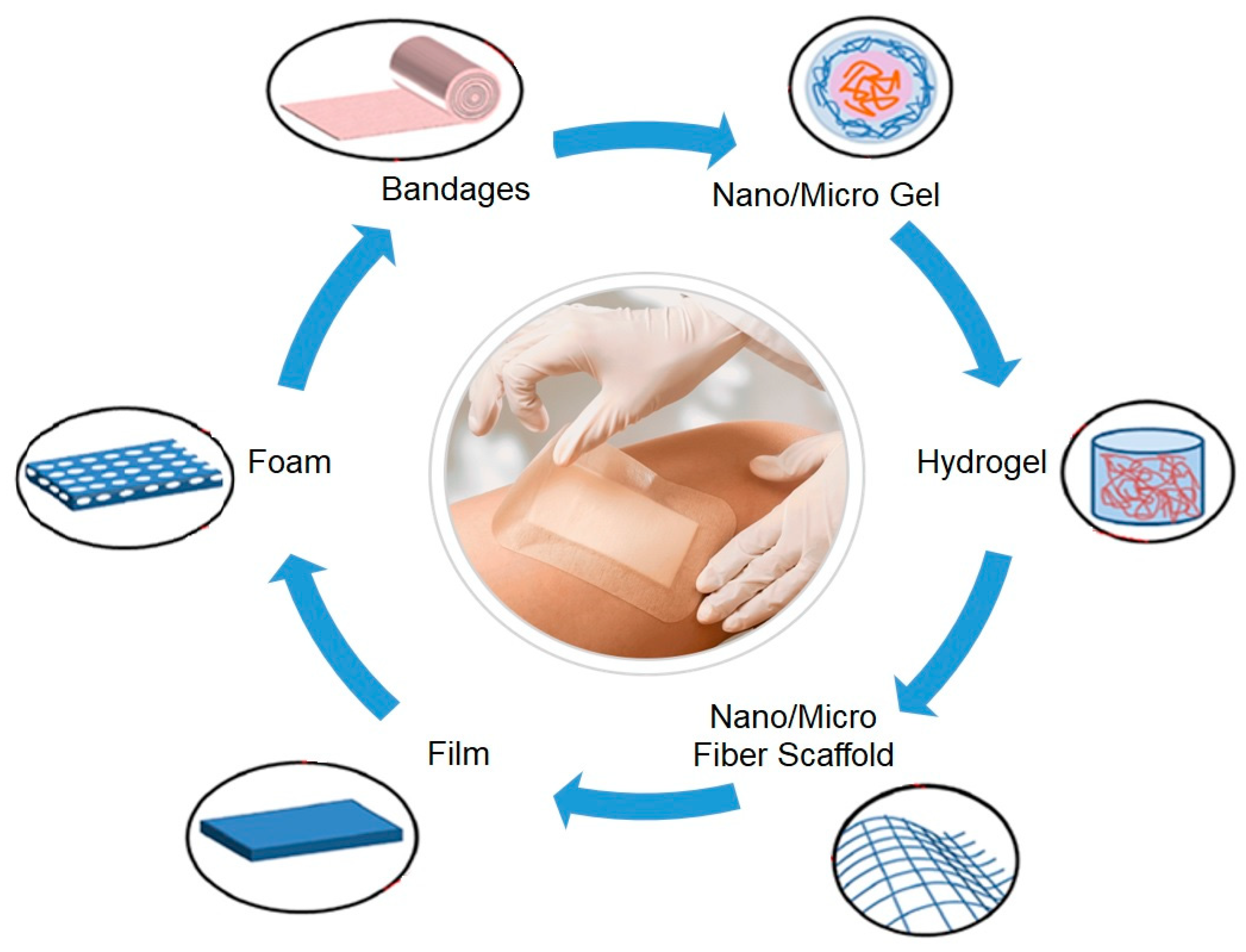
2. Nanostructured Wound Dressings
- ▪
- guaranteeing breathability;
- ▪
- maintaining a suitable physiological temperature;
- ▪
- ensuring a balanced moist environment, avoiding dehydration and cell death;
- ▪
- promoting debridement;
- ▪
- allowing proliferation and migration of fibroblasts and keratinocytes, and an enhanced collagen synthesis;
- ▪
- protecting the wound from bacteria and other external soiling; and,
- ▪
3. Cellulose and Its Derivatives
3.1. Cellulose
3.2. Cellulose Acetate (CA)
3.3. Nanocellulose
4. Application in Wound Healing: Synergistic Effect with Specialized Biomolecules
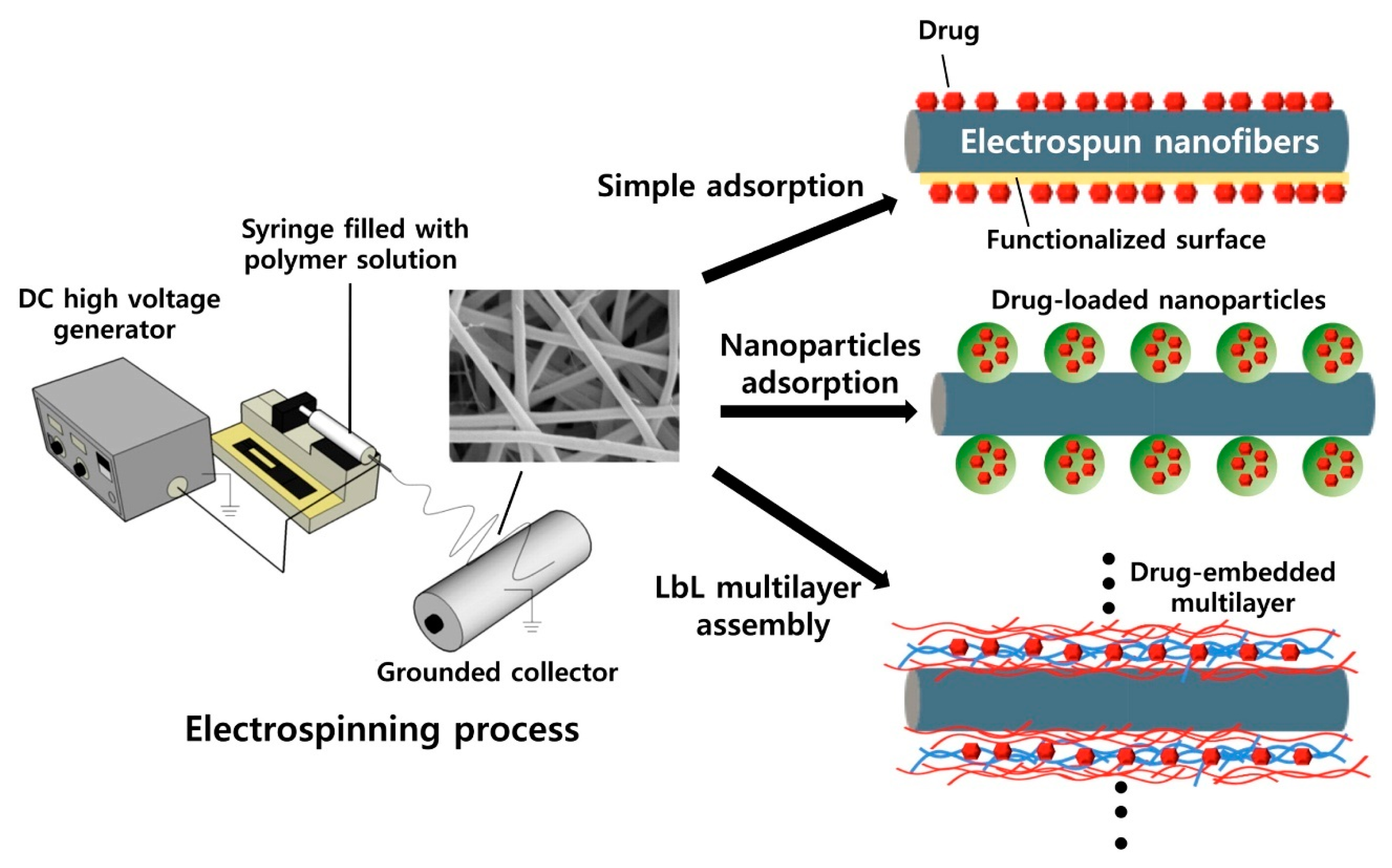
4.1. Drug Loading
4.2. Nanoparticles (NPs)
4.3. Natural Extracts
4.4. Wound Healing Alternative Methods Containing Cellulose-Based Compounds
5. Conclusions and Future Perspectives
Author Contributions
Funding
Acknowledgments
Conflicts of Interest
References
- Ghaffari-bohlouli, P.; Hamidzadeh, F.; Zahedi, P.; Fallah-darrehchi, M. Antibacterial nanofibers based on poly (l-lactide-co-d, l-lactide) and poly (vinyl alcohol) used in wound dressings potentially: A comparison between hybrid and blend properties. J. Biomater. Sci. Polym. Ed. 2020, 31, 219–243. [Google Scholar] [CrossRef] [PubMed]
- Simões, D.; Miguel, S.P.; Ribeiro, M.P.; Coutinho, P.; Mendonça, A.G.; Correia, I.J. Recent advances on antimicrobial wound dressing: A review. Eur. J. Pharm. Biopharm. 2018, 127, 130–141. [Google Scholar] [CrossRef] [PubMed]
- Adeli, H.; Khorasani, M.T.; Parvazinia, M. Wound dressing based on electrospun PVA/chitosan/starch nanofibrous mats: Fabrication, antibacterial and cytocompatibility evaluation and in vitro healing assay. Int. J. Biol. Macromol. 2019, 122, 238–254. [Google Scholar] [CrossRef] [PubMed]
- Wang, P.H.; Huang, B.S.; Horng, H.C.; Yeh, C.C.; Chen, Y.J. Wound healing. J. Chin. Med. Assoc. 2018, 81, 94–101. [Google Scholar] [CrossRef]
- Gupta, A.; Kowalczuk, M.; Heaselgrave, W.; Britland, S.T.; Martin, C.; Radecka, I. The production and application of hydrogels for Wound Management: A Review. Eur. Polym. J. 2019, 111, 134–151. [Google Scholar] [CrossRef]
- Berthet, M.; Gauthier, Y.; Lacroix, C.; Verrier, B.; Monge, C. Nanoparticle-Based Dressing: The Future of Wound Treatment? Trends Biotechnol. 2017, 35, 770–784. [Google Scholar] [CrossRef]
- Zahedi, P.; Rezaeian, I.; Ranaei-Siadat, S.O.; Jafari, S.H.; Supaphol, P. A review on wound dressings with an emphasis on electrospun nanofibrous polymeric bandages. Polym. Adv. Technol. 2010, 21, 77–95. [Google Scholar] [CrossRef]
- Naderi, N.; Karponis, D.; Mosahebi, A.; Seifalian, A.M. Nanoparticles in wound healing from hope to promise, from promise to routine. Front. Biosci. 2018, 23, 1038–1059. [Google Scholar]
- Unnithan, A.R.; Gnanasekaran, G.; Sathishkumar, Y.; Lee, Y.S.; Kim, C.S. Electrospun antibacterial polyurethane-cellulose acetate-zein composite mats for wound dressing. Carbohydr. Polym. 2014, 102, 884–892. [Google Scholar] [CrossRef]
- Scalise, A.; Bianchi, A.; Tartaglione, C.; Bolletta, E.; Pierangeli, M.; Torresetti, M.; Marazzi, M.; Di Benedetto, G. Microenvironment and microbiology of skin wounds: The role of bacterial biofilms and related factors. Semin. Vasc. Surg. 2015, 28, 151–159. [Google Scholar] [CrossRef]
- Ambekar, R.S.; Kandasubramanian, B. Advancements in nano fi bers for wound dressing: A review. Eur. Polym. J. 2019, 117, 304–336. [Google Scholar] [CrossRef]
- Felgueiras, H.P.; Teixeira, M.A.; Tavares, T.D.; Homem, N.C.; Zille, A.; Amorim, M.T.P. Antimicrobial action and clotting time of thin, hydrated poly (vinyl alcohol)/cellulose acetate films functionalized with LL37 for prospective wound-healing applications. Appl. Polym. 2019, 48626, 1–12. [Google Scholar] [CrossRef]
- Ghomi, E.R.; Khalili, S.; Khorasani, S.N.; Neisiany, R.E. Wound dressings: Current advances and future directions. Appl. Polym. 2019, 47738, 1–12. [Google Scholar]
- Jannesari, M.; Varshosaz, J.; Morshed, M.; Zamani, M. Composite poly(vinyl alcohol)/poly(vinyl acetate) electrospun nanofibrous mats as a novel wound dressing matrix for controlled release of drugs. Int. J. Nanomed. 2011, 6, 993–1003. [Google Scholar]
- Dart, A.; Bhave, M.; Kingshott, P. Antimicrobial Peptide-Based Electrospun Fibers for Wound Healing Applications. Macromol. Biosci. 2019, 1800488, 1–16. [Google Scholar] [CrossRef]
- Li, H.; Williams, G.R.; Wu, J.; Lv, Y.; Sun, X.; Wu, H.; Zhu, L.M. Thermosensitive nanofibers loaded with ciprofloxacin as antibacterial wound dressing materials. Int. J. Pharm. 2017, 517, 135–147. [Google Scholar] [CrossRef]
- Teixeira, M.A.; Amorim, M.T.P.; Felgueiras, H.P. Poly (Vinyl Alcohol) Based Nanofibrous Electrospun Scaffolds for Tissue Engineering Applications. Polymers 2020, 12, 7. [Google Scholar] [CrossRef] [Green Version]
- Ahmadi Majd, S.; Rabbani Khorasgani, M.; Moshtaghian, S.J.; Talebi, A.; Khezri, M. Application of Chitosan/PVA Nano fiber as a potential wound dressing for streptozotocin-induced diabetic rats. Int. J. Biol. Macromol. 2016, 92, 1162–1168. [Google Scholar] [CrossRef]
- Wang, J.; Windbergs, M. Functional electrospun fibers for the treatment of human skin wounds. Eur. J. Pharm. Biopharm. 2017, 119, 283–299. [Google Scholar] [CrossRef]
- Golizadeh, M.; Karimi, A.; Gandomi-ravandi, S.; Vossoughi, M.; Khafaji, M.; Joghataei, M.; Faghihi, F. Evaluation of cellular attachment and proliferation on di ff erent surface charged functional cellulose electrospun nano fi bers. Carbohydr. Polym. 2019, 207, 796–805. [Google Scholar] [CrossRef]
- Chen, J.; Xu, J.; Wang, K.; Cao, X.; Sun, R. Cellulose acetate fibers prepared from different raw materials with rapid synthesis method. Carbohydr. Polym. 2016, 137, 685–692. [Google Scholar] [CrossRef] [PubMed]
- Khoshnevisan, K.; Maleki, H.; Samadian, H.; Shahsavari, S.; Sarrafzadeh, M.H.; Larijani, B.; Dorkoosh, F.A.; Haghpanah, V.; Khorramizadeh, M.R. Cellulose acetate electrospun nanofibers for drug delivery systems: Applications and recent advances. Carbohydr. Polym. 2018, 198, 131–141. [Google Scholar] [CrossRef] [PubMed]
- Soares, R.M.D.; Siqueira, N.M.; Prabhakaram, M.P.; Ramakrishna, S. Electrospinning and electrospray of bio-based and natural polymers for biomaterials development. Mater. Sci. Eng. C 2018, 92, 969–982. [Google Scholar] [CrossRef] [PubMed]
- Ghorani, B.; Kadkhodaee, R.; Rajabzadeh, G.; Tucker, N. Assembly of odour adsorbent nanofilters by incorporating cyclodextrin molecules into electrospun cellulose acetate webs. React. Funct. Polym. 2019, 134, 121–132. [Google Scholar] [CrossRef]
- Samadian, H.; Salehi, M.; Farzamfar, S.; Vaez, A.; Sahrapeyma, H.; Goodarzi, A.; Ghorbani, S. In vitro and in vivo evaluation of electrospun cellulose acetate/gelatin/hydroxyapatite nanocomposite mats for wound dressing applications. Artif. Cells Nanomed. Biotechnol. 2018, 1401, 964–974. [Google Scholar] [CrossRef] [Green Version]
- Tsekova, P.B.; Spasova, M.G.; Manolova, N.E.; Markova, N.D.; Rashkov, I.B. Electrospun curcumin-loaded cellulose acetate/polyvinylpyrrolidone fibrous materials with complex architecture and antibacterial activity. Mater. Sci. Eng. C 2017, 73, 206–214. [Google Scholar] [CrossRef]
- De Freitas, R.R.M.; Senna, A.M.; Botaro, V.R. Influence of degree of substitution on thermal dynamic mechanical and physicochemical properties of cellulose acetate. Ind. Crop. Prod. 2017, 109, 452–458. [Google Scholar] [CrossRef]
- Wang, D.; Yue, Y.; Wang, Q.; Cheng, W.; Han, G. Preparation of cellulose acetate-polyacrylonitrile composite nanofibers by multi-fluid mixing electrospinning method: Morphology, wettability, and mechanical properties. Appl. Surf. Sci.. 2020, 510. [Google Scholar] [CrossRef]
- Abrigo, M.; McArthur, S.L.; Kingshott, P. Electrospun nanofibers as dressings for chronic wound care: Advances, challenges, and future prospects. Macromol. Biosci. 2014, 14, 772–792. [Google Scholar] [CrossRef]
- Felgueiras, H.P.; Teixeira, M.A.; Tavares, T.D.; Amorim, M.T.P. New method to produce poly (vinyl alcohol)/cellulose acetate films with improved antibacterial action. Mater. Today Proc. 2019, 10–13. [Google Scholar] [CrossRef] [Green Version]
- Teixeira, M.A.; Amorim, M.T.P.; Felgueiras, H.P. Cellulose Acetate in Wound Dressings Formulations: Potentialities and Electrospinning Capability. In Proceedings of the XV Mediterranean Conference on Medical and Biological Engineering and Computing—MEDICON 2019, Coimbra, Portugal, 26–28 September 2019; Springer: Berlin/Heidelberg, Germany, 2020; Volume 76, pp. 1515–1525. [Google Scholar]
- Poonguzhali, R.; Basha, S.K.; Kumari, V.S. Novel asymmetric chitosan/PVP/nanocellulose wound dressing: In vitro and in vivo evaluation. Int. J. Biol. Macromol. 2018, 112, 1300–1309. [Google Scholar] [CrossRef] [PubMed]
- Felgueiras, H.P.; Teixeira, M.A.; Amorim, M.T.P. Potentialities of LL37 for Wound Healing Applications: Study of Its Activity in Synergy with Biodegradable Composites Made of PVA and CA. In Proceedings of the XV Mediterranean Conference on Medical and Biological Engineering and Computing—MEDICON 2019, Coimbra, Portugal, 26–28 September 2019; Springer: Berlin/Heidelberg, Germany, 2020; Volume 76, pp. 1515–1525. [Google Scholar]
- Sheikhi, A.; Hayashi, J.; Eichenbaum, J.; Gutin, M.; Kuntjoro, N.; Khorsandi, D.; Khademhosseini, A. Recent advances in nanoengineering cellulose for cargo delivery. J. Control. Release 2019, 294, 53–76. [Google Scholar] [CrossRef] [PubMed]
- Zizovic, I.; Senerovic, L.; Moric, I.; Adamovic, T.; Jovanovic, M.; Kalagasidis, M.; Misic, D.; Stojanovic, D.; Milovanovic, S. Utilization of supercritical carbon dioxide in fabrication of cellulose acetate films with anti-bio film effects against Pseudomonas aeruginosa and Staphylococcus aureus. J. Supercrit. Fluids 2018, 140, 11–20. [Google Scholar] [CrossRef]
- Sun, F.; Nordli, H.R.; Pukstad, B.; Gamstedt, E.K.; Chinga, G. Mechanical characteristics of nanocellulose-PEG bionanocomposite wound dressings in wet conditions. J. Mech. Behav. Biomed. Mater. 2017, 69, 377–384. [Google Scholar] [CrossRef]
- Moura, L.I.F.; Dias, A.M.A.; Carvalho, E.; De Sousa, H.C. Recent advances on the development of wound dressings for diabetic foot ulcer treatment—A review. Acta Biomater. 2013, 9, 7093–7114. [Google Scholar] [CrossRef] [Green Version]
- Liu, M.; Duan, X.P.; Li, Y.M.; Yang, D.P.; Long, Y.Z. Electrospun nanofibers for wound healing. Mater. Sci. Eng. C 2017, 76, 1413–1423. [Google Scholar] [CrossRef]
- Huang, Y.; Dan, N.; Dan, W.; Zhao, W.; Bai, Z.; Chen, Y.; Yang, C. Bilayered Antimicrobial Nanofiber Membranes for Wound Dressings via in Situ Cross-Linking Polymerization and Electrospinning. Ind. Eng. Chem. Res. 2018, 57, 17048–17057. [Google Scholar] [CrossRef]
- Hajipour, M.J.; Fromm, K.M.; Akbar Ashkarran, A.; Jimenez de Aberasturi, D.; de Larramendi, I.R.; Rojo, T.; Serpooshan, V.; Parak, W.J.; Mahmoudi, M.; Santos, C.L.; et al. Coated Cotton Gauze with Ag/ZnO/chitosan Nanocomposite as a Modern Wound Dressing. J. Ind. Text. 2012, 9, 143–154. [Google Scholar]
- Davis, J.; Mclister, A.; Cundell, J.S.; Dewar, F. Smart Bandage Technologies Design and Application; Academic Press: Cambridge, MA, USA, 2016; ISBN 9780128037621. [Google Scholar]
- Weller, C.; Weller, C.; Team, V. Interactive Dressings and Their Role in Moist Wound Management, 2nd ed.; Elsevier: Amsterdam, The Netherlands, 2019; ISBN 9780081021927. [Google Scholar]
- Weller, C. Interactive Dressings and Their Role in Moist Wound Management, 2nd ed.; Elsevier: Amsterdam, The Netherlands, 2009; ISBN 9781845692711. [Google Scholar]
- Shankhwar, N.; Kumar, M.; Mandal, B.B.; Robi, P.S.; Srinivasan, A. Electrospun polyvinyl alcohol-polyvinyl pyrrolidone nanofibrous membranes for interactive wound dressing application. J. Biomater. Sci. Polym. Ed. 2016, 27, 247–262. [Google Scholar] [CrossRef]
- Felgueiras, H.P.; Amorim, M.T.P. Electrospun polymeric dressings functionalized with antimicrobial peptides and collagen type i for enhanced wound healing. IOP Conf. Ser. Mater. Sci. Eng. 2017, 254, 062004. [Google Scholar] [CrossRef]
- Skórkowska-Telichowska, K.; Czemplik, M.; Kulma, A.; Szopa, J. The local treatment and available dressings designed for chronic wounds. J. Am. Acad. Dermatol. 2013, 68, e117–e126. [Google Scholar] [CrossRef] [PubMed]
- Hasatsri, S.; Pitiratanaworanat, A.; Swangwit, S.; Boochakul, C.; Tragoonsupachai, C. Comparison of the Morphological and Physical Properties of Different Absorbent Wound Dressings. Dermatol. Res. Pract. 2018, 2018. [Google Scholar] [CrossRef] [PubMed] [Green Version]
- Felgueiras, H.P.; Amorim, M.T.P. Functionalization of electrospun polymeric wound dressings with antimicrobial peptides. Colloids Surf. B Biointerfaces 2017, 156, 133–148. [Google Scholar] [CrossRef] [PubMed]
- Pang, Q.; Zheng, X.; Luo, Y.; Ma, L.; Gao, C. A photo-cleavable polyprodrug-loaded wound dressing with UV-responsive antibacterial property. J. Mater. Chem. B 2017, 5, 8975–8982. [Google Scholar] [CrossRef]
- Jones, V.; Grey, J.E.; Harding, K.G. ABC of wound healing Wound dressings. Practice 2006, 332, 777–780. [Google Scholar]
- Gupta, A.; Low, W.L.; Radecka, I.; Britland, S.T.; Mohd Amin, M.C.I.; Martin, C. Characterisation and in vitro antimicrobial activity of biosynthetic silver-loaded bacterial cellulose hydrogels. J. Microencapsul. 2016, 33, 725–734. [Google Scholar] [CrossRef] [PubMed]
- Yang, J.M.; Yang, J.H.; Tsou, S.C.; Ding, C.H.; Hsu, C.C.; Yang, K.C.; Yang, C.C.; Chen, K.S.; Chen, S.W.; Wang, J.S. Cell proliferation on PVA/sodium alginate and PVA/poly(γ-glutamic acid) electrospun fiber. Mater. Sci. Eng. C 2016, 66, 170–177. [Google Scholar] [CrossRef]
- Ahmed, E.M. Hydrogel: Preparation, characterization, and applications: A review. J. Adv. Res. 2015, 6, 105–121. [Google Scholar] [CrossRef] [Green Version]
- Braunberger, T.L.; Fatima, S.; Vellaichamy, G.; Nahhas, A.F.; Parks-Miller, A.; Hamzavi, I.H. Dress for Success: A Review of Dressings and Wound Care in Hidradenitis Suppurativa. Curr. Dermatol. Rep. 2018, 7, 269–277. [Google Scholar] [CrossRef]
- Hilton, J.R.; Williams, D.T.; Beuker, B.; Miller, D.R.; Harding, K.G. Wound Dressings in Diabetic Foot Disease. Clin. Infect. Dis. 2004, 39, S100–S103. [Google Scholar] [CrossRef] [Green Version]
- Momoh, F.U.; Boateng, J.S.; Richardson, S.C.W.; Chowdhry, B.Z.; Mitchell, J.C. Development and functional characterization of alginate dressing as potential protein delivery system for wound healing. Int. J. Biol. Macromol. 2015, 81, 137–150. [Google Scholar] [CrossRef] [PubMed] [Green Version]
- Colobatiu, L.; Gavan, A.; Mocan, A.; Bogdan, C.; Mirel, S.; Tomuta, I. Development of bioactive compounds-loaded chitosan films by using a QbD approach—A novel and potential wound dressing material. React. Funct. Polym. 2019, 138, 46–54. [Google Scholar] [CrossRef]
- Sharma, S.; Dua, A.; Malik, A. Third Generation Materials for Wound Dressings. Int. J. Pharm. Sci. Res. 2014, 5, 2113–2124. [Google Scholar]
- Fahmy, H.M.; Aly, A.A.; Abou-Okeil, A. A non-woven fabric wound dressing containing layer-by-layer deposited hyaluronic acid and chitosan. Int. J. Biol. Macromol. 2018, 114, 929–934. [Google Scholar] [CrossRef] [PubMed]
- Liao, N.; Rajan, A.; Kumar, M.; Prasad, A.; Tshool, S.; Park, C.; Sang, C. Electrospun bioactive poly (E-caprolactone)–cellulose acetate—Dextran antibacterial composite mats for wound dressing applications. Colloids Surf. A Physicochem. Eng. Asp. 2015, 469, 194–201. [Google Scholar] [CrossRef]
- Miguel, S.P.; Figueira, D.R.; Simões, D.; Ribeiro, M.P.; Coutinho, P.; Ferreira, P.; Correia, I.J. Electrospun polymeric nanofibres as wound dressings: A review. Colloids Surf. B Biointerfaces 2018, 169, 60–71. [Google Scholar] [CrossRef]
- Song, K.; Wu, Q.; Qi, Y.; Karki, T. Electrospun Nanofibers with Antimicrobial Properties; Woodhead Publishing: Cambridge, UK, 2017; ISBN 9780081009079. [Google Scholar]
- Alavarse, A.C.; de Oliveira Silva, F.W.; Colque, J.T.; da Silva, V.M.; Prieto, T.; Venancio, E.C.; Bonvent, J.J. Tetracycline hydrochloride-loaded electrospun nanofibers mats based on PVA and chitosan for wound dressing. Mater. Sci. Eng. C 2017, 77, 271–281. [Google Scholar] [CrossRef]
- Abdullah, M.F.; Nuge, T.; Andriyana, A.; Ang, B.C.; Muhamad, F. Core-Shell fibers: Design, roles, and controllable release strategies in tissue engineering and drug delivery. Polymers 2019, 11. [Google Scholar] [CrossRef] [Green Version]
- Chen, Y.; Qiu, Y.; Chen, W.; Wei, Q. Electrospun thymol-loaded porous cellulose acetate fibers with potential biomedical applications. Mater. Sci. Eng. C 2020, 109, 110536. [Google Scholar] [CrossRef]
- Awal, A.; Sain, M.; Chowdhury, M. Preparation of cellulose-based nano-composite fibers by electrospinning and understanding the effect of processing parameters. Compos. Part B Eng. 2011, 42, 1220–1225. [Google Scholar] [CrossRef]
- Liakos, I.L.; Holban, A.M.; Carzino, R.; Lauciello, S.; Grumezescu, A.M. Electrospun Fiber Pads of Cellulose Acetate and Essential Oils with Antimicrobial Activity. Nanomaterials 2017, 7, 84. [Google Scholar] [CrossRef] [PubMed] [Green Version]
- Hamano, F.; Seki, H.; Ke, M.; Gopiraman, M. Cellulose acetate nano fiber mat with honeycomb-like surface structure. Mater. Lett. 2016, 169, 33–36. [Google Scholar] [CrossRef]
- Lukanina, K.I.; Grigoriev, T.E.; Krasheninnikov, S.V.; Mamagulashvilli, V.G.; Kamyshinsky, R.A.; Chvalun, S.N. Multi-hierarchical tissue-engineering ECM-like scaffolds based on cellulose acetate with collagen and chitosan fillers. Carbohydr. Polym. 2018, 191, 119–126. [Google Scholar] [CrossRef] [PubMed]
- Zhijiang, C.; Yi, X.; Haizheng, Y.; Jia, J.; Liu, Y. Poly(hydroxybutyrate)/cellulose acetate blend nano fiber scaffolds: Preparation, characterization and cytocompatibility. Mater. Sci. Eng. C 2016, 58, 757–767. [Google Scholar] [CrossRef]
- Lin, N.; Dufresne, A. Nanocellulose in biomedicine: Current status and future prospect. Eur. Polym. J. 2014, 59, 302–325. [Google Scholar] [CrossRef] [Green Version]
- Salas, C.; Nypelö, T.; Rodriguez-abreu, C.; Carrillo, C.; Rojas, O.J. Nanocellulose properties and applications in colloids and interfaces. Curr. Opin. Colloid Interface Sci. 2014, 19, 383–396. [Google Scholar] [CrossRef]
- Jorfi, M.; Foster, E.J. Recent advances in nanocellulose for biomedical applications. Appl. Polym. 2015, 41719, 1–19. [Google Scholar] [CrossRef]
- Gopi, S.; Balakrishnan, P.; Chandradhara, D.; Poovathankandy, D.; Thomas, S. General scenarios of cellulose and its use in the biomedical field. Mater. Today Chem. 2019, 13, 59–78. [Google Scholar] [CrossRef]
- Fahimirad, S.; Ajalloueian, F. Naturally-derived electrospun wound dressings for target delivery of bio- active agents. Int. J. Pharm. 2019, 566, 307–328. [Google Scholar] [CrossRef]
- Ko, S.W.; Soriano, J.P.E.; Rajan Unnithan, A.; Lee, J.Y.; Park, C.H.; Kim, C.S. Development of bioactive cellulose nanocrystals derived from dominant cellulose polymorphs I and II from Capsosiphon Fulvescens for biomedical applications. Int. J. Biol. Macromol. 2018, 110, 531–539. [Google Scholar] [CrossRef]
- Moon, R.J.; Martini, A.; Nairn, J.; Youngblood, J.; Martini, A.; Nairn, J. Cellulose Nanomaterials Review: Structure, Properties and Nanocomposites. Chem. Soc. Rev. 2011, 40, 3941–3994. [Google Scholar] [CrossRef] [PubMed]
- Klemm, D.; Heublein, B.; Fink, H.; Bohn, A. Polymer Science Cellulose: Fascinating Biopolymer and Sustainable Raw Material Angewandte. Angew. Chem. 2005, 44, 3358–3393. [Google Scholar] [CrossRef] [PubMed]
- Tausif, M.; Jabbar, A.; Naeem, M.S.; Basit, A.; Ahmad, F.; Cassidy, T. Cotton in the new millennium: Advances, economics, perceptions and problems. Text. Prog. 2018, 50, 1–66. [Google Scholar] [CrossRef]
- Alam, M.N.; Christopher, L.P. A novel, cost-effective and eco-friendly method for preparation of textile fibers from cellulosic pulps. Carbohydr. Polym. 2017, 173, 253–258. [Google Scholar] [CrossRef]
- Trache, D.; Hussin, M.H.; Haafiz, M.K.M.; Thakur, V.K. Recent progress in cellulose nanocrystals: Sources and production. Nanoscale 2017, 9, 1763–1786. [Google Scholar] [CrossRef] [PubMed] [Green Version]
- An, S.; Jeon, B.; Bae, J.H.; Kim, I.S.; Paeng, K.; Kim, M.; Lee, H. Thiol-based chemistry as versatile routes for the effective functionalization of cellulose nanofibers. Carbohydr. Polym. 2019, 226, 115259. [Google Scholar] [CrossRef]
- Sindhu, K.A.; Prasanth, R.; Kumar, V. Medical Applications of Cellulose and Its Derivatives: Present and Future. Nanocellul. Polym. Nanocompos. 2014, 437–478. [Google Scholar]
- Courtenay, J.C.; Sharma, R.I.; Scott, J.L. Recent advances in modified cellulose for tissue culture applications. Molecules 2018, 23, 654. [Google Scholar] [CrossRef] [Green Version]
- Ahn, Y.; Hu, D.; Hyung, J.; Hyun, S.; Joo, H.; Kim, H. Effect of co-solvent on the spinnability and properties of electrospun cellulose nanofiber. Carbohydr. Polym. 2012, 89, 340–345. [Google Scholar] [CrossRef]
- Chakraborty, P.K.; Adhikari, J.; Saha, P. Facile fabrication of electrospun regenerated cellulose nano fiber scaffold for potential bone-tissue engineering application. Int. J. Biol. Macromol. 2019, 122, 644–652. [Google Scholar] [CrossRef]
- Cui, C.; Xiang, C.; Geng, L.; Lai, X.; Guo, R.; Zhang, Y. Flexible and ultrathin electrospun regenerate cellulose nano fibers and d-Ti3C2Tx(MXene) composite film for electromagnetic interference shielding. J. Alloy. Compd. 2019, 788, 1246–1255. [Google Scholar] [CrossRef]
- Yousef, S.; Tatariants, M.; Tichonovas, M.; Sarwar, Z.; Jonuškienė, I. A new strategy for using textile waste as a sustainable source of recovered cotton. Resour. Conserv. Recycl. 2019, 145, 359–369. [Google Scholar] [CrossRef]
- Yousef, S.; Tatariants, M.; Tichonovas, M.; Kliucininkas, L.; Lukošiūtė, S.I.; Yan, L. Sustainable green technology for recovery of cotton fibers and polyester from textile waste. J. Clean. Prod. 2020, 254, 120078. [Google Scholar] [CrossRef]
- Liu, W.; Liu, S.; Liu, T.; Liu, T.; Zhang, J.; Liu, H. Eco-friendly post-consumer cotton waste recycling for regenerated cellulose fi bers. Carbohydr. Polym. 2019, 206, 141–148. [Google Scholar] [CrossRef] [PubMed]
- Lv, F.; Wang, C.; Zhu, P.; Zhang, C. Isolation and recovery of cellulose from waste nylon/cotton blended fabrics by 1-allyl-3-methylimidazolium chloride. Carbohydr. Polym. 2015, 123, 424–431. [Google Scholar] [CrossRef] [PubMed]
- Ma, Y.; Zeng, B.; Wang, X.; Byrne, N. Circular Textiles: Closed Loop Fiber to Fiber Wet Spun Process for Recycling Cotton from Denim. ACS Sustain. Chem. Eng. 2019, 7, 11937–11943. [Google Scholar] [CrossRef]
- Haule, L.V.; Carr, C.M.; Rigout, M. Preparation and physical properties of regenerated cellulose fibres from cotton waste garments. J. Clean. Prod. 2016, 112, 4445–4451. [Google Scholar] [CrossRef]
- Haslinger, S.; Hummel, M.; Anghelescu-hakala, A.; Määttänen, M.; Sixta, H. Upcycling of cotton polyester blended textile waste to new man-made cellulose fibers. Waste Manag. 2019, 97, 88–96. [Google Scholar] [CrossRef]
- Tavker, N.; Sharma, M. Designing of waste fruit peels extracted cellulose supported molybdenum sulfide nanostructures for photocatalytic degradation of RhB dye and industrial effluent. J. Environ. Manag. 2020, 255, 109906. [Google Scholar] [CrossRef]
- Yiin, C.L.; Ho, S.; Yusup, S.; Quitain, A.T.; Chan, Y.H.; Loy, A.C.M.; Gwee, Y.L. Recovery of cellulose fibers from oil palm empty fruit bunch for pulp and paper using green delignification approach. Bioresour. Technol. 2019, 290, 121797. [Google Scholar] [CrossRef]
- Konwarh, R.; Karak, N.; Misra, M. Electrospun cellulose acetate nanofibers: The present status and gamut of biotechnological applications. Biotechnol. Adv. 2013, 31, 421–437. [Google Scholar] [CrossRef] [PubMed]
- Goswami, M.; Moni, A. Synthesis and characterization of a biodegradable Cellulose acetate montmorillonite composite for effective adsorption of Eosin Y. Carbohydr. Polym. 2019, 206, 863–872. [Google Scholar] [CrossRef] [PubMed]
- Li, W.; Li, T.; Li, G.; An, L.; Li, F.; Zhang, Z. Electrospun H4SiW12O40/cellulose acetate composite nanofibrous membrane for photocatalytic degradation of tetracycline and methyl orange with different mechanism. Carbohydr. Polym. 2017, 168, 153–162. [Google Scholar] [CrossRef] [PubMed]
- Huang, H.; Dean, D. 3-D printed porous cellulose acetate tissue scaffolds for additive manufacturing. Addit. Manuf. 2020, 31, 100927. [Google Scholar] [CrossRef]
- Chainoglou, E.; Karagkiozaki, V.; Choli-papadopoulou, T. Development of Biofunctionalized Cellulose Acetate Nanoscaffolds for Heart Valve Tissue Engineering. World J. Nano Sci. Eng. 2016, 6, 129–152. [Google Scholar] [CrossRef] [Green Version]
- Ghareeb, H.O.; Radke, W. Characterization of cellulose acetates according to DS and molar mass using two-dimensional chromatography. Carbohydr. Polym. 2013, 98, 1430–1437. [Google Scholar] [CrossRef] [PubMed]
- Ghareeb, H.O.; Radke, W. Separation of cellulose acetates by degree of substitution. Polymer 2013, 54, 2632–2638. [Google Scholar] [CrossRef]
- Othman, H.; Malz, F.; Kilz, P.; Radke, W. Molar mass characterization of cellulose acetates over a wide range of high DS by size exclusion chromatography with multi-angle laser light scattering detection. Carbohydr. Polym. 2012, 88, 96–102. [Google Scholar]
- Rosli, W.; Daud, W.; Muhammad, F. Cellulose acetate from oil palm empty fruit bunch via a one step heterogeneous acetylation. Carbohydr. Polym. 2015, 132, 252–260. [Google Scholar]
- Candido, R.G.; Gonc, A.R. Synthesis of cellulose acetate and carboxymethylcellulose from sugarcane straw. Carbohydr. Polym. 2016, 152, 679–686. [Google Scholar] [CrossRef]
- Cao, J.; Sun, X.; Lu, C.; Zhou, Z.; Zhang, X.; Yuan, G. Water-soluble cellulose acetate from waste cotton fabrics and the aqueous processing of all-cellulose composites. Carbohydr. Polym. 2016, 149, 60–67. [Google Scholar] [CrossRef] [PubMed] [Green Version]
- Cerqueira, D.A.; Filho, G.R.; Meireles, S. Optimization of sugarcane bagasse cellulose acetylation. Carbohydr. Polym. 2007, 69, 579–582. [Google Scholar] [CrossRef]
- Andrade, J.A.; Lisboa, M.D.; Cintra, C.; Ramirez, J.L.; Signini, R.; Martins, D.; Cavalcante, S.M.; Ramirez, D.P. Sorghum straw: Pulping and bleaching process optimization and synthesis of cellulose acetate. Int. J. Biol. Macromol. 2019, 135, 877–886. [Google Scholar] [CrossRef] [PubMed]
- Amaral, H.R.; Cipriano, D.F.; Santos, M.S.; Schettino, M.A.; Ferreti, J.V.T.; Meirelles, C.S.; Pereira, V.S.; Cunha, A.G.; Emmerich, F.G.; Freitas, J.C.C. Production of high-purity cellulose, cellulose acetate and cellulose-silica composite from babassu coconut shells. Carbohydr. Polym. 2019, 210, 127–134. [Google Scholar] [CrossRef]
- Sun, X.; Lu, C.; Zhang, W.; Tian, D.; Zhang, X. Acetone-soluble cellulose acetate extracted from waste blended fabrics via ionic liquid catalyzed acetylation. Carbohydr. Polym. 2013, 98, 405–411. [Google Scholar] [CrossRef]
- Candido, R.G.; Godoy, G.G.; Gonc, A.R. Characterization and application of cellulose acetate synthesized from sugarcane bagasse. Carbohydr. Polym. J. 2017, 167, 280–289. [Google Scholar] [CrossRef] [Green Version]
- Cao, L.; Luo, G.; Tsang, D.C.W.; Chen, H.; Zhang, S.; Chen, J. A novel process for obtaining high quality cellulose acetate from green landscaping waste. J. Clean. Prod. 2018, 176, 338–347. [Google Scholar] [CrossRef]
- Ass, B.A.P.; Ciacco, G.T.; Frollini, E. Cellulose acetates from linters and sisal: Correlation between synthesis conditions in DMAc/LiCl and product properties. Bioresour. Technol. 2006, 97, 1696–1702. [Google Scholar] [CrossRef]
- Das, A.M.; Ali, A.A.; Hazarika, M.P. Synthesis and characterization of cellulose acetate from rice husk: Eco-friendly condition. Carbohydr. Polym. 2014, 112, 342–349. [Google Scholar] [CrossRef]
- Cheng, H.N.; Dowd, M.K.; Selling, G.W.; Biswas, A. Synthesis of cellulose acetate from cotton byproducts. Carbohydr. Polym. 2010, 80, 449–452. [Google Scholar] [CrossRef]
- Fan, G.; Wang, M.; Liao, C.; Fang, T.; Li, J.; Zhou, R. Isolation of cellulose from rice straw and its conversion into cellulose acetate catalyzed by phosphotungstic acid. Carbohydr. Polym. 2013, 94, 71–76. [Google Scholar] [CrossRef] [PubMed]
- Nabili, A.; Fattoum, A.; Christine, M.; Salon, B.; Bras, J. Synthesis of cellulose triacetate—I from microfibrillated date seeds cellulose (Phoenix dactylifera L.). Iran. Polym. J. 2017, 26, 137–147. [Google Scholar] [CrossRef]
- Hussain, M.A.; Liebert, T.; Heinze, T.; Jena, D. Acylation of Cellulose with N, N-Carbonyldiimidazole-Activated Acids in the Novel Solvent Dimethyl Sulfoxide/Tetrabutylammonium Fluoride. Macromol. Rapid Commun. 2004, 101, 916–920. [Google Scholar] [CrossRef]
- Kargarzadeh, H.; Huang, J.; Lin, N.; Ahmad, I.; Mariano, M.; Dufresne, A.; Thomas, S.; Gał, A. Recent Developments in Nanocellulose-based Biodegradable Polymers, Thermoplastic Polymers, and Porous Nanocomposites. Prog. Polym. Sci. 2018, 87, 197–227. [Google Scholar] [CrossRef]
- Luo, H.; Cha, R.; Li, J.; Hao, W. Advances in tissue engineering of nanocellulose-based scaffolds: A review. Carbohydr. Polym. 2019, 224, 115144. [Google Scholar] [CrossRef]
- Carlström, I.E.; Rashad, A.; Campodoni, E.; Sandri, M.; Syverud, K.; Bolstad, A.I.; Mustafa, K. Cross-linked gelatin-nanocellulose scaffolds for bone tissue engineering. Mater. Lett. 2020, 264, 1–5. [Google Scholar] [CrossRef]
- Mishra, R.K.; Sabu, A.; Tiwari, S.K. Materials chemistry and the futurist eco-friendly applications of nanocellulose: Status and prospect. J. Saudi Chem. Soc. 2018, 22, 949–978. [Google Scholar] [CrossRef]
- Poonguzhali, R.; Basha, S.K.; Kumari, V.S. Synthesis and characterization of chitosan-PVP-nanocellulose composites for in-vitro wound dressing application. Int. J. Biol. Macromol. 2017, 105, 111–120. [Google Scholar] [CrossRef]
- Gopakumar, D.A.; Thomas, S.; Grohens, Y. Nanocelluloses as Innovative Polymers for Membrane Applications; Elsevier: Amsterdam, The Netherlands, 2016. [Google Scholar]
- Xue, Y.; Mou, Z.; Xiao, H. Nanocellulose as a sustainable biomass material: Structure, properties, present status and future prospects in biomedical applications. Nanoscale 2017, 9, 14758–14781. [Google Scholar] [CrossRef]
- Löbmann, K.; Svagan, A.J. Cellulose nanofibers as excipient for the delivery of poorly soluble drugs. Int. J. Pharm. 2017, 533, 285–297. [Google Scholar] [CrossRef]
- Blanco, A.; Monte, M.C.; Campano, C.; Balea, A.; Merayo, N.; Negro, C. Nanocellulose for Industrial Use: Cellulose Nanofibers (CNF), Cellulose Nanocrystals (CNC), and Bacterial Cellulose (BC); Elsevier: Amsterdam, The Netherlands, 2018; ISBN 9780128133514. [Google Scholar]
- Eyley, S.; Thielemans, W. Surface modification of cellulose nanocrystals. Nanoscale 2014, 6, 7764–7779. [Google Scholar] [CrossRef] [PubMed] [Green Version]
- Vijayakumar, V.; Samal, S.K.; Mohanty, S.; Nayak, S.K. Recent advancements in biopolymer and metal nanoparticle-based materials in diabetic wound healing management. Int. J. Biol. Macromol. 2019, 122, 137–148. [Google Scholar] [CrossRef] [PubMed]
- Ni, X.; Cheng, W.; Huan, S.; Wang, D.; Han, G. Electrospun cellulose nanocrystals/poly(methyl methacrylate) composite nanofibers: Morphology, thermal and mechanical properties. Carbohydr. Polym. 2019, 206, 29–37. [Google Scholar] [CrossRef] [PubMed]
- Sharma, C.; Bhardwaj, N.K. Bacterial nanocellulose: Present status, biomedical applications and future perspectives. Mater. Sci. Eng. C 2019, 104, 109963. [Google Scholar] [CrossRef]
- Rojas, O.J. Cellulose Chemistry and Properties: Fibers, Nanocelluloses and Advanced Materials; Springer: Berlin/Heidelberg, Germany, 2016; ISBN 9783319260136. [Google Scholar]
- Eslahi, N.; Mahmoodi, A.; Mahmoudi, N.; Zandi, N. Processing and Properties of Nanofibrous Bacterial Cellulose-Containing Polymer Composites: A Review of Recent Advances for Biomedical Applications Processing and Properties of Nanofibrous Bacterial Cellulose-Containing Polymer Composites: A Review of R. Polym. Rev. 2020, 60, 144–170. [Google Scholar] [CrossRef]
- Bian, H.; Gao, Y.; Luo, J.; Jiao, L.; Wu, W.; Fang, G.; Dai, H. Lignocellulosic nanofibrils produced using wheat straw and their pulping solid residue: From agricultural waste to cellulose nanomaterials. Waste Manag. 2019, 91, 1–8. [Google Scholar] [CrossRef]
- Chandra, J.; George, N.; Narayanankutty, S.K. Isolation and characterization of cellulose nanofibrils from arecanut husk fibre. Carbohydr. Polym. 2016, 142, 158–166. [Google Scholar]
- Zimmermann, T.; Bordeanu, N.; Strub, E. Properties of nanofibrillated cellulose from different raw materials and its reinforcement potential. Carbohydr. Polym. 2010, 79, 1086–1093. [Google Scholar] [CrossRef]
- Ahuja, D.; Kaushik, A.; Singh, M. Simultaneous extraction of lignin and cellulose nanofibrils from waste jute bags using one pot pre-treatment. Int. J. Biol. Macromol. 2018, 107, 1294–1301. [Google Scholar] [CrossRef]
- Ling, C.; Shi, S.; Hou, W.; Yan, Z. Separation of waste polyester/cotton blended fabrics by phosphotungstic acid and preparation of terephthalic acid. Polym. Degrad. Stab. 2019, 161, 157–165. [Google Scholar] [CrossRef]
- Marcos, R.; Pires, W.; Neto, F.; Alves, H.; Ferreira, D.; Oliveira, N.; Pasquini, D. Cellulose nanocrystals from pineapple leaf, a new approach for the reuse of this agro-waste. Ind. Crop. Prod. 2013, 50, 707–714. [Google Scholar]
- Singh, S.; Gaikwad, K.K.; Park, S.; Lee, Y.S. Microwave-assisted step reduced extraction of seaweed (Gelidiella aceroso) cellulose nanocrystals. Int. J. Biol. Macromol. 2017, 99, 506–510. [Google Scholar] [CrossRef] [PubMed]
- Bano, S.; Negi, Y.S. Studies on cellulose nanocrystals isolated from groundnut shells. Carbohydr. Polym. 2017, 157, 1041–1049. [Google Scholar] [CrossRef] [PubMed]
- Hong, F.; Guo, X.; Zhang, S.; Han, S.; Yang, G.; Jönsson, L.J. Bacterial cellulose production from cotton-based waste textiles: Enzymatic saccharification enhanced by ionic liquid pretreatment. Bioresour. Technol. 2012, 104, 503–508. [Google Scholar] [CrossRef] [PubMed]
- Abdelraof, M.; Hasanin, M.S.; El-Saied, H. Ecofriendly green conversion of potato peel wastes to high productivity bacterial cellulose. Carbohydr. Polym. 2019, 211, 75–83. [Google Scholar] [CrossRef]
- Chen, L.; Hong, F.; Yang, X.; Han, S. Biotransformation of wheat straw to bacterial cellulose and its mechanism. Bioresour. Technol. 2013, 135, 464–468. [Google Scholar] [CrossRef]
- Wu, M.; Chen, W.; Hu, J.; Tian, D.; Shen, F.; Zeng, Y.; Yang, G. Valorizing kitchen waste through bacterial cellulose production towards a more sustainable biore finery. Sci. Total Environ. 2019, 695, 133898. [Google Scholar] [CrossRef]
- Serra, R.; Grande, R.; Butrico, L.; Rossi, A.; Caroleo, B.; Amato, B.; Gallelli, L.; Franciscis, S. De Chronic wound infections: The role of Pseudomonas aeruginosa and Staphylococcus aureus. Expert Rev. Anti Infect. Ther. 2015, 13, 605–613. [Google Scholar] [CrossRef]
- Buch, P.J.; Chai, Y.; Goluch, D. Treating Polymicrobial Infections in Chronic Diabetic Wounds. Clin. Microbiol. Rev. 2019, 32, e00091-18. [Google Scholar] [CrossRef] [Green Version]
- Unnithan, A.R.; Barakat, N.A.M.; Pichiah, P.B.T.; Gnanasekaran, G.; Nirmala, R.; Cha, Y.; Jung, C.; El-newehy, M.; Yong, H. Wound-dressing materials with antibacterial activity from electrospun polyurethane—Dextran nanofiber mats containing ciprofloxacin HCl. Carbohydr. Polym. 2012, 90, 1786–1793. [Google Scholar] [CrossRef]
- Ardila, N.; Medina, N.; Arkoun, M.; Heuzey, M.C.; Ajji, A.; Panchal, C.J. Chitosan–bacterial nanocellulose nanofibrous structures for potential wound dressing applications. Cellulose 2016, 23, 3089–3104. [Google Scholar] [CrossRef]
- Xu, H.; Bronner, T.; Yamamoto, M.; Yamane, H. Regeneration of cellulose dissolved in ionic liquid using laser-heated melt-electrospinning. Carbohydr. Polym. 2018, 201, 182–188. [Google Scholar] [CrossRef] [PubMed]
- Rezaei, A.; Nasirpour, A.; Fathi, M. Application of Cellulosic Nanofibers in Food Science Using Electrospinning and Its Potential Risk. Compr. Rev. Food Sci. Food Saf. 2015, 14, 269–284. [Google Scholar] [CrossRef]
- Nada, A.A.; Hassan, F.; Abdellatif, H.; Soliman, A.A.F.; Shen, J.; Hudson, S.M. Fabrication and bioevaluation of a medicated electrospun mat based on azido-cellulose acetate via click chemistry. Cellulose 2019, 26, 9721–9736. [Google Scholar] [CrossRef]
- Li, R.; Jiang, Q.; Ren, X.; Xie, Z.; Huang, T. Electrospun non-leaching biocombatible antimicrobial cellulose acetate nanofibrous mats. J. Ind. Eng. Chem. 2015, 27, 315–321. [Google Scholar] [CrossRef]
- Ndong, G.M.A.; Granet, R.; Pierre, J.; Brégier, F.; Léger, D.Y.; Fidanzi-dugas, C.; Lequart, V.; Joly, N.; Liagre, B.; Chaleix, V.; et al. Development of curcumin—Cyclodextrin/cellulose nanocrystals complexes: New anticancer drug delivery systems. Bioorg. Med. Chem. Lett. 2015, 26, 10–14. [Google Scholar]
- Bacakova, L.; Pajorova, J.; Bacakova, M.; Skogberg, A.; Kallio, P.; Kolarova, K.; Svorcik, V. Versatile Application of Nanocellulose: From Industry to Skin Tissue Engineering and Wound Healing. Nanomaterials 2019, 9, 164. [Google Scholar] [CrossRef] [Green Version]
- Naeem, M.; Siddiqui, Q.; Leroy, A.; Khan, M.R.; Wei, Q. The production and characterization of microbial cellulose—Electrospun membrane hybrid nano-fabrics. J. Ind. Text. 2019. [Google Scholar] [CrossRef]
- Kalia, S.; Boufi, S.; Celli, A.; Kango, S. Nanofibrillated cellulose: Surface modification and potential applications. Colloid Polym. Sci. 2014, 292, 5–31. [Google Scholar] [CrossRef]
- Esmaeili, A.; Haseli, M. Electrospinning of thermoplastic carboxymethyl cellulose/poly (ethylene oxide) nano fibers for use in drug-release systems. Mater. Sci. Eng. C 2017, 77, 1117–1127. [Google Scholar] [CrossRef]
- Li, H.; Zhang, Z.; Godakanda, V.U.; Chiu, Y.; Angkawinitwong, U.; Patel, K.; Stapleton, P.G.; De Silva, R.M.; De Silva, K.M.N.; Zhu, L.; et al. The effect of collection substrate on electrospun ciprofloxacin-loaded poly (vinylpyrrolidone) and ethyl cellulose nanofibers as potential wound dressing materials. Mater. Sci. Eng. C 2019, 104, 109917. [Google Scholar] [CrossRef] [PubMed]
- Journal, A.I.; Gencturk, A.; Kahraman, E.; Güngör, S.; Özhan, G.; Özsoy, Y.; Sarac, A.S.; Kahraman, E.; Güngör, S.; Özhan, G.; et al. Polyurethane/hydroxypropyl cellulose electrospun nanofiber mats as potential transdermal drug delivery system: Characterization studies and in vitro assays. Artif. Cells Nanomed. Biotechnol. 2017, 45, 655–664. [Google Scholar]
- Shi, D.; Wang, F.; Lan, T.; Zhang, Y.; Shao, Z.; Nanofiber, E.Á.; Ag, Á. Convenient fabrication of carboxymethyl cellulose electrospun nanofibers functionalized with silver nanoparticles. Cellulose 2016, 23, 1899–1909. [Google Scholar] [CrossRef]
- Jatoi, A.W.; Kim, I.S.; Ni, Q.Q. A comparative study on synthesis of AgNPs on cellulose nanofibers by thermal treatment and DMF for antibacterial activities. Mater. Sci. Eng. C 2019, 98, 1179–1195. [Google Scholar] [CrossRef]
- Darbasizadeh, B.; Fatahi, Y.; Feyzi-barnaji, B.; Arabi, M.; Motasadizadeh, H.; Farhadnejad, H.; Moraffah, F.; Rabiee, N. Crosslinked-polyvinyl alcohol-carboxymethyl cellulose/ZnO nanocomposite fibrous mats containing erythromycin (PVA-CMC/ZnO-EM): Fabrication, characterization and in-vitro release and anti-bacterial properties. Int. J. Biol. Macromol. 2019, 141, 1137–1146. [Google Scholar] [CrossRef] [PubMed]
- De Melo Brites, M.; Cerón, A.A.; Costa, S.M.; Oliveira, R.C.; Ferraz, H.G.; Catalani, L.H.; Costa, S.A. Bromelain immobilization in cellulose triacetate nanofiber membranes from sugarcane bagasse by electrospinning technique. Enzym. Microb. Technol. 2020, 132, 109384. [Google Scholar] [CrossRef] [PubMed]
- Liu, X.; Yang, Y.; Yu, D.; Zhu, M.; Zhao, M.; Williams, G.R. Tunable zero-order drug delivery systems created by modified triaxial electrospinning. Chem. Eng. J. 2019, 356, 886–894. [Google Scholar] [CrossRef] [Green Version]
- Brako, F.; Luo, C.; Craig, D.Q.M.; Edirisinghe, M. An Inexpensive, Portable Device for Point-of-Need Generation of Silver-Nanoparticle Doped Cellulose Acetate Nanofibers for Advanced Wound Dressing. Macromol. Mater. Eng. 2018, 303, 1700586. [Google Scholar] [CrossRef] [Green Version]
- Liakos, I.; Rizzello, L.; Hajiali, H.; Brunetti, V.; Carzino, R.; Pompa, P.P. Fibrous wound dressings encapsulating essential oils as natural antimicrobial agents. J. Mater. Chem. B 2015, 3, 1583–1589. [Google Scholar] [CrossRef]
- Yang, Y.; Li, W.; Yu, D.; Wang, G.; Williams, G.R. Tunable drug release from nano fibers coated with blank cellulose acetate layers fabricated using tri-axial electrospinning. Carbohydr. Polym. 2019, 203, 228–237. [Google Scholar] [CrossRef]
- Wahab Jatoi, A.; Kim, I.S.; Ni, Q. Cellulose acetate nano fibers embedded with AgNPs anchored TiO2 nanoparticles for long term excellent antibacterial applications. Carbohydr. Polym. 2019, 207, 640–649. [Google Scholar] [CrossRef] [PubMed]
- Yu, D.; Yu, J.; Chen, L.; Williams, G.R.; Wang, X. Modified coaxial electrospinning for the preparation of high-quality ketoprofen-loaded cellulose acetate nanofibers. Carbohydr. Polym. 2012, 90, 1016–1023. [Google Scholar] [CrossRef] [PubMed]
- Wahab, A.; Soo Kim, I.; Ogasawara, H.; Ni, Q. Characterizations and application of CA/ZnO/AgNP composite nano fibers for sustained antibacterial properties. Mater. Sci. Eng. C 2019, 105, 110077. [Google Scholar]
- Castillo-Ortega, M.; Nájera-Luna, A.; Rodríguez-Félix, D.E.; Encinas, J.C.; Rodríguez-Félix, F.; Romero, J.; Herrera-Franco, P.J. Preparation, characterization and release of amoxicillin from cellulose acetate and poly (vinyl pyrrolidone) coaxial electrospun fibrous membranes Preparation, characterization and release of amoxicillin from cellulose acetate and poly (vinyl pyrroli). Mater. Sci. Eng. C 2011, 31, 1772–1778. [Google Scholar] [CrossRef]
- Quirós, J.; Gonzalo, S.; Jalvo, B.; Boltes, K.; Perdigón-melón, J.A.; Rosal, R. Electrospun cellulose acetate composites containing supported metal nanoparticles for antifungal membranes. Sci. Total Environ. 2016, 563, 912–920. [Google Scholar] [CrossRef]
- Gomaa, S.F.; Madkour, T.M.; Moghannem, S.; El-Sherbiny, I.M. New polylactic acid/cellulose acetate-based antimicrobial interactive single dose nanofibrous wound dressing mats. Int. J. Biol. Macromol. 2017, 105, 1148–1160. [Google Scholar] [CrossRef]
- Khan, M.Q.; Kharaghani, D.; Shahzad, A.; Saito, Y.; Yamamoto, T.; Ogasawara, H.; Kim, I.S. Fabrication of antibacterial electrospun cellulose acetate/silver-sulfadiazine nanofibers composites for wound dressings applications. Polym. Test. 2019, 74, 39–44. [Google Scholar] [CrossRef]
- Rajan, A.; Gnanasekaran, G.; Sathishkumar, Y.; Soo, Y.; Sang, C. Electrospun antibacterial—Cellulose acetate—Zein composite mats for wound dressing. Carbohydr. Polym. 2014, 102, 884–892. [Google Scholar]
- Suwantong, O.; Ruktanonchai, U.; Supaphol, P. Electrospun cellulose acetate fiber mats containing asiaticoside or Centella asiatica crude extract and the release characteristics of asiaticoside. Polymer 2008, 49, 4239–4247. [Google Scholar] [CrossRef]
- Suwantong, O.; Opanasopit, P.; Ruktanonchai, U.; Supaphol, P. Electrospun cellulose acetate fiber mats containing curcumin and release characteristic of the herbal substance. Polymer 2007, 48, 7546–7557. [Google Scholar] [CrossRef]
- Phiriyawirut, M.; Phaechamud, T. Gallic Acid-loaded Cellulose Acetate Electrospun Nanofibers: Thermal Properties, Mechanical Properties, and Drug Release Behavior. Open J. Polym. Chem. 2012, 2012, 21–29. [Google Scholar] [CrossRef]
- Chantarodsakun, T.; Vongsetskul, T. [6]-Gingerol-loaded cellulose acetate electrospun fibers as a topical carrier for controlled release. Polym. Bull. 2014, 71, 3163–3176. [Google Scholar] [CrossRef]
- Edikresnha, D.; Suciati, T.; Munir, M.M.; Khairurrijal, K. Polyvinylpyrrolidone/cellulose acetate electrospun composite nanofibres loaded by glycerine and garlic extract with in vitro antibacterial activity and release behaviour test. RSC Adv. 2019, 9, 26351–26363. [Google Scholar] [CrossRef] [Green Version]
- Cheng, M.; Qin, Z.; Hu, S.; Dong, S.; Ren, Z.; Yu, H. Achieving Long-Term Sustained Drug Delivery for Electrospun Biopolyester Nano fibrous Membranes by Introducing Cellulose Nanocrystals. ACS Biomater. Sci. Eng. 2017, 3, 1666–1676. [Google Scholar] [CrossRef]
- Hivechi, A.; Bahrami, S.H.; Siegel, R.A. Drug release and biodegradability of electrospun cellulose nanocrystal reinforced polycaprolactone. Mater. Sci. Eng. C 2019, 94, 929–937. [Google Scholar] [CrossRef] [PubMed]
- Yu, H.; Wang, C.; Abdalkarim, S. Cellulose nanocrystals/polyethylene glycol as bifunctional reinforcing/compatibilizing agents in poly (lactic acid) nanofibers for controlling long-term in vitro drug release. Cellulose 2017, 24, 4461–4467. [Google Scholar] [CrossRef]
- Abdalkarim, S.Y.H.; Yu, H.Y.; Wang, D.; Yao, J. Electrospun poly(3-hydroxybutyrate-co-3-hydroxy-valerate)/cellulose reinforced nanofibrous membranes with ZnO nanocrystals for antibacterial wound dressings. Cellulose 2017, 24, 2925–2938. [Google Scholar] [CrossRef]
- Alvarado, N.; Romero, J.; Torres, A.; Carol, L.; Guarda, A. Supercritical impregnation of thymol in poly (lactic acid) filled with electrospun poly (vinyl alcohol) cellulose nanocrystals nano fibers: Development an active food packaging material. J. Food Eng. J. 2018, 217, 1–10. [Google Scholar] [CrossRef]
- Lu, Z.; Gao, J.; He, Q.; Wu, J.; Liang, D.; Yang, H.; Chen, R. Enhanced antibacterial and wound healing activities of microporous chitosan-Ag/ZnO composite dressing. Carbohydr. Polym. 2017, 156, 460–469. [Google Scholar] [CrossRef]
- Tian, J.; Wong, K.K.Y.; Ho, C.; Lok, C.; Yu, W.; Che, C.; Chiu, J.; Tam, P.K.H. Topical Delivery of Silver Nanoparticles Promotes Wound Healing. ChemMedChem 2007, 2, 129–136. [Google Scholar] [CrossRef]
- Zhijiang, C.; Ping, X.; Shiqi, H.; Cong, Z. Soy protein nanoparticles modified bacterial cellulose electrospun nanofiber membrane scaffold by ultrasound-induced self-assembly technique: Characterization and cytocompatibility. Cellulose 2019, 26, 6133–6150. [Google Scholar] [CrossRef]
- Azarniya, A.; Eslahi, N.; Mahmoudi, N.; Simchi, A. Effect of graphene oxide nanosheets on the physico-mechanical properties of chitosan/bacterial cellulose nanofibrous composites. Compos. Part A Appl. Sci. Manuf. 2016, 85, 113–122. [Google Scholar] [CrossRef] [Green Version]
- Azarniya, A.; Tamjid, E.; Eslahi, N.; Simchi, A. Modification of bacterial cellulose/keratin nanofibrous mats by a tragacanth gum-conjugated hydrogel for wound healing. Int. J. Biol. Macromol. 2019, 134, 280–289. [Google Scholar] [CrossRef] [PubMed]
- Gao, Y.; Truong, Y.B.; Zhu, Y.; Kyratzis, I.L. Electrospun Antibacterial Nanofibers: Production, Activity, and In Vivo Applications. J. Appl. Polym. 2014, 40797, 1–13. [Google Scholar]
- Sang, H.; Gyoung, T.; Gwan, T. Surface-functionalized electrospun nano fibers for tissue engineering and drug delivery. Adv. Drug Deliv. Rev. 2009, 61, 1033–1042. [Google Scholar]
- Sajid, M.; Płotka-wasylka, J. Nanoparticles: Synthesis, characteristics, and applications in analytical and other sciences. Microchem. J. 2020, 154, 104623. [Google Scholar] [CrossRef]
- Gallo, J.; Panacek, A.; Prucek, R.; Kriegova, E.; Hradilova, S.; Hobza, M.; Holinka, M. Silver Nanocoating Technology in the Prevention of Prosthetic Joint Infection. Materials 2016, 9, 337. [Google Scholar] [CrossRef]
- Wilkinson, L.J.; White, R.J.; Chipman, J.K. Silver and nanoparticles of silver in wound dressings: A review of efficacy and safety. J. Wound Care 2013, 20, 543–549. [Google Scholar] [CrossRef]
- Rubina, M.S.; Said-galiev, E.E.; Naumkin, A.V.; Shulenina, A.V.; Belyakova, O.A.; Yu, A. Preparation and Characterization of Biomedical Collagen—Chitosan Scaffolds With Entrapped Ibuprofen and Silver Nanoparticles. Polym. Eng. Sci. 2019, 59, 2479–2487. [Google Scholar] [CrossRef]
- Beom, S.; Dananjaya, S.H.S.; Nikapitiya, C.; Keun, B.; Gooneratne, R.; Kim, T.; Lee, J.; Kim, C.; Zoysa, M. De Silver nanoparticles enhance wound healing in zebra fish (Danio rerio). Fish Shellfish Immunol. 2017, 68, 536–545. [Google Scholar]
- Kharaghani, D.; Qamar, M.; Tamada, Y.; Ogasawara, H.; Inoue, Y.; Saito, Y.; Hashmi, M.; Soo, I.; Fusion, N.; Fibers, F.; et al. Fabrication of electrospun antibacterial PVA/Cs nanofibers loaded with CuNPs and AgNPs by an in-situ method. Polym. Test. 2018, 72, 315–321. [Google Scholar] [CrossRef]
- Liu, D.; Liu, L.; Yao, L.; Peng, X.; Li, Y.; Jiang, T.; Kuang, H. Synthesis of ZnO nanoparticles using radish root extract for effective wound dressing agents for diabetic foot ulcers in nursing care. J. Drug Deliv. Sci. Technol. 2020, 55, 101364. [Google Scholar] [CrossRef]
- Ying, W.; Tan, J.; Chen, C.; Sun, T.; Wang, S.; Zhang, M. Biofabrication of silver nanoparticles and its application for development of wound dressing system in nursing care for burn injuries in children. J. Drug Deliv. Sci. Technol. 2019, 54, 101236. [Google Scholar] [CrossRef]
- Son, W.K.; Youk, J.H.; Lee, T.S.; Park, W.H. Preparation of Antimicrobial Ultrafine Cellulose Acetate Fibers with Silver Nanoparticles. Macromol. Rapid Commun. 2004, 25, 1632–1637. [Google Scholar] [CrossRef]
- Spagnol, C.; Fragal, E.H.; Pereira, A.G.B.; Nakamura, C.V.; Muniz, E.C.; Follmann, H.D.M.; Silva, R.; Rubira, A.F. Cellulose nanowhiskers decorated with silver nanoparticles as an additive to antibacterial polymers membranes fabricated by electrospinning. J. Colloid Interface Sci. 2018, 531, 705–715. [Google Scholar] [CrossRef]
- Huang, S.; Zhou, L.; Li, M.; Wu, Q.; Kojima, Y. Preparation and Properties of Electrospun Poly (Vinyl Pyrrolidone)/Cellulose Nanocrystal/Silver. Materials 2016, 9, 523. [Google Scholar] [CrossRef]
- Mandla, S.; Huyer, L.; Radisic, M. Review: Multimodal bioactive material approaches for wound healing. APL Bioeng. 2018, 2, 021503. [Google Scholar] [CrossRef]
- Percival, S.L.; Bowler, P.G.; Russell, D. Bacterial resistance to silver in wound care. J. Hosp. Infect. 2005, 60, 1–7. [Google Scholar] [CrossRef]
- Sridhar, R.; Lakshminarayanan, R.; Madhaiyan, K.; Barathi, V.A.; Hsiu, K.; Lim, C.; Ramakrishna, S. Electrosprayed nanoparticles and electrospun nanofibers based on natural materials: Applications in tissue regeneration, drug delivery and pharmaceuticals. Chem. Soc. Rev. 2015, 44, 790–814. [Google Scholar] [CrossRef] [Green Version]
- Miguel, S.P.; Sequeira, R.S.; Moreira, A.F.; Cabral, C.S.D. An overview of electrospun membranes loaded with bioactive molecules for improving the wound healing process. Eur. J. Pharm. Biopharm. 2019, 139, 1–22. [Google Scholar] [CrossRef]
- Suwantong, O.; Ruktanonchai, U.; Supaphol, P. In vitro biological evaluation of electrospun cellulose acetate fiber mats containing asiaticoside or curcumin. J. Biomed. Mater. Res. A 2010, 94, 1216–1225. [Google Scholar]
- Guebitz, G.M.; Nyanhongo, G.S. Enzymes as Green Catalysts and Interactive Biomolecules in Wound Dressing Hydrogels. Trends Biotechnol. 2018, 36, 1040–1053. [Google Scholar] [CrossRef] [PubMed]
- Huang, W.; Li, X.; Xue, Y.; Huang, R.; Deng, H.; Ma, Z. Antibacterial multilayer films fabricated by LBL immobilizing lysozyme and HTCC on nanofibrous mats. Int. J. Biol. Macromol. 2013, 53, 26–31. [Google Scholar] [CrossRef] [PubMed]
- Li, W.; Li, X.; Wang, Q.; Pan, Y.; Wang, T.; Wang, H.; Song, R.; Deng, H. Antibacterial activity of nanofibrous mats coated with lysozyme-layered silicate composites via electrospraying. Carbohydr. Polym. 2014, 99, 218–225. [Google Scholar] [CrossRef] [PubMed]
- Maver, T.; Virant, N.; Ojstrs, A.; Kurec, M.; Gradis, L.; Kleinschek, K.S. A multifunctional electrospun and dual nano-carrier biobased system for simultaneous detection of pH in the wound bed and controlled release of benzocaine. Springer 2018, 0123456789, 7277–7297. [Google Scholar]
- Taepaiboon, P.; Rungsardthong, U.; Supaphol, P. Vitamin-loaded electrospun cellulose acetate nanofiber mats as transdermal and dermal therapeutic agents of vitamin A acid and vitamin E. Eur. J. Pharm. Biopharm. 2007, 67, 387–397. [Google Scholar] [CrossRef] [PubMed]
- Cui, Z.; Lin, J.; Zhan, C.; Wu, J.; Shen, S.; Si, J.; Wang, Q. Biomimetic composite scaffolds based on surface modification of polydopamine on ultrasonication induced cellulose nanofibrils (CNF) adsorbing onto electrospun thermoplastic polyurethane (TPU) nanofibers. J. Biomater. Sci. Polym. Ed. 2020, 1–17. [Google Scholar] [CrossRef] [PubMed]
- Kolakovic, R.; Laaksonen, T.; Peltonen, L.; Laukkanen, A.; Hirvonen, J. Spray-dried nanofibrillar cellulose microparticles for sustained drug release. Int. J. Pharm. 2012, 430, 47–55. [Google Scholar] [CrossRef]


| Raw material | Solvents | Methods | Observations | Ref. |
|---|---|---|---|---|
| Black 100% cotton jeans Blue 80/20% cotton/polyester jeans | DMSO | Pretreatment: (1) to dissolve the dyes, HNO3 (0.5–1.0 to 1.5–2.0 M) was used at 50 °C for 20 min. Cellulose recovery: (1) PES and other organic contaminants were dissolved in DMSO at 50 °C; (2) the bleaching process resorted to NaClO diluted in HCl for 2 h at 40 °C. | 1.0 and 1.5 M HNO3 were sufficient to dissolve the dyes in 20 min; The complete dissolution of PES and other organic contaminants took 6 h for the blue and 10 h for the black samples; The solvents used were recovered as well as the extracted PES, turning the entire process highly sustainable. | [88] |
| Black 100% cotton samples Blue 80/20% cotton/polyester samples | HNO3 DMCHA | Pretreatment: (1) for dye removal various concentrations of HNO3 were applied to the samples at 50 °C; (2) to regenerate the acid from the solution, dyes were absorbed with activated carbon. Dissolution and extraction of PES: (1) pre-treated fabrics were exposed to various amounts of DMCHA at 50 °C to dissolve PES; (2) after, filtration was done with CO2 for 1 h to extract the solidified polymer and the solvent was regenerated. Recovery of cellulose: (1) the portion of cotton resultant from the PES dissolution was washed and dried. | 1.0 M HNO3 applied for 15 min at 50 °C was sufficient for dye removal from the blue sample; To remove dye from the black sample HNO3 was used at 1.5 M for 20 min at 50 °C; 100% cotton samples required 10 h for PES and organic contaminants dissolution, while 80/20% cotton/PES needed 6 h; High purity cotton and PES fibers were recovered from the textile waste. | [89] |
| Post-consumer cotton waste: white and colored cotton wastes | Alkali/urea aqueous system: NaOH/ CH₄N₂O and LiOH/ CH₄N₂O; | Pretreatment: (1) cotton shirts were cut in small pieces; (2) these were hydrolyzed in H2SO4 and autoclaved at 120 °C for 12 min. Wet spinning: (1) dried hydrolyzed cotton was dissolved in two aqueous solutions, LiOH/Urea/dH2O and NaOH/urea/dH2O, at concentrations of 3.25% and 5%. | Uniform regenerated fibers were obtained with diameters ranging from 23.9 to 33.0 μm; A structural shift from cellulose I in the original/hydrolyzed cotton fibers to cellulose II in the regenerated fibers was observed; A small amount of dye was lost during hydrolysis but no dye leaching was observed during spinning; The intrinsic color of the regenerated fibers eliminates the need for dyeing processes. | [90] |
| Waste nylon/cotton blended fabrics (WNCFs) | [AMIM]Cl | Pretreatment: (1) WNCFs were subjected to cutting and shredding processes; (2) the pieces of WNCFs were dewaxed in Soxhlet apparatus with NaOH solution (2 wt.%) for 2 h at 80 °C; (3) dried WNCFs were immersed in boiling water for 2 h, and then dried again at 80 °C in a vacuum oven for 24 h. Cellulose recovery: (1) dried blended fabrics were mixed with IL, at 110 °C under stirring, until complete dissolution of cellulose; (2) the solution was filtered; (3) a cotton cellulose/[AMIM]Cl mixture was obtained; (4) the precipitate was washed with dH2O, and dried at 50 °C for 48 h. | [AMIM]Cl showed to be an effective solvent to extract cellulose from WNCFs;Optimal operation conditions were attained with 3 wt.% waste fabrics and 110 °C for 80 min; The crystal structure of cotton cellulose from WNCFs was transformed from cellulose I into cellulose II after separation from nylon 6 by [AMIM]Cl; The highest yield obtained from the regenerated cellulose films was of ≈ 58%. | [91] |
| Waste denim | [Bmim]OAc DMSO | Pretreatment: (1) samples were ground into powder; (2) to attain a DP of ≈ 1000, the powder was treated with 10% NaOH for various time periods; (3) the pretreated substrates were washed with dH2O until neutral pH was reached, and dried in an oven at 60 °C overnight; Cellulose recovery: (1) fibers are wet spun at a polymer concentration of 6 wt.% in the binary solvent system of [Bmim]OAc and DMSO at a ratio of 20/80; (2) filaments were extruded through the spinneret into a coagulation bath containing dH2O at RT; (3) the fibers were washed in warm dH2O (60 °C) for 2 h and air dried at RT. | The addition of an aprotic solvent (DMSO) accelerated dissolution of the cellulosic materials (pre-swelling) while reducing the viscosity of the spinning dope; Use of binary solvent system of IL and DMSO at high concentration (1/4) reduces the overall process cost; The regenerated discolored cellulose fibers had similar morphology and mechanical properties to those of viscose fibers. | [92] |
| Cotton waste garments (CWG) | NMMO | Pretreatment: (1) CWG or denim were prepared and purified; (2) the purified samples were deconstructed into a pulp; (3) to produce fibers designated by ReCell, both pulps either from cotton waste or wood pulp were combined: ReCell-1, pulp from fabrics washed 50 times with ECE-phosphate based detergent, to mimic the effect of domestic washing cycles; ReCell-2, prepared from a blend of 20% cellulose recovered after purification of treated cotton fabrics (easy care finished cotton fabric was washed 50 times with ECE-phosphate based detergent and subsequently purified in acid-alkali) and 80% wood pulp; ReCell-Denim fibers, pulp from waste denim was washed once with ECE-phosphate based detergent; Lyocell, fibers were produced from purified CWG in NMMO solution without wood pulp; Dissolution and fiber spinning: (1) pulp from different fibers was mixed with NMMO at increasing temperatures and under vacuum conditions; (2) the spinning temperature was established at 115 °C. | The surface of all studied fibers appeared to be smooth; Fibers spun from CWG had higher molecular weight than standard lyocell fibers; ReCell-2 exhibited superior mechanical and molecular properties in relation to the typical fibers regenerated from wood pulp. | [93] |
| Bleached softwood kraft pulp (BSWK) | Pretreatment: (1) periodate oxidation of BSWK was performed resorting to NaIO4 and NaCl under stirring at RT for 12 h; (2) the modified pulp was filtered and washed three times with dH2O; (3) modified cellulose was dispersed in NaOH solution at temperatures < 0 °C for 10 min under stirring; (4) chitosan was added to the cellulose dispersion at RT and 300 rpm for 30 min to induce the fibers crosslinking; Fiber extrusion: (1) the solution was extruded in the form of fibers into a coagulation bath of H2SO4/Na2SO4 at RT; (2) fibers were washed to remove excess of acid. | The fibers tenacity, in result of chitosan crosslinking, was comparable to that of viscose rayon; Crosslinked cellulose fibers become less hydrophilic, a desirable property for high-quality textile applications; Toxic CS2 were avoided;The entire process is water-based, simple and environmentally friendly, without requiring cellulose purification and removal of hemicellulose. | [80] | |
| White postconsumer textiles (cotton/polyester blend) | [DBNH][OAc] | Pretreatment: (1) cotton/PES samples were shredded and blended to obtain a mixture with a concentration of 50 wt.% cotton and 50 wt.% PES; (2) the samples suffered alkaline washing to remove silicate; (3) cotton/PES blends were submitted to O3 and H2O2 to adjust the viscosity and to bleach the material, respectively; (4) acid washing was performed to remove the metals present; Recovery of dry-jet wet spun textile grade cellulose [M] and PES [S] fibers: (1) [M1], [S1], [S2]: cotton/PES blends were mixed with [DBNH] [OAc] for 1h at 80 °C with a concentration of cotton of 6.5 wt.%; (2) [M2]: similar conditions but higher amount of cotton, 10.5 wt.%. | Spun fibers displayed properties similar to Lyocell, with linear densities between 0.75–2.95 dtex, breaking tenacities of 27–48 cN/tex, and elongations of 7–9%; PES undergoes visible degradation once dispersed in [DBNH][OAc], this is evident by the decrease of its MMD and tensile properties. | [94] |
| Waste fruit peels (WFP) | Isolation of cellulose: (1) different seasonal fruits were used and fruit bran was prepared to extract cellulose; (2) to remove hemicellulose and lignin content an alkali hydrolysis was done with KOH at RT; (3) samples were bleached in NaClO2 at 70 °C for 1 h; (4) to disintegrate fibrils and form finest cellulose an acid hydrolysis was done with H2SO4 at 80 °C for 1 h; (5) at each step the suspension was neutralized, washed and centrifuged. | A photocatalyst cellulose/MoS2 was developed by in situ hydrothermal approach with high photocatalytic activity; Increase in photodegradation efficacy results from the existence of cellulose as support for MoS2, which causes a delay in the recombination of photo-generated charge carriers. | [95] | |
| Empty fruit brunch (EFB) | LTTMs: mixture of L-malic acid-sucrose-dH2O at molar ratio of 2/4/2 (w/w/w) or mixture of cactus malic acid-sucrose-dH2O at molar ratio of 2/4/5 (w/w/w) | Delignification of EFB: (1) the EFB was pretreated with LTTMs in a ratio of 1/20 (w/w) at 80 °C for 6 h in an oil bath with magnetic stirring; (2) cellulose fibers were washed with dH2O for the precipitation of lignin; (3) the precipitated lignin and cellulose fibers were separated by filtration and then dried. | The EFB recovered cellulose fibers using cactus malic acid-LTTMs showed the lowest lignin content; LTTMs-delignified EFB displays a great potential for producing specialty papers for pulp and paper industries. | [96] |
| Raw material | Solvents | Catalyst | Acetylating Agent | Methods | Observations | Ref. |
|---|---|---|---|---|---|---|
| Waste cotton fabrics (WCFs) | [Hmim]HSO4 | (CH3CO)2O | Pretreatment: (1) WCFs were cut and shredded, and used without further purification or bleaching; Acetylation: (1) WCFs, (CH3CO)2O and 0.1–0.4 molar equivalents of ionic liquids (ILs) were mixed and heated at 100 °C for 1–5 h; (2) the mixture was poured into ethanol and stirred for 30 min; (3) the solid consisting of CA and unreacted cellulose was filtered and washed with ethanol three times and then dried at 60 °C for 24 h; (4) the sample was then refluxed for 24 h by the Soxhlet extraction method using dH2O; (5) the filtrate was dried in a vacuum oven at 60 °C for 24 h to obtain the water-soluble CA. | There is no water-soluble CA without an ILs catalyst; Conversion of water-soluble CA increases significantly with the increase content of ILs in a 1 h reaction time; Conversion of water-soluble CA decreases with ILs amount when the reaction time is 2, 3, 4 and 5 h. This relates to the increase of DS values and, consequent, decrease in solubility; Highest conversion was obtained with 0.2 molar equivalents of ILs in a 3 h reaction. | [107] | |
| Cotton burr Cotton seed hull | Iodine | (CH3CO)2O | Pretreatment: (1) samples were pulverized with a hammer mill; (2) scouring step: samples were suspended in 6% solution of NaOH, heated in a water bath for 35 min, filtered, and washed with water at 95 °C; (3) bleaching step: the material was suspended in a NaOH solution at pH 12.0 with 1.5% H2O2 for 1 h, in 95 °C water bath; (4) water and caustic were removed by filtration and the pH was adjusted to 7.0; (5) the resulting powder was dried at 40 °C overnight. Acetylation: (1) samples, (CH3CO)2O and iodine were heated at 80–100 °C for 20–24 h; (2) the mixture was cooled to RT and treated with a saturated solution of Na2S2O3, while stirring; (3) the mixture was poured into ethanol and stirred for 30 min; (4) the solid, which contained CA, was filtered, washed and dried at 60 °C; (5) CA was dissolved in CH2Cl2 and filtered; (6) the filtrate was evaporated under vacuum at RT. | The process was optimized by varying the temperature and the amounts of (CH3CO)2O and iodine; The best yields obtained were of 15–24%, which corresponded to a conversion of 50–80% of the starting cellulose. | [116] | |
| Rice straw (RS) | H3PW12O40 | (CH3CO)2O | Pretreatment: (1) RS was cut and washed, dried and crushed into powder by a grinder; (2) powder was Soxhlet extracted using a toluene-ethanol mixture for 24 h to remove wax, pigments and oils, followed by drying; (3) the dewaxed powder was stirred in KOH solution with H2O2; (4) the mixture was then cooled to RT, filtered and washed until the filtrate became neutral, and finally dried. Acetylation: (1) samples, CH3COOH, (CH3CO)2O, CH2Cl2, and H3PW12O40 were mixed; (2) the mixture was refluxed; (3) the mixture was filtered and the residue collected; (4) acetone was added, the material was filtered and the filtrate was evaporated after stirring; (5) the solid was dried overnight at 80 °C. | 83 wt.% content of cellulose was obtained after pretreatment with 4% KOH and immersion in CH3COOH for 5 h; Acetone-soluble CA with DS values around 2.2 were obtained by changing the amount of H3PW12O40 and the acetylation time. | [117] | |
| Green landscaping waste (GLW) | CH3COOH | H2SO4 | (CH3CO)2O | Pretreatment: (1) GLWs and H3PO4 solution were loaded into a reactor at 150 °C for 15 min and under stirring to carry out the hydrolysis process; (2) the final product was filtered. Acetylation: (1) CH3COOH, (CH3CO)2O and H2SO4 were mixed with GLWs; (2) the mixture was heated to 60 °C under stirring; (3) the reacted mixture was cooled to RT, filtered and evaporated to recover CA; (4) CA was dried at 80 °C for 12 h. | Diluted H3PO4 disrupted the crystalline structure of cellulose and increased the amorphous region, rendering the cellulose more accessible to (CH3CO)2O, leading to a more effective acylation; Acetylation of pinewood without pretreatment registered an 8.3% yield of CA (low); High acetylation levels were obtained with pretreatment at 150 °C, 1.8 h, 8 mL/g, 100 mL, 1.67 wt.% of H3PO4 in solution, and 150 rpm. | [113] |
| Microcrystalline cellulose (MCC) Cotton linter pulp Wheat straw pulp Bamboo pulp Bleached softwood sulfite dissolving pulp Bleached hardwood kraft pulp (HP) | DMSO | NaOH | C4H6O2 | Pretreatment: delignification with NaClO2 and KOH; Acetylation (transesterification): (1) cellulose was dissolved in DMSO; (2) NaOH was added dropwise to activate the -OH groups; (3) C4H6O2 was poured into the mixture under stirring for 15 min to obtain CA. | Cellulose was esterified within 15 min; CA-MCC solution displayed the lowest viscosity, while the CA-HP solution had the highest values, showing also higher DPs, which hindered the DS; DS values for all CA samples were above 2.52, confirming a successful synthesis; - Most of the obtained fibers were triacetate fibers with DS higher than 2.75; CA fibers with high DPs exhibited the lowest DS; The yields of the obtained subtracts were: CA-MCC 89.21%, CA-CP 84.75%, CA-WP 72.38%, CA-BP 68.83%, CA-SP 66.28%, and CA-HP 58.59%. | [21] |
| Babassu coconut shells (BCS) | CH3COOH | H2SO4 | (CH3CO)2O | Pretreatment (organosolv process): (1) pretreatment of endocarp of BCS; (2) reaction of raw material with 80% ethanol/20% HNO3 v/v for 3 h under reflux (at ≈ 100 °C); (3) reaction with NaOH for 1 h at RT; (4) obtained samples were washed to reach pH 7.0. Acetylation: (1) CH3COOH was added to the obtained cellulose (30 m at RT); (2) H2SO4 was added and stirred for 25 min, followed by the addition of (CH3CO)2O which was stirred for the same time; (3) stirring for 24 h at RT; (4) water was added to stop the reaction, the precipitated CA was filtered and washed with dH2O; (5) neutralization with 10% Na2CO3 (pH 7.0); (6) CA was washed for 2 days using dialysis tubing (water replaced every 6 h) and dried at 90 °C for 4 h. | The organosolv extraction was rapid, effective (with yields of 70–95%) and eco-friendly;The yield of the acetylation reaction was estimated in 76%; The CA DS was determined at 2.63 ± 0.01. | [110] |
| Sugarcane straw (SCS) | Glacial CH3COOH | H2SO4 | (CH3CO)2O | Pretreatment: (1) (acid) SCS was treated with H2SO4 (10% v/v) at 100 °C for 1 h; (2) (alkaline) SCS was treated with NaOH (5% w/v) at 100°C for 1 h; (3) (chelating) SCS was treated with 0.5% C10H16N2O8 for 30 min at 70 °C; (4) (bleaching) SCS was treated with 5% (v/v) H2O2 and 0.1% MgSO4. Acetylation: (1) CH3COOH was added to SCS cellulose and stirred at 37.8 °C for 1 h; (2) glacial CH3COOH and H2SO4 were added to the mixture for 45 min; (3) (CH3CO)2O and H2SO4 were added after the mixture was cooled to 18.3 °C; (4) the temperature was increased to 35 °C and the mixture was stirred for 1.5 h; (5) water and glacial CH3COOH were added and stirred for 1 h; (6) the material obtained was washed with dH2O until reaching pH 7.0. | Cellulose with 90% purity was obtained; CA presented a DS of 2.72 ± 0.19 and a percentage of acetyl groups of 41.05 ± 2.77%, characteristic of a triacetate. | [106] |
| Sorghum straw (SS) | CH3COOH | H2SO4 | (CH3CO)2O | Pretreatment (extraction): different cooking times (1.5–2.5 h) and alkali solutions (NaOH) (0.75–1.25% w/v) were applied at a ratio of 1/20 (w/v) of SS/NaOH at 90 °C; (2) samples were washed several times with dH2O until NaOH was completely removed, followed by drying at 50 °C for 12 h in oven; (3) (bleaching) SS acetate buffer (pH 4.5) and 2 wt.% NaClO2 were combined at 80 °C for 0–35 min and 20–25 mL; (4) samples were dried at 50 °C for 12 h. Acetylation: time ranged from 6 to 16 h; (1) bleached pulp was added to CH3COOH solution; (2) after 30 min, H2SO4 and (CH3CO)2O were added and stirred for 25 min; (3) (CH3CO)2O was added to the mixture and stirred for 30 min; (4) the mixture was left to rest for 6, 7, 8, 9, 10, 11, 13, 15 and 16 h, at 25 °C; (5) CA was precipitated in water and filtered; (6) the material was washed to remove the excess of CH3COOH. | CA with the highest DS was obtained by acetylating cellulose with (CH3CO)2O for 16 h at RT; CA reached a DS of 2.6–2.7. | [109] |
| Microfibrillated date seeds cellulose | CH3COOH | H2SO4 | (CH3CO)2O | Acetylation: (1) (swelling) seeds were mixed with CH3COOH at RT for 2 h; (2) the mixture was poured in a cooled solution of (CH3CO)2O, CH3COOH and H2SO4; (3) dH2O was poured to the reaction at constant stirring to precipitate CA; (4) the residue was washed with dH2O until neutral pH was reached; (5) the obtained material was dried in an air oven at 50 °C. | A yield of 79% was obtained for cellulose triacetate. | [118] |
| Untreated sisal Treated sisal (mercerized) Mercerized cotton linters | DMAc/LiCl | (CH3CO)2O | Pretreatment (mercerization): (1) samples were mercerized in 20% NaOH solution at 0 °C for 1 h; (2) alkali-swollen material was washed in dH2O until a constant pH was reached. Acetylation: (1) cellulose and DMAc were mixed, heated at 150 °C and stirred for 1 h; (2) LiCl was added and the mixture was heated to 170 °C; (3) (CH3CO)2O was added dropwise at 110 °C for 1 or 4 h; (4) precipitation was induced with CH3OH followed by purification via Soxhlet extraction and drying at 50 °C. | LiCl did not influence the DS but affected aggregation during filtration; High LiCl content induced separation of the cellulose chains, which in turn reduced aggregation; Mercerized products reached higher DS values than untreated samples. | [114] | |
| Waste polyester/cotton blended fabrics (WBFs) | [Hmim]HSO4 | (CH3CO)2O | Pretreatment: (1) WBFs were cut and shredded. Acetylation: (1) (CH3CO)2O and [Hmim]HSO4 were added to WBFs powders at 100 °C for 12 h; (2) the mixture was poured into ethanol; (3) the solid, which consisted of CA and PET was filtered, washed and dried; (4) to extract acetone-soluble CA, part of the sample was refluxed using acetone; (5) the filtrate was dried and refluxed using DMF. | [Hmim]HSO4 at 0.4 molar equivalents of IL was the most acetone-soluble formulation; The extraction yield of acetone-soluble CA was 49.3%, which corresponded to a conversion of 84.5% of WBFs original cellulose; 96.2% of the original PET were recovered. | [111] | |
| Sugarcane bagasse (SB) | CH3COOH | H2SO4 | (CH3CO)2O | Pretreatment (purification): (1) the material was mixed with NaOH at RT for 18 h; (2) the mixture was filtered and washed with dH2O; (3) the material was refluxed in a HNO3/ethanol solution at 20% v/v for 3 h (solution changed every hour); (4) the bagasse was washed with dH2O and oven dried at 105 °C for 3 h; Acetylation: (1) SB was mixed with CH3COOH and stirred for 30 min; (2) H2SO4 and CH3COOH were added to the system; (3) the mixture was filtered and (CH3CO)2O was added; (4) the solution was returned to the bagasse container and stirred for 30 min; (5) the mixture stood at 28 °C and dH2O was added to stop the reaction and precipitate CA; (6) CA was washed in dH2O and dried at RT overnight. | After sugarcane bagasse purification, 75% of α-cellulose was attained; The CA viscosity-average molecular weight increased from 5.5 × 10³ to 55.5 × 10³ g/mol. | [108] |
| Commercial cellulose | DMSO/TBAF | CDI | C11H16O2, CH3COOH, C18H36O2, and C5H4O3 | Acetylation: (1) esterification of cellulose using carboxylic acids, activated in situ with CDI; (2) 15 min at RT was enough to obtain a clear solution. | Cellulose esters were prepared with DS values up to 1.9, without any required pretreatment; Esterification with C11H16O2, and C5H4O3 was the most effective. | [119] |
| Type | Raw material | Main Agent | Methods | Observations | Ref. |
|---|---|---|---|---|---|
| CNF | Wheat straw (WS) Waste wheat straw (WWS) | p-TsOH | Fractionation of WS and WWS using p-TsOH: (1) WS or WWS were added to the concentrated acid solution at continuous stirring; (2) after, it was filtered. Mechanical fibrillation: (1) two hydrolyzed fiber samples were mechanically fibrillated to produce LCNF. Alkaline peroxide post-treatment: (1) bleaching was conducted at 60 °C by adding the obtained LCNF suspension to a H2O2 solution (stirring); (2) the pH of the suspension was adjusted to 11.5 with 4 M NaOH; (3) the resultant purified LCNF (P-LCNF) was dialyzed using dH2O until the pH was constant. | Alkaline peroxide post-treatment was further conducted to obtain purified lignocellulosic nanofibrils (P-LCNF) with low lignin content and thin diameters; The low-temperature fractionation process on WS and WWS fibers could yield cellulose nanomaterials with potential value-added for a variety of applications and uncover a new efficient processing tool for agricultural wastes. | [135] |
| Arecanut husk (AH) | HCl, NaOH | Isolation of cellulose nanofibrils: (1) the dried AH fibers were dewaxed with a mixture of toluene and ethanol for 48 h at 50 °C, followed by washing with boiling water and dried in air; (2) the dried fibers were then cut; (3) to remove lignin and hemicelluloses, a treatment with NaOH was applied at 50 °C for 4 h; (4) samples were washed to remove the alkali compounds and treated with HCl to break the cell walls and separate the microfibrils; (5) fibers were washed with dH2O to eliminate any acid traces; (6) fibers were grinded into a pulp form and treated again with alkali to remove the remaining non-cellulosic components, followed by acid hydrolysis; (7) the delignification was further carried out by bleaching with NaClO2 and glacial acetic acid for 2 h at 60 °C. | Highly crystalline and thermally stable cellulose nanofibrils, with very high aspect ratio, were prepared from AH fibers by HCl hydrolysis followed by mechanical fibrillation. | [136] | |
| Softwood sulfite pulp (SSP) Wheat straw (WSP1) Refined fibrous wheat straw cellulose suspension (WSP2) Refined beech wood (BWP1) Refined fibrous beech wood pulp suspension (BWP2) | Mechanical pretreatment: (1) SSP, WSP1 and WSP2 were milled; Mechanical high-shear disintegration: (1) mechanical treatment under high pressure was performed to separate the nanofibrillated cellulose from the suspensions. | The homogeneity of the NFC material was determined as more important for its reinforcement potential than the DP. | [137] | ||
| Waste jute bags (WJB) | Toluene/ethanol, NaOH, C2H6O, H2O2, HCl | Pretreatment (isolation of lignin and cellulose nanofibrils): (1) the WJB were chopped into small pieces, washed and dried; (2) the samples were dewaxed in a soxhlet apparatus using toluene/ethanol; Lignin and cellulose removal: (1) the pretreated jute fibers were subjected to soda cooking at high temperatures; (2) temperature was reduced to separate the fibers; (3) to precipitate lignin the pH was lowered and the samples filtered; (4) the mixture was subjected to C2H6O solution to increase its purity by dissolving the hemicellulose; (5) jute fibers pulp were bleached with H2O2 and the residual lignin dissolved; (6) bleached pulp was hydrolyzed with HCl resulting in defibrillation of the cellulose. | It was possible to isolate cellulose nanofibrils and extract lignin by discarding the hemicellulose using a soda cooking pretreatment followed by fiber defibrillation by acidic hydrolysis. | [138] | |
| CNCs | Waste polyester/cotton blended fabrics (WBFs) | H3PW12O40 | Separation treatment: (1) the WBFs were mixed with H3PW12O40 aqueous solution and heated to 120–170 °C for 3–8 h; (2) the solution was filtered and MCC were oven-dried in a vacuum oven at 60 °C for 6 h, and stored for further processing. | The optimal conditions for the separation treatment were determined as follows: 3.47 mmol/L of HPW concentration, solid/liquid ratio of 1/20, reaction temperature of 140 °C, and reaction time of 6 h; The yields of MCC and PES were 85.12% and 99.77%, respectively. | [139] |
| Pineapple leaf (PL) | H2SO4 | Pretreatment: (1) raw PL was ground; (2) the powder was treated with a NaOH aqueous solution for 4 h at 100 °C; (3) samples were bleached in acetate buffer and NaClO2 at 80 °C for 4 h; Isolation of cellulose nanocrystals: (1) treated PL was milled with a blender; (2) the samples were submitted to hydrolysis at 45 °C for 5 min in H2SO4; (3) the resulting suspension was ultrasonicated for 10 min and stored at 4 °C. | The most successful extraction of high crystalline cellulose was attained with a hydrolysis process of 30 min. | [140] | |
| Seaweed | H2SO4C6H11ClN2 | Pretreatment: (1) the powdered seaweed samples were treated with NaOH under microwave irradiation for 30 min at 360 W; (2) to ensure complete delignification, the alkali-pretreated sample was bleached using H2O2 for 4 h at 55 °C; (3) the bleached sample was subjected to hydrolysis using H2SO4 and C6H11ClN2 for 30 min at 95 °C to remove the amorphous parts of the sample. | CNCs can be successfully isolated from Gelidiella aceroso via microwave irradiation, which is an alternative energy source for alkali treatment. | [141] | |
| Groundnut shells (GNS) | H2SO4 | Pretreatment: (1) GNS were cleaned by washing in dH2O, dried and milled; (2) powdered shells were submitted to soxhlet extraction for 8 h using benzene/methanol; (3) the dewaxed shells were bleached with NaClO2 to remove lignin at 70 °C for 2 h, and then filtered; (4) the holocellulose obtained was treated with 1 M NaOH solution at 65 °C for 2 h to remove hemicelluloses; (5) the extracted product was dried for 24 h at 100 °C; Isolation of cellulose nanocrystals: (1) a certain amount of cellulose was treated with H2SO4 for 75 min at 45 °C; (2) in the end the samples were washed. | CNCs were successfully isolated from groundnut shells, after purification and acid hydrolysis treatment, reaching a yield of 12%. | [142] | |
| BC | Undyed cotton-based textile wastes | [AMIM]Cl | Pretreatment: (1) the waste cotton was cut into small pieces; (2) these were added to an IL solution at 90, 110 or 130 °C; (3) dH2O was used as an anti-solvent for regenerated cellulose; Enzymatic hydrolysis: (1) cellulose regenerated and untreated cotton were immersed in citrate buffer containing cellulase and incubated at 50 °C; (2) the amount of IL affecting the polymer yield was analyzed. | Pretreatment with [AMIM]Cl is very efficient in increasing the hydrolytic rate of cotton cloth, since after 4 h the yields of the reduced sugar from pretreated and untreated cotton cloth were 22.4% and 4.0%, respectively; Higher BC yields (40–65%) were obtained in cotton enzymatic hydrolysate cultures; BC production decreased at IL concentration of 0.001 g/mL. | [143] |
| Potato peel waste (PPW) | HNO3; H2SO4; HCl; H3PO4 | Production of PPW acid hydrolysate: (1) PPW was added to solutions of HNO3, H2SO4, HCl and H3PO4 at 100 °C for 2, 3, 4 and 6 h; (2) the pH of each mixture was neutralized to 6 with 1 M NaOH; PPW as alternative media for BC production: five factors were tested to optimize BC production, initial pH (7–11), media volume (mL), inoculum size (4–12%), temperature (25–45 °C), and incubation time (2–6 days); BC purification: (1) the produced BC was collected, rinsed in dH2O, and immersed in 1 N NaOH at 60 °C for 90 min to remove attached cells and impurities; (2) pellicles were rinsed with methanol, washed with the dH2O and dried at 60 °C for 24 h. | Maximum BC yield was achieved using PPW-nitric acid hydrolysate at 2.61 g/L followed by PPW-sulfuric acid hydrolysate at 2.18 g/L; Optimal BC production conditions were determined as pH 9 with 8% inoculum size and volume of 55 mL, at 35 °C and incubation of 6 days. | [144] | |
| Wheat straw (WS) | [AMIM]Cl | Pretreatment: (1) WS was mixed with IL; (2) the mixture was heated from 90 to 120 °C and incubated for different times under 500 rpm; (3) dH2O was added to straw/IL solution to regenerate the straw; Enzymatic hydrolysis: (1) WS regenerated was placed in acetate buffer (pH 5.0) containing cellulase and was incubated at 50 °C at 80 rpm. | The hydrolytic efficiency of regenerated straw increased compared to untreated materials; The yield of the straw was 71.2% after pretreatment in [AMIM]Cl at 110 °C for 1.5 h, with a 3 wt.% straw dosage, which was 3.6 times higher than that of untreated straw (19.6%); BC yield obtained from straw hydrolysates was higher than that from glucose-based media. | [145] | |
| Kitchen waste (KW) | α-amylase; amylglucosidase | Pretreatment: (1) samples were subjected to a washing process using tap water to separate the KW into solid fraction (starch-rich solid) and liquid fraction (oil/water mixture); (2) the solid fraction was sterilized at 121 °C for 15 min; Enzymatic saccharification of the solid fraction: (1) samples were hydrolyzed using α-amylase and amylglucosidase at 55 °C for 24 h, at 150 rpm; BC production: (1) the glucose concentration of the resultant hydrolysate was diluted to 50 g/L; (2) then 5 g/L peptone, 5 g/L yeast extract, 1.15 g/L citric acid and 2.7 g/L disodium hydrogen phosphate were added to prepare the BC production media; (3) the seed culture was incubated at 30 °C and 150 rpm for 2 days; (4) 10 mL of the cultured seed were inoculated in 100 mL of production media (pH of 5.0), which was cultivated at 30 °C under static conditions for 15 days; (5) at 1, 4, 8, 12 and 15 days the concentrations of glucose and glycerol were measured. | The washing with dH2O during pretreatment removed oil and NaCl from samples, increasing the BC yield. | [146] |
| Subtract | Drugs | Nanoparticles | Natural Extracts | Ref. |
|---|---|---|---|---|
| Cellulose | Tetracycline hydrochloride (TH) Ciprofloxacin (CIF) Donepezil hydrochloride (DNP) | Silver NPs (AgNPs) Zinc oxide NPs (ZnONPs) | Bromelain | [159,160,161,162,163,164,165] |
| CA | TH Ferulic acid (FA) Ibuprofen (IBU) Ketoprofen (KET) Amoxicillin Thymoquinone (TQ) Silver salt of sulfadiazine (SSD) | Silver Titanium dioxide Zinc oxide Copper | Cinnamon (CN); Lemongrass (LG); Peppermint (PM) Rosemary; Oregano Thymol Zein Asiaticoside (AC) Curcumin (Curc) Acid gallic Gingerol Garlic extract | [60,65,67]; [166,167,168,169,170,171,172,173,174,175,176,177,178,179,180,181,182] |
| CNC | TH | ZnO AgNPs | Thymol | [183,184,185,186,187]; [188,189] |
| BC | Soy protein particles Graphene oxide (GO) | Tragacanth gum (TG) | [190,191,192] |
| Drugs | Polymer(s) and solvent(s) | Processing conditions | Observations | Ref. |
|---|---|---|---|---|
| Cellulose | ||||
| TH | 3% w/v of TCMC in DMF; 1% w/v of PEO in CHCl₃ | Single nozzle and core-shell electrospinning; Graft copolymerization: NaCMC was grafted with MA originating NaCMC-co-MA copolymer (TCMC); Single nozzle: 5% w/w TH (in relation to methanol concentration) was added to TCMC/PEO and processed at 15 kV, distance of 20 cm and feed rate of 3 mL/h; Core-shell: TCMC was used at the shell and 5% w/w TH/PEO was used at the core, fibers were produced using potential of 15 kV, distance of 18 cm and feed rate of 0.4 mL/h. | Fibers produced from polymer blend were more uniform and bead free than those generated from core-shell; The TH release profile in core-shell nanofibers was more efficient, with an initial burst release of only 26% (first 30 min), and a 92% released within 72 h; TH-loaded TCMC/PEO core-shell nanofibers revealed excellent antibacterial effects against Gram-positive bacteria. | [159] |
| CIF | 13% w/v of EC or PVP in HFIP | Single nozzle electrospinning; 5% and 15% w/w of CIF (with respect to the polymer concentration) was added to PVP and to EC; Fibers were produced using potential of 20 kV, distance of 16 cm and a feed rate of 0.8 mL/h. Fibers were collected from an aluminum foil and from a gauze covering the foil. The following samples were produced: S1: control with PVP; S2: PVP/CIF (5%) in foil; S2G: PVP/CIF (5%) in gauze; S3: PVP/CIF (15%) in foil; S3G: PVP/CIF (15%) in gauze; S4: control of EC; S5: EC/CIF (5%) in foil; S5G: EC/CIF (5%) in gauze; S6: EC/CIF (15%) in foil; S6G: EC/CIF (15%) in gauze. | Neat PVP fibers generated the largest diameters (832 ± 241 nm), which decreased after CIF addition; Neat EC fibers displayed diameters of 597 ± 214 nm; while S5 and S6 attained diameters of 435 ± 137 nm and 368 ± 108 nm, respectively; Drug release was slower on EC than on PVP fibers; After 480 min, both sets of fibers had released 90% of their CIF loading; Samples showed no toxicity towards cells; Inhibition zones of the CIF-loaded PVP fibers (S2 and S3) for E. coli and S. aureus after 24 h contact were 5.30–5.71 cm and for CIF-loaded EC fibers were 4.29–4.72 cm. | [160] |
| DNP | 12.5% w/v of PU in DMF; 1.2, 2.5, 5.0, and 10.0% w/v of HPC in DMF | Single nozzle electrospinning; PU was blended with various concentrations of HPC and DNP at 1.25% w/v (RT); Fibers were produced using potential of 15 kV, distance of 15 cm and a feed rates of 1.0 mL/h. | Mats presented a uniform, non-beaded, and smooth morphology, with diameters ranging from 464 ± 24 to 995 ± 14 nm; PU/HPC/DNP mats portrayed generally smooth nanofibers, with the exception of ratios 10/4/1 and 10/8/1 which displayed some beads; Nanofibers composed of PU/HPC/DNP at ratios 10/0/1, 10/1/1, 10/2/1, and 10/4/1 revealed an initial burst release of 66, 66, 61, and 71%, respectively; The total amount of DNP on the fibers ranged 85–90%;In vitro cytotoxicity analysis indicated that PU/HPC mats were well tolerated by the skin and the DNP was not irritant. | [161] |
| Cellulose acetate | ||||
| TH | 18% w/w CA in acetone/ DMAc at 2/1 v/v; 10% w/w PCL in DMF/ THF at 1/1 v/v; CA/PCL were mixed at 1/1, 2/1 and 3/1 v/v; 1% w/w dextran was added to CA/PCL | Single nozzle electrospinning; 1% w/w THC was added to CA/PCL/dextran; Fibers were produced using potential of 15 kV, distance of 15 cm and feed rate of 1.0 mL/h. | Fiber diameters varied from 0.28 to 2.20 µm; The CA/PCL/Dextran/THF were very smooth; Higher amounts of PCL produced more uniform fibers; Fibers modified with dextran were dense, uniform and revealed smaller diameters; THC loaded nanofibers were very biocompatibility, accelerating 3T3 fibroblasts proliferation and differentiation; Drug loaded mats were effective against S. aureus and E. coli bacteria. | [60] |
| FA | Core: 16% w/v of gliadin in HFIP/TFA at 8/2 v/v; Middle layer: 6% w/v CA in acetone/acetic acid at 2/1 v/v; Outer layer: acetone and acetate acid at 2/1 v/v | Triaxial electrospinning; FA: 4% w/v in 8/2 v/v HFIP/TFA and mixture with the 16% w/v gliadin (core); Four different fibers were produced using potential of 15 kV, distance of 20 cm and feed rates of 0.3 outer, 0.1–0.5 middle and 2 inner. | Fibers were linear, cylindrical and with a smooth surface; As feed rates increased diameters decreased and the sheath thickness decreased; Thicker CA coatings increased the release time; The sheath prevented the initial burst release; After the first hour, continued drug release was still observed. | [166] |
| IBU | Core: 16% w/v of gliadin in HFIP/TFA at 8/2 v/v; Middle layer: 0, 1, 3 and 5% w/v CA in acetone/acetic acid at 2/1 v/v; Outer layer: acetone and acetate acid at 2/1 v/v | Triaxial electrospinning; IBU: 4% w/v in 8/2 v/v HFIP/TFA and mixture with the 16% w/v gliadin (core); Four different fibers were produced using potential of 15 kV, distance of 20 cm and feed rates of 0.3 outer, 0.3 middle and 2 inner. | Fibers were linear, cylindrical and with a smooth surface; Diameters increased with the increased content of CA in the middle layer: 540 (0%), 660 (1%), 720 (3%), and 870 (5%) nm; Higher CA concentrations also increased the sheath thickness to 1.82 (1%), 5.85 (3%), and 11.60 (5%) nm; Time for IBU complete release increases with the fiber sheath thickness; In the first hour, release of IBU was determined at 34.2 ± 4.5% (0%), 8.3 ± 4.6% (1%), 5.4 ± 4.1% (3%), and 2.7 ± 3.1% (5%). | [169] |
| KET | Core and Sheath: 11% w/v CA in acetone/DMAc/ethanol at 4/1/1 v/v | Coaxial electrospinning; KET: 2% w/v (in relation to the polymers mass) was mixture with 11% w/v CA; Fibers were produced using potential of 15 kV, distance of 15 cm and a feed rate at the core of 1.0 mL/h and at the sheath at 0.0, 0.2 and 0.4 mL/h. | As the feed rate at the sheath increased the diameters decreased and the fibers became smoother and uniform; Fiber produced with a 0.2 mL/h feeding rate averaged 240 nm and were capable of sustaining a more controlled release profile of KET. | [171] |
| Amoxicillin | 8% w/v CA in acetone/water at 80/20 v/v 8% w/v PVP in ethanol/water at 85/15 v/v. | Coaxial electrospinning; Two different nanofibers were produced: CA/PVP/CA: PVP-core and PVP/CA/PVP: CA-core; Fibers were produced using potential of 15 kV, distance of 15 cm and a feed rates between 0.3 and 1.0 mL/h; After electrospinning, dried rectangular-shaped samples were immersed in a 1 M aqueous solution of amoxicillin for 90 min. | CA/PVP/CA after being washed in water showed the existence of cylindrical fibers; PVP/CA/PVP washed with water showed lower diameters (due to dissolution of PVP); Fibers diameters ranged from 0.5 to 2.0 μm; Young’s Modulus and the strain at break of CA/PVP/CA are slightly higher than PVP/CA/PVA; Drug release kinetics was dependent on the media pH; Time release of amoxicillin was of ≈ 15 days and was accelerated at basic pHs (pH = 7.2). | [173] |
| TQ | 6% w/v PLA/CA in DCM/DMF at 7/3 v/v, at ratios 9/1 and 7/3 w/w | Single nozzle electrospinning; 3% w/w TQ (in relation to the polymers mass) was mixture with PLA/CA; Fibers were produced using potentials of 20–24 kV and feed rates of 1.5–3.0 mL/h. | Fiber diameters reduced with increased CA content; Presence of TQ reduced even more the diameters; 7/3 PLA/CA loaded with TQ revealed the most porous structure, with an initial burst of TQ that lasted 24 h, followed by a more sustained release of the drug for 9 successive days; 7/3 PLA/CA loaded with TQ promoted the most fibroblasts proliferation and collagen deposition and was the most effective against bacteria. | [175] |
| SSD | 24% w/w CA in DMF/acetone at 6/4 v/v | Single nozzle electrospinning; SSD was mixed with CA solution at 0.125, 0.25, 0.37 and 0.50% w/w; Fibers were produced using potential of 12 kV and distance of 15 cm. | SSD was uniformly distributed along the fibers; The average fiber diameters decreased with the increasing loading of SSD, from ≈ 292 nm to ≈ 286 nm; 0.5% w/w SSD was the most effective concentration against bacteria. | [176] |
| Cellulose Nanocrystalline | ||||
| TH | 10% w/v PHBV in chloroform/DMF at 9/1 w/w | Single nozzle electrospinning; 1, 3, 6, 9 and 10% w/w CNCs were added to the PHBV solution; 5, 15, and 25% w/w TH were added to the PHBV/CNCs solutions; Fibers were produced using potential of 15 kV with a distance of 18 cm and a feed rate of 1.0 mL/h (during 6 h). | Addition of 3 to 6% w/w CNCs to the PHBV nanofibers (1025 ± 96 nm) decreased the fibers from 748 ± 62 to 620 ± 33 nm, respectively; The tensile strength and Young’s modulus increased with the increased CNCs content, and reached a maximum with 6% w/w CNCs; The higher CNCs content improved the hydrophilicity of PHBV nanocomposite; The percentage of drug loaded and the loading efficiency were 25.0 and 98.8%, respectively (≈ 86% HF was delivered within 540 h for nanofibrous containing 6% w/w CNCs). | [183] |
| 16% w/w PCL in acetic acid/dH2O 90/10 v/v | Single nozzle electrospinning; Synthesis of CNC: (1) high molecular weight cellulose was extracted from cotton waste; (2) cellulose was hydrolyzed in H2SO4; 1% w/w TH was dissolved in 90% acetic acid; 0, 0.5, 1.0, 1.5, 2.5, 4% CNCs were added to the TH solution and then mixed with PCL; Fibers were produced using potential of 17 kV with a distance of 16 cm and a feed rate of 0.9 mL/h. | The lowest fiber diameters were obtained with 4% CNCs; The highest tensile stress was obtained was with 1.5% CNCs; During biodegradation studies the weight loss of CNCs-incorporated samples was much higher than for pure PCL nanofibers; Drug release was slower with increasing amounts of CNCs in the PCL nanofibers. | [184] | |
| 10% w/w PLA in chloroform/DMF at 9/1 w/w | Single nozzle electrospinning; Synthesis of CNC: MCC was hydrolyzed in H2SO4; PEG/CNCs were mixed at 1/1; PLA was mixed with PEG/CNCs at 1–10% w/w; 3, 10, 15, 20 and 30% w/w TH were added to the polymeric blend; Fibers were produced using potential of 18 kV with a distance of 15 cm and a feed rate of 1 mL/h. | The diameter of the PLA nanofibers was 2.5 ± 0.1 µm and decreased to 1.2 ± 0.1 µm with the addition of 10% w/w PEG/CNCs; Increased drug loading reduced the fibers diameters; The water contact angle was significantly reduced with the incorporation of 10% w/w PEG/CNCs; Composite nanofibers containing 15–30% TH delivered more than 95.7% of their content within 1032 h, while neat PLA nanofibers only released 13% of the drug; Composite nanofibers showed good biocompatibility with MG63 cells. | [185] | |
| Nanoparticles | Polymer(s) and solvent(s) | Processing conditions | Results | Ref. |
|---|---|---|---|---|
| Cellulose | ||||
| AgNPs | 4% w/v CMC and 4% PEO w/v in water | Single nozzle electrospinning; Fibers were produced using potential of 22 kV, with distance of 15 cm and feed rate of 2 mL/h; After, electrospinning CMC/PEO mats were carefully immersed in AgNO3 solution (0.1 mol/L, to substitute Na+ with Ag+) and irradiated with UV-light. | The average diameter of CMC/AgNPs fibers (89 ± 23 nm) was smaller than that of CMC/PEO fibers (103 ± 30 nm); CMC/AgNPs nanofiber mats were 100% effective against S. aureus and E. coli. | [162] |
| 17% w/w CA in DMF/acetone at 1/2 v/v | Single nozzle electrospinning; Cellulose nanofibers were prepared from CA nanofibrous mats by a simple alkaline treatment with NaOH and coated with silver by immersion in AgNO3, forming CEAgNP; Fibers were produced using potential of 15 kV, with distance of 15 cm and feed rate of 0.06 mL/h. | CA nanofibers showed a smooth and regular morphology with an average diameter of 291 nm, and cellulose displayed diameters averaging 289 nm; All CEAgNP samples were 100% bactericidal, being effective in preventing growth of E. coli and S. aureus strains. | [163] | |
| ZnO NPs | 2% w/v CMC and 10% w/v PVA/dH2O | Single nozzle electrospinning; 1/1 w/w PVA/CMC was combined with 3% w/w of ZnO NPs (relative to PVA/CMC blend) and then with EM at 5% w/w (relative to PVA/CMC blend) and mixed until a homogenous mixture was obtained; Fibers were produced using potential of 16 kV, with distance of 20 cm and feed rate of 0.3 mL/h; - Crosslinking was performed with 2% glutaraldehyde vapor in a desiccator for 48 h and then dipped in 3% AlCl3 in ethanol. | PVA/CMC nanofibers ranged 214.5 ± 26.0 nm, while PVA/CMC/EM averaged 238.9 ± 18.0 nm; The average size of the fibers was determined in 193.5 ± 20.0 nm and 234.9 ± 28.0 nm for PVA/CMC/ZnO and EM-loaded PVA/CMC/ZnO nanocomposites, respectively; The PVA/CMC/EM nanofibrous mat showed a high initial burst release of EM (58%) Incorporation of 3% w/w ZnO NPs decreased the initial burst release of EM; EM-loaded PVA/CMC/ZnO nanocomposites were effective against S. aureus and E. coli. | [164] |
| Cellulose Acetate | ||||
| AgNPs | 10% w/w CA in acetone/water at 4/1 v/v | Single nozzle electrospinning; AgNPs were added to CA solution at 0.0, 0.75 and 1.50% w/w; Fibers were produced using potential of 15 kV, distance of 10 cm and a feed rate of 3.0 mL/h; | Fiber diameters increased with increasing content of AgNPs, from ≈ 568 nm (pure CA) to ≈ 614 nm (1.50% w/w). | [167] |
| Titanium dioxide (TiO2)/AgNPs | 17% w/v CA in DMF/acetone at 1/2 v/v | Single nozzle electrospinning; TiO2/AgNPs production: (1) 2/1% w/v DOPA in 1M Tris HCl buffer were used to coat TiO2 NPs; (2) DOPA-coated TiO2 were then added to 0.2 M AgNO3 and stirred for 18 h; (3) TiO2/AgNPs nanocomposite particles were centrifuged and dried at 60 °C for 12 h; 5% and 10% w/w TiO2/AgNPs were added to CA; Fibers were produced using potential of 15 kV and distance of 15 cm. | TiO2/AgNPs nanocomposite particles had spherical and rod-like shapes and sizes between 20 and 100 nm (average of ≈ 36.12 nm); As the NPs content increased so did the fibers diameters; Both studied NPs concentrations showed good antibacterial activities against E. coli and S. aureus. | [170] |
| ZnO/AgNPs | 17% w/w CA in DMF/acetone at 1/2w/w | Single nozzle electrospinning; 5% and 10% w/w ZnO/AgNPs were mixed with CA; Fibers were produced using potential of 15 kV and distance of 15 cm. | CA, CA/ZnO and CA/ZnO/AgNP nanofibers were regular and bead free; Addition of AgNPs to CA/ZnO reduced the fibers diameters; CA/ZnO/AgNPs nanofibers were effective against E. coli and S. aureus bacteria; Nanocomposites containing 10% w/w ZnO/AgNPs yielded 0% viable bacteria cells in relative cell viability experiments. | [172] |
| Ag/Cupper (Cu) loaded onto sepiolite (SEP) and mesoporous silica | 9% w/w CA in acetone/dH2O at 80/20 v/v | Single nozzle electrospinning; Two NPs were produced: NPs of silica SBA-15 contained 8.9% w/w Cu and 3.5% w/w Ag, and raw SEP NPs containing 24.4% w/w Ag and 18.5% w/w Cu; 5% w/w particles (in relation to the polymer and NPs mass) were added to CA; Fibers were produced using potential of 23 kV, distance of 15 cm and feed rate of 0.8 mL/h. | NPs became entrapped within the fibers during production; NPs were found well dispersed with occasional aggregates randomly distributed along the fibers; Diameters varied between 400 and 500 nm; All metal-loaded CA nanocomposites impaired significantly the growth of Aspergillus niger; The amount of metal NPs released daily by the nanocomposite represented ≈ 1% of the total amount of Ag or Cu. | [174] |
| Ag ions/AgNPs | 10% w/w CA in acetone/water at 80/20 w/w | Single nozzle electrospinning; 0.0, 0.05, 0.30 and 0.50% w/w AgNO3 were added to CA; Fibers were produced using potential of 17 kV, distance of 10 cm and feed rate of 3 mL/h; Silver ions on the electrospun CA fibers were submitted to UV irradiation (photoreduction). | Fiber diameters decreased with AgNO3 increased content; Silver ions in ultrafine CA fibers were successfully photoreduced into AgNPs; The average diameters of the AgNPs were in the range of 3–16 nm; Both AgNO3 (non-reduced) and AgNPs (photoreduced) ultrafine CA fibers showed very strong antimicrobial activity. | [203] |
| Cellulose Nanocrystalline | ||||
| ZnO | 10 % w/v PHBV in chloroform/DMF at 90/10 v/v | Single nozzle electrospinning; CNCs were prepared by acid hydrolysis in 9/1 v/v C6H8O7/ HCl at 80 °C for 6 h; Zn (NO3)26H2O were added at ½ into CNCs; NaOH was added drop-wise to precipitate Zn2+; 0, 3, 5, 10 and 15 w/w% CNC/ZnO to PHBV and mixed for 24 h prior to spinning; Fibers were produced using potential of 18 kV, distance of 16 cm and feed rate of 1 mL/h. | Fiber diameters became narrower with higher loads of CNC/ZnO; The uniformity and porosity of the mats also increased with the higher incorporation of CNC/ZnO; The tensile strength and Young’s modulus were the most important with 5 w/w% CNC/ZnO; Mats with 5 w/w% CNC/ZnO had the highest water absorbency and exhibited the best antibacterial activity. | [186] |
| AgNPs | 6% w/v PVA in dH2O | Single nozzle electrospinning; Synthesis of CNCs: (1) cellulose-rich cotton fibers were immersed in a NaOH solution (2% w/v) to remove impurities; (2) samples were hydrolyzed in HCl; CNCs were surface modified with succinic anhydride (SA) for 24 h; Modified CNCs (0.5 g) and AgNO3 at 0.05 M were mixed for 15 h, filtered and washed, and finally added to PVA; Fibers were produced using potential of 15 kV, distance of 15 cm and feed rate of 0.3 mL/h. | Films were smooth, highly flexible and displayed a highly homogeneous appearance; AgNPs coupled to the CNC were more effective against P. aeruginosa. | [204] |
| 16.6% w/w PVP in DMF | Single nozzle electrospinning; Synthesis of CNCs: CNCs were isolated from corn stalk using 60 w/w% sulfuric acid hydrolysis and mechanical treatments; AgNO3 and freeze-dried CNCs were dispersed in PVP at continuous stirring for 24 h at RT; Prepared samples: pure PVP, PVP/CNC-2%, PVP/CNC-4%, PVP/AgNO3-0.17%, PVP/AgNO3-0.34%, PVP/CNC-2%/AgNO3-0.17%, and PVP/CNC-2%/AgNO3-0.34% suspensions; Fibers were produced using potential of 18 kV, distance of 20 cm and feed rate of 1 mL/h. | Fiber diameters were the smallest for PVP/CNC-4%/AgNO3-0.34% (131 ± 46 nm); Upon addition of 4 w/w% CNCs, the ultimate tensile strength of pure PVP increased 0.8 MPa; PVP/CNC-4%/AgNO3-0.34% composites acted as excellent antimicrobial agents against both E. coli and S. aureus. | [205] | |
| Bacterial Cellulose (BC) | ||||
| Soy protein NPs | 5% w/v BC in TFA | Single nozzle electrospinning; Fibers were produced using potential of 30 kV, distance of 20 cm and feed rate of 0.2 mL/h; Surface functionalization: (1) 2.5% w/v of soy protein was dispersed in dH2O; (2) BC electrospun nanofiber scaffolds were immersed in soy protein solution and ultrasonicated for 1 h at 300 W for ultrasound-induced self-assembly process; (3) nanofibers were washed three times with ethanol/water mixture (70/30, v/v) to remove free soy protein molecules. | Nanofibers had a multi-size distribution with diameters ranging from 80 to 360 nm; After soy protein surface modification, nanofibers became more stretchable, increasing the elongation at break; Nanofibrous with soy protein NPs showed superior biocompatibility compared to pure BC electrospun nanofibers. | [190] |
| GO | 3% w/v chitosan (CS) in acetic acid solution and 5% w/v BC prepared at 1/1, 4.5/1 and 8/1; 5% w/v PEO was added to the mixtures at different amounts | Single nozzle electrospinning; 0, 3, 6 and 10 v/v% PEO were added to CS/BC; PEO/CS/BC fibers were produced using potential of 20 kV, distance of 12 cm and feed rate of 0.3 mL/h; 0, 0.5, 1, 1.5 and 2 w/w% GO were added to CS/BC; GO/CS/BC fibers were produced using potential of 22 kV, distance of 10. | Mats with uniform morphologies were attained with 1.5% GO, however with 2% GO smaller diameters were generated; High amounts of GO increased the scaffold mechanical strength; A reduction in the hydrophilicity of the electrospun nanofibers and their water vapor permeability with the addition of GO was also reported. | [191] |
| Natural Extracts | Polymer Concentration/Ratio/Solvent | Incorporation of Agent and Production Conditions | Results | Ref. |
|---|---|---|---|---|
| Cellulose | ||||
| Bromelain | 15% w/w CA in acetone/DMF at 85/15 w/w 15% w/w CTAc in acetone/DMF at 85/15 w/w 15% w/w 70%CA + 30%CTAB in acetone/DMF at 85/15 w/w | Single nozzle electrospinning; CTA was produced from CTAc and CTAB through traditional acetylation process with H2SO4 and C4H6O3; 0.0264 g of bromelain were added to 15% w/w 70%CA + 30%CTAB in acetone/DMF; Fibers were produced using potential of 25 kV, distance of 10 cm and feed rate of 4 mL/h; Bromelain was also immobilized via crosslinking on control fibers by immersion in 3-aminopropyl triethoxysilane and 1% v/v glutaraldehyde. | The acetyl content of CA was 41.9%, which corresponded to a D of 2.8; CTAC and CTAB solutions could not be electrospun because of their improper molar mass; CA fibers reached diameters of 470–755 nm and the CA+CTAB of 93–206 nm; Nanofibers immersed in a solution mimicking basic sweat had the lowest mass loss rate, not exceeding 9%, while in acid solutions they had the highest, ≈28%; In vitro controlled release tests were performed to semi-quantitatively evaluate the release profile of bromelain, which was completed in 3 days; Crosslinking was more effective than pos-electrospinning immobilization. | [165] |
| Cellulose acetate | ||||
| Cinnamon (CN); Lemongrass (LG); Peppermint (PM) | 15% w/v CA in acetone | Single nozzle electrospinning; 5% v/v of selected EO in CA solution; Fibers were produced using potential of 15 kV, distance of 15 cm (maintained for all combinations) and feed rate of 5 mL/h for pristine CA, 25 kV and 3 mL/h for CA/CN, and 20 kV and 5 mL/h for both CA/LG and CA/PM. | The produced fibers were smooth, with diameters averaging ≈ 4.2 μm for CA, ≈ 0.9 μm for CA/CN, ≈ 2.8 μm for CA/LG and ≈ 2.3 μm for CA/PM; Fibers encapsulating 6.2 to 25.0% w/w of EOs were able to effectively stop proliferation of E. coli; EOs loaded mats were only effective against C. albicans with concentrations above 40% w/w; No cytotoxic effects were observed against fibroblasts and human keratinocyte cell lines. | [168] |
| Rosemary; Oregano | 15% w/v of CA in acetone | Single nozzle electrospinning; 5% v/v of selected EO in CA solution; Fibers were produced using potential of −120 kV, distance of 15 cm and feed rate of 2 mL/h. | Fibers loaded with EOs revealed larger diameters because of the solution increased viscosity; Oregano oil was more effective than rosemary oil against bacteria; Rosemary oil was more efficient against the yeasts C. albicans than oregano oil. | [67] |
| Thymol (THY) | Porous mats: 5.75% w/v CA in acetone/DCM at 1/4 v/v; Nonporous mats: 15% w/w CA in acetone/DMAc at 3/2 v/v | Single nozzle electrospinning; Porous and nonporous mats: 0, 5, 10 and 15% w/w of THY (in relation to the polymer mass) mixed in the CA solution; Fibers were produced using potential of 18 kV, distance of 15 cm and feed rate of 2 mL/h. | Fibers from porous CA mats attained diameters of 2.95–4.66 μm; Fibers from nonporous CA mats exhibited smooth surface morphologies with diameters ranging 450–850 nm; Porous THY-loaded mats had a slower initial EO release, prolonging it over time, and reveling a superior antibacterial activity and cytocompatibility compared with the nonporous THY-loaded mats. | [65] |
| Zein; Streptomycin sulfate | 15% w/w CA and 10% w/w polyurethane (PU), at 1/1, 2/1 and 3/1 v/v, in DMF/MEK at 50/50 w/w | Single nozzle electrospinning; 2% w/w of zein and 1% w/w of streptomycin sulfate were added to the CA/PU solutions; Fibers were produced using potential of 18 kV, distance of 15 cm and feed rate of 0.5 mL/h. | 1/1 and 2/1 CA/Pu ratios registered bead formations on the surface; At 3/1 CA/PU fibers were more uniform exhibiting diameters of 400–700 nm; Loaded CA/PU accelerated blood clotting and enhanced fibroblasts growth, while displayed excellent bactericidal activity against Bacillus subtilis and E. coli bacteria. | [177] |
| Asiaticoside in the form of pure substance (PAC) and crude extract (CACE) | 17% w/v CA in acetone/DMAc at 2/1 v/v; For comparison purposes, films were also produced by solvent-casting at 4% w/v CA in acetone/DMAc at 2/1 v/v | Single nozzle electrospinning; 40% w/w of PAC or CACE (in relation to the polymer mass) were added to the CA solutions, both for electrospinning or solvent-casting; Fibers were produced using potential of 17.5 kV, distance of 15 cm and feed rate of 1 mL/h. | Produced fibers were smooth even with the addition of the plant extracts; The average fiber diameter increased from 485 nm for PAC loaded to 545 nm for CACE loaded spun mats; Loaded electrospun mats showed higher capacity to retain water and resist weight loss than those films produced by solvent casting; All extract-loaded films were nontoxic to cells, the only exception being the highest concentration of CACE which was seen to lower cell viability. | [178] |
| Curc | 17% w/v CA in acetone/DMAc at 2/1 v/v; For comparison purposes, films were also produced by solvent-casting at 4% w/v CA in acetone/DMAc at 2/1 v/v. | Single nozzle electrospinning; 5, 10, 15 and 20% w/w of Curc (in relation to the polymer mass) were added to the CA solutions, both for electrospinning and solvent-casting; Fibers were produced using potential of 17.5 kV, distance of 15 cm and feed rate of 1 mL/h. | Curc loading did not affect the electrospun mats morphology; The fiber diameter of Curc loaded CA fibers averaged 314–340 nm; The Curc loaded nanostructured mats antioxidant activity was superior to the casted films; Presence of Curc decreased cell viability but was not significant to pose any threats to the normal function of the human dermal fibroblast. | [179] |
| 10% w/w CA in acetone/water at 80/20 v/v; 10% w/w polyvinylpyrrolidone (PVP) in acetone/water at 50/50 v/v; 10% w/w CA/ PVP in acetone/water at 70/30 v/v. | One-pot electrospinning using the dual spinneret technique; 10% w/w of Curc (in relation to the polymer mass) were added to the CA, PVP or CA/PVP solutions; Fibers were produced using potential of 25 kV, distance of 15 cm and feed rate of 3 mL/h. | Diverse fiber diameters were obtained: ≈ 780 nm for neat CA, ≈ 495 for neat PVP, ≈ 1150 for Curc/CA, ≈ 570 for Curc/PVP, and ≈ 1560 for Curc/CA/PVP; Incorporation of PVP increased the fibers hydrophilicity and accelerated Curc release; Mats prepared by dual-spinneret electrospinning, namely Curc/CA+Curc/PVP, exhibited the highest antibacterial activity against S. aureus. | [26] | |
| Asiaticoside in form of PAC and CACE; Curc | 17% w/v CA in acetone/DMAc at 2/1 v/v. | Single nozzle electrospinning; 5, 10, 15 and 20% w/w of Curc (in relation to the polymer mass) were added to the CA solutions; 2, 40% w/w of PAC or CACE (in relation to the polymer mass) were added to the CA solutions; Fibers were produced using potential of 17.5 kV, distance of 15 cm and feed rate of 1 mL/h. | As-loaded herbal mats remain stable up to 4 months of storage, either at RT or 40 °C; Curc loaded mats showed superior antioxidant capacity compared to PAC or CACE containing mats; PAC and CACE loaded structures were more biocompatible the Curc loaded counterparts; 40% w/w PAC loaded surfaces supported the most attachment and proliferation of fibroblasts; Higher syntheses of collagen was observed for cells cultured on CA fibers that containing either 2% w/w CACE or 40% w/w PAC. | [210] |
| Gallic acid (GA) | 17% w/v CA in acetone/DMAc at 2/1 v/v | Single nozzle electrospinning; 2.5–10% w/w of GA (in relation to the polymer mass) were added to the CA solutions; Fibers were produced using potential of 12 kV, distance of 12.5 cm and feed rate of 0.1 mL/h. | Fiber diameters increased linearly with the amount of GA; GA aggregation of GA was observed on surfaces loaded with 7.5–10% v/v GA; GA was successfully released from the electrospun mats. | [180] |
| Gingerol | 12% w/v CA in acetone for 2 h at 25 °C; For comparison purposes, films were also produced by solvent-casting at 12% w/v CA in acetone | Single nozzle electrospinning; 6% w/w of gingerol were added to the CA solutions, both for electrospinning and solvent-casting; Fibers were produced using potential of 7.5 kV, distance of 10 cm and feed rate of 0.7 mL/h. | Fibers were smooth, varying from ≈ 475 nm (pristine) to 375 nm (loaded) in diameter, and with a very small number of beads being detected; ≈ 97% of the loaded gingerol could be released from the fibers at 37 °C; The release rate of gingerol increased drastically in the first 4 h (≈ 92%) and remained constant after that period; 2,2-diphenyl-1-picrylhydrazyl (DPPH) scavenging assays and in vitro cytotoxicity tests showed the antioxidant activity of the prepared fibers and a viability above 60% for L-929 mouse fibroblast-like cells. | [181] |
| Garlic extract | 9.6% w/v CA and 9% w/v PVP in 98% acetic acid | Single nozzle electrospinning; Garlic extraction: (1) the garlic was crushed and macerated in ethanol at 1/1 w/w for two nights at 4 °C; CA solution was mixed with PVP at 8:5, which made the ratio of the dry weight of PVP to CA of 3/2; For every 13 g of PVP/CA 1 g of glycerine was added (PVP/CA/glycerine) or 1 g of garlic extract (PVP/CA/garlic); combinations of the two were also made; Fibers were produced using potential of 15 kV and distance of 12 cm. | The composite nanofibrous mats were uniform, bead-free with a size ranging from 350 nm to 900 nm; Release of garlic extract from PVP/CA/glycerine/garlic was the most important due to the large diameter of the fibers; The antibacterial activity of the PVP/CA/garlic nanofibrous mat was effective against both S. aureus and P. aeruginosa; PVP/CA/glycerine/garlic fibers were the most antimicrobial. | [182] |
| Cellulose Nanocrystalline | ||||
| Thymol | 9% w/v PVA in dH2O | Single nozzle electrospinning; 30% w/w CNCs (in regard to PVA concentration) were prepared in dH2O/H2SO4 and added to PVA; Fibers were produced using potential of 10 kV, distance of 10 cm and feed rate of 0.25 mL/h. Electrospun PVA/CNCs was mixed with PLA in CHCl3 to obtain blends with a final concentration of 1 % w/w; nanocomposite films were impregnated with thymol dissolved in supercritical carbon dioxide (scCO2). | PVA/CNCs nanofibers impregnated with thymol registered a yield of 20%, while the PLA films obtained 24%; The release rate of thymol was significantly slower when PVA/CNCs were incorporated within a PLA matrix. | [187] |
| Bacterial Cellulose | ||||
| Tragacanth gum (TG) | 7.7% w/w of keratin/PEO at 70/30 in dH2O; | 0, 1, 3 and 5% w/w of BC were added to the keratin/PEO solution; Fibers were produced using potential of 22 kV, with distance of 10 cm and feeding rate of 0.1 mL/h; TG was incorporated by electrospraying as the nanofibers were being electrospun. | The mean fiber diameter of the mats composed by keratin/PEO was 243 ± 57 nm and reduced to 150 ± 43 nm with the addition of 1% or higher % of BC; BC (1%) significantly reduced the hydrophobicity of the mat; TG and BC modified mats promoted cell attachment and proliferation on the surface of the nanofibers. | [192] |
© 2020 by the authors. Licensee MDPI, Basel, Switzerland. This article is an open access article distributed under the terms and conditions of the Creative Commons Attribution (CC BY) license (http://creativecommons.org/licenses/by/4.0/).
Share and Cite
Teixeira, M.A.; Paiva, M.C.; Amorim, M.T.P.; Felgueiras, H.P. Electrospun Nanocomposites Containing Cellulose and Its Derivatives Modified with Specialized Biomolecules for an Enhanced Wound Healing. Nanomaterials 2020, 10, 557. https://0-doi-org.brum.beds.ac.uk/10.3390/nano10030557
Teixeira MA, Paiva MC, Amorim MTP, Felgueiras HP. Electrospun Nanocomposites Containing Cellulose and Its Derivatives Modified with Specialized Biomolecules for an Enhanced Wound Healing. Nanomaterials. 2020; 10(3):557. https://0-doi-org.brum.beds.ac.uk/10.3390/nano10030557
Chicago/Turabian StyleTeixeira, Marta A., Maria C. Paiva, M. Teresa P. Amorim, and Helena P. Felgueiras. 2020. "Electrospun Nanocomposites Containing Cellulose and Its Derivatives Modified with Specialized Biomolecules for an Enhanced Wound Healing" Nanomaterials 10, no. 3: 557. https://0-doi-org.brum.beds.ac.uk/10.3390/nano10030557