Article

Anti-Osteoarthritic Effects of a Mixture of Dried Pomegranate Concentrate Powder, Eucommiae Cortex, and Achyranthis Radix 5:4:1 (g/g) in a Surgically Induced Osteoarthritic Rabbit Model

1
Department of Foodscience and Biotechnology, Sungkyunkwan University, Suwon-si, Gyeonggi-do 16419, Korea
2
Department of Histology and Anatomy, College of Korean Medicine, Daegu Haany University, Gyeongsan-si, Gyeongsangbuk-do 38610, Korea
3
The Medical Research Center for Herbal Convergence on Liver Disease, Daegu Haany University, Gyeongsan-si, Gyeongsangbuk-do 38610, Korea
4
HLscience Co., Ltd. Uiwang-si, Gyeonggi-do 16004, Korea
5
Department of Preventive Medicine, College of Korean Medicine, Daegu Haany University, Gyeongsan-si, Gyeongsangbuk-do 38610, Korea
*
Authors to whom correspondence should be addressed.
These authors contributed equally to this work.
Submission received: 21 February 2020 / Revised: 13 March 2020 / Accepted: 19 March 2020 / Published: 22 March 2020
(This article belongs to the Section Phytochemicals and Human Health)

Abstract

:
In this study, we aimed to determine the synergistic effects of a formula consisting of dried pomegranate concentrate powder, Eucommiae Cortex, and Achyranthis Radix 5:4:1 (g/g) (PCP:EC:AR) in a surgically induced osteoarthritis (OA) rabbit model. PCP:EC:AR was orally administered once per day. Knee thickness, maximum extension of the knee joint, gross articular defect area, and the histopathological appearance of the cartilage were monitored, along with serum collagen type II C-telopeptide (CTX-II), cartilage oligomeric matrix protein (COMP), matrix metalloproteinase (MMP)-3, tumor necrosis factor (TNF)-α, interleukin (IL)-1β, and subchondral IL-1β and TNF-α levels. Roentgenographic images were also evaluated. PCP:EC:AR significantly inhibited the surgically induced increase in knee thickness, maximum extension of both knees, knee thickness after capsule exposure, gross femoral and tibial articular defect areas, loss of the knee joint area, serum and synovial COMP, CTX-II, and MMP expression, and synovial IL-1β, and TNF-α expression. In addition, surgically induced narrowing of the knee bones, loss of the joint area, cartilage damage, and osteophyte formation were reduced. PCP:EC:AR suppressed the surgically induced increases in the Mankin score, and subchondral IL-1β and TNF-α immunolabeled cell numbers. PCP:EC:AR exerted potent OA protective effects in a surgically induced OA rabbit model.

1. Introduction

Osteoarthritis (OA), a slowly progressive disease that causes functional disability, affects the majority of older people [1]. OA-related symptoms include joint pain, stiffness, limited movement with occasional effusion, inflammation, and structural changes [2,3]. The articular cartilage (AC) extracellular matrix (ECM) is composed of proteoglycan, collagen, and aggrecan [4,5]. Under normal conditions, collagen type 2 in the cartilaginous matrix contributes to the tensile strength of the tissue [6]. The composition and structure of the ECM changes with aging, and when the AC is exposed to mechanical damage [7]. Collagen has also been shown to change, from type 2 to type 1, in response to OA [8]. These changes in composition affect the mechanical stability of the ECM network. Inflammation is a critical factor in the development of OA [6,9]. The quality of the ECM is controlled by inflammatory cytokines and proteases. Various cytokines and chemokines, including tumor necrosis factor-α (TNF-α), interleukin-1β (IL-1β), IL-12, and IL-15, significantly increase the matrix-degrading proteins expression, including matrix metalloproteinases (MMPs) [10,11,12,13,14].
During tissue turnover, molecular biomarkers of OA are derived from tissue matrices released into fluids (i.e., blood, urine, or synovial fluid) [15,16]. They reflect bone or cartilage turnover and synovial metabolism, and can be used as an assessment of pathophysiological processes [17,18]. Higher baseline levels of collagen type II C-telopeptide (CTX-II) and cartilage oligomeric matrix protein (COMP) have been associated with the incidence of joint space narrowing, osteophytes, and knee OA [19,20]. MMP-3 levels have been also associated with the loss of volume [21]. Therefore, the value of CTX-II, COMP, and MMP-3 has been used as biomarkers of early-stage OA [15,16,22].
Pomegranate, which has been widely used in a therapeutic capacity [23], contains sugars, pectin, several tannins, and crude fiber [24]. Pomegranate seed juice and oil consist of anthocyanidins and flavonoids [25,26]. A large body of evidence shows that the pomegranate exerts antioxidant, anticarcinogenic, and anti-inflammatory effects [27,28,29,30]. In addition, molecular evidence supports that the pomegranate inhibits the IL-1β pathway in chondrocytes [31]. Eucommia ulmoides Oliver (EC) has been used in Korean medicine due to its antiviral, antihypertensive, and anti-inflammatory effects, as well as for its invigorating effects on the kidney and hepatoprotective action (without side-effects) [32,33,34]. In particular, EC protects cartilage by improving its metabolism, and inhibiting degradation of the ECM [35]. EC has been used as an anti-arthritic component in herbal formulations [36,37]. Achyranthes japonica Nakai (AR) has been commonly used to treat osteodynia of the knee and lumbar region, as well as spasm and flaccidity of the limbs, in traditional Korean medicine [38]. The bioactive compounds in AR, including inokosterone, ecdysterone, oleanolic acid, bisdesmoside, and saponins scavenge free radicals [39,40]. According to previous in vitro and in vivo studies, AR has anti-inflammatory, antioxidant, and anti-arthritic effects [41,42].
Mouse and rat models of OA have been developed [43]. These rodent models initially concerned the temporomandibular joint [44], but were then extended to encompass other synovial joints [45]. Furthermore, rodent models of chemically induced OA (using papain [46] or sodium iodoacetate [47], for example) have also been developed. The cartilage tissue of rats is insufficient for a multidisciplinary study; moreover, unlike humans, rats have open epiphyses. In contrast, due to their larger size, rabbit cartilage tissue is sufficient for cross-disciplinary studies, and the surgically induced OA shows a similar gross appearance between the rabbit and human knee. Thus. rabbit models, including anterior cruciate ligament transection and partial meniscectomy models [48,49], have been used to assess the anti-osteoarthritic potential of various drugs [50,51]. To successfully examine the potential effects of drugs against OA, selection of an appropriate animal model is important. In this experiment, we determined the synergistic effects of a mixed formula, consisting of dried pomegranate concentrate powder (PCP), Eucommiae Cortex (EC), and Achyranthis Radix (AR) 5:4:1 (g/g) (PCP:EC:AR) in a surgically induced OA rabbit model. We aimed to determine whether there are species-specific anti-OA effects, where we previously demonstrated anti-OA effects in a surgically induced OA rat model.
Synergistic increases in the biological activities of individual components can often be achieved using mixtures [52,53]. Therefore, we expected that a mixture of PCP, EC, and AR would show high efficacy against OA. The PCP:EC:AR mixture has shown anti-inflammatory activity in rat chondrocytes [54]. We previously demonstrated that PCP:EC:AR exerts greater anti-OA effects in a surgically induced OA rat model than each component individually. In this study, our objective was to confirm the anti-OA potential of PCP:EC:AR in a surgically induced OA rabbit model [48,49] to confirm its therapeutic potential against OA.
PCP:EC:AR mixtures were orally administered to rabbits after transecting the anterior cruciate ligament and performing partial medial meniscectomy. Knee thickness, maximum extension of the knee joint, gross articular defect area, serum CTX-II, COMP, and MMP-3 levels, synovial fluid CTX-II, COMP, IL-1β, MMP-3, and tumor necrosis factor (TNF)-α levels, the histopathological appearance of cartilage, Mankin score, thickness of the cartilage, number of chondrocytes in tibial and femoral articular regions, and subchondral IL-1β and TNF-α immunoreactive cell numbers were monitored as histomorphometric indices. Roentgenographic images were also assessed.

2. Materials and Methods

2.1. Animals and Husbandry

Sixty healthy male New Zealand White rabbits (11 weeks old, body weights ranged from 1.8–2.2 kg upon receipt; Shinyang Rabbit Farm, Hwasung, Korea) were used after 1 week of acclimatization to the laboratory environment. Four animals per stainless wire cages were allocated in a 20–25 °C temperature- and 45%–55% humidity-controlled room. There were 50 OA-induced rabbits; the remaining 10 rabbits were sham-operated controls. Five weeks after the OA operation, eight rabbits in each group were assessed in terms of knee thickness and body weight. Animals were maintained based on the national regulations of the usage and welfare of laboratory animals, and approved by the Institutional Animal Care and Use Committee in Kyungpook National University (Daegu, Korea) prior to the experiment [Approval No. KNU2015-0144].

2.2. Preparation and Administration of Test Substances

PCP (ASYA Meyve Suyu ve Gıda San. A.Ş., Ankara, Turkey) contained 1.15 mg/g ellagic acid, EC contained 1.58 mg/g pinoresinol diglucoside, and AR contained 0.26 mg/g ecdysterone as active components, respectively (Health Love Ltd., Anyang, Korea). PCP:EC:AR doses of 50, 100, and 200 mg/kg represented the high-, medium-, and low-dose conditions, respectively, as in our previous study on dose-dependent efficacy in a surgically induced OA rat model [55]. The PCP:EC:AR mixed formula (50, 100, and 200 mg/kg) was administered orally (once a day for 56 days, beginning 5 weeks after OA surgery) in a volume of 5 mL/kg distilled water using a gastric tube attached to a 20 mL syringe. Diclofenac (1 mL/kg; 2 mg/mL sterilized saline) was injected subcutaneously into the dorsal skin with a 26-G needle, in accordance with our previous rat studies [52,56]. The PCP:EC:AR mixed formula was prepared by directly dissolving the PCP, EC, and AR using distilled water. Equal volumes of vehicle were orally administered to sham and OA control rabbits.

2.3. OA Induction by Anterior Cruciate Ligament Transection and Partial Medial Meniscectomy

Surgery was performed in rabbits to induce OA by anterior cruciate ligament transection and partial medial meniscectomy [48,49]. All animals were anesthetized by an intraperitoneal injection of 25 mg/kg Zoletil (Zoletile 50; Virbac Lab., Carros, France), and were maintained on 1%–1.5% isoflurane in a mixture of 70% N2O and 28.5% O2 using an inhalation anesthesia apparatus (Surgivet, Waukesha, WI, USA) and a rodent ventilator (Model 687, Harvard Apparatus, Cambridge, UK). The OA treatment group underwent open surgery of the left knee joint, which involved anterior cruciate ligament transection and partial medial meniscectomy via an incision on the medial aspect of the joint capsule, anterior to the medial collateral ligament. The incision was closed in two layers. The joint capsule was sutured independently from the peripheral tissues using dissolvable 5-0 Vicryl sutures, and the skin was closed with interrupted silk sutures. This treatment was used to induce OA. Conversely, the right knee joint was used as the contralateral treatment. The second group of rabbits underwent a sham operation, in which a similar incision was made in the joint capsule, but the anterior cruciate ligament was not transected and no partial medial meniscectomy was performed (according to established methods) [48,49].

2.4. Body Weight Measurements

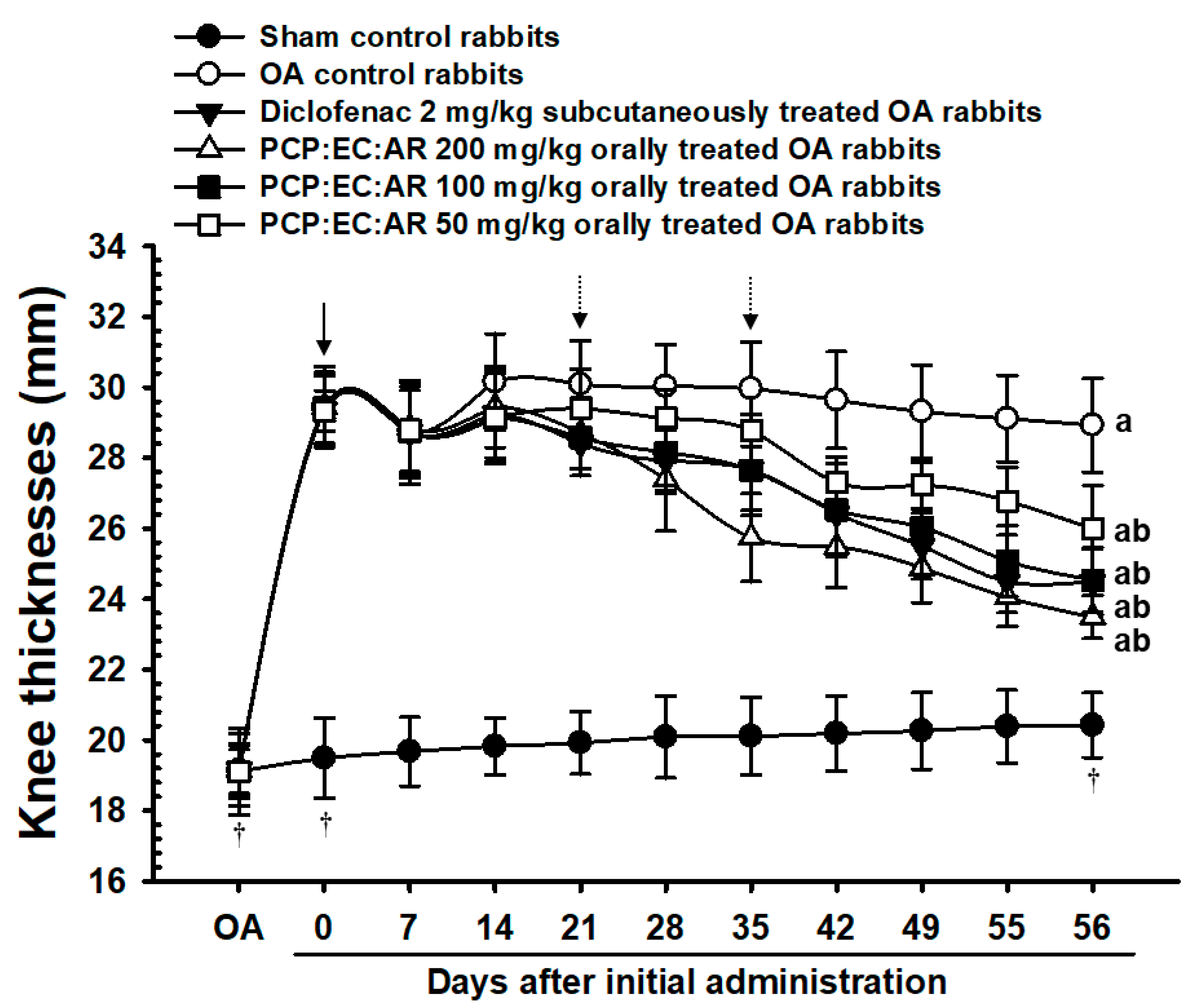
Body weight was measured at the time of OA surgery, at the time of initial treatment administration, and 7, 14, 21, 28, 35, 42, 49, 55, and 56 days thereafter using an electronic balance (Precisa Instrument, Dietikon, Switzland). All animals were fasted overnight (water was not) before weighing to reduce the effects of feeding on the measurements.

2.5. Knee Thickness Measurements

The thicknesses of the OA-operated hind knees were measured using digital calipers (Mytutoyo, Tokyo, Japan), and after exposing the joint capsule at euthanization (to reduce the influence of surrounding tissues, according to our previously established method) [52,56]. Changes in knee thickness after OA surgery were calculated.

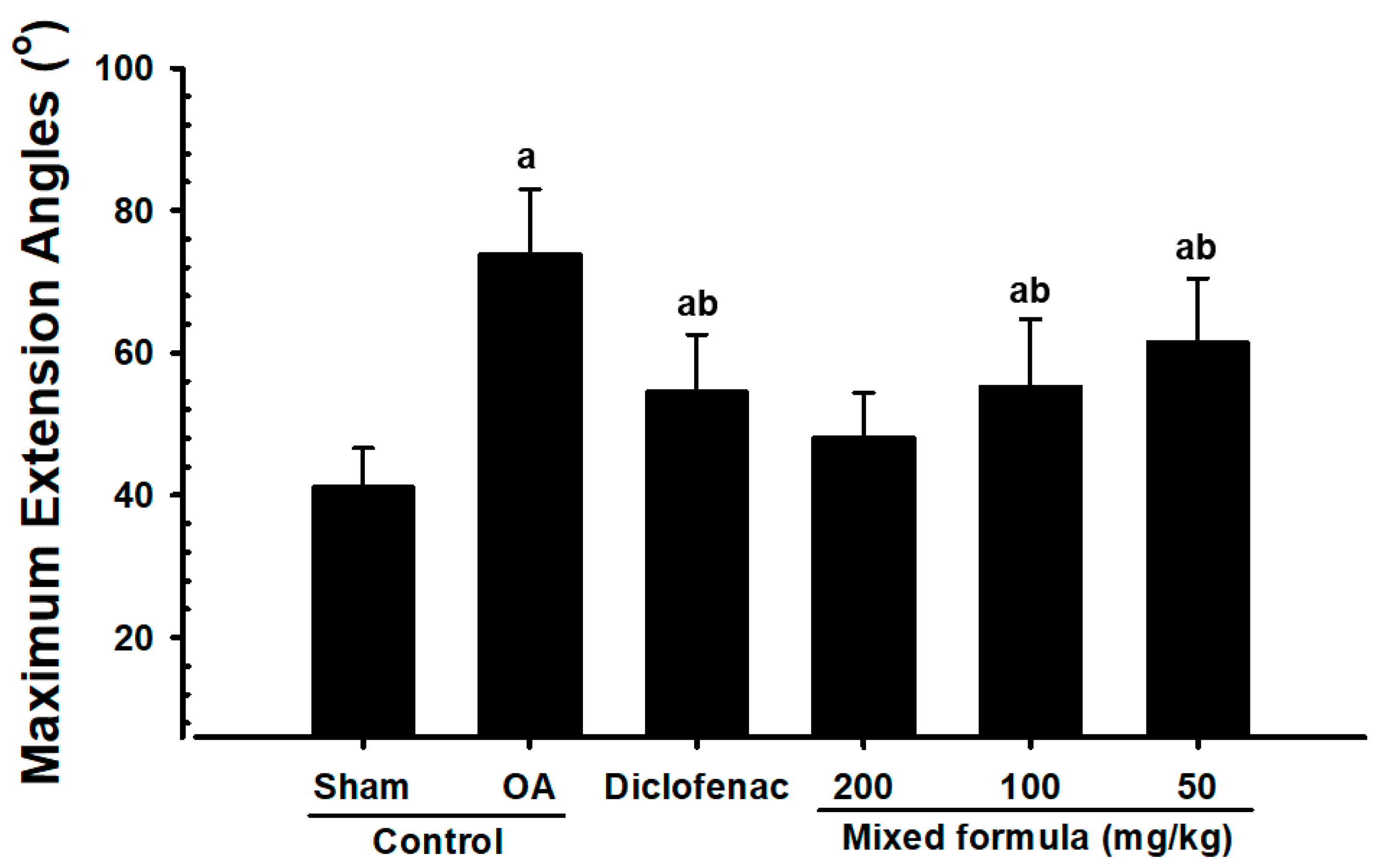
2.6. Measurement of Maximum Knee Extension

The OA-operated knees were dissected from the coxofemoral region to the ankle region, leaving the articular capsule intact. After dissection, the maximum extension of each knee was measured according to previous methods [52,56,57], with 0° corresponding to the maximum possible extension. All measurements were performed by the same veterinarian to minimize bias.

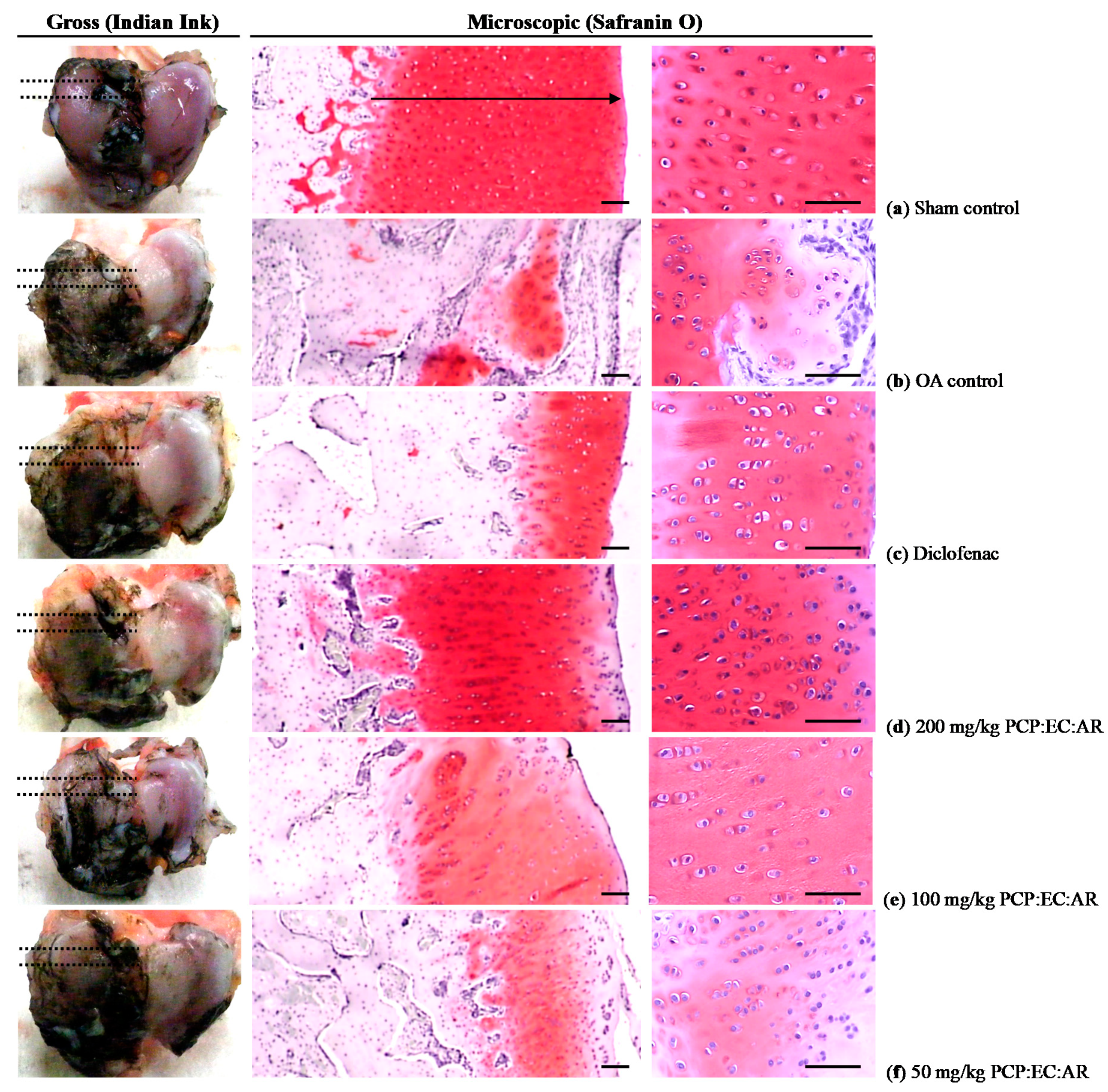
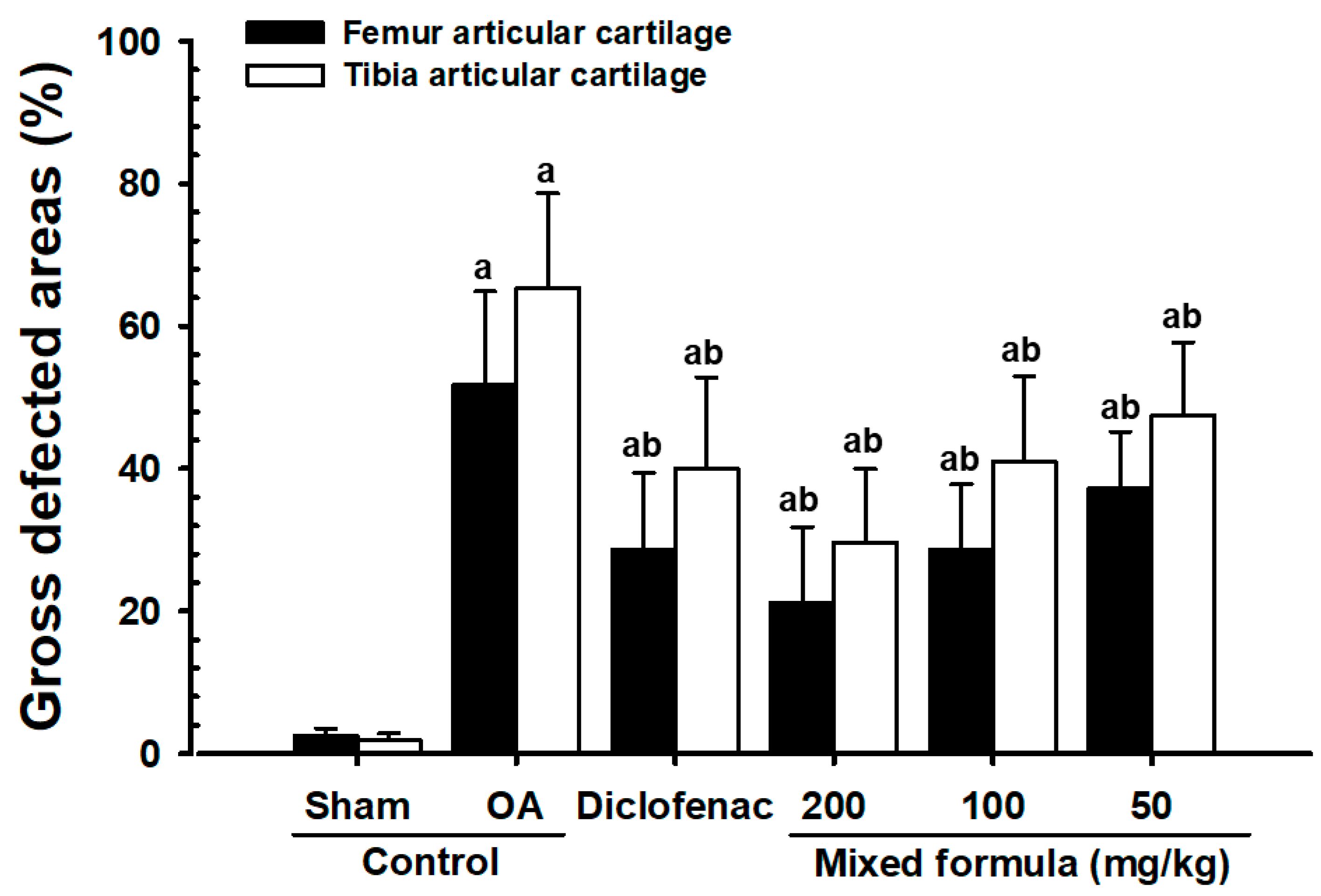
2.7. Measurement of Gross Articular Defect Area

After measuring the maximum extensor angle, the femoral and tibial articular surfaces were exposed, and the OA-induced defect area (% of defect cartilages) was measured using a computer-assisted image analysis program (iSolution FL ver 9.1; IMT i-solution Inc., Vancouver, Quebec, Canada) after staining with black India ink (Office center, Seoul, Korea).

2.8. Measurement of Serum COMP, CTX-II, and MMP-3 Levels

Approximately 10 mL of whole blood was collected from the vena cava at euthanization, and the serum was separated by centrifugation at 3000 rpm for 10 min. Serum concentrations of COMP, CTX-II, and MMP-3 were measured using sandwich enzyme-linked immunosorbent assays (ELISAs) in accordance with previous reports [15,16]. A rabbit COMP ELISA kit (MBS721182, MyBioSource, San Diego, CA, USA), rabbit CTX-II ELISA kit (MBS705896, MyBioSource, San Diego, CA, USA) and rabbit MMP-3 ELISA kit (MBS2502146, MyBioSource, San Diego, CA, USA) were used in this measurement. All ELISA procedures were performed according to the manufacturer’s instructions, except for the dilution ratio. Standards and samples (100 μL) were added to the appropriate wells and incubated for 2 h at 37 °C. The wells were aspirated, and a 1× biotin-labeled antibody (100 μL) was added to each well, followed by incubation for 1 h at 37 °C. The wells were then aspirated again, washed three times, and incubated with 1× horseradish peroxidase-conjugated avidin (100 μL) for 1 h at 37 °C. 3,3′,5,5′-Tetramethylbenzidine substrate (90 μL) was added to each well, followed by incubation for 15–30 min at 37 °C. Stop solution (50 μL) was then added to each well, and absorbance was measured at 450 nm using a microplate reader (Tecan, Männedorf, Switzerland).

2.9. Measurement of Synovial COMP, CTX-II, MMP-3, IL-1β, and TNF-α Levels

Synovial fluid was collected from the operated knee before euthanization and centrifuged at 12,500 rpm for 10 min at 4 °C. Synovial concentrations of COMP, CTX-II, MMP-3, IL-1β, and TNF-α were measured using ELISAs in accordance with previous reports [15,16,49,58], using methods similar to those described for the serum measurements. A rabbit COMP ELISA kit (MBS721182, MyBioSource, San Diego, CA, USA), rabbit CTX-II ELISA kit (MBS705896, MyBioSource, San Diego, CA, USA), rabbit MMP-3 ELISA kit (MBS2502146, MyBioSource, San Diego, CA, USA), rabbit IL-1β ELISA kit (LS-F23299, LSBio Seattle, WA, USA) and rabbit TNF-α ELISA kit (MBS2500169, MyBioSource, San Diego, CA, USA) were used.

2.10. Histology

Knee joint parts were obtained while preserving the joint capsules, fixed in 10% neutral buffered formalin, and then decalcified in decalcifying solution [24.4% formic acid, and 0.5N sodium hydroxide] for 5 days (mixed decalcifying solution was changed once a day for 5 days). Each knee joint was longitudinally trimmed, embedded in paraffin, and sectioned (3–4 μm) using a tungsten bladder equipped with n Polycut microtome (Model RM2255, Leica, Wetzlar, Germany). Cartilaginous tissue was stained with Safranin O [52,56,59]. The histological results were analyzed using a light microscope (Model Eclipse 80i, Nikkon, Tokyo, Japan) by a researcher who was blinded to the group assignments.

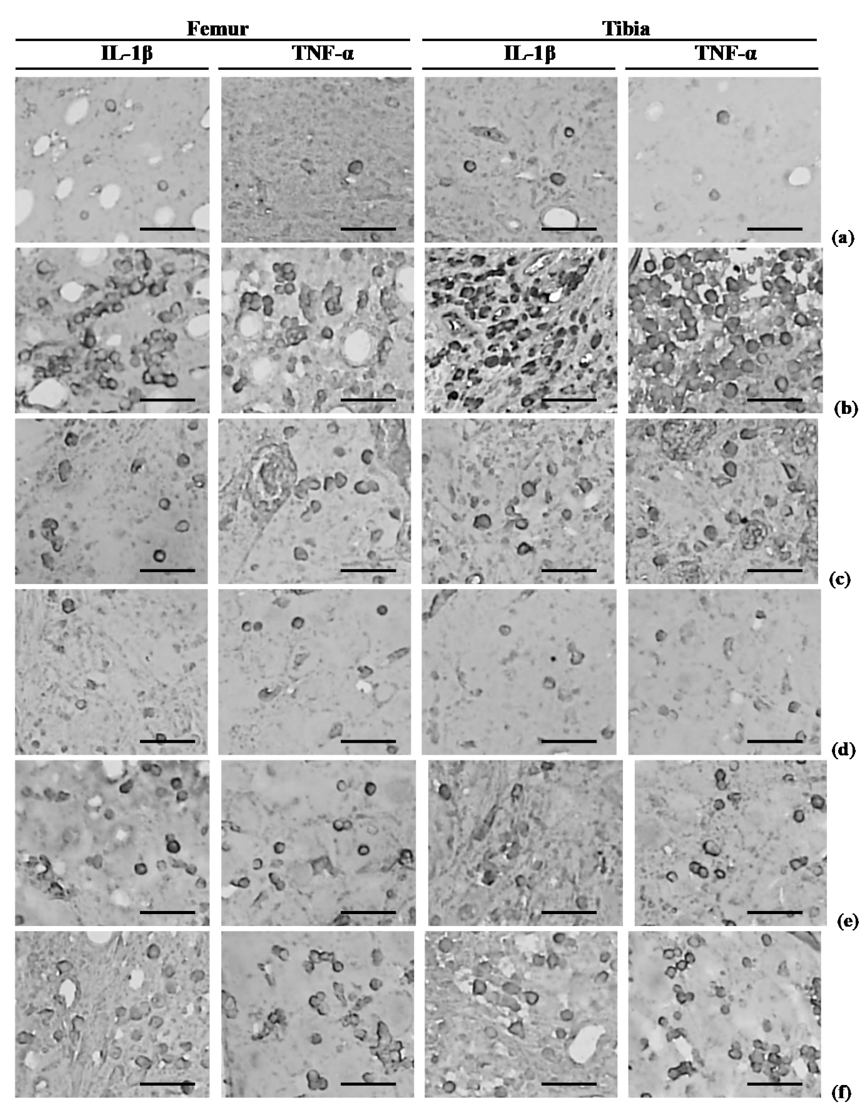
2.11. Immunohistochemistry

Immunoreactivity of the proinflammatory cytokines, IL-1β and TNF-α [60,61], was assessed. Briefly, endogenous peroxidase activity was blocked by incubating tissue sections in methanol and 0.3% H2O2 for 30 min, and non-specific binding of immunoglobulin was blocked with horse serum blocking solution for 1 h in a humidity chamber, after epitope retrieval by pretreatment of trypsin (Sigma-Aldrich, St. Louise, MO, USA) and 2 N HCl on unstained sections, as described previously [52,56]. The sections were incubated with primary antisera overnight at 4 °C in a humidity chamber, and then with biotinylated universal secondary antibody and ABC reagents for 1 h at room temperature in a humidity chamber. Finally, the sections were reacted with a peroxidase substrate kit for 3 min at room temperature.

2.12. Histomorphometry

Femoral and tibial AC in the knees was evaluated using the Mankin scoring system and Safranin O staining [52,56]. In the Mankin scoring system, higher scores indicate more severe OA (Semiquantative scores; Max = 12). The entire histological evaluation was performed by the same pathologist. In addition, the thickness of the tibial and femoral AC (μm/cartilage), and the numbers of femoral and tibial AC chondrocytes (cells/mm2), were measured in longitudinally trimmed samples using a computer-based automated image analyzer according to our previously established method [52,56,62,63]. The histopathologist was blinded to the group assignments. Cells with more than 20% immunoreactivity, as indicated by IL-1β and TNF-α antibodies, were regarded as positive [52,56], and the numbers of immunoreactive cells were counted separately in the femoral and tibial subchondral regions using an automated digital image analyzer (cells/mm2). One histological region of femoral and tibial AC was assessed in each rabbit (a total of eight fields in each group).

2.13. Statistical Analyses

All values are expressed as mean ± SD of eight rabbits. Multiple comparison tests for different dose groups were conducted. Homogeneity of variance was performed using the Levene test. If the Levene test indicated an equal variance, the obtained data were analyzed using a one way ANOVA test with a least-significant differences (LSD) multi-comparison test, as a post-hoc test. If the homogeneity of variance was violated, the Kruskal-Wallis H test was conducted. When a rejection of the null hypothesis is observed, the Mann-Whitney U (MW) test was performed. Statistical analyses were performed using SPSS (Release 14.0K, SPSS Inc., Chicago, IL, USA). In addition, the changes in percentage between sham and OA control rabbits were calculated to observe the severities of induced OA signs, and the changes of percentage as compared with OA control and test substance administered rabbits were also calculated to help the understanding of the efficacy of test materials.

3. Results

3.1. Effects of Treatment on Body Weight and Knee Thickness

At 35 days after surgery, the average weight of eight rabbits in each of the sham and OA groups was 2.39 ± 0.15 and 2.38 ± 0.12 kg, respectively. No significant difference in body weight was observed between the two groups (Table 1).
Eight rabbits from each group exhibiting increased knee thickness at 35 days after the OA operation were compared with sham control rabbits (arrow). The OA control rabbits showed increased knee thickness compared with the sham control rabbits, while the PCP:EC:AR (100 and 200 mg/kg)-treated rabbits showed an decrease in knee thickness beginning 21 days after initial administration of the treatment compared with the OA control rabbits, and from 35 days after initial administration in the PCP:EC:AR mixed-formula 50 mg/kg treated rabbits as compared with OA control rabbits, respectively (dot arrows). PCP:EC:AR treatment (50, 100, and 200 mg/kg) led to a dose-dependent decrease in knee thickness (Table 2, Figure 1).

3.2. Effects of Treatment on Knee Thickness after Capsule Exposure

Capsule-exposed knee thicknesses were greater in OA control rabbits than in sham control rabbits. However, significantly decreased knee thickness was observed after exposing the capsule in all PCP:EC:AR (50, 100, and 200 mg/kg)-treated rabbits compared with the OA control rabbits, in a dose-dependent manner (Figure 2).

3.3. Effects of Treatment on Maximum Knee Extension

Significant increases in maximum knee extension were observed in OA control rabbits compared with the sham control rabbits. However, the maximum extension of the knee decreased significantly in the PCP:EC:AR (50, 100, and 200 mg/kg)-treated rabbits compared with the OA control rabbits, in a dose-dependent manner (Figure 3).

3.4. Changes in Gross Articular Defect Areas

The femoral and tibial gross articular defect areas increased in the OA control, compared with sham control rabbits. In contrast, all PCP:EC:AR (50, 100, and 200 mg/kg)-treated rabbits showed dose-dependent decreases in gross femoral and tibial AC defect areas (Figure 4, Figure 5 and Figure 6).

3.5. Changes in Serum COMP, CTX-II, and MMP-3 Levels

Serum COMP, CTX-II, and MMP-3 levels were higher in the OA control rabbits than in the sham control rabbits. However, PCP:EC:AR (50, 100, and 200 mg/kg) decreased serum COMP, CTX-II, and MMP-3 levels compared with those in OA control rabbits, in a dose-dependent manner (Table 3).

3.6. Changes in Synovial COMP, CTX-II, MMP-3, IL-1β, and TNF-α Levels

Synovial COMP, CTX-II, MMP-3, IL-1β, and TNF-α levels were higher in OA control rabbits than in sham control rabbits. However, PCP:EC:AR (50, 100, and 200 mg/kg) decreased synovial COMP, CTX-II, MMP-3, IL-1β, and TNF-α levels compared with those of OA control rabbits, in a dose-dependent manner (Table 4).

3.7. Changes in Femoral and Tibial AC Mankin Scores

Marked increases in surface cartilage damage, a decreased number of chondrocytes, clone formation, and decreased Safranin O staining intensity were detected in the femoral and tibial AC of OA control rabbits. Consequently, the femoral and tibial AC Mankin scores were higher in OA control rabbits than in sham control rabbits. However, the femoral and tibial AC Mankin scores were lower in all PCP:EC:AR treatment groups (50, 100, and 200 mg/kg) than in OA control rabbits, in a dose-dependent manner (Table 5, Figure 4 and Figure 5).

3.8. Histopathological Changes in the Femoral and Tibial AC

Femoral AC thickness decreased significantly in OA control rabbits compared with sham control rabbits. In contrast, significant dose-dependent increases in femoral AC thickness were observed in all PCP:EC:AR treatment groups (50, 100, and 200 mg/kg) compared with OA control rabbits.
Tibial AC thickness decreased significantly in OA control rabbits. In contrast, tibial AC thickness increased significantly in all PCP:EC:AR (50, 100, and 200 mg/kg)-treated rabbits compared with OA control rabbits, in a dose-dependent manner (Table 6, Figure 4).
A significant decrease in the number of femoral AC chondrocytes was detected in OA control rabbits compared with sham control rabbits; however, a significant dose-dependent increase was observed in all PCP:EC:AR treatment groups (50, 100, and 200 mg/kg) compared with OA control rabbits (Table 6, Figure 4).
Fewer tibial AC chondrocytes were observed in the OA control rabbits than in the sham control rabbits; however, the number of tibial AC chondrocytes increased significantly in all PCP:EC:AR treatment groups (50, 100, and 200 mg/kg) compared with the OA control rabbits, in a dose-dependent manner (Table 6, Figure 5).

3.9. Effects of Treatment on IL-1β-Immunoreactive Cells in Femoral and Tibial Subchondral Regions

Significant increases in the numbers of femoral and tibial subchondral IL-1β-immunopositive cells were detected in OA control rabbits. However, significant dose-dependent decreases in femoral and tibial subchondral IL-1β-immunoreactive cells were observed in all PCP:EC:AR (50, 100, and 200 mg/kg)-treated rabbits compared with OA control rabbits (Table 6, Figure 7).

3.10. Effects of Treatment on TNF-α-Immunoreactive Cells in Femoral and Tibial Subchondral Regions

The numbers of femoral and tibial subchondral TNF-α-immunopositive cells increased in OA control rabbits, whereas they decreased in all PCP:EC:AR treatment groups (50, 100, and 200 mg/kg) in a dose-dependent manner (Table 6, Figure 7).

4. Discussion

The anti-OA potential of PCP:EC:AR was demonstrated in a surgically induced OA rabbit model [48,49]. The anti-OA effects of PCP:EC:AR were similar to those seen in rat and mouse models of OA; thus, PCP:EC:AR effectively relieves OA symptoms in various animal models. The PCP:EC:AR formula used herein was informed by our previous in vitro [54,64] and in vivo [55,65] experiments on OA rats.
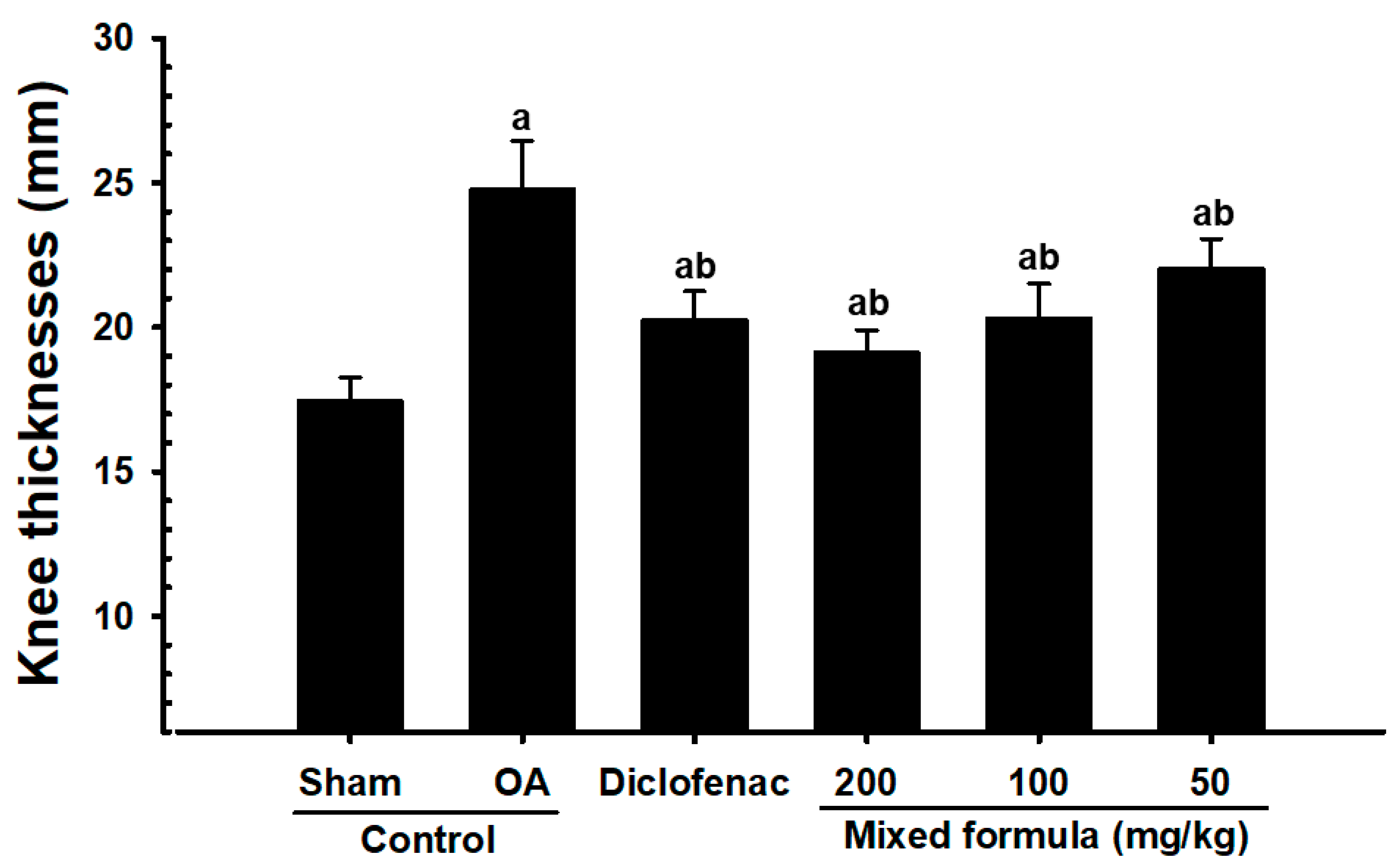
OA is a debilitating disease that affects the AC and subchondral bone. Inflammation is closely involved in all steps of OA progression, where cartilage damage triggers prostaglandin and leukotriene activation by cyclooxygenase-2 and 5-lipoxygenase, respectively. In addition, the inflammatory factors TNF-α and IL-1β contribute to OA progression. The inflammatory response results in swelling of the tissues surrounding the joints, in association with increased joint thickness, irregular bone growth, and osteophyte formation [66,67,68]. In the present study, OA-induced rabbits exhibited significantly increased knee thickness, decreases in AC thickness of the femur and tibia and the number of chondrocytes, significantly increased synovial TNF-α and IL-1β levels, and significantly higher numbers of femoral and tibial subchondral IL-1β and TNF-α-immunolabeled cells. However, these OA-related signs of inflammation in OA rabbits were dose-dependently and significantly inhibited by 56 days of administration with PCP:EC:AR (50, 100, and 200 mg/kg). These findings represent reliable evidence that a PCP:EC:AR mixed formula has favorable anti-inflammatory effects on surgically induced OA rabbits.
X-ray images have been widely used to detect loss of cartilage erosion, joint region, and osteophyte formation during the development of OA [62,69]. Thus, anti-OA agents should inhibit cartilage damage on X-ray images to indicate efficacy [49,70]. In our experiment, articular space narrowing, loss of the femoral and tibial AC erosion, joint region, and osteophyte formation were detected on roentgenographic images of the OA control rabbits. In addition, significant increments of gross articular defect areas in the femur and tibia were also detected in OA control rabbits. However, these changes were inhibited dose-dependently and significantly by 56 days of continuous treatment with PCP:EC:AR (50, 100, and 200 mg/kg), similar to the effects of subcutaneous diclofenac (2 mg/kg) treatment. These results show that PCP:EC:AR exerted anti-OA effects in a surgically induced OA rabbit model (Supplementary Data, Figure S1).
Fibrosis and inflammation have been linked to OA progression, and anti-fibrotic drugs have shown promise in the clinical treatment of OA. The anti-fibrotic and anti-inflammatory drug pirfenidone shows efficacy against kidney and lung fibrosis. Pirfenidone may encourage tissue repair and prevent progression of post-traumatic OA [71]. The fibrosis that occurs in OA due to chronic inflammation limits joint movement, where stiffness of the joints is a major symptom of OA. Joint stiffness is evaluated based on the maximum extension of the joint, where a 0 degree corresponds to the maximum extension, that is, better knee function [52,56,57,62]. Thus, the significant increases in maximum extension of the knees of our OA control rabbits indicate that OA was well-induced. However, these OA-related increases in maximum knee extension were also reduced dose-dependently and significantly by 56 days of continuous treatment with all three doses (50, 100, and 200 mg/kg) of PCP:EC:AR. These findings represent direct evidence that PCP:EC:AR inhibits joint stiffness induced by OA.
COMP, a thrombospondin gene family member, is widely expressed in the ECM of AC [72,73]. COMP binds collagen types Ⅰ, II, and Ⅳ, and is involved in the regulation of chondrocytes [74]. Kuhne et al. [75] reported that the serum COMP level in serum increases in patients after traumatic knee injury. COMP may be instructive regarding cartilage degradation status, and has been used as a marker of OA [15,16,76]. This protein is a constituent of AC, but is also synthesized by tendon synoviocytes and fibroblasts [76,77]. Marked increases in synovial and serum COMP levels were detected in our OA control rabbits. However, significant dose-dependent decrements in synovial and serum COMP levels were detected in the PCP:EC:AR (50, 100, and 200 mg/kg)-treated rabbits, indicating a favorable AC-preserving effect.
Type II collagen is found mainly in cartilage, of which it is a major structural component [78]. The normal cartilage matrix consists of more collagen type II than collagen type I [6,79,80]. Collagen type II shows strong interactions with proteoglycans, so the ECM network is affected by collagen type II content. Collagen type II content decreases during the development of OA, such that ECM integrity is inevitably reduced. Hence, measurements of constituents of this protein, such as CTX-II, may have utility as an index of cartilage degradation [76]. Increased CTX-II has also proven valuable as a biomarker for early OA [15,16]. In the present study, noticeable increases in serum and synovial CTX-II levels were observed in OA control rabbits, which corresponded well to the increases in COMP. However, dose-dependent significant decreases in synovial and serum CTX-II levels were detected in the PCP:EC:AR 5:4:1 (50, 100, and 200 mg/kg)-treated rabbits. These findings represent additional indirect evidence that the PCP:EC:AR mixture has favorable AC-preserving effects.
MMPs are involved in degradation of the ECM (mainly proteoglycans) [81], although their expression can be suppressed by metalloproteinase inhibitors [82]. In particular, the MMP-3 level is a reliable predictor of cartilage loss during the development of OA [21]. MMP-3 increases in the synovium and chondrocytes in surgically induced OA rabbit models [83]. Davidson et al. reported that less severe OA in joints was observed in MMP-3 knockout mice [84,85]. In the present study, significant increases in serum and synovial MMP-3 levels were observed in OA control rabbits, but these increases were inhibited dose-dependently and significantly by 56 days of continuous treatment of 50, 100, and 200 mg/kg PCP:EC:AR. These findings represent additional evidence that the anti-OA activity of PCP:EC:AR may be mediated by MMP-3 inhibition.
Mankin’s 14-point histopathological grading system of cartilage deterioration is based on cartilage surface damage, the number of chondrocytes, and Safranin O staining. In this study, normal cartilage was scored as 0; the most severe case of cartilage deterioration had a score of 12 [52,56,57,86]. In addition, decreases in AC thickness and the number of chondrocytes have been consistently detected in animal models of OA [52,56]. In the present experiment, these increments induce by OA in Mankin scores of femoral and tibial AC, and the decreases in chondrocyte numbers and cartilage thickness, were inhibited dose-dependently and significantly by 56 days of continuous treatment with PCP:EC:AR (50, 100, and 200 mg/kg) compared with those of OA control rabbits. These findings indicate that the PCP:EC:AR mixture has chondrocyte-protective effects in surgically induced OA rabbits, at least under the conditions of the present experiment.

5. Conclusions

PCP:EC:AR (50, 100, and 200 mg/kg) consistently showed significant dose-dependent activity against OA in a rabbit model, similar to subcutaneous diclofenac (2 mg/kg) treatment. Taken together, the results indicated that the PCP:EC:AR mixed formula is a promising novel agent for relieving OA symptoms.

Supplementary Materials

The following are available online at https://0-www-mdpi-com.brum.beds.ac.uk/2072-6643/12/3/852/s1, Figure S1: Representative X-ray images of the knee joints, taken from sham-operated or OA rabbits.

Author Contributions

Conceptualization, B.-R.C. and S.-K.K.; methodology, S.-K.K.; experiment, S.-J.K., Y.-J.L. and S.-K.K.; preparation and analysis of materials, B.-R.C. and J.-L.K.; data curation, S.-K.K. and Y.-J.L.; writing—original draft preparation, S.-K.K. and S.J.K.; writing—review and editing, B.-R.C. and Y.-J.L. All authors have read and agreed to the published version of the manuscript.

Funding

This work was supported by the National Research Foundation of Korea grant funded by the Korea government (MSIT) (No.2018R1A5A2025272).

Acknowledgments

This work was supported by the National Research Foundation of Korea grant funded by the Korea government (MSIT) (No.2018R1A5A2025272).

Conflicts of Interest

The authors declare that they have no conflict of interest.

References

  1. Dougados, M.; Nguyen, M.; Berdah, L.; Mazières, B.; Vignon, E.; Lequesne, M.; Group, E.I.S. Evaluation of the structure-modifying effects of diacerein in hip osteoarthritis: ECHODIAH, a three-year, placebo-controlled trial. Evaluation of the Chondromodulating Effect of Diacerein in OA of the Hip. Arthritis Rheum. 2001, 44, 2539–2547. [Google Scholar] [CrossRef]
  2. Goldring, M.B.; Goldring, S.R. Osteoarthritis. J. Cell. Physiol. 2007, 213, 626–634. [Google Scholar] [CrossRef] [PubMed]
  3. Felson, D.T. Developments in the clinical understanding of osteoarthritis. Arthritis Res. Ther. 2009, 11, 203. [Google Scholar] [CrossRef] [PubMed] [Green Version]
  4. Qin, J.; Liu, Y.S.; Liu, J.; Li, J.; Tan, Y.; Li, X.J.; Magdalou, J.; Mei, Q.B.; Wang, H.; Chen, L.B. Effect of Angelica sinensis Polysaccharides on Osteoarthritis In Vivo and In Vitro: A Possible Mechanism to Promote Proteoglycans Synthesis. Evid. Based Complement. Altern. Med. 2013, 2013, 794761. [Google Scholar] [CrossRef] [PubMed] [Green Version]
  5. Na, J.Y.; Song, K.B.; Kim, S.; Kwon, Y.B.; Kim, D.G.; Lee, J.K.; Jo, H.K.; Kwon, J. Effects of HPL-04 on degenerative osteoarthritis. J. Korean Soc. Food Sci. Nutr. 2014, 43, 30–39. [Google Scholar]
  6. Pearle, A.D.; Warren, R.F.; Rodeo, S.A. Basic science of articular cartilage and osteoarthritis. Clin. Sports Med. 2005, 24, 1–12. [Google Scholar] [CrossRef] [PubMed]
  7. Maldonado, M.; Nam, J. The role of changes in extracellular matrix of cartilage in the presence of inflammation on the pathology of osteoarthritis. Biomed Res. Int. 2013, 2013, 284873. [Google Scholar] [CrossRef] [PubMed] [Green Version]
  8. Lahm, A.; Mrosek, E.; Spank, H.; Erggelet, C.; Kasch, R.; Esser, J.; Merk, H. Changes in content and synthesis of collagen types and proteoglycans in osteoarthritis of the knee joint and comparison of quantitative analysis with Photoshop-based image analysis. Arch. Orthop. Trauma Surg. 2010, 130, 557–564. [Google Scholar] [CrossRef]
  9. Goldring, M.B. Articular cartilage degradation in osteoarthritis. HSS J. 2012, 8, 7–9. [Google Scholar] [CrossRef] [Green Version]
  10. Pfander, D.; Heinz, N.; Rothe, P.; Carl, H.D.; Swoboda, B. Tenascin and aggrecan expression by articular chondrocytes is influenced by interleukin 1b: A possible explanation for the changes in matrix synthesis during osteoarthritis. Ann. Rheum. Dis. 2004, 63, 240–244. [Google Scholar] [CrossRef] [Green Version]
  11. Meulenbelt, I.; Seymour, A.B.; Nieuwland, M.; Huizinga, T.W.; van Duijn, C.M.; Slagboom, P.E. Association of the interleukin-1 gene cluster with radiographic signs of osteoarthritis of the hip. Arthritis Rheum. 2004, 50, 1179–1186. [Google Scholar] [CrossRef] [PubMed]
  12. Sakkas, L.I.; Johanson, N.A.; Scanzello, C.R.; Platsoucas, C.D. Interleukin-12 is expressed by infiltrating macrophages and synovial lining cells in rheumatoid arthritis and osteoarthritis. Cell. Immunol. 1998, 188, 105–110. [Google Scholar] [CrossRef] [PubMed]
  13. Scanzello, C.R.; Umoh, E.; Pessler, F.; Diaz-Torne, C.; Miles, T.; Dicarlo, E.; Potter, H.G.; Mandl, L.; Marx, R.; Rodeo, S.; et al. Local cytokine profiles in knee osteoarthritis: Elevated synovial fluid interleukin-15 differentiates early from end-stage disease. Osteoarthr. Cartil. 2009, 17, 1040–1048. [Google Scholar] [CrossRef] [PubMed] [Green Version]
  14. Campo, G.M.; Avenoso, A.; D’Ascola, A.; Scuruchi, M.; Prestipino, V.; Calatroni, A.; Campo, S. Hyaluronan in part mediates IL-1beta-induced inflammation in mouse chondrocytes by up-regulating CD44 receptors. Gene 2012, 494, 24–35. [Google Scholar] [CrossRef] [PubMed]
  15. Chu, X.Q.; Wang, J.J.; Dou, L.D.; Zhao, G. Cartilage oligomeric matrix protein and matrix metalloproteinase-3 expression in the serum and joint fluid of a reversible osteoarthritis rabbit model. Genet. Mol. Res. 2015, 14, 14207–14215. [Google Scholar] [CrossRef] [PubMed]
  16. Jiao, Q.; Wei, L.; Chen, C.; Li, P.; Wang, X.; Li, Y.; Guo, L.; Zhang, C.; Wei, X. Cartilage oligomeric matrix protein and hyaluronic acid are sensitive serum biomarkers for early cartilage lesions in the knee joint. Biomarkers 2016, 21, 146–151. [Google Scholar] [CrossRef]
  17. Bauer, D.C.; Hunter, D.J.; Abramson, S.B.; Attur, M.; Corr, M.; Felson, D.; Heinegard, D.; Jordan, J.M.; Kepler, T.B.; Lane, N.E.; et al. Classification of osteoarthritis biomarkers: A proposed approach. Osteoarthr. Cartil. 2006, 14, 723–727. [Google Scholar] [CrossRef] [Green Version]
  18. Henrotin, Y.; Gharbi, M.; Mazzucchelli, G.; Dubuc, J.E.; De Pauw, E.; Deberg, M. Fibulin 3 peptides Fib3-1 and Fib3-2 are potential biomarkers of osteoarthritis. Arthritis Rheum. 2012, 64, 2260–2267. [Google Scholar] [CrossRef]
  19. Golightly, Y.M.; Marshall, S.W.; Kraus, V.B.; Renner, J.B.; Villaveces, A.; Casteel, C.; Jordan, J.M. Biomarkers of incident radiographic knee osteoarthritis: Do they vary by chronic knee symptoms? Arthritis Rheum. 2011, 63, 2276–2283. [Google Scholar] [CrossRef] [Green Version]
  20. Kraus, V.B.; Kepler, T.B.; Stabler, T.; Renner, J.; Jordan, J. First qualification study of serum biomarkers as indicators of total body burden of osteoarthritis. PLoS ONE 2010, 5, e9739. [Google Scholar] [CrossRef]
  21. Pelletier, J.P.; Raynauld, J.P.; Caron, J.; Mineau, F.; Abram, F.; Dorais, M.; Haraoui, B.; Choquette, D.; Martel-Pelletier, J. Decrease in serum level of matrix metalloproteinases is predictive of the disease-modifying effect of osteoarthritis drugs assessed by quantitative MRI in patients with knee osteoarthritis. Ann. Rheum. Dis. 2010, 69, 2095–2101. [Google Scholar] [CrossRef] [PubMed]
  22. Mobasheri, A. Osteoarthritis year 2012 in review: Biomarkers. Osteoarthr. Cartil. 2012, 20, 1451–1464. [Google Scholar] [CrossRef] [PubMed] [Green Version]
  23. Langley, P. Why a pomegranate? BMJ 2000, 321, 1153–1154. [Google Scholar] [CrossRef] [PubMed]
  24. Gil, M.I.; Tomás-Barberán, F.A.; Hess-Pierce, B.; Holcroft, D.M.; Kader, A.A. Antioxidant activity of pomegranate juice and its relationship with phenolic composition and processing. J. Agric. Food Chem. 2000, 48, 4581–4589. [Google Scholar] [CrossRef] [PubMed]
  25. Noda, Y.; Kaneyuki, T.; Mori, A.; Packer, L. Antioxidant activities of pomegranate fruit extract and its anthocyanidins: Delphinidin, cyanidin, and pelargonidin. J. Agric. Food Chem. 2002, 50, 166–171. [Google Scholar] [CrossRef] [PubMed]
  26. Singh, R.P.; Chidambara Murthy, K.N.; Jayaprakasha, G.K. Studies on the antioxidant activity of pomegranate (Punica granatum) peel and seed extracts using in vitro models. J. Agric. Food Chem. 2002, 50, 81–86. [Google Scholar] [CrossRef]
  27. Ahmed, S.; Wang, N.; Hafeez, B.B.; Cheruvu, V.K.; Haqqi, T.M. Punica granatum L. extract inhibits IL-1b-induced expression of matrix metalloproteinases by inhibiting the activation of MAP kinases and NF-kB in human chondrocytes in vitro. J. Nutr. 2005, 135, 2096–2102. [Google Scholar] [CrossRef] [Green Version]
  28. Shukla, M.; Gupta, K.; Rasheed, Z.; Khan, K.A.; Haqqi, T.M. Bioavailable constituents/metabolites of pomegranate (Punica granatum L) preferentially inhibit COX2 activity ex vivo and IL-1beta-induced PGE2 production in human chondrocytes in vitro. J. Inflamm. 2008, 5, 9. [Google Scholar] [CrossRef] [Green Version]
  29. Hadipour-Jahromy, M.; Mozaffari-Kermani, R. Chondroprotective effects of pomegranate juice on monoiodoacetate-induced osteoarthritis of the knee joint of mice. Phytother. Res. 2010, 24, 182–185. [Google Scholar] [CrossRef]
  30. Rasheed, Z.; Akhtar, N.; Haqqi, T.M. Pomegranate extract inhibits the interleukin-1b-induced activation of MKK-3, p38a-MAPK and transcription factor RUNX-2 in human osteoarthritis chondrocytes. Arthritis Res. Ther. 2010, 12, R195. [Google Scholar] [CrossRef] [Green Version]
  31. Rasheed, Z. Intake of Pomegranate Prevents the Onset of Osteoarthritis: Molecular Evidences. Int. J. Health Sci. 2016, 10, V–VIII. [Google Scholar] [CrossRef]
  32. Hsieh, C.L.; Yen, G.C. Antioxidant actions of du-zhong (Eucommia ulmoides Oliv.) toward oxidative damage in biomolecules. Life Sci. 2000, 66, 1387–1400. [Google Scholar] [CrossRef]
  33. Kwan, C.Y.; Chen, C.X.; Deyama, T.; Nishibe, S. Endothelium-dependent vasorelaxant effects of the aqueous extracts of the Eucommia ulmoides Oliv. leaf and bark: Implications on their antihypertensive action. Vasc. Pharmacol. 2003, 40, 229–235. [Google Scholar] [CrossRef] [PubMed]
  34. Zhao, Y.; Li, Y.; Wang, X.; Sun, W. The experimental study of Cortex Eucommiae on meridian tropsim: The distribution study of aucubin in rat tissues. J. Pharm. Biomed. Anal. 2008, 46, 368–373. [Google Scholar] [CrossRef] [PubMed]
  35. Lu, H.; Jiang, J.; Xie, G.; Liu, W.; Yan, G. Effects of an aqueous extract of Eucommia on articular cartilage in a rat model of osteoarthritis of the knee. Exp. Ther. Med. 2013, 6, 684–688. [Google Scholar] [CrossRef]
  36. Eum, H.A.; Lee, W.Y.; Kim, S.H.; Kim, J.Y.; Park, S.W.; Lee, J.S.; Choi, S.M.; Pyo, S.; Kang, M.J.; Min, Y.D.; et al. Anti-inflammatory activity of CML-1: An herbal formulation. Am. J. Chin. Med. 2005, 33, 29–40. [Google Scholar] [CrossRef]
  37. Lee, S.Y.; Kwon, H.K.; Lee, S.M. SHINBARO, a new herbal medicine with multifunctional mechanism for joint disease: First therapeutic application for the treatment of osteoarthritis. Arch. Pharmacal Res. 2011, 34, 1773–1777. [Google Scholar] [CrossRef]
  38. Li, J.; Li, H.J.; Li, P.; Qi, H. Simultaneous qualitation and quantification of four phytoecdysones in Radix Achyranthis Bidentatae by high-performance liquid chromatography with diode array detection. Biomed. Chromatogr. BMC 2007, 21, 823–828. [Google Scholar] [CrossRef]
  39. Lee, S.G.; Lee, E.J.; Park, W.D.; Kim, J.B.; Kim, E.O.; Choi, S.W. Anti-inflammatory and anti-osteoarthritis effects of fermented Achyranthes japonica Nakai. J. Ethnopharmacol. 2012, 142, 634–641. [Google Scholar] [CrossRef]
  40. Liu, M.; Dai, Y.; Yao, X.; Li, Y.; Luo, Y.; Xia, Y.; Gong, Z. Anti-rheumatoid arthritic effect of madecassoside on type II collagen-induced arthritis in mice. Int. Immunopharmacol. 2008, 8, 1561–1566. [Google Scholar] [CrossRef]
  41. Yu, F.; Li, X.; Cai, L.; Li, H.; Chen, J.; Wong, X.; Xu, H.; Zheng, C.; Liu, X.; Ye, H. Achyranthes bidentata polysaccharides induce chondrocyte proliferation via the promotion of the G1/S cell cycle transition. Mol. Med. Rep. 2013, 7, 935–940. [Google Scholar] [CrossRef] [PubMed]
  42. Weng, X.; Lin, P.; Liu, F.; Chen, J.; Li, H.; Huang, L.; Zhen, C.; Xu, H.; Liu, X.; Ye, H.; et al. Achyranthes bidentata polysaccharides activate the Wnt/b-catenin signaling pathway to promote chondrocyte proliferation. Int. J. Mol. Med. 2014, 34, 1045–1050. [Google Scholar] [CrossRef] [PubMed] [Green Version]
  43. Schwartz, E.R.; Greenwald, R.A. Experimental models of osteoarthritis. Bull. Rheum. Dis. 1979, 30, 1030–1033. [Google Scholar] [PubMed]
  44. Livne, E.; von der Mark, K.; Silbermann, M. Morphologic and cytochemical changes in maturing and osteoarthritic articular cartilage in the temporomandibular joint of mice. Arthritis Rheum. 1985, 28, 1027–1038. [Google Scholar] [CrossRef] [PubMed]
  45. McDevitt, C.; Gilbertson, E.; Muir, H. An experimental model of osteoarthritis; early morphological and biochemical changes. J. Bone Jt. Surg. Br. 1977, 59, 24–35. [Google Scholar] [CrossRef]
  46. Kopp, S.; Mejersjo, C.; Clemensson, E. Induction of osteoarthrosis in the guinea pig knee by papain. Oral Surg. Oral Med. Oral Pathol. 1983, 55, 259–266. [Google Scholar] [CrossRef]
  47. Kalbhen, D.A. Chemical model of osteoarthritis--a pharmacological evaluation. J. Rheumatol. 1987, 14, 130–131. [Google Scholar]
  48. Hanashi, D.; Koshino, T.; Uesugi, M.; Saito, T. Effect of femoral nerve resection on progression of cartilage degeneration induced by anterior cruciate ligament transection in rabbits. J. Orthop. Sci. Off. J. Jpn. Orthop. Assoc. 2002, 7, 672–676. [Google Scholar] [CrossRef]
  49. Jiang, D.; Zou, J.; Huang, L.; Shi, Q.; Zhu, X.; Wang, G.; Yang, H. Efficacy of intra-articular injection of celecoxib in a rabbit model of osteoarthritis. Int. J. Mol. Sci. 2010, 11, 4106–4113. [Google Scholar] [CrossRef] [Green Version]
  50. Miles, J.E.; Wenck, A.; Fricker, C.; Svalastoga, E.L. Modulation of the intramedullary pressure responses by calcium dobesilate in a rabbit knee model of osteoarthritis. Acta Orthop. 2011, 82, 622–627. [Google Scholar] [CrossRef] [Green Version]
  51. Takeshita, N.; Yoshimi, E.; Hatori, C.; Kumakura, F.; Seki, N.; Shimizu, Y. Alleviating effects of AS1892802, a Rho kinase inhibitor, on osteoarthritic disorders in rodents. J. Pharmacol. Sci. 2011, 115, 481–489. [Google Scholar] [CrossRef] [PubMed] [Green Version]
  52. Choi, J.S.; Shin, H.S.; Kim, K.Y.; Ku, S.K.; Choi, I.S.; Kim, J.W. Effect of Polycalcium, a mixture of Polycan and calcium lactate-gluconate in a 1:9 weight ratio, on rats with surgery-induced osteoarthritis. Exp. Ther. Med. 2015, 9, 1780–1790. [Google Scholar] [CrossRef] [PubMed] [Green Version]
  53. Kang, S.J.; Choi, B.R.; Kim, S.H.; Yi, H.Y.; Park, H.R.; Kim, D.C.; Choi, S.H.; Han, C.H.; Park, S.J.; Song, C.H.; et al. Dried pomegranate potentiates anti-osteoporotic and anti-obesity activities of red clover dry extracts in ovariectomized rats. Nutrients 2015, 7, 2622–2647. [Google Scholar] [CrossRef] [PubMed] [Green Version]
  54. Choi, B.R.; Ku, S.K.; Kang, S.J.; Park, H.R.; Sung, M.S.; Lee, Y.J.; Park, K.M. Concentration-dependent in vitro Anti-osteoarthritis Effects of Mixed Formula-Pomegranate Concentrate Powder: Eucommiae Cortex: Achyranthis Radix 5: 4: 1 (g/g) on the Primary Cultured Rat Articular Chondrocytes. J. Physiol. Pathol. Korean Med. 2019, 33, 131–140. [Google Scholar] [CrossRef]
  55. Choi, B.R.; Ku, S.K.; Kang, S.J.; Park, H.R.; Sung, M.S.; Lee, Y.J.; Park, K.M. Anti-osteoarthritic effects of a combination of pomegranate concentrate powder, Eucommiae cortex and Achyranthis radix in rats. J. Korean Med. 2018, 39, 86–113. [Google Scholar] [CrossRef]
  56. Moon, C.H.; Kwon, O.; Woo, C.H.; Ahn, H.D.; Kwon, Y.S.; Park, S.J.; Song, C.H.; Ku, S.K. Therapeutic effect of irradiation of magnetic infrared laser on osteoarthritis rat model. Photochem. Photobiol. 2014, 90, 1150–1159. [Google Scholar] [CrossRef]
  57. Rezende, M.U.; Gurgel, H.M.; Vilaca Junior, P.R.; Kuroba, R.K.; Lopes, A.S.; Phillipi, R.Z.; Hernandez, A.J. Diacerhein versus glucosamine in a rat model of osteoarthritis. Clinics 2006, 61, 461–466. [Google Scholar] [CrossRef] [Green Version]
  58. Chen, L.; Ling, P.X.; Jin, Y.; Zhang, T.M. Hyaluronic acid in combination with chondroitin sulfate and hyaluronic acid improved the degeneration of synovium and cartilage equally in rabbits with osteoarthritis. Drug Discov. Ther. 2011, 5, 190–194. [Google Scholar] [CrossRef] [Green Version]
  59. Tran, D.; Golick, M.; Rabinovitz, H.; Rivlin, D.; Elgart, G.; Nordlow, B. Hematoxylin and safranin O staining of frozen sections. Dermatol. Surg. 2000, 26, 197–199. [Google Scholar] [CrossRef]
  60. Hwangbo, M.; Jung, J.Y.; Ki, S.H.; Park, S.M.; Jegal, K.H.; Cho, I.J.; Lee, J.H.; Kang, S.H.; Park, S.D.; Ku, S.K.; et al. U-Bang-Haequi Tang: A Herbal Prescription that Prevents Acute Inflammation through Inhibition of NF-kB-Mediated Inducible Nitric Oxide Synthase. Evid. Based Complement. Altern. Med. 2014, 2014, 542825. [Google Scholar] [CrossRef] [Green Version]
  61. Lee, C.W.; Park, S.M.; Kim, Y.S.; Jegal, K.H.; Lee, J.R.; Cho, I.J.; Ku, S.K.; Lee, J.Y.; Ahn, Y.T.; Son, Y.; et al. Biomolecular evidence of anti-inflammatory effects by Clematis mandshurica Ruprecht root extract in rodent cells. J. Ethnopharmacol. 2014, 155, 1141–1155. [Google Scholar] [CrossRef] [PubMed]
  62. Kim, J.W.; Cho, H.R.; Ku, S.K. Efficacy test of Polycan, a beta-glucan originated from Aureobasidium pullulans SM-2001, on anterior cruciate ligament transection and partial medial meniscectomy-induced-osteoarthritis rats. J. Microbiol. Biotechnol. 2012, 22, 274–282. [Google Scholar] [CrossRef] [PubMed] [Green Version]
  63. Kang, S.J.; Lee, J.E.; Lee, E.K.; Jung, D.H.; Song, C.H.; Park, S.J.; Choi, S.H.; Han, C.H.; Ku, S.K.; Lee, Y.J. Fermentation with Aquilariae Lignum enhances the anti-diabetic activity of green tea in type II diabetic db/db mouse. Nutrients 2014, 6, 3536–3571. [Google Scholar] [CrossRef] [PubMed] [Green Version]
  64. Choi, B.R.; Ku, S.K.; Kang, S.J.; Park, H.R.; Sung, M.S.; Lee, Y.J.; Park, K.M. Anti-osteoarthritis effects of Pomegranate, Eucommiae cortex and Achyranthis radix extracts on the primary cultured rat articular chondrocytes. Soc. Prev. Korean Med. 2017, 21, 87–98. [Google Scholar] [CrossRef]
  65. Choi, B.-R.; Ku, S.-K.; Kang, S.-J.; Park, H.-R.; Sung, M.-S.; Lee, Y.-J.; Park, K.-M. Selection of the optimal herbal composition of pomegranate concentrated powder from aqueous extracts of and to treat osteoarthritis in rats. J. Korean Med. 2017, 38, 11–40. [Google Scholar] [CrossRef] [Green Version]
  66. Guo, J.S.; Ou, L.; Zhou, J.; Wang, X.J.; Guo, X. Impact on the model of rat osteoarthritis of jingu tablet. Zhongguo Zhongyao Zazhi China J. Chin. Mater. Med. 2006, 31, 232–235. [Google Scholar]
  67. Hardy, M.M.; Seibert, K.; Manning, P.T.; Currie, M.G.; Woerner, B.M.; Edwards, D.; Koki, A.; Tripp, C.S. Cyclooxygenase 2-dependent prostaglandin E2 modulates cartilage proteoglycan degradation in human osteoarthritis explants. Arthritis Rheum. 2002, 46, 1789–1803. [Google Scholar] [CrossRef]
  68. Nam, D.E.; Kim, O.K.; Shim, T.J.; Kim, J.H.; Lee, J. Effect of Boswellia serrata extracts on degenerative osteoarthritis in vitro and in vivo models. J. Korean Soc. Food Sci. Nutr. 2014, 43, 631–640. [Google Scholar] [CrossRef] [Green Version]
  69. Sakano, Y.; Terada, N.; Ueda, H.; Fujii, Y.; Hamada, Y.; Akamatsu, N.; Ohno, S. Histological study of articular cartilage in experimental rat knee arthritis induced by intracapsular injection of cationic polyethyleneimine. Med. Electron Microsc. Off. J. Clin. Electron Microsc. Soc. Jpn. 2000, 33, 246–257. [Google Scholar] [CrossRef]
  70. Kim, J.K.; Park, S.W.; Kang, J.W.; Kim, Y.J.; Lee, S.Y.; Shin, J.; Lee, S.; Lee, S.M. Effect of GCSB-5, a Herbal Formulation, on Monosodium Iodoacetate-Induced Osteoarthritis in Rats. Evid. Based Complementary Altern. Med. Ecam 2012, 2012, 730907. [Google Scholar] [CrossRef]
  71. Chan, D.D.; Li, J.; Luo, W.; Predescu, D.N.; Cole, B.J.; Plaas, A. Pirfenidone reduces subchondral bone loss and fibrosis after murine knee cartilage injury. J. Orthop. Res. Off. Publ. Orthop. Res. Soc. 2018, 36, 365–376. [Google Scholar] [CrossRef] [PubMed]
  72. Briggs, M.D.; Rasmussen, I.M.; Weber, J.L.; Yuen, J.; Reinker, K.; Garber, A.P.; Rimoin, D.L.; Cohn, D.H. Genetic linkage of mild pseudoachondroplasia (PSACH) to markers in the pericentromeric region of chromosome 19. Genomics 1993, 18, 656–660. [Google Scholar] [CrossRef]
  73. Newton, G.; Weremowicz, S.; Morton, C.C.; Copeland, N.G.; Gilbert, D.J.; Jenkins, N.A.; Lawler, J. Characterization of human and mouse cartilage oligomeric matrix protein. Genomics 1994, 24, 435–439. [Google Scholar] [CrossRef] [PubMed]
  74. Acharya, C.; Yik, J.H.; Kishore, A.; Van Dinh, V.; Di Cesare, P.E.; Haudenschild, D.R. Cartilage oligomeric matrix protein and its binding partners in the cartilage extracellular matrix: Interaction, regulation and role in chondrogenesis. Matrix Biol. J. Int. Soc. Matrix Biol. 2014, 37, 102–111. [Google Scholar] [CrossRef] [PubMed]
  75. Kuhne, S.A.; Neidhart, M.; Everson, M.P.; Hantzschel, H.; Fine, P.R.; Gay, S.; Hauselmann, H.J.; Gay, R.E. Persistent high serum levels of cartilage oligomeric matrix protein in a subgroup of patients with traumatic knee injury. Rheumatol. Int. 1998, 18, 21–25. [Google Scholar] [CrossRef]
  76. Christgau, S.; Garnero, P.; Fledelius, C.; Moniz, C.; Ensig, M.; Gineyts, E.; Rosenquist, C.; Qvist, P. Collagen type II C-telopeptide fragments as an index of cartilage degradation. Bone 2001, 29, 209–215. [Google Scholar] [CrossRef]
  77. Hummel, K.M.; Neidhart, M.; Vilim, V.; Hauser, N.; Aicher, W.K.; Gay, R.E.; Gay, S.; Häuselmann, H.J. Analysis of cartilage oligomeric matrix protein (COMP) in synovial fibroblasts and synovial fluids. Br. J. Rheumatol. 1998, 37, 721–728. [Google Scholar] [CrossRef] [Green Version]
  78. Prockop, D.J.; Kivirikko, K.I. Collagens: Molecular biology, diseases, and potentials for therapy. Annu. Rev. Biochem. 1995, 64, 403–434. [Google Scholar] [CrossRef]
  79. Martel-Pelletier, J.; Boileau, C.; Pelletier, J.P.; Roughley, P.J. Cartilage in normal and osteoarthritis conditions. Best Pract. Res. Clin. Rheumatol. 2008, 22, 351–384. [Google Scholar] [CrossRef]
  80. Poole, A.R.; Kobayashi, M.; Yasuda, T.; Laverty, S.; Mwale, F.; Kojima, T.; Sakai, T.; Wahl, C.; El-Maadawy, S.; Webb, G.; et al. Type II collagen degradation and its regulation in articular cartilage in osteoarthritis. Ann. Rheum. Dis. 2002, 61, 78–81. [Google Scholar] [CrossRef] [Green Version]
  81. Nagase, H.; Woessner, J.F., Jr. Matrix metalloproteinases. J. Biol. Chem. 1999, 274, 21491–21494. [Google Scholar] [CrossRef] [Green Version]
  82. Bresnihan, B. Pathogenesis of joint damage in rheumatoid arthritis. J. Rheumatol. 1999, 26, 717–719. [Google Scholar] [PubMed]
  83. Mehraban, F.; Lark, M.W.; Ahmed, F.N.; Xu, F.; Moskowitz, R.W. Increased secretion and activity of matrix metalloproteinase-3 in synovial tissues and chondrocytes from experimental osteoarthritis. Osteoarthr. Cartil. 1998, 6, 286–294. [Google Scholar] [CrossRef] [PubMed] [Green Version]
  84. Blaney Davidson, E.N.; van der Kraan, P.M.; van den Berg, W.B. TGF-b and osteoarthritis. Osteoarthr. Cartil. 2007, 15, 597–604. [Google Scholar] [CrossRef] [Green Version]
  85. Blom, A.B.; van Lent, P.L.; Libregts, S.; Holthuysen, A.E.; van der Kraan, P.M.; van Rooijen, N.; van den Berg, W.B. Crucial role of macrophages in matrix metalloproteinase-mediated cartilage destruction during experimental osteoarthritis: Involvement of matrix metalloproteinase 3. Arthritis Rheum. 2007, 56, 147–157. [Google Scholar] [CrossRef] [PubMed]
  86. Armstrong, S.; Read, R.; Ghosh, P. The effects of intraarticular hyaluronan on cartilage and subchondral bone changes in an ovine model of early osteoarthritis. J. Rheumatol. 1994, 21, 680–688. [Google Scholar] [PubMed]
Figure 1. Knee thickness changes in sham-operated or OA rabbits. Significant (p < 0.01) increases of knee thicknesses were demonstrated in OA control rabbits from 35 days after OA (one day before initial administration) as compared with sham control rabbits, throughout the whole experiment periods in the present study (arrow). Significant (p < 0.01 or p < 0.05) decreases of knee thicknesses were demonstrated in diclofenac, PCP: EC:AR mixed formula 100 and 200 mg/kg treated rabbits from 21 days after initial administration as compared with OA control rabbits, and from 35 days after initial administration in PCP:EC:AR mixed-formula 50 mg/kg treated rabbits as compared with OA control rabbits, respectively (dot arrows). Values are expressed as mean ± S.D. of eight rabbits, mm. OA = Osteoarthritis; AR = Aqueous extracts of Achyranthis Radix; EC = Aqueous extracts of Eucommiae Cortex; PCP = Dried Pomegranate Juice Concentrated Powder. OA means at OA surgery; 0 means at start of administration, at 35 days after OA operation; 56 means 56 days after initiation of administration, at euthanization. All animals were fasted overnight before OA surgery, the first administration, and euthanization, respectively (†). a p < 0.01 as compared with sham control; b p < 0.01 as compared with OA control.
Nutrients 12 00852 g001
Figure 2. Knee thickness after capsule exposure in sham-operated or OA rabbits. Values are expressed as mean ± S.D. of eight rabbits, mm. OA = Osteoarthritis; AR = Aqueous extracts of Achyranthis Radix; EC = Aqueous extracts of Eucommiae Cortex; PCP = Dried Pomegranate Juice Concentrated Powder. a p < 0.01 as compared with sham control; b p < 0.01 as compared with OA control.
Nutrients 12 00852 g002
Figure 3. Maximum knee extension angles in sham-operated or OA rabbits. Values are expressed as mean ± S.D. of eight rabbits, in degrees (°). AR = Aqueous extracts of Achyranthis Radix; OA = Osteoarthritis; EC = Aqueous extracts of Eucommiae Cortex; PCP = Dried Pomegranate Juice Concentrated Powder. a p < 0.01 as compared with sham control; b p < 0.01 as compared with OA control.
Nutrients 12 00852 g003
Figure 4. Representative gross and general histological images of the femur articular surface cartilages, taken from sham-operated or OA rabbits. (a) Sham vehicle control (sham-operated and distilled water orally administered rabbits); (b) OA control (OA-surgery and distilled water orally administered rabbits); (c) Diclofenac (OA-surgery and diclofenac sodium 2 mg/kg subcutaneously treated rabbits); (d) PCP:EC:AR mixed-formula 200 mg/kg orally administrated OA rabbits; (e) PCP:EC:AR mixed formula 100 mg/kg orally administrated OA rabbits; (f) PCP:EC:AR mixed formula 50 mg/kg orally administrated OA rabbits. OA = Osteoarthritis; AR = Aqueous extracts of Achyranthis Radix; EC = Aqueous extracts of Eucommiae Cortex; PCP = Dried Pomegranate Juice Concentrated Powder. Arrows indicated the thicknesses of femur articular cartilage; Dot lines indicated the regions of histological observations. Scale bars = 200 µm.
Nutrients 12 00852 g004
Figure 5. Representative gross and general histological images of the tibia articular surface cartilages, taken from sham-operated or OA rabbits. (a) Sham vehicle control (sham-operated and distilled water orally administered rabbits); (b) OA control (OA-surgery and distilled water orally administered rabbits); (c) Diclofenac (OA-surgery and diclofenac sodium 2 mg/kg subcutaneously treated rabbits); (d) PCP:EC:AR mixed formula 200 mg/kg orally administrated OA rabbits; (e) PCP:EC:AR mixed formula 100 mg/kg orally administrated OA rabbits; (f) PCP:EC:AR mixed formula 50 mg/kg orally administrated OA rabbits. OA = Osteoarthritis; AR = Aqueous extracts of Achyranthis Radix; EC = Aqueous extracts of Eucommiae Cortex; PCP = Dried Pomegranate Juice Concentrated Powder. Arrows indicated the thicknesses of tibia articular cartilage; Dot lines indicated the regions of histological observations. Scale bars = 200 µm.
Nutrients 12 00852 g005
Figure 6. Changes on the femur and tibia articular cartilage defect areas in sham-operated or OA rabbits. Values are expressed mean ± S.D. of eight rabbits, %. OA = Osteoarthritis; AR = Aqueous extracts of Achyranthis Radix; EC = Aqueous extracts of Eucommiae Cortex; PCP = Dried Pomegranate Juice Concentrated Powder. a p < 0.01 as compared with sham control; b p < 0.01 and c p < 0.05 as compared with OA control.
Nutrients 12 00852 g006
Figure 7. Representative immunohistochemistrical images of the femur and tibia subchondral IL-1β and TNF-α-immunoreactive cells, taken from sham-operated or OA rabbits. (a) Sham vehicle control (sham-operated and distilled water orally administered rabbits); (b) OA control (OA-surgery and distilled water orally administered rabbits); (c) Diclofenac (OA-surgery and diclofenac sodium 2 mg/kg subcutaneously treated rabbits); (d) PCP:EC:AR mixed formula 200 mg/kg orally administrated OA rabbits; (e) PCP:EC:AR mixed formula 100 mg/kg orally administrated OA rabbits; (f) PCP:EC:AR mixed formula 50 mg/kg orally administrated OA rabbits. OA = Osteoarthritis; AR = Aqueous extracts of Achyranthis Radix; EC = Aqueous extracts of Eucommiae Cortex; PCP = Dried Pomegranate Juice Concentrated Powder; ABC = Avidin-biotin complex; IL = Interleukin; TNF = Tumor necrosis factor. All ABC immunostain. Scale bars = 100 µm.
Nutrients 12 00852 g007
Table 1. Body weight gains in sham-operated or OA rabbits.
Periods GroupsBody Weights (kg) atBody Weight Gains (g)
OA [A] *First Treatment [B] *Last TreatmentEuthanize [C] *OA Induced [B-A]Treatment [C-B]
Controls
Sham1.98 ± 0.092.39 ± 0.152.66 ± 0.122.50 ± 0.130.41 ± 0.080.11 ± 0.10
OA1.96 ± 0.112.39 ± 0.142.66 ± 0.222.51 ± 0.200.43 ± 0.090.13 ± 0.20
Diclofenac1.96 ± 0.112.38 ± 0.132.68 ± 0.132.54 ± 0.140.41 ± 0.060.16 ± 0.18
Mixed formula—PCP:EC:AR
200 mg/kg1.99 ± 0.082.38 ± 0.072.65 ± 0.182.55 ± 0.180.39 ± 0.100.18 ± 0.21
100 mg/kg1.96 ± 0.112.38 ± 0.132.69 ± 0.152.54 ± 0.180.41 ± 0.040.16 ± 0.12
50 mg/kg1.96 ± 0.132.36 ± 0.172.73 ± 0.122.58 ± 0.150.40 ± 0.050.21 ± 0.21
Values are expressed mean ± SD of eight rabbits, kg. OA = Osteoarthritis; AR = Aqueous extracts of Achyranthis Radix; EC = Aqueous extracts of Eucommiae Cortex; PCP = Dried Pomegranate Juice Concentrated Powder. * All animals were overnight fasted.
Table 2. Knee thickness changes in sham-operated or OA rabbits.
Periods GroupsKnee Thicknesses (mm) atChanges after OA Surgery (mm) [B-A]
OA [A] *Euthanize [B] *
Controls
Sham19.11 ± 1.2320.43 ± 0.921.31 ± 0.67
OA19.14 ± 0.7628.93 ± 1.33 a9.79 ± 1.33 a
Diclofenac19.15 ± 0.7124.49 ± 0.92 ab5.34 ± 1.20 ab
Mixed formula—PCP:EC:AR
200 mg/kg19.16 ± 1.0223.49 ± 0.61 ab4.33 ± 1.01 ab
100 mg/kg19.10 ± 0.7624.54 ± 0.92 ab5.44 ± 1.24 ab
50 mg/kg19.11 ± 0.6326.00 ± 1.22 ab6.89 ± 1.02 ab
Values are expressed mean ± SD of eight rabbits, mm. OA = Osteoarthritis; AR = Aqueous extracts of Achyranthis Radix; EC = Aqueous extracts of Eucommiae Cortex; PCP = Dried Pomegranate Juice Concentrated Powder. * All animals were overnight fasted. a p < 0.01 as compared with sham control; b p < 0.01 as compared with OA control.
Table 3. Serum COMP, CTX-II and MMP-3 levels in sham-operated or OA rabbits.
Items GroupsSerum Levels (μg/L)
COMPCTX-IIMMP-3
Controls
Sham3.12 ± 0.1235.71 ± 3.101.50 ± 0.50
OA4.03 ± 0.35 a64.30 ± 12.49 a3.88 ± 0.18 a
Diclofenac3.40 ± 0.11 ac47.36 ± 7.24 ac2.74 ± 0.51 ac
Mixed formula—PCP:EC:AR
200 mg/kg3.24 ± 0.08 bc41.73 ± 4.17 ac2.36 ± 0.39 ac
100 mg/kg3.42 ± 0.07 ac47.31 ± 7.99 ad2.80 ± 0.51 ac
50 mg/kg3.66 ± 0.16 ac50.63 ± 3.79 ac3.40 ± 0.30 ad
Values are expressed mean ± SD of eight rabbits, μg/L. OA = Osteoarthritis; AR = Aqueous extracts of Achyranthis Radix; EC = Aqueous extracts of Eucommiae Cortex; PCP = Dried Pomegranate Juice Concentrated Powder; COMP = Cartilage oligomeric matrix protein; CTX-II = Collagen type II C-telopeptide; MMP = Matrix metalloproteinase. a p < 0.01 and b p < 0.05 as compared with sham control; c p < 0.01 and d p < 0.05 as compared with OA control.
Table 4. Synovial COMP, CTX-II, MMP-3, IL-1β, and TNF-α content in sham-operated or OA rabbits.
Items GroupsSynovial Contents (μg/L)
COMPCTX-IIMMP-3IL-1βTNF-α
Controls
Sham 5.24 ± 0.47123.56 ± 22.90 4.71 ± 0.54 13.39 ± 2.95 7.37 ± 1.95
OA 37.20 ± 5.85 a325.50 ± 50.20 a 39.94 ± 2.44 a 88.77 ± 14.63 a 75.85 ± 8.93 a
Diclofenac 24.94 ± 3.61 ab206.73 ± 51.26 ab 26.07 ± 4.34 ab 58.00 ± 10.89 ab 45.49 ± 11.00 ab
Mixed formula—PCP:EC:AR
200 mg/kg 17.65 ± 4.37 ab171.19 ± 42.48 b 22.25 ± 5.05 ab 37.52 ± 11.91 ab 30.04 ± 10.42 ab
100 mg/kg 25.56 ± 4.15 ab216.05 ± 23.29 ab 26.96 ± 4.46 ab 56.49 ± 10.86 ab 44.04 ± 13.89 ab
50 mg/kg 30.93 ± 2.95 ab245.86 ± 25.75 ab 31.85 ± 5.89 ab 65.98 ± 10.93 ab 55.71 ± 13.52 ab
Values are expressed mean ± SD of eight rabbits, μg/L. OA = Osteoarthritis; AR = Aqueous extracts of Achyranthis Radix; EC = Aqueous extracts of Eucommiae Cortex; PCP = Dried Pomegranate Juice Concentrated Powder; COMP = Cartilage oligomeric matrix protein; CTX-II = Collagen type II C-telopeptide; MMP = Matrix metalloproteinase; IL = Interleukin; TNF = Tumor necrosis factor. a p < 0.01 as compared with sham control; b p < 0.01 as compared with OA control.
Table 5. Femur and tibia articular cartilage Mankin scores in sham-operated or OA rabbits.
Groups ItemsControlDiclofenacMixed Formula—PCP:EC:AR
ShamOA200 mg/kg100 mg/kg50 mg/kg
Femur
Surface0.50 ± 0.533.00 ± 0.00 a1.75 ± 0.46 ac0.75 ± 0.46 c1.88 ± 0.64 ac2.25 ± 0.46 ad
Cellularity0.25 ± 0.462.63 ± 0.52 a1.63 ± 0.52 ac0.63 ± 0.52 c1.75 ± 0.46 ac1.88 ± 0.83 ad
Clone0.00 ± 0.002.75 ± 0.46 a1.13 ± 0.83 bc0.63 ± 0.52 bc1.50 ± 0.76 ac1.88 ± 0.83 ad
Stain intensity0.38 ± 0.522.00 ± 0.76 a1.50 ± 0.52 a1.00 ± 0.76 c1.63 ± 0.52 a2.13 ± 0.64 a
Total *1.13 ± 0.8310.83 ± 1.30 a6.00 ± 1.07 ac3.00 ± 1.31 ac6.75 ± 1.75 ac8.13 ± 1.55 ac
Tibia
Surface0.38 ± 0.522.50 ± 0.53 a1.00 ± 0.76 c0.88 ± 0.83 c1.50 ± 0.53 ac2.25 ± 0.46 a
Cellularity0.50 ± 0.532.50 ± 0.53 a1.50 ± 0.53 bd0.63 ± 0.52 c1.13 ± 0.35 c1.63 ± 0.52 ad
Clone0.00 ± 0.002.13 ± 0.83 a1.13 ± 0.83 bd0.50 ± 0.53 c1.00 ± 0.00 ad1.38 ± 0.92 a
Stain intensity0.38 ± 0.522.13 ± 0.64 a1.00 ± 0.76 bc0.75 ± 0.46 c1.13 ± 0.35 bc1.75 ± 0.71 a
Total *1.25 ± 1.289.25 ± 1.04 a4.63 ± 1.60 ac2.75 ± 1.28 bc4.75 ± 0.89 ac7.00 ± 1.41 ac
Values are expressed mean ± SD of eight rabbits, score (* Max of totalized scores = 12). OA = Osteoarthritis; AR = Aqueous extracts of Achyranthis Radix; EC = Aqueous extracts of Eucommiae Cortex; PCP = Dried Pomegranate Juice Concentrated Powder. a p < 0.01 and b p < 0.05 as compared with sham control; c p < 0.01 and d p < 0.05 as compared with OA control.
Table 6. Femur and tibia articular cartilage general histomorphometrical analysis in sham-operated or OA rabbits.
Groups ItemsControlDiclofenacMixed Formula—PCP:EC:AR
ShamOA200 mg/kg100 mg/kg50 mg/kg
Femur articular cartilage
Thickness 11372.82 ± 175.61531.29 ± 108.43 a892.33 ± 144.88 ab1025.15 ± 188.58 ab858.59 ± 104.91 ab738.97 ± 90.30 ab
Chondrocytes 2494.88 ± 69.5095.63 ± 23.49 a204.63 ± 38.14 ab296.00 ± 27.56 ab192.88 ± 32.95 ab136.63 ± 23.38 ab
Subchondral IL-1β 223.38 ± 5.53361.25 ± 70.56 a88.88 ± 25.91 ab65.88 ± 19.25 ab113.13 ± 18.59 ab214.00 ± 47.99 ab
Subchondral TNF-α 217.25 ± 4.23429.00 ± 100.94 a143.63 ± 43.55 ab76.50 ± 17.36 ab143.13 ± 19.86 ab275.38 ± 71.06 ab
Tibia articular cartilage
Thickness 11896.66 ± 176.58577.70 ± 108.46 a866.53 ± 157.81 ab1105.09 ± 182.50 ab845.26 ± 170.82 ab728.59 ± 64.47 ac
Chondrocytes 2328.75 ± 61.2671.50 ± 14.63 a136.38 ± 29.69 ab218.88 ± 37.52 ab126.63 ± 37.75 ab100.63 ± 18.19 ab
Subchondral IL-1β 224.38 ± 10.93270.88 ± 25.39 a110.63 ± 32.63 ab102.75 ± 21.99 ab127.25 ± 23.09 ab215.13 ± 35.06 ab
Subchondral TNF-α 238.50 ± 12.80309.00 ± 44.82 a110.13 ± 26.94 ab81.00 ± 11.76 ab134.88 ± 26.44 ab219.88 ± 49.99 ab
Values are expressed mean ± SD of eight rabbits. 1 μm; 2 cells/mm2. OA = Osteoarthritis; AR = Aqueous extracts of Achyranthis Radix; EC = Aqueous extracts of Eucommiae Cortex; PCP = Dried Pomegranate Juice Concentrated Powder; AC = Articular cartilage; IL = Interleukin; TNF = Tumor necrosis factor. a p < 0.01 as compared with sham control; b p < 0.01 and c p < 0.05 as compared with OA control.

