Review

Comprehension of the Relationship between Autophagy and Reactive Oxygen Species for Superior Cancer Therapy with Histone Deacetylase Inhibitors

Department of Food Science and Nutrition, Nara Women’s University, Nara 630-8506, Japan
*
Author to whom correspondence should be addressed.
Submission received: 30 June 2021 / Revised: 19 July 2021 / Accepted: 22 July 2021 / Published: 25 July 2021

Abstract

:
Epigenetics contains various mechanisms by which cells employ to regulate the transcription of many DNAs. Histone acetylation is an obvious example of the epigenetic mechanism regulating the expression of several genes by changing chromatin accessibility. Histone deacetylases (HDACs) are a class of enzymes that play a critical role in the epigenetic regulation by deacetylation of histone proteins. Inhibitors of the histone deacetylase could result in hyperacetylation of histones, which eventually induce various cellular consequences such as generation of reactive oxygen species (ROS), activation of apoptotic pathways, and initiating autophagy. In particular, excessive levels of ROS have been proposed to contribute to the pathophysiology of various diseases including cancer. Cancers are, as it were, a class of redox diseases. Low levels of ROS are beneficial for cells, however, cancer cells generally have high levels of ROS, which makes them more susceptible than normal cells to the further increases of ROS levels. Cancer cells exhibit metabolic alterations for managing to sustain these oxidative stresses. There is a growing interest in the use of HDAC inhibitors as promising cancer therapeutics with potentiating the activity of established therapeutic applications. Therefore, it should be important to understand the underlying relationship between the regulation of HDACs, ROS production, and cancer cell biology.

1. Introduction

Excessive production of reactive oxygen species (ROS) may cause oxidative stress that is involved in aging and in the pathogenesis of various human diseases including cancer [1,2]. Although intracellular ROS levels are higher in cancer cells than in normal cells, cancer cells might manage to protect themselves from oxidative stress through the upregulation of various antioxidant pathways. Cellular redox homeostasis is essential for normal physiological processes and plays an important role in the cell survival and therapy resistance of cancer cells [3]. Failure of cancer therapy is a major concern nowadays. Among the failure, cancer therapy resistance is the most serious. While conventional cancer therapy has been successful to some extent, the problem in therapeutics is the development of therapy resistance [4]. The resistance may be acquired by various mechanisms. Altered redox balance and consequent disruption of redox signaling are implicated in the resistance to chemo and/or radiation cancer therapy [5]. In particular, the expression of antioxidant enzymes is likely to contribute to the maintenance of the redox environment, which is advantageous for the development of therapy resistance [6]. While the generation of ROS is an explanation for the source of DNA damages, DNA damages could also be independently induced by changes in the DNA repair activity and chromatin remodeling factors [7], which is also unsafe to cancer cells and normal cells alike. In addition, certain situations such as hypoxia play a pivotal role in the acceleration of intracellular production of ROS, which could promote an undifferentiated state of cancer cells [8]. A better understanding of the molecular mechanisms underlying these intricate roles of ROS and oxidative stress as a determining factor for therapy resistance in the field of cancer biology is now necessary. Summarizing the current understanding of the molecular mechanisms by which ROS regulate a matter of cell life or cell death (cell destiny), here, we discuss lines of evidence indicating that the use of histone deacetylase (HDAC) inhibitors for cancer therapy may elicit histone acetylation and increases in ROS and/or DNA damages in cancer cells. Consequently, the usage of appropriate HDAC inhibitors could be more effective for DNA damaging in cancer cells. This would be of importance in order to integrate approaches for effective cancer therapy in the future.

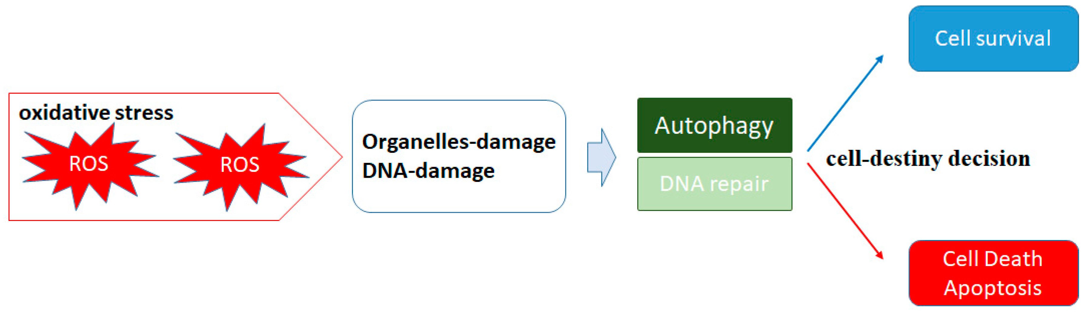
2. ROS and Autophagy

ROS are oxygen-containing molecules that are generated by redox reactions during cellular metabolism, which are comprised of radicals and nonradical derivatives including superoxide anion, hydroxyl radical, and hydrogen peroxide, largely derived from oxidative metabolism in physiological processes and/or inflammatory reactions [9]. During cells’ lifetime, various biological molecules are involved in signal transduction and/or energy metabolism. Additionally, ROS are formed as byproducts in those processes [10]. ROS were previously considered to function as molecules all harmful to cellular components. For example, high levels of ROS are known to cause DNA damages [2,11]. Subsequently, DNA damages could be repaired with a cell cycle arrest in affected cells. If the damage is effectively repaired, cells may continue to survive. However, if the damage is too severe to be repaired, cells may undergo apoptosis and/or cell death [12] (Figure 1). In this case, ROS consequently increases the expression of the proapoptotic proteins and caspases, leading to the activation of apoptosis [13]. However, accumulated superoxide anion molecules in cells would happen to be converted by superoxide dismutase (SOD) into hydrogen peroxide [14]. As the hydrogen peroxide is still toxic to cells, it is quickly converted into harmless water in the presence of catalase or glutathione peroxidase [15]. Many studies have accepted that ROS may also play a critical role in physiological processes. For example, different degrees of ROS would influence cell energetics and intracellular signaling pathways to regulate mRNA and protein expression, which determines the cell’s destiny, selecting either cell survival or cell death [16] (Figure 1).
One of the most important biological responses regulated by oxidative stress in cells is autophagy, which is an evolutionarily conserved process of degradation by lysosomal hydrolases [17]. Autophagy was firstly identified as a multistep catabolic process that promotes lysosome-mediated degradation of damaged cellular components [18], which is initiated by the formation and accumulation of autophagosomes [19]. Autophagy provides an intracellular recycling system that may provide cellular defense, by which autophagy may contribute to disease prevention and disorders management [20]. By the action of autophagy, the degraded components including free fatty acids and amino acids are recycled to synthesize new molecules. Autophagy even eliminates and recycles damaged organelles including peroxisomes, lysosomes, endoplasmic reticulum, mitochondria, and nucleus [21,22]. Therefore, autophagy is involved in the clearance and recycling of damaged organelles and aggregated proteins and lipids in order to keep cellular homeostasis [21,22]. High levels of ROS may induce autophagy-mediated cell protection [23]. Afterward, autophagy reduces ROS levels by eliminating damaged mitochondria, a process called mitophagy, which maintains a functional healthy mitochondrial group. Therefore, autophagy could eliminate ROS and ROS-damaged proteins, inhibiting cell death and enhancing cell survival [24]. Interaction between ROS and autophagy may protect cells from oxidative damage and/or cell death [25]. Autophagy generally provides a protective role, but on the other hand, autophagy is also closely associated with cell death, which has been characterized as programmed cell death, apoptosis, and/or necrosis [26] (Figure 1). A wide variety of internal and external cellular stresses would activate autophagy as an adaptive response to struggle with stresses, which could break the initial stress conditions later. As is well known, the common stimuli for autophagy are oxygen deprivation and nutrient scarcity [27], which may initially induce the production of ROS [28]. As an essential molecular step for autophagy induction, the activity of mammalian target of rapamycin (mTOR) downstream effector of the phosphoinositide 3-kinase (PI3K)/AKT signaling should be inhibited under nutrient starvation and/or oxygen deprivation [29,30]. Autophagy could also be stimulated by activated AMP-activated protein kinase (AMPK) during energy loss. The mTOR is a main suppressive component of autophagy signaling [31]. Therefore, mTOR inhibition has been shown a trigger during autophagic induction followed by ROS accumulation in cells [32]. Autophagy has also been reported to occur in the condition of mTOR deactivation due to extensive intracellular ROS production [32,33]. Of course, ROS induction could initiate autophagy even in tumor cells [34].

3. Hypoxia and ROS in Tumor Progression

Hypoxia plays an important role in the cells-microenvironment including several tumors [35]. During deprivation of sufficient oxygen supply, cells cannot keep adequate antioxidant capability resulting in increased ROS levels [36]. Therefore, cells must adapt to the consequences of reduced oxygen availability. Mitochondria may release superoxide in hypoxic conditions to the intermembrane space, where it is converted to hydrogen peroxide by SOD [37]. The hydrogen peroxide then enters the cytosol, where it may activate multiple responses including autophagy to cells [38]. Therefore, hypoxia is involved in the regulatory roles of autophagy [39]. As mentioned above, autophagy is a cell’s challenge to deal with oxidative stresses for cell survival, which may help both cancerous and normal cells to overcome necrosis and/or apoptosis by recycling damaged molecules or organelles. As the survival role of autophagy may depend on the repression of the necrosis and/or apoptosis pathways, the level of ROS is tightly controlled by inducible antioxidant signaling. For example, ROS has been shown to induce molecular markers of angiogenesis, such as hypoxia-inducible factor (HIF) and/or vascular endothelial growth factor (VEGF) [40,41]. Angiogenesis is often mediated by hypoxia that results in increased HIF1α and its transcriptional signaling target VEGF, which may eventually mediate transcriptional responses in both normal tissues and tumors. In the course of tumorigenesis, HIFs activate genes that induce tumor proliferation, invasion, and migration [42]. In fact, HIF2α promotes hypoxic cells proliferation by increasing cMyc proto-oncogene transcription [43]. In this regard, hypoxia might contribute to the maintenance of cancer stem cells [44] which are self-renewing tumor cells. In addition, ROS have been known to increase HIFs stability in inflammatory cells [45]. HIF1 activation mediates its nuclear translocation to activate the expression of HIF-dependent genes [46]. A key role of HIF1 is to increase the ability to activate signaling for promoting ATP production in hypoxic cells. Consequently, the HIF activation might prevent unnecessary ROS production in hypoxic cells [47]. Likewise, hypoxic environments seem to be supportive in various ways for maintaining all of normal, cancerous, and stem cells.
In response to hypoxia, cancer cells may activate numerous hypoxia-inducible genes and thus promote angiogenesis to enhance proliferation. Hence, tumor hypoxia is usually associated with poor patient prognosis [48]. For example, previous studies have shown a link between hypoxia and CA19-9 that is a poor prognostic marker in cancers [49]. In addition, hypoxia is a common feature of solid tumors, and it is defined as one of the most important causes for radiation-therapy failure [50]. Radiation therapy kills tumors by generating ROS since ROS are the effector molecules of the radiation contributing to radiation-induced DNA damages and cancer cell death [50]. Enhancing the radioresponsiveness of cancers should be essential for improving the prognosis. However, consequent damage to surrounding healthy normal cells by excessive radiation therapy could finally lead to the failure of the cancer treatment (Figure 2). In addition, hypoxia-stimulating HIF1 and VEGF expression has been shown to induce radiation therapy resistance [51]. Tumor hypoxia is consequently associated with therapy resistance and poor prognosis [52]. Remarkably, hypoxia also blocks HDAC inhibitor-induced differentiation of breast cancer cells, suggesting that tumor hypoxia should be considered as a theoretically important factor that may affect therapeutic efficiency with HDAC inhibitors [53] (Figure 2). On the other hand, hyperoxia may converse the hypoxia-induced radiation therapy-resistance in cancer [54] (Figure 3). Surprisingly, hyperoxia has enhanced radiosensitivity by decreasing the level of hypoxia-induced HIF1 and VEGF in HeLa cells [54]. Accordingly, hyperoxia could suppress the hypoxia-activated signaling pathways for proliferation in tumor cells. In general, high oxygen concentration has been considered a good radio-therapeutic sensitizer [55].

4. HDAC Inhibitors in Cancer Therapy

Histones are the main structural protein of eukaryotic chromatin, whose acetylation/ deacetylation is a ubiquitous epigenetic posttranslational modification [56]. These changes in DNA structure affect the action of transcription factors that could induce or repress gene transcription [56]. In transcriptionally active chromatin, histones are usually hyperacetylated, while hypoacetylated histones are equivalent to silenced chromatin [57]. Therefore, the histone acetyltransferases (HATs) catalyzing acetylation are related to activated gene transcription [58] (Figure 2). Conversely, the activity of HDACs is mainly involved in silencing gene expression [59]. Disturbance of the HDACs activity may cause abnormally increased transcription of key genes regulating cancer signaling pathways such as cell proliferation, migration, and/or invasion [60]. In addition, the activity and/or gene transcription of HDACs have been shown to be upregulated in response to hypoxia [61].
Cancer initiation and progression may be the result of genetic and/or epigenetic alterations [62]. Therefore, acetylation-mediated histone protein modification indeed plays an important role in epigenetic regulation of cancer development [63]. In fact, the increased expression and activation of HDACs are frequently found in cancers [64]. Moreover, an imbalance between the activities of HAT and HDAC enzymes is also associated with various forms of cancer [65]. Particularly, dysregulation of the HDACs would be critical to the development and/or progression of advanced tumors [66]. Accordingly, HDAC inhibitors could induce epigenetic changes in normal and cancerous cells, and their therapeutic potential for various cancers has been shown [67] (Figure 2). As shown in Figure 2, oxidative DNA damages could be prone to occur at transcribed genomic sites treated with HDAC inhibitor rather than nontranscribed genomic sites without HDAC inhibitor. This epigenetic modification induces ROS-effector-susceptible mechanisms eventually causing cell death in the HDAC inhibitor treatment. However, the vast cells death may bring about the success of cancer therapy but also cause significant damages to normal tissues. Therefore, combining mild HDACs inhibitors with other anticancer agents/radiation therapy might provide a rationale for the promising effective treatment of cancers [68]. These combinations might enhance the efficacy and reduce the toxicity or resistance to therapy (Figure 3). The major classes of HDAC inhibitors include short-chain fatty acids (SCFAs), hydroxamic acid derivatives, synthetic benzamide derivatives, and cyclic tetrapeptides [69]. Butyrate, one of the short-chain fatty acids produced by gut microbiota during anaerobic fermentation, could induce apoptosis in cancer cells and inhibit tumor progression via suppressing HDACs [70]. Butyrate did not increase radiation-induced cell death in normal cells after irradiation [71]. In addition, butyrate could enhance the efficacy of radiation therapy while protecting the normal mucosa, minimalizing the associated toxicity of the therapy [72]. Too much toxicity of cancer therapy due to the normal-cell-associated side effects may develop secondary malignancies, radiation-induced fibrosis, and infertility, actually limiting the safety of the treatment [67,73]. In addition, these factors significantly disturb the quality of life (QOL) of patients [74,75]. To find an appropriate (not too high but not too low) level of effectiveness in cancer therapy should be important and required for the good QOL of individuals.

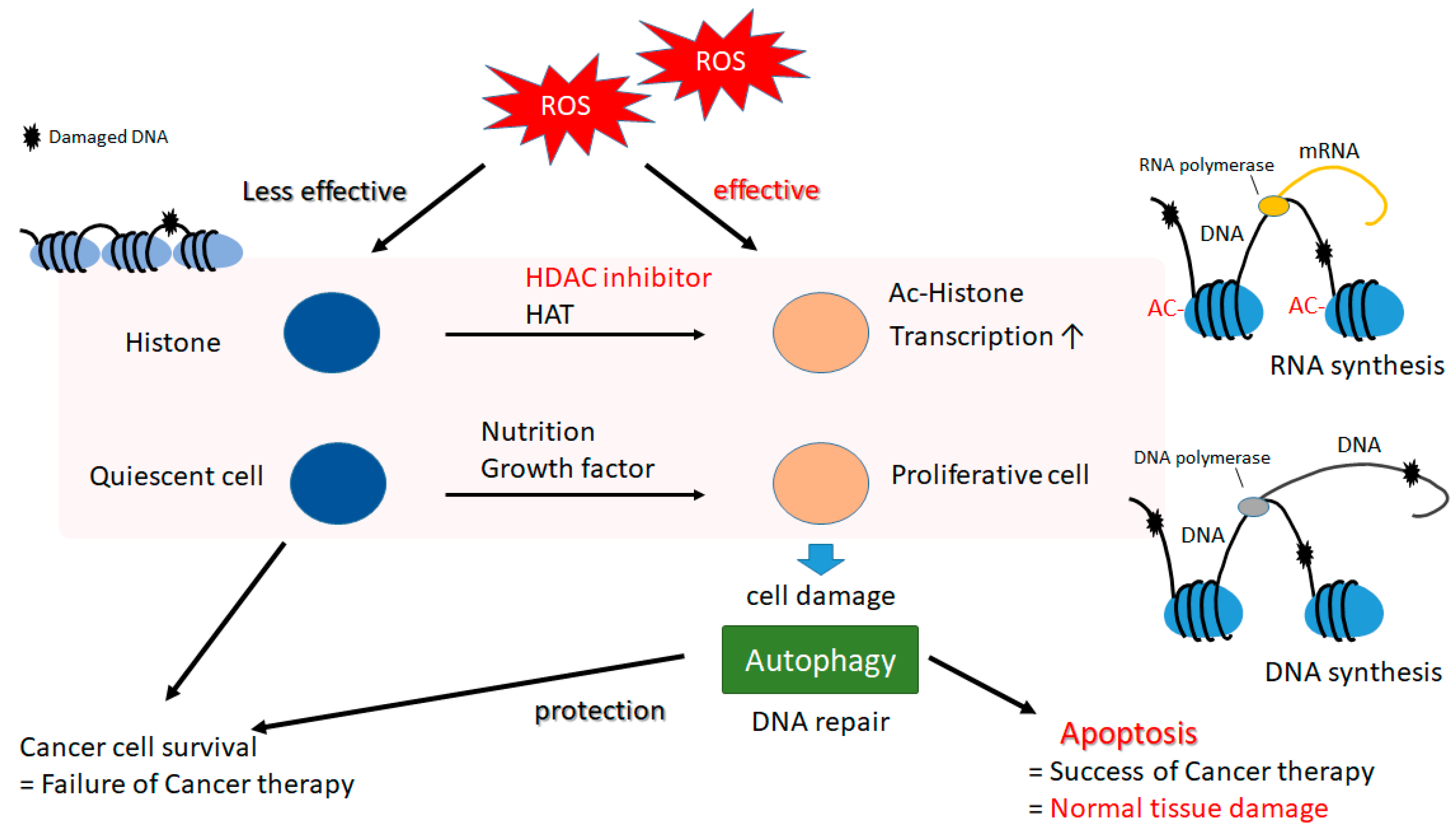
5. A Matter of Cell Life or Cell Death Could Be Determined by Autophagy in Both Cancerous and Normal Types of Cells

HDACs inhibitors work by promoting acetylation of histones in a cell, leading to activation of a variety of genes implicated in the regulation of cell survival, differentiation, proliferation, and cell death/apoptosis. The biological activity of these HDACs inhibitors also includes significant antifungal and/or antiviral applications [76]. Screening of natural sources has yielded new molecules that have been identified as potent HDAC inhibitors. In fact, natural compounds such as tetrapeptide, polyketides, terpenoids, and hydroxamic acid have been reported to show potential HDAC inhibitory activity [77]. Some of the HDAC inhibitors from the natural dietary origin are protocatechuic aldehyde, kaempferol, butein, resveratrol, diallyl disulfide, sinapinic acid, and zerumbone [77]. Epidemiological studies have suggested that vegetables, fruits, grains, and fatty acids may provide certain protection against some diseases including cancer without any detectable side effects [78] (Figure 3). In particular, HDAC inhibitors may exhibit their antitumor effects by the activation of ROS generation, autophagy, and mitotic cell death within cancer cells [79]. Paradoxically, HDAC-inhibitor-mediated autophagy has been attributed with a critical role in HDAC-inhibitor resistance [80]. If performed properly, however, treatment with mild HDAC inhibitors as the combination of radiation therapy/chemotherapy or inhibitors of autophagy would be established as an advanced therapeutic approach to resensitize cancer cells. (Figure 3). For satisfactory achievement, the definition of critical factors that can determine the direction of autophagy toward tumor cell death would be indispensable.
The tumor microenvironment is hypoxic and inflammatory with ROS, which is known to be favorable for the induction of autophagy [81]. In certain situations, however, autophagy could help induce both apoptosis and survival [82,83]. The outcome of this regulation is controversial, which could be correlated with different responses observed between normal and cancer cells. Cell-destiny decisions of whether to live or to die correlating with either health or disease might be regulated by an intricate system of balanced signaling pathways. (Figure 1). Cells need to cope with a multitude of variable stress stimuli, which may be eventually linked to either cytoprotection or cytotoxicity. Compared to normal cells, cancer cells may adapt faster to microenvironmental modifications [84]. Hypoxia activates additional pathways including autophagy, which might be a regulated program for cell-destiny decisions in the hypoxia-induced tumor response (Figure 1). In turn, autophagy might coordinate cancer cell plasticity for vitality and homeostasis.

6. Histone Modification in Cancer and Future Perspectives

A nucleosome is the basic repeating unit of the chromatin, whose structure is a histone fold domain with two tail domains. The tail domains are rich in lysine residues that are exposed to posttranslational modifications such as methylation, acetylation, and/or ubiquitination. Therefore, histone modifications are involved in the regulation of many biological processes such as transcription and DNA damage repair [85]. Cancer cells exhibit persistent replication stress during uncontrolled cell proliferation. This fundamental difference between cancerous and healthy normal cells makes replication stress a promising target for anticancer therapies. Since replication stress occurs in the context of chromatin or nucleosome, advances in the understanding of how histone dynamics are associated with replication stress could lead to novel cancer therapeutics. For example, chemotherapeutic resistance has also been considered a major challenge in cancer therapy that also affects HDAC inhibitors’ treatment [86]. Explanations could be found in cancer-cell-specific effects influenced by tumor-specific mutations and/or microenvironmental oxidative stress conditions. Exact mechanistic insights related to the effectors and resistance mechanisms of HDAC inhibitors supporting a reliable suggestion of the treatment outcome are still inadequate. Although autophagy is a potential target for cancer therapy, autophagy is associated with a confusing role in diverse stages of tumorigenesis. A better understanding of the roles of autophagy in the oncology field is particularly crucial. This information might allow the design of improved antitumor agents, which include the targeting of autophagy. It would be of major interest to improve current knowledge on the effects of targeting autophagy to impact antitumor therapies in clinical settings. Autophagy also plays a key role in other diseases including obesity and diabetes, cardiovascular disease, and bacterial infections [87]. For the treatment of those diseases, single-cell studies would be valuable in optimizing the treatment’s efficacy [88,89]. While comparable to apoptosis, autophagy is engaged to intricate signaling pathways even to cell survival. A focus of emerging studies should investigate the clarification of unexplained molecular mechanisms of HDAC inhibitors associated with intracellular ROS formation.

Author Contributions

Conceptualization, Y.I. and S.M.; writing—original draft preparation, Y.I., N.N., A.T., K.T., Y.K. and S.M.; writing—review and editing, Y.I., N.N., A.T., K.T., Y.K. and S.M.; visualization, Y.I., N.N., A.T., K.T., Y.K. and S.M.; supervision, S.M. All authors have read and agreed to the published version of the manuscript.

Funding

This research received no external funding.

Institutional Review Board Statement

Not applicable.

Informed Consent Statement

Not applicable.

Conflicts of Interest

The authors declare that they have no competing financial interest.

References

  1. Bernatoniene, J.; Kopustinskiene, D.M. The Role of Catechins in Cellular Responses to Oxidative Stress. Molecules 2018, 23, 965. [Google Scholar] [CrossRef] [Green Version]
  2. Son, J.M.; Lee, C. Mitochondria: Multifaceted regulators of aging. BMB Rep. 2019, 52, 13–23. [Google Scholar] [CrossRef] [PubMed] [Green Version]
  3. Pal, D.; Rai, A.; Checker, R.; Patwardhan, R.S.; Singh, B.; Sharma, D.; Sandur, S.K. Role of protein S-Glutathionylation in cancer progression and development of resistance to anti-cancer drugs. Arch. Biochem. Biophys. 2021, 704, 108890. [Google Scholar] [CrossRef] [PubMed]
  4. Adampourezare, M.; Dehghan, G.; Hasanzadeh, M.; Hosseinpoure Feizi, M.A. Application of lateral flow and microfluidic bio-assay and biosensing towards identification of DNA-methylation and cancer detection: Recent progress and challenges in biomedicine. Biomed. Pharmacother. 2021, 141, 111845. [Google Scholar] [CrossRef] [PubMed]
  5. Ebrahimi, S.; Hashemy, S.I. MicroRNA-mediated redox regulation modulates therapy resistance in cancer cells: Clinical perspectives. Cell. Oncol. 2019, 42, 131–141. [Google Scholar] [CrossRef] [PubMed]
  6. Brown, C.O.; Salem, K.; Wagner, B.A.; Bera, S.; Singh, N.; Tiwari, A.; Choudhury, A.; Buettner, G.R.; Goel, A. Interleukin-6 counteracts therapy-induced cellular oxidative stress in multiple myeloma by up-regulating manganese superoxide dismutase. Biochem. J. 2012, 444, 515–527. [Google Scholar] [CrossRef] [PubMed] [Green Version]
  7. Karakaidos, P.; Karagiannis, D.; Rampias, T. Resolving DNA Damage: Epigenetic Regulation of DNA Repair. Molecules 2020, 25, 2496. [Google Scholar] [CrossRef]
  8. Das, B.; Pal, B.; Bhuyan, R.; Li, H.; Sarma, A.; Gayan, S.; Talukdar, J.; Sandhya, S.; Bhuyan, S.; Gogoi, G.; et al. MYC Regulates the HIF2alpha Stemness Pathway via Nanog and Sox2 to Maintain Self-Renewal in Cancer Stem Cells versus Non-Stem Cancer Cells. Cancer Res. 2019, 79, 4015–4025. [Google Scholar] [CrossRef] [PubMed]
  9. Smallwood, M.J.; Nissim, A.; Knight, A.R.; Whiteman, M.; Haigh, R.; Winyard, P.G. Oxidative stress in autoimmune rheumatic diseases. Free Radic. Biol. Med. 2018, 125, 3–14. [Google Scholar] [CrossRef]
  10. He, L.; He, T.; Farrar, S.; Ji, L.; Liu, T.; Ma, X. Antioxidants Maintain Cellular Redox Homeostasis by Elimination of Reactive Oxygen Species. Cell. Physiol. Biochem. 2017, 44, 532–553. [Google Scholar] [CrossRef] [PubMed]
  11. Moloney, J.N.; Cotter, T.G. ROS signalling in the biology of cancer. Semin. Cell Dev. Biol. 2018, 80, 50–64. [Google Scholar] [CrossRef] [PubMed]
  12. Yao, Y.; Zang, Y.; Qu, J.; Tang, M.; Zhang, T. The Toxicity of Metallic Nanoparticles on Liver: The Subcellular Damages, Mechanisms, And Outcomes. Int. J. Nanomed. 2019, 14, 8787–8804. [Google Scholar] [CrossRef] [Green Version]
  13. Lieberman, J. Granzyme A activates another way to die. Immunol. Rev. 2010, 235, 93–104. [Google Scholar] [CrossRef] [Green Version]
  14. Miao, L.; St. Clair, D.K. Regulation of superoxide dismutase genes: Implications in disease. Free Radic. Biol. Med. 2009, 47, 344–356. [Google Scholar] [CrossRef] [Green Version]
  15. Cheng, C.C.; Sofiyatun, E.; Chen, W.J.; Wang, L.C. Life as a Vector of Dengue Virus: The Antioxidant Strategy of Mosquito Cells to Survive Viral Infection. Antioxidants 2021, 10, 395. [Google Scholar] [CrossRef]
  16. Meijles, D.N.; Zoumpoulidou, G.; Markou, T.; Rostron, K.A.; Patel, R.; Lay, K.; Handa, B.S.; Wong, B.; Sugden, P.H.; Clerk, A. The cardiomyocyte “redox rheostat”: Redox signalling via the AMPK-mTOR axis and regulation of gene and protein expression balancing survival and death. J. Mol. Cell. Cardiol. 2019, 129, 118–129. [Google Scholar] [CrossRef] [PubMed] [Green Version]
  17. Shpilka, T.; Welter, E.; Borovsky, N.; Amar, N.; Shimron, F.; Peleg, Y.; Elazar, Z. Fatty acid synthase is preferentially degraded by autophagy upon nitrogen starvation in yeast. Proc. Natl. Acad. Sci. USA 2015, 112, 1434–1439. [Google Scholar] [CrossRef] [Green Version]
  18. Che, J.; Wang, W.; Huang, Y.; Zhang, L.; Zhao, J.; Zhang, P.; Yuan, X. miR-20a inhibits hypoxia-induced autophagy by targeting ATG5/FIP200 in colorectal cancer. Mol. Carcinog. 2019, 58, 1234–1247. [Google Scholar] [CrossRef]
  19. Meliton, L.N.; Zhu, X.; Brown, M.; Epshtein, Y.; Kawasaki, T.; Letsiou, E.; Dudek, S.M. Degradation of group V secretory phospholipase A(2) in lung endothelium is mediated by autophagy. Microvasc. Res. 2020, 129, 103954. [Google Scholar] [CrossRef]
  20. Mei, Y.; Thompson, M.D.; Cohen, R.A.; Tong, X. Autophagy and oxidative stress in cardiovascular diseases. Biochim. Biophys. Acta 2015, 1852, 243–251. [Google Scholar] [CrossRef] [PubMed] [Green Version]
  21. Sica, V.; Galluzzi, L.; Bravo-San Pedro, J.M.; Izzo, V.; Maiuri, M.C.; Kroemer, G. Organelle-Specific Initiation of Autophagy. Mol. Cell 2015, 59, 522–539. [Google Scholar] [CrossRef] [PubMed] [Green Version]
  22. Farrugia, M.A.; Puglielli, L. Nepsilon-lysine acetylation in the endoplasmic reticulum—A novel cellular mechanism that regulates proteostasis and autophagy. J. Cell Sci. 2018, 131, jcs221747. [Google Scholar] [CrossRef] [PubMed] [Green Version]
  23. Lee, J.; Song, C.H. Effect of Reactive Oxygen Species on the Endoplasmic Reticulum and Mitochondria during Intracellular Pathogen Infection of Mammalian Cells. Antioxidants 2021, 10, 872. [Google Scholar] [CrossRef] [PubMed]
  24. Cao, S.; Tang, J.; Huang, Y.; Li, G.; Li, Z.; Cai, W.; Yuan, Y.; Liu, J.; Huang, X.; Zhang, H. The Road of Solid Tumor Survival: From Drug-Induced Endoplasmic Reticulum Stress to Drug Resistance. Front. Mol. Biosci. 2021, 8, 620514. [Google Scholar] [CrossRef] [PubMed]
  25. Yang, N.; Guan, Q.W.; Chen, F.H.; Xia, Q.X.; Yin, X.X.; Zhou, H.H.; Mao, X.Y. Antioxidants Targeting Mitochondrial Oxidative Stress: Promising Neuroprotectants for Epilepsy. Oxid. Med. Cell. Longev. 2020, 2020, 6687185. [Google Scholar] [CrossRef] [PubMed]
  26. Wang, K. Autophagy and apoptosis in liver injury. Cell Cycle 2015, 14, 1631–1642. [Google Scholar] [CrossRef] [Green Version]
  27. Packer, M. Role of Impaired Nutrient and Oxygen Deprivation Signaling and Deficient Autophagic Flux in Diabetic CKD Development: Implications for Understanding the Effects of Sodium-Glucose Cotransporter 2-Inhibitors. J. Am. Soc. Nephrol. 2020, 31, 907–919. [Google Scholar] [CrossRef]
  28. Song, S.B.; Hwang, E.S. High Levels of ROS Impair Lysosomal Acidity and Autophagy Flux in Glucose-Deprived Fibroblasts by Activating ATM and Erk Pathways. Biomolecules 2020, 10, 761. [Google Scholar] [CrossRef]
  29. Chen, A.; Xiong, L.J.; Tong, Y.; Mao, M. Neuroprotective effect of brain-derived neurotrophic factor mediated by autophagy through the PI3K/Akt/mTOR pathway. Mol. Med. Rep. 2013, 8, 1011–1016. [Google Scholar] [CrossRef] [Green Version]
  30. Wei, W.; Lu, M.; Lan, X.B.; Liu, N.; Su, W.K.; Dushkin, A.V.; Yu, J.Q. Neuroprotective Effects of Oxymatrine on PI3K/Akt/mTOR Pathway After Hypoxic-Ischemic Brain Damage in Neonatal Rats. Front. Pharmacol. 2021, 12, 642415. [Google Scholar] [CrossRef]
  31. Paquette, M.; El-Houjeiri, L.; Pause, A. mTOR Pathways in Cancer and Autophagy. Cancers 2018, 10, 18. [Google Scholar] [CrossRef] [Green Version]
  32. Gammoh, N.; Lam, D.; Puente, C.; Ganley, I.; Marks, P.A.; Jiang, X. Role of autophagy in histone deacetylase inhibitor-induced apoptotic and nonapoptotic cell death. Proc. Natl. Acad. Sci. USA 2012, 109, 6561–6565. [Google Scholar] [CrossRef] [PubMed] [Green Version]
  33. Yoshii, S.R.; Mizushima, N. Monitoring and Measuring Autophagy. Int. J. Mol. Sci. 2017, 18, 1865. [Google Scholar] [CrossRef] [PubMed]
  34. Scherz-Shouval, R.; Elazar, Z. Regulation of autophagy by ROS: Physiology and pathology. Trends Biochem. Sci. 2011, 36, 30–38. [Google Scholar] [CrossRef] [PubMed]
  35. Huang, Z.; Wang, S.; Zhang, H.J.; Zhou, Y.L.; Tang, X.; Shi, J.H. Characteristics of hypoxic tumor microenvironment in non-small cell lung cancer, involving molecular patterns and prognostic signature. Transl. Lung Cancer Res. 2021, 10, 2132–2147. [Google Scholar] [CrossRef]
  36. Yadav, A.K.; Yadav, P.K.; Chaudhary, G.R.; Tiwari, M.; Gupta, A.; Sharma, A.; Pandey, A.N.; Pandey, A.K.; Chaube, S.K. Autophagy in hypoxic ovary. Cell. Mol. Life Sci. 2019, 76, 3311–3322. [Google Scholar] [CrossRef]
  37. Gill, T.; Levine, A.D. Mitochondria-derived hydrogen peroxide selectively enhances T cell receptor-initiated signal transduction. J. Biol. Chem. 2013, 288, 26246–26255. [Google Scholar] [CrossRef] [PubMed] [Green Version]
  38. Levonen, A.L.; Hill, B.G.; Kansanen, E.; Zhang, J.; Darley-Usmar, V.M. Redox regulation of antioxidants, autophagy, and the response to stress: Implications for electrophile therapeutics. Free Radic. Biol. Med. 2014, 71, 196–207. [Google Scholar] [CrossRef] [Green Version]
  39. Sha, S.; Tan, J.; Miao, Y.; Zhang, Q. The Role of Autophagy in Hypoxia-Induced Neuroinflammation. DNA Cell Biol. 2021, 40, 733–739. [Google Scholar] [CrossRef]
  40. Engin, A. Adipose Tissue Hypoxia in Obesity and Its Impact on Preadipocytes and Macrophages: Hypoxia Hypothesis. Adv. Exp. Med. Biol. 2017, 960, 305–326. [Google Scholar] [PubMed]
  41. Vriend, J.; Reiter, R.J. Melatonin and the von Hippel-Lindau/HIF-1 oxygen sensing mechanism: A review. Biochim. Biophys. Acta 2016, 1865, 176–183. [Google Scholar] [CrossRef]
  42. Jing, Y.; Liu, L.Z.; Jiang, Y.; Zhu, Y.; Guo, N.L.; Barnett, J.; Rojanasakul, Y.; Agani, F.; Jiang, B.H. Cadmium increases HIF-1 and VEGF expression through RO.S.; ER.K.; and AKT signaling pathways and induces malignant transformation of human bronchial epithelial cells. Toxicol. Sci. 2012, 125, 10–19. [Google Scholar] [CrossRef]
  43. Feng, L.; Sun, C.; Sun, X.; Zhao, Y.; Yu, R.; Kang, C. Identification of inhibitors targeting HIF-2alpha/c-Myc by molecular docking and MM-GBSA technology. J. Recept. Signal Transduct. 2020, 1–9. [Google Scholar] [CrossRef] [PubMed]
  44. Tong, W.W.; Tong, G.H.; Liu, Y. Cancer stem cells and hypoxia-inducible factors (Review). Int. J. Oncol. 2018, 53, 469–476. [Google Scholar] [CrossRef] [PubMed] [Green Version]
  45. Chen, R.; Lai, U.H.; Zhu, L.; Singh, A.; Ahmed, M.; Forsyth, N.R. Reactive Oxygen Species Formation in the Brain at Different Oxygen Levels: The Role of Hypoxia Inducible Factors. Front. Cell Dev. Biol. 2018, 6, 132. [Google Scholar] [CrossRef] [Green Version]
  46. Lee, H.J.; Han, H.J. Role of Microtubule-Associated Factors in HIF1alpha Nuclear Translocation. Adv. Exp. Med. Biol. 2020, 1232, 271–276. [Google Scholar]
  47. Minet, E.; Michel, G.; Remacle, J.; Michiels, C. Role of HIF-1 as a transcription factor involved in embryonic development, cancer progression and apoptosis (review). Int. J. Mol. Med. 2000, 5, 253–259. [Google Scholar] [CrossRef] [PubMed]
  48. Skwarska, A.; Calder, E.D.D.; Sneddon, D.; Bolland, H.; Odyniec, M.L.; Mistry, I.N.; Martin, J.; Folkes, L.K.; Conway, S.J.; Hammond, E.M. Development and pre-clinical testing of a novel hypoxia-activated KDAC inhibitor. Cell Chem. Biol. 2021. [Google Scholar] [CrossRef] [PubMed]
  49. Ohta, S.; Morine, Y.; Imura, S.; Ikemoto, T.; Arakawa, Y.; Iwahashi, S.; Saito, Y.U.; Yamada, S.; Wada, Y.; Yamashita, S.; et al. Carbohydrate Antigen 19–9 Is a Prognostic Factor Which Correlates with HDAC1 and HIF-1alpha for Intrahepatic Cholangiocarcinoma. Anticancer Res. 2019, 39, 6025–6033. [Google Scholar] [CrossRef]
  50. Wang, H.; Jiang, H.; Van De Gucht, M.; De Ridder, M. Hypoxic Radioresistance: Can ROS Be the Key to Overcome It? Cancers 2019, 11, 112. [Google Scholar] [CrossRef] [Green Version]
  51. Fu, Z.; Chen, D.; Cheng, H.; Wang, F. Hypoxia-inducible factor-1alpha protects cervical carcinoma cells from apoptosis induced by radiation via modulation of vascular endothelial growth factor and p53 under hypoxia. Med. Sci. Monit. 2015, 21, 318–325. [Google Scholar]
  52. Ahmad, I.M.; Dafferner, A.J.; O’Connell, K.A.; Mehla, K.; Britigan, B.E.; Hollingsworth, M.A.; Abdalla, M.Y. Heme Oxygenase-1 Inhibition Potentiates the Effects of Nab-Paclitaxel-Gemcitabine and Modulates the Tumor Microenvironment in Pancreatic Ductal Adenocarcinoma. Cancers 2021, 13, 2264. [Google Scholar] [CrossRef]
  53. Kim, H.; Lin, Q.; Yun, Z. BRCA1 regulates the cancer stem cell fate of breast cancer cells in the context of hypoxia and histone deacetylase inhibitors. Sci. Rep. 2019, 9, 9702. [Google Scholar] [CrossRef] [PubMed] [Green Version]
  54. Dong, D.; Fu, Y.; Chen, F.; Zhang, J.; Jia, H.; Li, J.; Wang, H.; Wen, J. Hyperoxia sensitizes hypoxic HeLa cells to ionizing radiation by downregulating HIF-1alpha and VEGF expression. Mol. Med. Rep. 2021, 23, 62. [Google Scholar] [CrossRef]
  55. De Ridder, M.; Van Esch, G.; Engels, B.; Verovski, V.; Storme, G. Hypoxic tumor cell radiosensitization: Role of the iNOS/NO pathway. Bull. Cancer 2008, 95, 282–291. [Google Scholar]
  56. Asfaha, Y.; Schrenk, C.; Alves Avelar, L.A.; Hamacher, A.; Pflieger, M.; Kassack, M.U.; Kurz, T. Recent advances in class IIa histone deacetylases research. Bioorg. Med. Chem. 2019, 27, 115087. [Google Scholar] [CrossRef]
  57. Liu, L.K.; Gao, R.L.; Gao, Y.; Xu, J.Y.; Guo, L.M.; Wang, K.J.; Liu, H.P. A histone K-lysine acetyltransferase CqKAT2A-like gene promotes white spot syndrome virus infection by enhancing histone H3 acetylation in red claw crayfish Cherax quadricarinatus. Dev. Comp. Immunol. 2020, 107, 103640. [Google Scholar] [CrossRef] [PubMed]
  58. Wapenaar, H.; Dekker, F.J. Histone acetyltransferases: Challenges in targeting bi-substrate enzymes. Clin. Epigenetics 2016, 8, 59. [Google Scholar] [CrossRef] [PubMed] [Green Version]
  59. Verza, F.A.; Das, U.; Fachin, A.L.; Dimmock, J.R.; Marins, M. Roles of Histone Deacetylases and Inhibitors in Anticancer Therapy. Cancers 2020, 12, 1664. [Google Scholar] [CrossRef] [PubMed]
  60. Bian, X.; Liang, Z.; Feng, A.; Salgado, E.; Shim, H. HDAC inhibitor suppresses proliferation and invasion of breast cancer cells through regulation of miR-200c targeting CRK.L. Biochem. Pharmacol. 2018, 147, 30–37. [Google Scholar] [CrossRef]
  61. Ellis, L.; Hammers, H.; Pili, R. Targeting tumor angiogenesis with histone deacetylase inhibitors. Cancer Lett. 2009, 280, 145–153. [Google Scholar] [CrossRef] [PubMed] [Green Version]
  62. Gagliano, T.; Brancolini, C. Epigenetic Mechanisms beyond Tumour-Stroma Crosstalk. Cancers 2021, 13, 914. [Google Scholar] [CrossRef]
  63. Markouli, M.; Strepkos, D.; Basdra, E.K.; Papavassiliou, A.G.; Piperi, C. Prominent Role of Histone Modifications in the Regulation of Tumor Metastasis. Int. J. Mol. Sci. 2021, 22, 2778. [Google Scholar] [CrossRef]
  64. Garcia, T.B.; Uluisik, R.C.; van Linden, A.A.; Jones, K.L.; Venkataraman, S.; Vibhakar, R.; Porter, C.C. Increased HDAC Activity and c-MYC Expression Mediate Acquired Resistance to WEE1 Inhibition in Acute Leukemia. Front. Oncol. 2020, 10, 296. [Google Scholar] [CrossRef] [PubMed]
  65. Myzak, M.C.; Ho, E.; Dashwood, R.H. Dietary agents as histone deacetylase inhibitors. Mol. Carcinog. 2006, 45, 443–446. [Google Scholar] [CrossRef] [Green Version]
  66. Ellis, L.; Pili, R. Histone Deacetylase Inhibitors: Advancing Therapeutic Strategies in Hematological and Solid Malignancies. Pharmaceuticals 2010, 3, 2411–2469. [Google Scholar] [CrossRef]
  67. Hontecillas-Prieto, L.; Flores-Campos, R.; Silver, A.; de Álava, E.; Hajji, N.; García-Domínguez, D.J. Synergistic Enhancement of Cancer Therapy Using HDAC Inhibitors: Opportunity for Clinical Trials. Front. Genet. 2020, 11, 578011. [Google Scholar] [CrossRef] [PubMed]
  68. Booth, L.; Roberts, J.L.; Sander, C.; Lee, J.; Kirkwood, J.M.; Poklepovic, A.; Dent, P. The HDAC inhibitor AR42 interacts with pazopanib to kill trametinib/dabrafenib-resistant melanoma cells in vitro and in vivo. Oncotarget 2017, 8, 16367–16386. [Google Scholar] [CrossRef] [Green Version]
  69. Rosato, R.R.; Grant, S. Histone deacetylase inhibitors in cancer therapy. Cancer Biol. Ther. 2003, 2, 30–37. [Google Scholar] [CrossRef] [Green Version]
  70. Pant, K.; Yadav, A.K.; Gupta, P.; Islam, R.; Saraya, A.; Venugopal, S.K. Butyrate induces ROS-mediated apoptosis by modulating miR-22/SIRT-1 pathway in hepatic cancer cells. Redox. Biol. 2017, 12, 340–349. [Google Scholar] [CrossRef]
  71. Perona, M.; Thomasz, L.; Rossich, L.; Rodriguez, C.; Pisarev, M.A.; Rosemblit, C.; Cremaschi, G.A.; Dagrosa, M.A.; Juvenal, G.J. Radiosensitivity enhancement of human thyroid carcinoma cells by the inhibitors of histone deacetylase sodium butyrate and valproic acid. Mol. Cell. Endocrinol. 2018, 478, 141–150. [Google Scholar] [CrossRef] [PubMed]
  72. Park, M.; Kwon, J.; Shin, H.J.; Moon, S.M.; Kim, S.B.; Shin, U.S.; Han, Y.H.; Kim, Y. Butyrate enhances the efficacy of radiotherapy via FOXO3A in colorectal cancer patient-derived organoids. Int. J. Oncol. 2020, 57, 1307–1318. [Google Scholar] [CrossRef]
  73. Hanania, A.N.; Mainwaring, W.; Ghebre, Y.T.; Hanania, N.A.; Ludwig, M. Radiation-Induced Lung Injury: Assessment and Management. Chest 2019, 156, 150–162. [Google Scholar] [CrossRef]
  74. Griffin, M.F.; Drago, J.; Almadori, A.; Kalavrezos, N.; Butler, P.E. Evaluation of the efficacy of lipotransfer to manage radiation-induced fibrosis and volume defects in head and neck oncology. Head Neck 2019, 41, 3647–3655. [Google Scholar] [CrossRef]
  75. Mahmood, J.; Connors, C.Q.; Alexander, A.A.; Pavlovic, R.; Samanta, S.; Soman, S.; Matsui, H.; Sopko, N.A.; Bivalacqua, T.J.; Weinreich, D.; et al. Cavernous Nerve Injury by Radiation Therapy May Potentiate Erectile Dysfunction in Rats. Int. J. Radiat. Oncol. Biol. Phys. 2017, 99, 680–688. [Google Scholar] [CrossRef] [PubMed]
  76. Thipparapu, G.; Ajumeera, R.; Venkatesan, V. Novel dihydropyrimidine derivatives as potential HDAC inhibitors: In silico study. Silico Pharmacol. 2017, 5, 10. [Google Scholar] [CrossRef]
  77. Singh, A.K.; Bishayee, A.; Pandey, A.K. Targeting Histone Deacetylases with Natural and Synthetic Agents: An Emerging Anticancer Strategy. Nutrients 2018, 10, 731. [Google Scholar] [CrossRef] [PubMed] [Green Version]
  78. Ivey, K.L.; Jensen, M.K.; Hodgson, J.M.; Eliassen, A.H.; Cassidy, A.; Rimm, E.B. Association of flavonoid-rich foods and flavonoids with risk of all-cause mortality. Br. J. Nutr. 2017, 117, 1470–1477. [Google Scholar] [CrossRef] [Green Version]
  79. Hui, K.F.; Yeung, P.L.; Chiang, A.K. Induction of MAPK- and ROS-dependent autophagy and apoptosis in gastric carcinoma by combination of romidepsin and bortezomib. Oncotarget 2016, 7, 4454–4467. [Google Scholar] [CrossRef] [PubMed] [Green Version]
  80. Dahabieh, M.S.; Huang, F.; Goncalves, C.; Flores González, R.E.; Prabhu, S.; Bolt, A.; Di Pietro, E.; Khoury, E.; Heath, J.; Xu, Z.Y.; et al. Silencing PEX26 as an unconventional mode to kill drug-resistant cancer cells and forestall drug resistance. Autophagy 2021, 1–19. [Google Scholar] [CrossRef]
  81. Medvedev, R.; Ploen, D.; Spengler, C.; Elgner, F.; Ren, H.; Bunten, S.; Hildt, E. HCV-induced oxidative stress by inhibition of Nrf2 triggers autophagy and favors release of viral particles. Free Radic. Biol. Med. 2017, 110, 300–315. [Google Scholar] [CrossRef]
  82. Chen, N.; Debnath, J. IkappaB kinase complex (IKK) triggers detachment-induced autophagy in mammary epithelial cells independently of the PI3K-AKT-MTORC1 pathway. Autophagy 2013, 9, 1214–1227. [Google Scholar] [CrossRef] [Green Version]
  83. Yang, M.; Feng, C.; Zhang, Y.; Liu, C.; Li, B.; Zhu, Q.; Huang, B.; Zhou, Y. Autophagy protects nucleus pulposus cells from cyclic mechanical tension-induced apoptosis. Int. J. Mol. Med. 2019, 44, 750–758. [Google Scholar] [CrossRef] [PubMed]
  84. Sobierajska, K.; Wieczorek, K.; Ciszewski, W.M.; Sacewicz-Hofman, I.; Wawro, M.E.; Wiktorska, M.; Boncela, J.; Papiewska-Pajak, I.; Kwasniak, P.; Wyroba, E.; et al. beta-III tubulin modulates the behavior of Snail overexpressed during the epithelial-to-mesenchymal transition in colon cancer cells. Biochim. Biophys. Acta 2016, 1863, 2221–2233. [Google Scholar] [CrossRef] [PubMed]
  85. Zhou, K.; Gaullier, G.; Luger, K. Nucleosome structure and dynamics are coming of age. Nat. Struct. Mol. Biol. 2019, 26, 3–13. [Google Scholar] [CrossRef] [PubMed]
  86. Kim, H.; Kim, S.N.; Park, Y.S.; Kim, N.H.; Han, J.W.; Lee, H.Y.; Kim, Y.K. HDAC inhibitors downregulate MRP2 expression in multidrug resistant cancer cells: Implication for chemosensitization. Int. J. Oncol. 2011, 38, 807–812. [Google Scholar] [CrossRef]
  87. McClung, J.M.; McCord, T.J.; Ryan, T.E.; Schmidt, C.A.; Green, T.D.; Southerland, K.W.; Reinardy, J.L.; Mueller, S.B.; Venkatraman, T.N.; Lascola, C.D.; et al. BAG3 (Bcl-2-Associated Athanogene-3) Coding Variant in Mice Determines Susceptibility to Ischemic Limb Muscle Myopathy by Directing Autophagy. Circulation 2017, 136, 281–296. [Google Scholar] [CrossRef]
  88. Fei, Q.; Ma, H.; Zou, J.; Wang, W.; Zhu, L.; Deng, H.; Meng, M.; Tan, S.; Zhang, H.; Xiao, X.; et al. Metformin protects against ischaemic myocardial injury by alleviating autophagy-ROS-NLRP3-mediated inflammatory response in macrophages. J. Mol. Cell. Cardiol. 2020, 145, 1–13. [Google Scholar] [CrossRef]
  89. Davidson, S.M.; Padró, T.; Bollini, S.; Vilahur, G.; Duncker, D.J.; Evans, P.C.; Guzik, T.; Hoefer, I.E.; Waltenberger, J.; Wojta, J.; et al. Progress in cardiac research—From rebooting cardiac regeneration to a complete cell atlas of the heart. Cardiovasc. Res. 2021, cvab200. [Google Scholar] [CrossRef]
Figure 1. Schematic diagram of cell-destiny decision after oxidative stresses. Reactive oxygen species (ROS) may cause DNA damages or organelles damage in a cell. The damages could be treated with autophagy or DNA repair systems to keep cellular homeostasis. If the damage is effectively repaired, cells may continue to survive. However, if the damage is too severe to be repaired, cells may undergo cell death.
Oxygen 01 00004 g001
Figure 2. Overview about effector mechanisms of histone deacetylase (HDAC) inhibitor in cancer therapy. HDAC and histone acetyltransferase (HAT) are enzymes that influence transcription by selectively acetylating (Ac-) or deacetylating histone proteins. HDAC inhibitors acetylate histones in normal and tumor cells to the same extent. Oxidative DNA damages could be prone to occur at a transcribed or replicative genomic sites (treated with HDAC inhibitor or with nutrition/growth factor rich condition, respectively) rather than nontranscribed or nonreplicative genomic sites. This modification induces ROS-effector-susceptible mechanisms including oxidative DNA damages eventually causing cell death, preferentially occurring in the HDAC inhibitor and/or nutrition-rich conditions. A great amount of cells’ death may lead to the success of cancer therapy but also cause normal tissue damages. Note that some critical pathways have been omitted for clarity.
Oxygen 01 00004 g002
Figure 3. Implication of hypothetical therapeutic strategy using mild HDAC inhibitors. Some HDAC inhibitors may represent a class of mild edible anticancer therapeutics. Using these HDAC inhibitors with the situation of low nutrition, low growth factors, or hyperoxia, cancer cells could be most sensitive to chemo- and radiation therapy, which might lead to successful cancer therapy with low damage of normal tissues.
Oxygen 01 00004 g003
Publisher’s Note: MDPI stays neutral with regard to jurisdictional claims in published maps and institutional affiliations.

