Article

Genotyping and Antimicrobial Susceptibility of Clostridium perfringens and Clostridioides difficile in Camel Minced Meat

by
Mahmoud Fayez
1,2,†,
Waleed R. El-Ghareeb
3,
Ahmed Elmoslemany
4,
Saleem J. Alsunaini
1,
Mohamed Alkafafy
5,
Othman M. Alzahrani
6,
Samy F. Mahmoud
5 and
Ibrahim Elsohaby
7,8,9,*,†
1
Al-Ahsa Veterinary Diagnostic Lab, Ministry of Environment, Water and Agriculture, Al-Ahsa 31982, Saudi Arabia
2
Department of Bacteriology, Veterinary Serum and Vaccine Research Institute, Ministry of Agriculture, Cairo 11381, Egypt
3
Department of Veterinary Public Health and Animal Husbandry, College of Veterinary Medicine, King Faisal University, P.O. Box 400, Al-Ahsa 31982, Saudi Arabia
4
Hygiene and Preventive Medicine Department, Faculty of Veterinary Medicine, Kafrelsheikh University, Kafr El-Sheikh 33516, Egypt
5
Department of Biotechnology, College of Science, Taif University, P.O. Box 11099, Taif 21944, Saudi Arabia
6
Department of Biology, College of Science, Taif University, P.O. Box 11099, Taif 21944, Saudi Arabia
7
Department of Animal Medicine, Faculty of Veterinary Medicine, Zagazig University, Zagazig 44511, Egypt
8
Department of Health Management, Atlantic Veterinary College, University of Prince Edward Island, Charlottetown, PE C1A 4P3, Canada
9
Department of Infectious Diseases and Public Health, Jockey Club of Veterinary Medicine and Life Sciences, City University of Hong Kong, Kowloon, Hong Kong
*
Author to whom correspondence should be addressed.
These authors contributed equally to this work.
Submission received: 13 November 2021 / Revised: 11 December 2021 / Accepted: 17 December 2021 / Published: 19 December 2021

Abstract

:
The present study aimed to determine the occurrence, genotypes, and antimicrobial resistance of Clostridium perfringens (C. perfringens) and Clostridioides difficile (C. difficile) in camel minced meat samples collected from small butcher shops and supermarkets in Al-Ahsa Governorate, Saudi Arabia. A total of 100 camel minced meat samples were randomly collected from small butcher’s shops (n = 50) and supermarkets (n = 50) in Al-Ahsa Governorate, Saudi Arabia. C. perfringens and C. difficile were isolated and identified using the VITEK-2 compact system and 16S rRNA gene amplification. Genotypes, toxin genes, and antimicrobial susceptibility of the isolates were determined. Moreover, ELISA was used to detect C. perfringens and C. difficile toxins. C. perfringens and C. difficile were isolated from 14% and 4% of the tested minced meat samples, respectively. Out of the 14 C. perfringens isolates, type A (64.3%), type B (7.1%), type C (21.5%), and type D (7.1%) were detected. However, out of the four C. difficile isolates, three (75%) were type A+B+ and one (25%) was type AB+. None of the C. perfringens or C. difficile toxins were identified using ELISA. C. perfringens and C. difficile isolates exhibited a high rate of resistance to tetracycline (56% and 75%, respectively). However, all isolates were susceptible to amoxicillin-clavulanate. Multidrug resistance was observed in three (21.4%) C. perfringens and one (25%) C. difficile isolates. In conclusion, camel minced meat was contaminated with C. perfringens and C. difficile, which present a potential risk of food poisoning. The majority of the isolates were resistant to at least one antimicrobial, and some isolates were multidrug-resistant. Therefore, food safety standards and frequent inspections of abattoirs, small butcher shops, and supermarkets should be enforced.

1. Introduction

Food hygiene is described as all the conditions and measures necessary to ensure that food is safe and fit for human consumption during production, processing, storage, distribution, and preparation [1]. Foodborne diseases are caused by food contamination and can occur at any stage of the food production, delivery, and consumption chain. There are over 200 foodborne hazards, including microbiological hazards such as bacteria, viruses, parasites, and chemical contaminants, that arise naturally or due to pollution, food processing, packaging, transportation, or storage [2]. Foodborne illnesses and outbreaks are among the leading causes of death globally [3,4]. According to studies, the magnitude of foodborne illnesses in 2010 included 600 million illnesses and 420,000 deaths worldwide [5,6]. Nausea, vomiting, retching, diarrhea, abdominal discomfort, prostration, abdominal cramps, fever, chills, headache, and arthralgia are just a few of the symptoms [7].
Clostridia spp. are anaerobic Gram-positive, spore-forming and non-motile bacteria commonly found in the intestinal tract of humans and animals and the soil [8]. Clostridium perfringens (C. perfringens) is the most widespread species and among the most common foodborne pathogens in industrial countries. There are several toxigenic types of C. perfringens, including A, B, C, D, and E. However, type A is primarily associated with foodborne illness [9]. C. perfringens food poisoning can occur when cooked meat is not adequately heated or refrigerated before serving. Illness often arises 8–15 h after consuming contaminated food. The symptoms include strong abdominal cramps, gas, and diarrhea (nausea and infrequent vomiting) [10]. Clostridioides difficile (C. difficile) is also associated with food poisoning, with symptoms ranging from mild diarrhea to life-threatening pseudomembranous colitis [11,12].
The recent rise and re-emergence of antimicrobial-resistant foodborne bacteria necessitate coordinated efforts, particularly in developing countries [13]. Antibacterial resistance can spread through the food chain, either directly or indirectly. Direct exposure happens when a human comes into contact with an animal or its blood, saliva, milk, sperm, feces, or urine. Indirect contact occurs, following consumption of infected food products such as eggs, meat, and dairy products [14,15].
Camel is a versatile animal that is used for milk, meat, wool, transportation, racing, tourism, agricultural work, and beauty contests. Camel meat is a major source of animal protein in many African and Asian countries, particularly in locations where the environment makes it difficult for other animals to produce. Camel meat is healthier than other meat animals since the carcass has less fat and has lower cholesterol levels in the fat. Camel meat also has a higher proportion of polyunsaturated fatty acids than beef [16,17].
In Saudi Arabia, a number of surveys have investigated the prevalence of camel meat contamination with some aerobic bacteria such as Staphylococcus aureus, E. coli, and Salmonella [18,19,20,21]. To the best of the authors’ knowledge, no literature exists that investigates the anaerobic contamination of camel meat with bacteria. Therefore, the main goal of this study is to determine the occurrence, genotypes and antimicrobial resistance of C. perfringens and C. difficile in camel minced meat samples collected from small butcher shops and supermarkets in Al-Ahsa Governorate, Saudi Arabia.

2. Results

2.1. Isolation and Identification of C. perfringens and C. difficile

Out of the 100 minced meat samples tested in the present study, 14% and 4% were contaminated with C. perfringens and C. difficile, respectively. More than 70% of the C. perfringens and C. difficile isolates were recovered from samples collected from butcher shops (Table 1).
The number of C. perfringens in positive samples ranged from 200 to 2 × 10³ CFU/g. Based on the 16S rRNA sequence analysis, the 14 C. perfringens and four C. difficile isolates were clustered with the reference C. perfringens (NR 121697, NR 113204, and NR 112169) and C. difficile (NR 112172), respectively, with a similarity level of 100% (Figure 1).

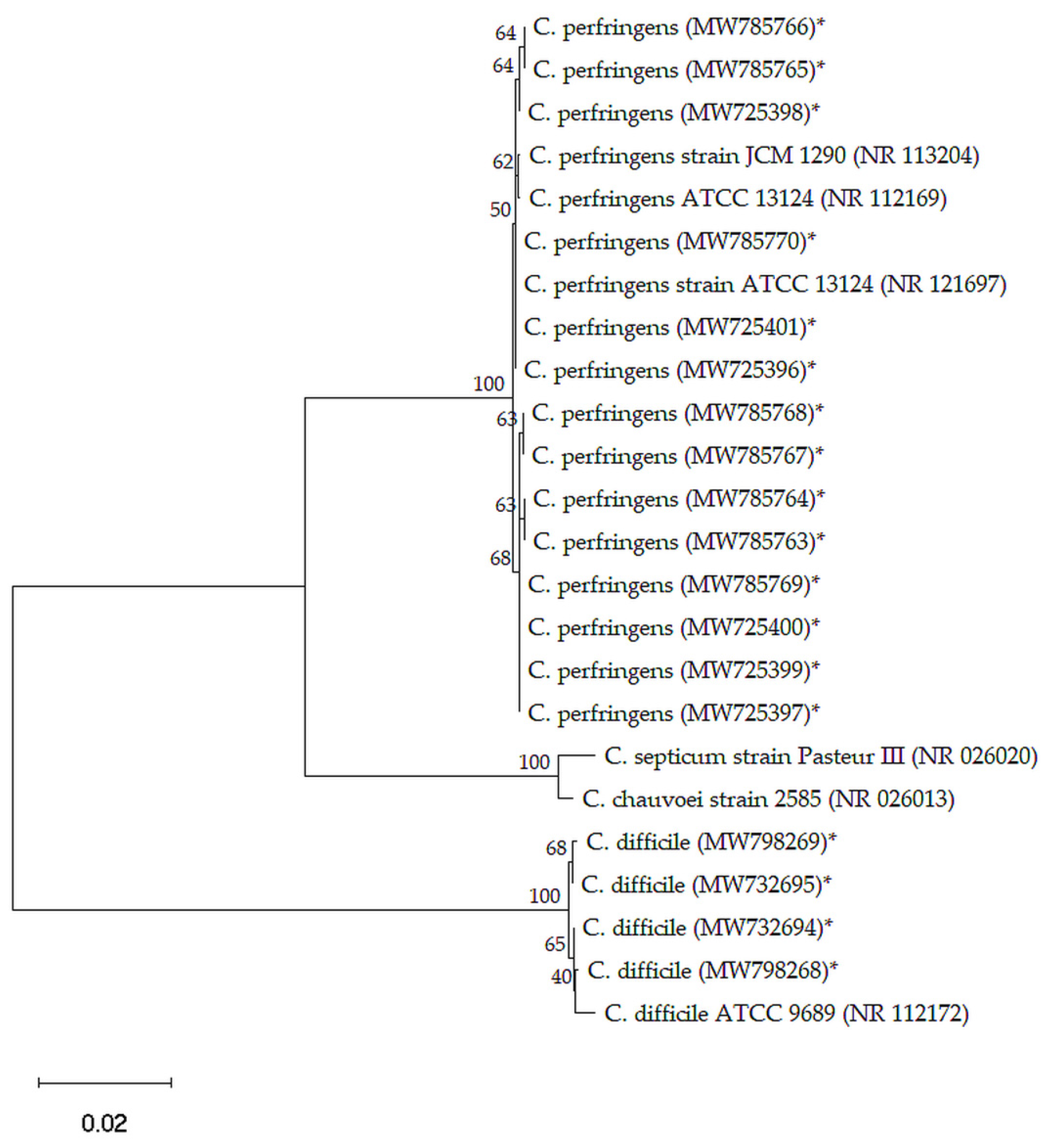
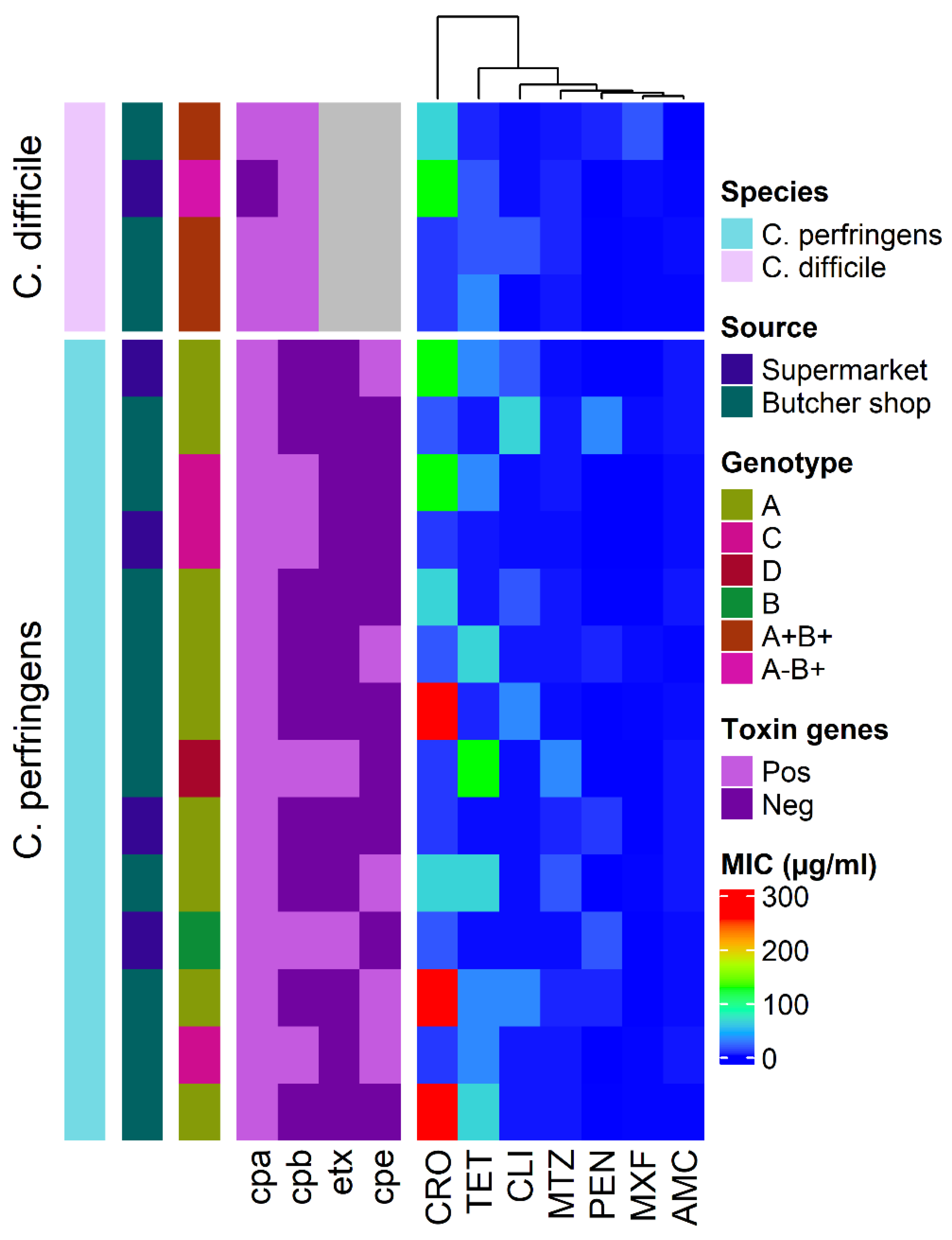
2.2. Genotyping of C. perfringens and C. difficile Isolates

PCR genotyping revealed that C. perfringens isolated from camel minced meat was related to type A, B, C, and D, whereas C. difficile isolates were related to type A+B+ and AB+ (Figure 2).
Table 2 shows the distribution of C. perfringens and C. difficile types and toxin genes in camel minced meat samples. Out of the 14 C. perfringens isolates, nine (64.3%) were type A, one (7.1%) was type B, three (21.5%) were type C, and one (7.1%) was type D. C. perfringens type A with only cpa+ gene was found in five (55.6%) isolates and cpa+ associated cpe+ genes were found in four (44.4%) isolates. However, out of the four C. difficile isolates, three (75%) were type A+B+ and one (25%) was type AB+.

2.3. Detection of C. perfringens and C. difficile Toxin by ELISA

None of the C. perfringens or C. difficile toxins were detected in the supernatant of homogenized meat samples using ELISA. The alpha toxin of C. perfringens was detected by ELISA in culture supernatants from all C. perfringens isolates, but the beta and epsilon toxins were detected in culture supernatants from four and one C. perfringens isolates, respectively. C. difficile alpha and beta toxins were detected in the culture supernatant of three isolates, but only beta toxin was detected in one isolate’s culture supernatant. Enterotoxin was found in the culture supernatant of four C. perfringens types A and one C. perfringens type C.

2.4. Antimicrobial Susceptibility

The distribution of minimum inhibitory concentration (MIC) values of different antimicrobials against the 14 C. perfringens and four C. difficile isolates recovered from camel minced meat is shown in Figure 2. C. perfringens and C. difficile isolates had a high frequency of tetracycline (TET) resistance (56% and 75%, respectively), followed by ceftriaxone (CRO) resistance (50% and 50%, respectively). All C. perfringens isolates were amoxicillin-clavulanate (AMC) and moxifloxacin (MXF) sensitive; however, all C. difficile isolates were only AMC sensitive. The frequency of antimicrobial resistance of C. perfringens and C. difficile genotypes isolated from camel minced meat is shown in Figure 3. Multidrug resistance was observed in three (21.4%) C. perfringens and one (25%) C. difficile isolates. The mean multiple antibiotic resistance (MAR) index for resistant C. perfringens isolates was 0.30 (range 0.14–0.57), and C. difficile isolates was 0.29 (range 0.14–0.43) (Table 3).

3. Discussion

Foodborne pathogens cause a wide range of diseases, with serious consequences for human health and the economy. C. perfringens is one of the most common foodborne pathogens that contaminate many types of retail meat products and cause food poisoning in humans and domestic animals [22]. Several studies have investigated the occurrence and genotypes of C. perfringens in retail food, including chicken [23], beef [24], and sheep [25] meats. However, few studies have determined C. perfringens genotypes and antimicrobial resistance in camel meat [26,27].
In the present study, C. perfringens was isolated from 14% of the tested minced meat samples. This finding is consistent with previous studies that reported the occurrence of C. perfringens in 33.7% [27] and 2.7% [26] of tested camel meat samples and swabs, respectively. On the other hand, previous studies have reported the occurrence of C. perfringens in 18% of tested minced meat in Turkey [28]; 21.2% of beef slaughtering and butchering processes in China [29]; 44.3% of goat, sheep, and cattle meat in Pakistan [30]; and 23.5% of raw processed meat in South Africa [31].
Clostridium perfringens types A, B, C, and D have been isolated in this study and type A (64.3%) was the most prevalent, followed by type C (21.5%). Several studies that investigated the distribution of C. perfringens types in retail food samples have reported that C. perfringens type A was the most common among the types recovered from camel [26,27], beef [24] and sheep [25] meats. However, a recent study in Korea detected only C. perfringens type A (33%) in tested chicken and beef meat samples [32]. In another study conducted in sheep meat, C. perfringens type A was not detected, and types B, C, and D were isolated [33]. All types of C. perfringens are commensal in the gastrointestinal tract of animals, therefore, contamination during slaughter and butchering could be the primary source of C. perfringens in meats [34,35].
Toxin genes (cpa, cpb, cpe, and etx) were found on the C. perfringens isolates recovered in our study. Although all C. perfringens isolates were found to be associated with cpa+ gene, only five (35.7%) isolates were associated with the enterotoxins (cpe+) gene, which is responsible for nearly all C. perfringens food poisoning outbreaks [25,36,37]. Similarly, previous studies found that all C. perfringens isolates from meat samples were positive for cap+ gene [38,39], suggesting that it might be a universal gene in C. perfringens isolated from meat samples [40]. Moreover, the cpe+ gene has been detected in 1.4% [25], 5% [41], and 27.2% [42] of C. perfringens isolated from raw and processed meats. In contrast, a recent study did not find cpe+ gene in any of the C. perfringens isolates recovered from meat samples in Korea [32].
Clostridium difficile infections have been increased globally in the last two decades, causing severe intestinal infections in humans [43,44]. Thus, the occurrence of C. difficile has been detected in 4% of camel minced meat used in this study, and all isolates were identified as toxigenic. This finding is consistent with previous studies that isolated C. difficile from 1.9% of ground beef samples in France [45], 5% of ground beef and hamburger samples in Sweden [46], and 6.3% of lamb meat samples in the Netherlands [47]. However, C. difficile was not detected in ground beef in Switzerland [48] and Austria [49]. Food animals are known carriers of C. difficile [50], and multiple reports show C. difficile shedding in animals at slaughter [51,52]. Contamination of meats may occur due to gut content leakage during evisceration or due to the accumulation of spores within the slaughterhouse environment [53]. The discovery of genetically identical C. difficile strains in food, livestock, and humans has raised awareness for C. difficile potential as an unspecific foodborne agent [54,55].
ELISA can detect C. perfringens and C. difficile toxins. In the current investigation, neither C. perfringens nor C. difficile toxins were found in camel minced meat. In comparison, C. perfringens toxins were found in 13% of minced meat samples in Turkey [28]. Although C. perfringens and C. difficile were isolated from some samples, they were toxin negative, which could be related to the fact that some clostridia are incapable of making toxins [25] or the toxin concentration in the samples may be below the ELISA detection limit (5 ng/mL).
In Saudi Arabia, antimicrobials have been used in livestock to promote growth and prevent several infectious diseases. However, their use has paradoxically increased the bacterial resistance to antimicrobials [56,57]. In this study, C. perfringens and C. difficile isolates exhibited a high resistance rate to TET (56% and 75%, respectively). However, all isolates were susceptible to AMC. Similarly, previous reports have shown that C. perfringens and C. difficile isolates are highly resistant to TET [39,57,58] and are susceptible to beta-lactams [58,59]. In South Korea, more than 93% of C. perfringens isolates from beef, poultry, duck, and pork meats were tetracycline resistant [60]. Furthermore, many investigations have found that beta-lactam antibiotics, such as ampicillin, can inhibit C. perfringens isolated from beef, poultry, and pork meats from the United States, Belgium, Scandinavia, and India [61,62].
Multidrug resistance has been reported in C. perfringens [62,63] and C. difficile [64,65] isolates from retail meats. In the present study, 21.4% of C. perfringens and 25% of C. difficile isolates from camel minced meat showed multidrug resistance, higher than the prevalence of multidrug-resistant observed in other studies [11,32,61]. Overuse of antimicrobials (over-the-counter antibiotics without a prescription), use of TET as a growth promoter, and extensive international travel to Saudi Arabia [66] are all possible explanations for the high resistance rate found in this study. In addition, the prevalence and/or emergence of multidrug-resistant C. perfringens has been increasing and poses a tremendous public health concern [67]. A recent study has reported that multidrug resistance bacteria in retail meats originates primarily in veterinary healthcare settings or on farms where animals are administered antibiotics in their feeds or treat diseases [68].
The limitation of this study is the small number of minced meat samples collected. However, the collected samples represent only one district and do not represent different regions of Saudi Arabia. Despite this limitation, the study identified that camel meat was contaminated with C. perfringens and C. difficile and further studies are warranted to determine their prevalence and zoonotic potential.

4. Materials and Methods

4.1. Sample Collection

A total of 100 camel minced meat samples were randomly collected from small butcher’s shops (n = 50) and supermarkets (n = 50) in Al-Ahsa Governorate, Saudi Arabia, from September 2019 to June 2020. Approximately 100 gm of minced meat was collected aseptically in sterile plastic bags and stored at 4 °C until processing within 12–24 h. For sample processing, 25 gm of each sample was aseptically placed in a sterile plastic bag containing 225 mL sterile peptone solution (Oxoid, Basingstoke, Hampshire, UK) and homogenized using a stomacher at low speed for 3 min.

4.2. Isolation and Counting of C. perfringens

A plate count of viable C. perfringens was performed according to Rhodehamel and Harmon [10]. Briefly, homogenized meat samples were serially diluted (10−1 to 10−6) using a sterile peptone diluent. Dilutions were thoroughly mixed by gentle shaking before each transfer. An aliquot of 0.1 mL of each dilution was inoculated into Tryptose-sulfite-cycloserine (TSC) agar (HiMedia, Mumbai, India) containing egg yolk emulsion (Oxoid, Basingstoke, Hampshire, UK), then spread over agar surface with sterile glass rod spreader. All plates were incubated at 37 °C under anaerobic conditions for 24 h. Plates showing 20–200 black colonies surrounded by white zone were selected for counting.
For isolation, five suspected colonies from each plate were selected and inoculated into a freshly prepared thioglycollate broth, then incubated under anaerobic conditions at 37 °C for 24 h. For each sample, a loopful was then sub-cultured into 5% sheep blood agar and incubated anaerobically for purification. Colonies were identified based on Gram staining and hemolysis on blood agar. Colonies showing Gram-positive rods and hemolysis on agar were selected for further automated biochemical identification by VITEK-2 compact system using VITEK-2 ANC card for Anaerobes (BioMerieux, Marcyl’Etoile, France). A reference strain of C. perfringens ATCC 19574 was used as a control. Purified isolates were sub-cultured into fluid thioglycollate broth and incubated for 24 h under anaerobic conditions to further detect C. perfringens toxins in the culture supernatant.

4.3. Isolation of C. difficile

The broth enrichment method was used to isolate C. difficile [69]. Briefly, 25 gm of minced meat was thoroughly homogenized with 25 mL phosphate-buffered saline (pH 7.4) in a sterile container. One mL of the homogenate was inoculated into 9 mL C. difficile moxalactam and norfloxacin broth (CDMN) (Oxoid, Basingstoke, Hampshire, UK), then incubated under anaerobic conditions at 37 °C for 48 h. An aliquot of the broth was subjected to alcohol shock by adding an equal volume of anhydrous ethanol for 1 h in a sterile tube. The tubes were then centrifuged at 1900 g for 10 min. Pellets were inoculated into CDMN agar and incubated anaerobically at 37 °C for 48 h. Five suspicious colonies were sub-cultured onto 5% sheep blood agar for purification. Isolates were identified based on Gram staining, colony morphology, and production of proline aminopeptidase. Further biochemical identification was performed by VITEK-2 compact system using VITEK-2 ANC card for Anaerobes (BioMerieux, Marcyl’Etoile, France). A reference strain of C. difficile ATCC 43596 was used as a control. Recovered isolates were sub-cultured in TY medium (3% w/v tryptose, 2% w/v yeast extract, and 0.1% w/v thioglycollate) for 48 h at 37 °C for detection of C. difficile toxins.

4.4. Molecular Identification and Genotyping

Genomic DND was extracted and purified from all C. perfringens and C. difficile isolates using QIAamp DNA mini kit (Qiagen, Courtaboeuf, France) according to the manufacturer’s instructions.

4.4.1. 16S rRNA Gene Amplification and Sequencing

The extracted DNA (2 μL) was amplified in 20 μL of the final volume of 2X HotStartTaq® Plus Master Mix Kit (QIAGEN, Germantown, MD, USA) and 0.5 mm of each forward (LPW58, 5′- AGGCCCGGGAACGTATTCAC-′3) and reverse (LPW81, 5′-TGGCG AACGGGTGAGTAA-′3) primers. Thermo-cycling conditions were performed in Bio-Rad iCycler Thermal Cycler (Bio-Rad Laboratories, Hercules, CA, USA) as described by Woo, et al. [70]. PCR products were purified by the QIA quick PCR purification kit (Qiagen, Courtaboeuf, France). Purified products were sequenced using an ABI 3500 Genetic analyzer (Applied Biosystems, Foster City, CA, USA). The 16S rRNA gene sequences were subjected to analysis via the National Center for Biological Information (NCBI) Basic Local Alignment Search Tool (https://blast.ncbi.nlm.nih.gov/Blast.cgi; accessed on 11 March 2021) and have been deposited in the GeneBank with accession numbers (MW725396–MW725401, MW732694, MW732695 and MW785763–MW798269).

4.4.2. Detection of C. perfringens and C. difficile Toxin Genes by Real-Time PCR

C. perfringens toxin genes including alpha (cpa), beta (cpb), epsilon (etx), and enterotoxin (cpe) toxin genes, were amplified by real-time PCR using specific primers and probes previously designed by Gurjar, et al. [71]. The 20 μL uniplex reaction mix containing 8 μL PCR grade water, 4 μL of 5X FastStart DNA Master Plus (Roche Diagnostics, Mannheim, Germany), 1 μL of forward and reverse primers, 1 μL of Taqman hybridization probe, and 5 μL DNA template. Cycling conditions comprised initial denaturation at 95 °C for 10 min followed by 40 cycles of denaturation (95 °C for 30 s) and annealing and extension (55 °C for 1 min) using Light cycler 2.0 (Roche Applied Science, Penzberg, Germany). Negative results were considered when no amplification was recorded or when Ct value was higher than 36 cycle.
C. difficile toxins A (tcdA) and B (tcdB) were amplified using a commercial kit (RealStar Clostridium difficile PCR Kit, Altona Diagnostics, Hamburg, Germany) according to the manufacturer’s instructions in Light cycler 2.0.

4.5. Detection of C. perfringens and C. difficile Toxin by ELISA

Minced meat samples were homogenized and centrifuged at 17,096 g for 10 min, and the supernatant was filtrated through a sterile 0.45 µm syringe filter in a sterile tube. Sandwich ELISA kits (Multiscreen AgELISA Enterotoxemia, Bio-X Diagnostics, Jemelle, Belgium) were used to detect C. perfringens toxins (alpha, beta and epsilon) according to the manufacturer’s instructions. However, for detection of enterotoxin, the overnight growth of C. perfringens in cooked meat media was heat-inactivated at 75 °C for 20 min then sub-cultured in modified Duncan and Strong Medium [72] and incubated at 37 °C for 24 h. Cells were removed by centrifugation, and the enterotoxin was detected in the supernatant by a commercial latex test (PET-RPLA Toxin Detection Kit, Oxoid, Basingstoke, Hampshire, UK) as per the manufacturer’s instructions. Commercial ELISA Kits (Ridascreen Clostridium difficile Toxin A/B, R Biopharm AG, Germany) were used to detect C. difficile toxin A/B following the manufacturer’s guidelines.

4.6. Antimicrobial Susceptibility Testing

The MIC was determined by broth microdilution methods. Seven different antimicrobials (penicillin (PEN, ≥2 μg/mL), amoxicillin-clavulanate (AMC, ≥16 μg/mL), ceftriaxone (CRO, ≥64 μg/mL), moxifloxacin (MXF, ≥8 μg/mL), clindamycin (CLI, ≥8 μg/mL), metronidazole (MTZ, ≥32 μg/mL), and tetracycline (TET, ≥16 μg/mL)) from seven different antimicrobial classes were used to assess the antimicrobial susceptibility of both C. perfringens and C. difficile. Antimicrobial solutions were prepared, and a double-fold dilution in brucella broth (0.125–256 μg/mL) was performed in a sterile microtiter plate. A fresh culture from overnight growth was adjusted to 0.5 McFarland standard (106 CFU/mL) and added to each dilution before being incubated at 37 °C for 48 h under anaerobic conditions. The MIC values were determined according to the Clinical Laboratory Standards Institute [73,74]. Sterile brucella broth and C. perfringens ATCC 19574 cultures were included in each run as negative and positive controls to assess the method reliability. The MAR index was calculated (number of antimicrobials that isolate showed resistance/total number of antimicrobials that isolate had been evaluated for susceptibility) [75]. However, multidrug resistance was defined as resistance to at least three antimicrobial classes [76].

4.7. Data Analysis

Collected data were visualized with R software (R Core Team, 2019; version 3.5.3), and the “Complex-Heatmap” R package was used to build heatmap [77]. Phylogenetic analysis was performed using MEGA (version 11) software. Multiple sequence alignments were then performed by ClustalW, and the neighbour-joining method with 1000 bootstrap was used to establish the phylogenetic tree.

5. Conclusions

Results of this work show that camel minced meat was contaminated with C. perfringens and C. difficile. Moreover, our results provide further evidence on the emergence of multidrug-resistant strains. Thus, food safety standards and frequent inspections of abattoirs, small butcher shops, and supermarkets should be enforced. Furthermore, proactive antimicrobial agent control measures should be developed to limit the spread of multidrug-resistant strains.

Author Contributions

Conceptualization, M.F., A.E. and I.E.; methodology, M.F., W.R.E.-G., S.J.A., M.A., O.M.A. and S.F.M.; software, M.F., A.E. and I.E.; validation, M.F., W.R.E.-G., S.J.A., M.A., O.M.A. and S.F.M.; formal analysis, M.F., A.E. and I.E.; investigation, M.F., W.R.E.-G., S.J.A., M.A., O.M.A. and S.F.M.; resources, M.F., S.J.A., M.A., O.M.A. and S.F.M.; data curation, M.F., W.R.E.-G., M.A., O.M.A. and S.F.M.; writing—original draft preparation, M.F. and I.E.; writing—review and editing, W.R.E.-G., A.E., S.J.A., M.A., O.M.A. and S.F.M.; visualization, I.E.; supervision, M.F. and S.J.A.; project administration, M.F. and S.J.A.; funding acquisition, O.M.A. All authors have read and agreed to the published version of the manuscript.

Funding

Taif University Researchers Supporting Project number (TURSP-2020/262), Taif University, P.O. Box 11099, Taif 21944, Saudi Arabia.

Institutional Review Board Statement

The Taif University Ethics Committee has approved the study protocol (TURSP-2020-262).

Informed Consent Statement

Not applicable.

Data Availability Statement

The data presented in this study are available on request from the corresponding author.

Acknowledgments

The authors would like to thank the Al-Ahsa Veterinary Diagnostic Laboratory staff, Saudi Arabia, for technical assistance and sample collection. The authors would also like to thank the Taif University Researchers Supporting Program (Project number: TURSP-2020/262), Taif University, Saudi Arabia for their support.

Conflicts of Interest

The authors declare no conflict of interest.

References

  1. World Health Organization. The Role of food Safety in Health and Development: Report of a Joint FAO/WHO Expert Committee on Food Safety; World Health Organization: Geneva, Switzerland, 1984; p. 79. [Google Scholar]
  2. Mead, P.S.; Slutsker, L.; Dietz, V.; McCaig, L.F.; Bresee, J.S.; Shapiro, C.; Griffin, P.M.; Tauxe, R.V. Food-related illness and death in the United States. Emerg. Infect. Dis. 1999, 5, 607. [Google Scholar] [CrossRef]
  3. Flint-Garcia, S.A.; Thuillet, A.C.; Yu, J.; Pressoir, G.; Romero, S.M.; Mitchell, S.E.; Doebley, J.; Kresovich, S.; Goodman, M.M.; Buckler, E.S. Maize association population: A high-resolution platform for quantitative trait locus dissection. Plant J. 2005, 44, 1054–1064. [Google Scholar] [CrossRef]
  4. McIntyre, L.; Vallaster, L.; Wilcott, L.; Henderson, S.B.; Kosatsky, T. Evaluation of food safety knowledge, attitudes and self-reported hand washing practices in FOODSAFE trained and untrained food handlers in British Columbia, Canada. Food Control 2013, 30, 150–156. [Google Scholar] [CrossRef]
  5. Havelaar, A.H.; Kirk, M.D.; Torgerson, P.R.; Gibb, H.J.; Hald, T.; Lake, R.J.; Praet, N.; Bellinger, D.C.; De Silva, N.R.; Gargouri, N. World Health Organization global estimates and regional comparisons of the burden of foodborne disease in 2010. PLoS Med. 2015, 12, e1001923. [Google Scholar] [CrossRef] [PubMed] [Green Version]
  6. Hoffmann, S.; Devleesschauwer, B.; Aspinall, W.; Cooke, R.; Corrigan, T.; Havelaar, A.; Angulo, F.; Gibb, H.; Kirk, M.; Lake, R. Attribution of global foodborne disease to specific foods: Findings from a World Health Organization structured expert elicitation. PLoS ONE 2017, 12, e0183641. [Google Scholar] [CrossRef] [Green Version]
  7. CDC. CDC, What Is a Foodborne Disease Outbreak and Why Do They Occur. 2012. Available online: http://www.cdc.gov/foodsafety/facts.html#whatisanoutbreak. (accessed on 20 August 2021).
  8. McClane, B.A.; Uzal, F.A.; Miyakawa, M.F.; Lyerly, D.; Wilkins, T. The enterotoxic clostridia. Prokaryotes 2006, 4, 698–752. [Google Scholar]
  9. Miyamoto, K.; Nagahama, M. Clostridium: Food poisoning by Clostridium perfringens. In Encyclopedia of Food and Health; Caballero, B., Finglas, P.M., Toldrá, F., Eds.; Academic Press: Oxford, UK, 2016; pp. 149–154. [Google Scholar]
  10. Rhodehamel, E.; Harmon, S. BAM: Clostridium perfringens; US FDA: Rockville, MD, USA, 2001. [Google Scholar]
  11. Rodriguez-Palacios, A.; Staempfli, H.R.; Duffield, T.; Weese, J.S. Clostridium difficile in retail ground meat, Canada. Emerg. Infect. Dis. 2007, 13, 485. [Google Scholar] [CrossRef]
  12. Songer, J.G.; Trinh, H.T.; Killgore, G.E.; Thompson, A.D.; McDonald, L.C.; Limbago, B.M. Clostridium difficile in retail meat products, USA, 2007. Emerg. Infect. Dis. 2009, 15, 819. [Google Scholar] [CrossRef] [PubMed]
  13. Odeyemi, O.; Sani, N. Antibiotic resistance and burden of foodborne diseases in developing countries. Future Sci. OA 2016, 2, FSO139. [Google Scholar] [CrossRef] [Green Version]
  14. Chang, Q.; Wang, W.; Regev-Yochay, G.; Lipsitch, M.; Hanage, W.P. Antibiotics in agriculture and the risk to human health: How worried should we be? Evol. Appl. 2015, 8, 240–247. [Google Scholar] [CrossRef] [Green Version]
  15. Liu, Y.-Y.; Wang, Y.; Walsh, T.R.; Yi, L.-X.; Zhang, R.; Spencer, J.; Doi, Y.; Tian, G.; Dong, B.; Huang, X. Emergence of plasmid-mediated colistin resistance mechanism MCR-1 in animals and human beings in China: A microbiological and molecular biological study. Lancet Infect. Dis. 2016, 16, 161–168. [Google Scholar] [CrossRef]
  16. Dawood, A.A.; Alkanhal, M.A. Nutrient composition of Najdi-camel meat. Meat Sci. 1995, 39, 71–78. [Google Scholar] [CrossRef]
  17. Al-Ani, F. Camel Management and Disease, 1st ed.; Al. Sharq Printing Press: Ar-Rayyan, Qatar; Dar-Ammar Book Publisher: Amman, Jordan, 2004. [Google Scholar]
  18. Bosilevac, J.M.; Gassem, M.A.; Al Sheddy, I.A.; Almaiman, S.A.; Al-Mohizea, I.S.; Alowaimer, A.; Koohmaraie, M. Prevalence of Escherichia coli O157: H7 and Salmonella in camels, cattle, goats, and sheep harvested for meat in Riyadh. J. Food Prot. 2015, 78, 89–96. [Google Scholar] [CrossRef] [Green Version]
  19. Raji, M.A.; Garaween, G.; Ehricht, R.; Monecke, S.; Shibl, A.M.; Senok, A. Genetic characterization of Staphylococcus aureus isolated from retail meat in Riyadh, Saudi Arabia. Front. Microbiol. 2016, 7, 911. [Google Scholar] [CrossRef] [PubMed]
  20. El-Ghareeb, W.R.; Almathen, F.S.; Fayez, M.M.; Alsultan, R.A. Methicillin resistant Staphylococcus aureus (MRSA) in camel meat: Prevalence and antibiotic susceptibility. Slov. Vet. Res. 2019, 56, 249–256. [Google Scholar] [CrossRef] [Green Version]
  21. El-Ghareeb, W.R.; Abdel-Raheem, S.M.; Al-Marri, T.M.; Alaql, F.A.; Fayez, M.M. Isolation and identification of extended spectrum β-lactamases (ESBLs) Escherichia coli from minced camel meat in Eastern province, Saudi Arabia. Thai J. Vet. Med. 2020, 50, 155–161. [Google Scholar]
  22. McClane, B.A.; Robertson, S.L.; Li, J.  Clostridium perfringens. In Food Microbiology: Fundamentals and Frontiers; Wiley & Sons: New York, NY, USA, 2012; pp. 465–489. [Google Scholar]
  23. Nowell, V.J.; Poppe, C.; Parreira, V.R.; Jiang, Y.-F.; Reid-Smith, R.; Prescott, J.F. Clostridium perfringens in retail chicken. Anaerobe 2010, 16, 314–315. [Google Scholar] [CrossRef]
  24. Miki, Y.; Miyamoto, K.; Kaneko-Hirano, I.; Fujiuchi, K.; Akimoto, S. Prevalence and characterization of enterotoxin gene-carrying Clostridium perfringens isolates from retail meat products in Japan. Appl. Environ. Microbiol. 2008, 74, 5366–5372. [Google Scholar] [CrossRef] [PubMed] [Green Version]
  25. Wen, Q.; McClane, B.A. Detection of enterotoxigenic Clostridium perfringens type A isolates in American retail foods. Appl. Environ. Microbiol. 2004, 70, 2685–2691. [Google Scholar] [CrossRef] [Green Version]
  26. Basma, S.; AM, E.M. Detection of enterotoxigenic Clostridium perfringens type: A in camel meat. Vet. Med. J. 2006, 54, 179–188. [Google Scholar]
  27. Mohamed, M.E.; I Suelam, I.; Saleh, M.A. The presence of toxin genes of Clostridium perfringens isolated from camels and humans in Egypt. Vet. Arh. 2010, 80, 383–392. [Google Scholar]
  28. Kamber, U.; Gokce, H.; Elmali, M. Clostridium perfringens and its toxins in minced meat from Kars, Turkey. Food Addit. Contam. 2007, 24, 673–678. [Google Scholar] [CrossRef] [PubMed]
  29. Jiang, Y.; Ma, Y.; Liu, Q.; Li, T.; Li, Y.; Guo, K.; Zhang, Y. Tracing Clostridium perfringens strains from beef processing of slaughter house by pulsed-field gel electrophoresis, and the distribution and toxinotype of isolates in Shaanxi province, China. Food Microbiol. 2022, 101, 103887. [Google Scholar] [CrossRef] [PubMed]
  30. Khan, M.A.; Khan, S.B.; Khan, I.U.; Sultan, A.; Khan, R.; Shahid, M.; Aqib, A.I.; Khan, S.; Khan, A.Z.; Hassan, M. The prevalence of Clostridium perfringens in retail meat of Mardan, Pakistan. Turk. J. Vet. Anim. Sci. 2020, 44, 618–623. [Google Scholar] [CrossRef]
  31. Madoroba, E.; Magwedere, K.; Chaora, N.S.; Matle, I.; Muchadeyi, F.; Mathole, M.A.; Pierneef, R. Microbial Communities of Meat and Meat Products: An Exploratory Analysis of the Product Quality and Safety at Selected Enterprises in South Africa. Microorganisms 2021, 9, 507. [Google Scholar] [CrossRef]
  32. Jang, Y.-S.; Kim, D.-H.; Bae, D.; Kim, S.-H.; Kim, H.; Moon, J.-S.; Song, K.-Y.; Chon, J.-W.; Seo, K.-H. Prevalence, toxin-typing, and antimicrobial susceptibility of Clostridium perfringens from retail meats in Seoul, Korea. Anaerobe 2020, 64, 102235. [Google Scholar] [CrossRef]
  33. Guran, H.S.; Vural, A.; Erkan, M.E. The prevalence and molecular typing of Clostridium perfringens in ground beef and sheep meats. J. Verbraucherschutz Lebensmittelsicherheit 2014, 9, 121–128. [Google Scholar] [CrossRef]
  34. Uzal, F.A.; Songer, J.G. Diagnosis of Clostridium perfringens intestinal infections in sheep and goats. J. Vet. Diagn. Investig. 2008, 20, 253–265. [Google Scholar] [CrossRef] [Green Version]
  35. Fayez, M.; Elsohaby, I.; Al-Marri, T.; Zidan, K.; Aldoweriej, A.; El-Sergany, E.; Elmoslemany, A. Genotyping and antimicrobial susceptibility of Clostridium perfringens isolated from dromedary camels, pastures and herders. Comp. Immunol. Microbiol. Infect. Dis. 2020, 70, 101460. [Google Scholar] [CrossRef]
  36. Collie, R.E.; McClane, B.A. Evidence that the enterotoxin gene can be episomal in Clostridium perfringens isolates associated with non-food-borne human gastrointestinal diseases. J. Clin. Microbiol. 1998, 36, 30–36. [Google Scholar] [CrossRef] [Green Version]
  37. Sparks, S.G.; Carman, R.J.; Sarker, M.R.; McClane, B.A. Genotyping of enterotoxigenic Clostridium perfringens fecal isolates associated with antibiotic-associated diarrhea and food poisoning in North America. J. Clin. Microbiol. 2001, 39, 883–888. [Google Scholar] [CrossRef] [Green Version]
  38. Brynestad, S.; Granum, P.E. Clostridium perfringens and foodborne infections. Int. J. Food Microbiol. 2002, 74, 195–202. [Google Scholar] [CrossRef]
  39. Llanco, L.; Nakano, V.; Ferreira, A.; Avila-Campos, M. Toxinotyping and antimicrobial susceptibility of Clostridium perfringens isolated from broiler chickens with necrotic enteritis. Int. J. Food Microbiol. Res. 2012, 4, 290. [Google Scholar]
  40. Petit, L.; Gibert, M.; Popoff, M.R. Clostridium perfringens: Toxinotype and genotype. Trends Microbiol. 1999, 7, 104–110. [Google Scholar] [CrossRef]
  41. Freedman, J.C.; Shrestha, A.; McClane, B.A. Clostridium perfringens enterotoxin: Action, genetics, and translational applications. Toxins 2016, 8, 73. [Google Scholar] [CrossRef] [Green Version]
  42. Yibar, A.; Cetin, E.; Ata, Z.; Erkose, E.; Tayar, M. Clostridium perfringens contamination in retail meat and meat-based products in Bursa, Turkey. Foodborne Pathog. Dis. 2018, 15, 239–245. [Google Scholar] [CrossRef]
  43. Lessa, F.C.; Mu, Y.; Bamberg, W.M.; Beldavs, Z.G.; Dumyati, G.K.; Dunn, J.R.; Farley, M.M.; Holzbauer, S.M.; Meek, J.I.; Phipps, E.C. Burden of Clostridium difficile infection in the United States. N. Engl. J. Med. 2015, 372, 825–834. [Google Scholar] [CrossRef] [Green Version]
  44. Kola, A.; Wiuff, C.; Akerlund, T.; van Benthem, B.H.; Coignard, B.; Lyytikäinen, O.; Weitzel-Kage, D.; Suetens, C.; Wilcox, M.H.; Kuijper, E.J. Survey of Clostridium difficile infection surveillance systems in Europe, 2011. Eurosurveillance 2016, 21, 30291. [Google Scholar] [CrossRef] [Green Version]
  45. Bouttier, S.; Barc, M.-C.; Felix, B.; Lambert, S.; Collignon, A.; Barbut, F. Clostridium difficile in ground meat, France. Emerg. Infect. Dis. 2010, 16, 733. [Google Scholar] [CrossRef]
  46. Von Abercron, S.M.M.; Karlsson, F.; Wigh, G.T.; Wierup, M.; Krovacek, K. Low occurrence of Clostridium difficile in retail ground meat in Sweden. J. Food Protect. 2009, 72, 1732–1734. [Google Scholar] [CrossRef]
  47. De Boer, E.; Zwartkruis-Nahuis, A.; Heuvelink, A.E.; Harmanus, C.; Kuijper, E.J. Prevalence of Clostridium difficile in retailed meat in the Netherlands. Int. J. Food Microbiol. 2011, 144, 561–564. [Google Scholar] [CrossRef] [PubMed]
  48. Hofer, E.; Haechler, H.; Frei, R.; Stephan, R. Low occurrence of Clostridium difficile in fecal samples of healthy calves and pigs at slaughter and in minced meat in Switzerland. J. Food Protect. 2010, 73, 973–975. [Google Scholar] [CrossRef] [PubMed]
  49. Jöbstl, M.; Heuberger, S.; Indra, A.; Nepf, R.; Köfer, J.; Wagner, M. Clostridium difficile in raw products of animal origin. Int. J. Food Microbiol. 2010, 138, 172–175. [Google Scholar] [CrossRef]
  50. Dubberke, E.R.; Haslam, D.B.; Lanzas, C.; Bobo, L.D.; Burnham, C.A.; Gröhn, Y.T.; Tarr, P.I. The ecology and pathobiology of Clostridium difficile infections: An interdisciplinary challenge. Zoonoses Public health 2011, 58, 4–20. [Google Scholar] [CrossRef] [Green Version]
  51. Rodriguez-Palacios, A.; Pickworth, C.; Loerch, S.; LeJeune, J.T. Transient fecal shedding and limited animal-to-animal transmission of Clostridium difficile by naturally infected finishing feedlot cattle. Appl. Environ. Microbiol. 2011, 77, 3391–3397. [Google Scholar] [CrossRef] [PubMed] [Green Version]
  52. Weese, J.S.; Rousseau, J.; Deckert, A.; Gow, S.; Reid-Smith, R.J. Clostridium difficile and methicillin-resistant Staphylococcus aureus shedding by slaughter-age pigs. BMC Vet. Res. 2011, 7, 1–7. [Google Scholar] [CrossRef] [PubMed] [Green Version]
  53. EFSA, E. European Food Safety Authority and European Centre for Disease Prevention and Control. EFSA J. 2015, 13, 4329. [Google Scholar]
  54. Janezic, S.; Zidaric, V.; Pardon, B.; Indra, A.; Kokotovic, B.; Blanco, J.L.; Seyboldt, C.; Diaz, C.R.; Poxton, I.R.; Perreten, V. International Clostridium difficile animal strain collection and large diversity of animal associated strains. BMC Microbiol. 2014, 14, 1–10. [Google Scholar] [CrossRef] [Green Version]
  55. Knight, D.R.; Elliott, B.; Chang, B.J.; Perkins, T.T.; Riley, T.V. Diversity and evolution in the genome of Clostridium difficile. Clin. Microbiol. Rev. 2015, 28, 721–741. [Google Scholar] [CrossRef] [PubMed] [Green Version]
  56. Immerseel, F.V.; Buck, J.D.; Pasmans, F.; Huyghebaert, G.; Haesebrouck, F.; Ducatelle, R. Clostridium perfringens in poultry: An emerging threat for animal and public health. Avian Pathol. 2004, 33, 537–549. [Google Scholar] [CrossRef] [PubMed]
  57. Hosseinzadeh, S.; Bahadori, M.; Poormontaseri, M.; Dehghani, M.; Fazeli, M.; Nazifi, S. Molecular characterization of Clostridium perfringens isolated from cattle and sheep carcasses and its antibiotic resistance patterns in Shiraz slaughterhouse, southern Iran. Vet. Arh. 2018, 88, 581–591. [Google Scholar] [CrossRef]
  58. Beran, V.; Chmelar, D.; Vobejdova, J.; Konigova, A.; Nemec, J.; Tvrdik, J. Sensitivity to antibiotics of Clostridium difficile toxigenic nosocomial strains. Folia Microbiol. 2014, 59, 209–215. [Google Scholar] [CrossRef]
  59. Hmood, A.M.; Al-Shukri, M.S.; Al-Charrakh, A.H. Molecular detection and antimicrobial resistance of Clostridium perfringens isolated from diabetic patients and bullet wounds. J. Appl. Biol. Biotechnol. 2019, 7, 54–59. [Google Scholar]
  60. Hu, W.-S.; Kim, H.; Koo, O.K. Molecular genotyping, biofilm formation and antibiotic resistance of enterotoxigenic Clostridium perfringens isolated from meat supplied to school cafeterias in South Korea. Anaerobe 2018, 52, 115–121. [Google Scholar] [CrossRef]
  61. Gharaibeh, S.; Al Rifai, R.; Al-Majali, A. Molecular typing and antimicrobial susceptibility of Clostridium perfringens from broiler chickens. Anaerobe 2010, 16, 586–589. [Google Scholar] [CrossRef] [PubMed]
  62. Yadav, J.P.; Das, S.C.; Dhaka, P.; Vijay, D.; Kumar, M.; Mukhopadhyay, A.K.; Chowdhury, G.; Chauhan, P.; Singh, R.; Dhama, K. Molecular characterization and antimicrobial resistance profile of Clostridium perfringens type A isolates from humans, animals, fish and their environment. Anaerobe 2017, 47, 120–124. [Google Scholar] [CrossRef]
  63. Kiu, R.; Hall, L.J. An update on the human and animal enteric pathogen Clostridium perfringens. Emerg. Microbes Infect. 2018, 7, 1–15. [Google Scholar] [CrossRef] [Green Version]
  64. Mooyottu, S.; Flock, G.; Kollanoor-Johny, A.; Upadhyaya, I.; Jayarao, B.; Venkitanarayanan, K. Characterization of a multidrug resistant C. difficile meat isolate. Int. J. Food Microbiol. 2015, 192, 111–116. [Google Scholar] [CrossRef] [PubMed]
  65. Zhang, W.-Z.; Li, W.-G.; Liu, Y.-Q.; Gu, W.-P.; Zhang, Q.; Li, H.; Liu, Z.-J.; Zhang, X.; Wu, Y.; Lu, J.-X. The molecular characters and antibiotic resistance of Clostridioides difficile from economic animals in China. BMC Microbiol. 2020, 20, 1–7. [Google Scholar] [CrossRef] [PubMed]
  66. Zowawi, H.M. Antimicrobial resistance in Saudi Arabia: An urgent call for an immediate action. Saudi Med. J. 2016, 37, 935. [Google Scholar] [CrossRef]
  67. Ngamwongsatit, B.; Tanomsridachchai, W.; Suthienkul, O.; Urairong, S.; Navasakuljinda, W.; Janvilisri, T. Multidrug resistance in Clostridium perfringens isolated from diarrheal neonatal piglets in Thailand. Anaerobe 2016, 38, 88–93. [Google Scholar] [CrossRef]
  68. Doyle, M.E. Multidrug-resistant pathogens in the food supply. Foodborne Pathog. Dis. 2015, 12, 261–279. [Google Scholar] [CrossRef] [PubMed]
  69. Weese, J.S.; Avery, B.P.; Rousseau, J.; Reid-Smith, R.J. Detection and enumeration of Clostridium difficile spores in retail beef and pork. Appl. Environ. Microbiol. 2009, 75, 5009–5011. [Google Scholar] [CrossRef] [Green Version]
  70. Woo, P.; Lau, S.; Chan, K.; Fung, A.; Tang, B.; Yuen, K. Clostridium bacteraemia characterised by 16S ribosomal RNA gene sequencing. J. Clin. Pathol. 2005, 58, 301–307. [Google Scholar] [CrossRef] [Green Version]
  71. Gurjar, A.; Hegde, N.; Love, B.; Jayarao, B. Real-time multiplex PCR assay for rapid detection and toxintyping of Clostridium perfringens toxin producing strains in feces of dairy cattle. Mol. Cell. Probes 2008, 22, 90–95. [Google Scholar] [CrossRef]
  72. Duncan, C.L.; Strong, D.H. Improved medium for sporulation of Clostridium perfringens. Appl. Microbiol. 1968, 16, 82–89. [Google Scholar] [CrossRef] [PubMed]
  73. CLSI. Clinical and Laboratory Standards Institute. Methods for Antimicrobial Susceptibility Testing of Anaerobic Bacteria, 9th ed.; CLSI standard M11; Clinical and Laboratory Standards Institute: Wayne, PA, USA, 2018. [Google Scholar]
  74. CLSI. Clinical and Laboratory Standards Institute. Performance Standards for Antimicrobial Susceptibility Testing, 30th ed.; CLSI Supplement M100; Clinical and Laboratory Standards Institute: Wayne, PA, USA, 2020. [Google Scholar]
  75. Singh, S.; Yadav, A.S.; Singh, S.M.; Bharti, P. Prevalence of Salmonella in chicken eggs collected from poultry farms and marketing channels and their antimicrobial resistance. Food Res. Int. 2010, 43, 2027–2030. [Google Scholar] [CrossRef]
  76. Magiorakos, A.-P.; Srinivasan, A.; Carey, R.; Carmeli, Y.; Falagas, M.; Giske, C.; Harbarth, S.; Hindler, J.; Kahlmeter, G.; Olsson-Liljequist, B. Multidrug-resistant, extensively drug-resistant and pandrug-resistant bacteria: An international expert proposal for interim standard definitions for acquired resistance. Clin. Microbiol. Infect. 2012, 18, 268–281. [Google Scholar] [CrossRef] [Green Version]
  77. Gu, Z.; Eils, R.; Schlesner, M. Complex heatmaps reveal patterns and correlations in multidimensional genomic data. Bioinformatics 2016, 32, 2847–2849. [Google Scholar] [CrossRef] [PubMed] [Green Version]
Figure 1. Phylogenetic tree based on 16S rRNA sequences of C. perfringens and C. difficile isolates recovered from camel minced meat samples. The asterisk (*) refers to C. perfringens and C. difficile isolates recovered in this study.
Pathogens 10 01640 g001
Figure 2. Heat map of the toxin genes and antimicrobial resistance profiles of C. perfringens and C. difficile genotypes recovered from camel minced meat collected from small butcher shops and supermarkets. Antimicrobials are ceftriaxone (CRO), tetracycline (TET), clindamycin (CLI), metronidazole (MTZ), penicillin (PEN), moxifloxacin (MXF), and amoxicillin-clavulanate (AMC).
Pathogens 10 01640 g002
Figure 3. Frequency of antimicrobial resistance of (a) C. perfringens genotypes, (b) C. perfringens enterotoxin gene, and (c) C. difficile genotypes recovered from camel minced meat samples. Antimicrobials are amoxicillin-clavulanate (AMC), clindamycin (CLI), ceftriaxone (CRO), metronidazole (MTZ), moxifloxacin (MXF), penicillin (PEN), and tetracycline (TET).
Pathogens 10 01640 g003
Table 1. The number of C. perfringens and C. difficile isolates recovered from camel minced meat samples collected from small butcher shops and supermarkets.
Sample Source No. of Tested SamplesNo. (%) of
C. perfringens C. difficile
Butcher shops 5010 (71.4)3 (75)
Supermarkets 504 (28.6)1 (25)
Total 10014 (14%)4 (4%)
Table 2. Distribution of C. perfringens and C. difficile types and toxin genes in camel minced meat samples.
Clostridium spp.TypeNToxin Gene No. (%)
C. perfringensA9cpa+5 (35.7)
cpa+, cpe+4 (28.6)
B1cpa+, cpb+, etx+1 (7.1)
C3cpa+, cpb+2 (14.3)
cpa+, cpb+, cpe+1 (7.1)
D1cpa+, etx+1 (7.1)
C. difficileA+B+3tcdA, tcdB3 (75)
AB+1tcdB1 (25)
Table 3. The antimicrobial-resistant profiles of C. perfringens and C. difficile isolates recovered from camel minced meat samples.
GenotypeSourceAccession No. Resistance Patterns 1MAR Index 2
C. perfringens type CSupermarketMW725399-0.00
C. perfringens type ASupermarketMW785765PEN0.14
C. perfringens type BSupermarketMW785767PEN0.14
C. perfringens type CButcher shopMW785769TET0.14
C. perfringens type AButcher shopMW725397PEN, CLI0.29
C. perfringens type CButcher shopMW725398CRO, TET0.29
C. perfringens type AButcher shopMW725400CRO, CLI0.29
C. perfringens type AButcher shopMW725401PEN, TET0.29
C. perfringens type AButcher shopMW785763CRO, CLI0.29
C. perfringens type DButcher shopMW785764MTZ, TET0.29
C. perfringens type AButcher shopMW785770CRO, TET0.29
C. perfringens type A SupermarketMW725396CRO, CLI, TET0.43
C. perfringens type AButcher shopMW785766CRO, MTZ, TET0.43
C. perfringens type AButcher shopMW785768PEN, CRO, CLI, TET0.57
C. difficile type A+B+Butcher shopMW798269TET0.14
C. difficile type A+B+Butcher shopMW798268CLI, TET0.29
C. difficile type AB+SupermarketMW732695CRO, TET0.29
C. difficile type A+B+Butcher shopMW732694PEN, CRO, MXF0.43
1 CRO: ceftriaxone; CLI: clindamycin; TET: tetracycline; PEN: penicillin; MTZ: metronidazole; MXF: moxifloxacin. 2 MAR: Multiple antibiotic resistance index.
Publisher’s Note: MDPI stays neutral with regard to jurisdictional claims in published maps and institutional affiliations.

