Article

Characterization of Pathogenic and Nonpathogenic Fusarium oxysporum Isolates Associated with Commercial Tomato Crops in the Andean Region of Colombia

by
Sandra L. Carmona
1,†,
Diana Burbano-David
1,†,
Magda R. Gómez
1,
Walter Lopez
2,
Nelson Ceballos
3,
Jairo Castaño-Zapata
3,
Jaime Simbaqueba
1 and
Mauricio Soto-Suárez
1,*
1
Corporación Colombiana de Investigación Agropecuaria. AGROSAVIA, Km 14 vía Mosquera-Bogotá, Mosquera 250047, Colombia
2
Departamento de Física y Química, Facultad de Ciencias Exactas y Naturales, Universidad Nacional de Colombia sede Manizales, Manizales 170004, Colombia
3
Facultad de Ciencias Agropecuarias, Universidad de Caldas, Manizales 170004, Colombia
*
Author to whom correspondence should be addressed.
These authors contributed equally to this work.
Submission received: 17 December 2019 / Revised: 7 January 2020 / Accepted: 15 January 2020 / Published: 20 January 2020
(This article belongs to the Special Issue Soil-Borne Plant Pathogenic Fungi)

Abstract

:
In Colombia, tomato production under protected conditions represents an important economic contribution to the agricultural sector. Fusarium wilt diseases, caused by pathogenic formae speciales of the soil-borne fungus Fusarium oxysporum Schltdl., cause significant yield losses in tomatoes throughout the world. Investigation of the F. oxysporum–tomato pathosystem in Colombia is required to develop appropriate alternative disease management. In this study, 120 fungal isolates were obtained from four different departments in the Central Andean Region in Colombia from tomato crops with symptoms of wilt disease. A molecular characterization of the fungal isolates was performed using the SIX1, SIX3, and SIX4 effector genes of Fusarium oxysporum f. sp. lycopersici W.C. Snyder & H.N. Hansen (Fol). Additionally, we developed a new specific marker to distinguish between Fusarium oxysporum f. sp. radicis-lycopersici Jarvis & Shoemaker (Forl) and Fol isolates. Furthermore, a phylogenetic analysis using the Translation Elongation Factor 1-alpha (EF1a) gene was performed with the collected isolates. Two isolates (named Fol59 and Fol-UDC10) were identified as Fol race 2, four isolates were identified as Forl, six isolates were identified as F. solani, and most of the isolates were grouped within the F. oxysporum species complex. The phylogenetic tree of EF1a showed that most of the isolates could potentially correspond to nonpathogenic strains of F. oxysporum. Additional pathogenicity assays carried out with Fol59 and Fol-UDC10 confirmed that both isolates were highly virulent strains. This study represents a contribution to the understanding of the local interaction between tomatoes and F. oxysporum in Colombia.

1. Introduction

Fusarium oxysporum is a ubiquitous species complex of fungi that includes soil-borne plant pathogenic lineages that are the causal agents of vascular wilt disease in a broad range of plant species, such as bananas, cotton, tomatoes, and legumes [1,2,3,4]. Pathogenic strains of F. oxysporum are grouped into formae speciales (ff. spp.), depending on the host species they infect [5,6,7]. A forma specialis (f. sp.) can be subdivided into races that are defined by the virulence patterns on the resistant or susceptible varieties of the host species [3].
Tomatoes are one of the most important vegetable crops worldwide [8]. However, their production is severely affected by diseases caused by pathogens. Among these, vascular wilt and crown root rot diseases caused by Fusarium oxysporum ff. spp. lycopersici (Fol) and radicis-lycopersici (Forl), respectively, are responsible for substantial yield losses (up to 80% in severe epidemics), and they coexist in the same fields as tomato crops [6,9,10,11,12].
The interaction between Fol and tomatoes is one of the best-studied F. oxysporum pathosystems, and its impact on tomato production worldwide has been reduced due to the effective deployment of major disease resistance (R) genes. In tomatoes, four R genes (named I for immunity), I, I-2, I-3, and I-7, have been isolated [13,14,15,16]. The proteins encoded by the I genes can recognize specific effector proteins secreted by Fol (e.g., secreted in xylem (SIX) proteins) during host colonization [17]. The genes SIX4 (AVR1), SIX3 (AVR2), and SIX1 (AVR1) are required for I-, I-2-, and I-3-mediated resistance to Fol races 1, 2, and 3, respectively [2,13,14,18,19]. Therefore, tomato-resistant materials carrying immunity genes (I, I-2, and I-3) have been developed and successfully deployed to control the three known races of Fol [17,20]. Moreover, SIX genes might be used to differentiate between Fol races and between Fol and other F. oxysporum ff. spp. [11,21].
Strains of F. oxysporum that infect different crop species have already been reported in Colombia. In the 1950s, Fusarium oxysporum f. sp. cubense (Foc) race 1 (i.e., the causal agent of Panama disease), was detected in the banana cultivar Gros Michael in the banana production region of Magdalena (Northern Colombia) [22]. Recently, Garcia-Bastidas et al. [23] reported a major outbreak of the highly aggressive f. sp. of Foc known as tropical race 4 (TR4), which struck banana plantations in Northern Colombia, threatening the entire banana production of the Americas. Fusarium oxysporum f. sp. dianthi, which infects carnations (i.e., the most limiting disease for this crop species), has been well characterized, with at least four pathogenic races identified, from which race 2 is the most frequent in this crop [24]. In another study, 12 isolates obtained from plant and animal tissues were identified as Fusarium spp., 7 of them grouped into the F. oxysporum species complex (FOSC), and were tested for their ability to infect different hosts (cross-infection) in plant hosts such as tomatoes, passionfruit, and carnations and in animal hosts. The isolates showed no cross-infection, suggesting host specificity for each f. sp. (e.g., Fol to tomatoes) [25].
In 2017, a total of 347,636 tons of tomato were produced in Colombia [26], representing 18.1% of national vegetable production. Despite the increase in production, there is a lack of knowledge about wilting diseases in the tomato cultivars used. Furthermore, outbreaks of vascular wilt disease (presumably caused by F. oxysporum) have been unofficially reported in tomato, cucumber, lentil, and chickpea crops from the Central Andean departments of Cundinamarca, Boyacá, and Valle del Cauca [22]. Currently, there is only one pathogenic isolate of Fol that has been identified by pathogenicity tests in 15 accessions of wild tomatoes [27]. However, information about the race of Fol or the presence of resistance genes in the plant accessions evaluated was not reported.
In Colombia, tomato production consists of the use of seeds of more than 60 different types of imported tomato hybrids, most of them with reported resistance to Fol races 1 and 2 [28]. Nevertheless, there is no information about the ff. spp. and physiological races affecting tomato crops. Therefore, the aim of this study was to perform an accurate identification of Fol strains from 120 Fusarium spp. isolates collected from wilted tomato plants in four different departments in the Central Colombian Andean Region (Cundinamarca, Boyacá, Antioquia, and Caldas) using a combination of morphological and molecular analyses. The morphological characterization was carried out by optical and scanning electron microscopy. The molecular analysis consisted of PCR screening of secretions in the xylem (SIX) effector genes, one new marker specific to Forl isolates and a phylogenetic analysis using the translation elongation factor 1-alpha (EF1a) gene of Fusarium species. Additionally, we performed pathogenicity assays to identify virulent strains of F. oxysporum on susceptible tomato plants. To the best of our knowledge, this is the first study describing the Fol races, Forl, and the nonpathogenic F. oxysporum isolates associated with the tomato cropping system in Colombia.

2. Results

2.1. Fungal Isolates from the Central Colombian Andean Region

A total of 120 fungal isolates were obtained from 15 commercial hybrid tomato genotypes, collected from 32 tomato field locations that were visited in four departments of the Colombian Andean Region, including Antioquia, Boyacá, Caldas, and Cundinamarca (Table 1). Samples were collected from infected plants at different developmental stages between the first flowering (30 days old, approximately) and last fruit harvest (7 months old, approximately). The plants exhibited the typical symptoms of vascular wilt disease: wilting, chlorosis in the oldest leaves, and vascular browning, which is typical for Fol infections (Figure 1). Most of the isolates were obtained from four commercial hybrid tomato varieties: Chonto Aslam (16), Roble (14), Libertador (15), and Milano-Nicolas (19) (Table 1).
In 2017, a total of 32 tomato fields with vascular wilting symptoms and necrosis of the xylem vessels were visited in the departments of Cundinamarca, Boyacá, Antioquia, and Caldas, located in the Central Colombian Andean Region (Figure 1A,B). In those visits, a total of 222 growers were surveyed using two questions regarding Fusarium wilt disease and the types of chemicals applied to control the wilting disease symptoms observed. The questions were: (i) Has your tomato crop been affected by Fusarium wilt in the last year? (ii) Which chemical products do you use to control the disease?
The data collected revealed that 24% of the tomato crops visited were affected by a wilt disease (presumably caused by Fusarium oxysporum) in 2017. Moreover, in all of the tomato crops affected by the wilting disease, the farmers used chemical products to control the wilting symptoms observed. Farmers reported a total number of 49 different commercial products they used to control the wilt disease. Interestingly, only 14% of these commercial products have an active ingredient to control Fusarium spp., but they do not specifically control Fol or Forl.

2.2. Identification of F. oxysporum Isolates

2.2.1. Morphology

Macroscopic analysis of the colony showed a variation in color between white, pale-to-dark violet, and magenta (Supplementary Materials, Figure S1A). Microscopic characteristics showed the formation of a slightly curved macroconidia over the sporodochia, with 3–4 septates and with a somewhat hooked apex and pedicellate base. The microconidia were oval, curved, or kidney-shaped, without or with one septate and formed over short monophialids and conforming pseudoheads. Microconidia were the most abundant structure in the culture media. All isolates also formed intercalary or terminal chlamydospores on hyphaes, which had a thickened cell wall. Microconidia ranged in size from 6.14 to 9.75 μm, macroconidia varied from 18.7 to 48.9 μm, and chlamydospores had a diameter from 7.5 to 8.4 μm (Supplementary Materials, Figure S1B). The fungal macroscopic characteristics and variations observed in the size and shape of the microconidia, macroconidia, and chlamydospores [29] suggested that the isolates collected may have corresponded to Fusarium spp.

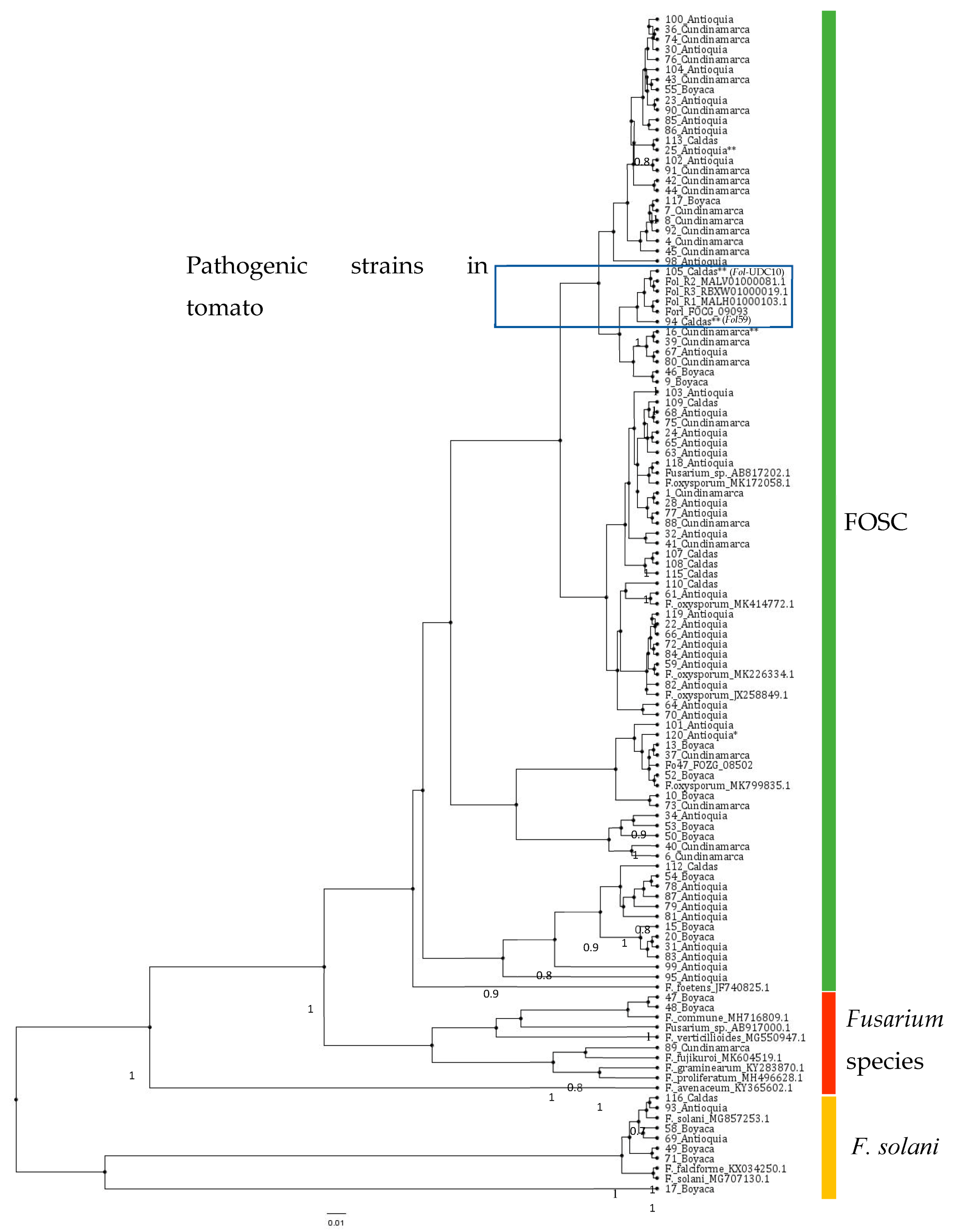
2.2.2. Molecular Identification of the Putative Fusarium Isolates

To further characterize the Fusarium isolates, we amplified the Pg1 fragment (Table 2) in the 120 isolates obtained. In this PCR analysis, 105 isolates amplified a fragment that corresponded to the Pg1 (Supplementary Materials, Figure S2). This result, together with morphology and microscopic analyses, suggested that 105 isolates out of the 120 collected might have been grouped into the F. oxysporum species complex. Moreover, in order to further support this evidence, we used the primers for the EF1a gene, designed by Cobo-Diaz et al. [30] (Table 2). The EF1a gene has been widely used for phylogenetic studies with isolates of the Fusarium genus [4,30,31]. In this study, 113 of the 120 isolates were amplified with EF1a (Supplementary Materials, Figure S3). This group of 113 isolates was used for further analyses with the EF1a gene. The PCR products of the EF1a obtained from the isolates was sequenced using the SANGER platform, and a total of 95 sequences were selected based on quality and length (≥350 bp), while 18 sequences were not considered for further analysis. A phylogenetic analysis was performed for the 95 fungal isolates with an EF1a sequence of good quality (Table 1). The analysis included 22 EF1a reference sequences of different Fusarium species and 10 EF1a reference sequences of F. oxysporum isolates, which were associated with tomato crops (Table 3, Figure 2). This analysis showed that the isolates were not grouped according to geographical location. Six of the ninety-five fungal isolates analyzed (49_Boyacá, 58_Boyacá, 69_Antioquia, 71_Boyacá, 93_Antioquia, and 116_Caldas) could be related to the F. solani clade, while three isolates (47_Boyacá, 48_Boyacá, and 89_Cundinamarca) were grouped close to Fusarium species different from F. oxysporum. The remaining fungal isolates were grouped with reference sequences of the F. oxysporum species complex (Figure 2).

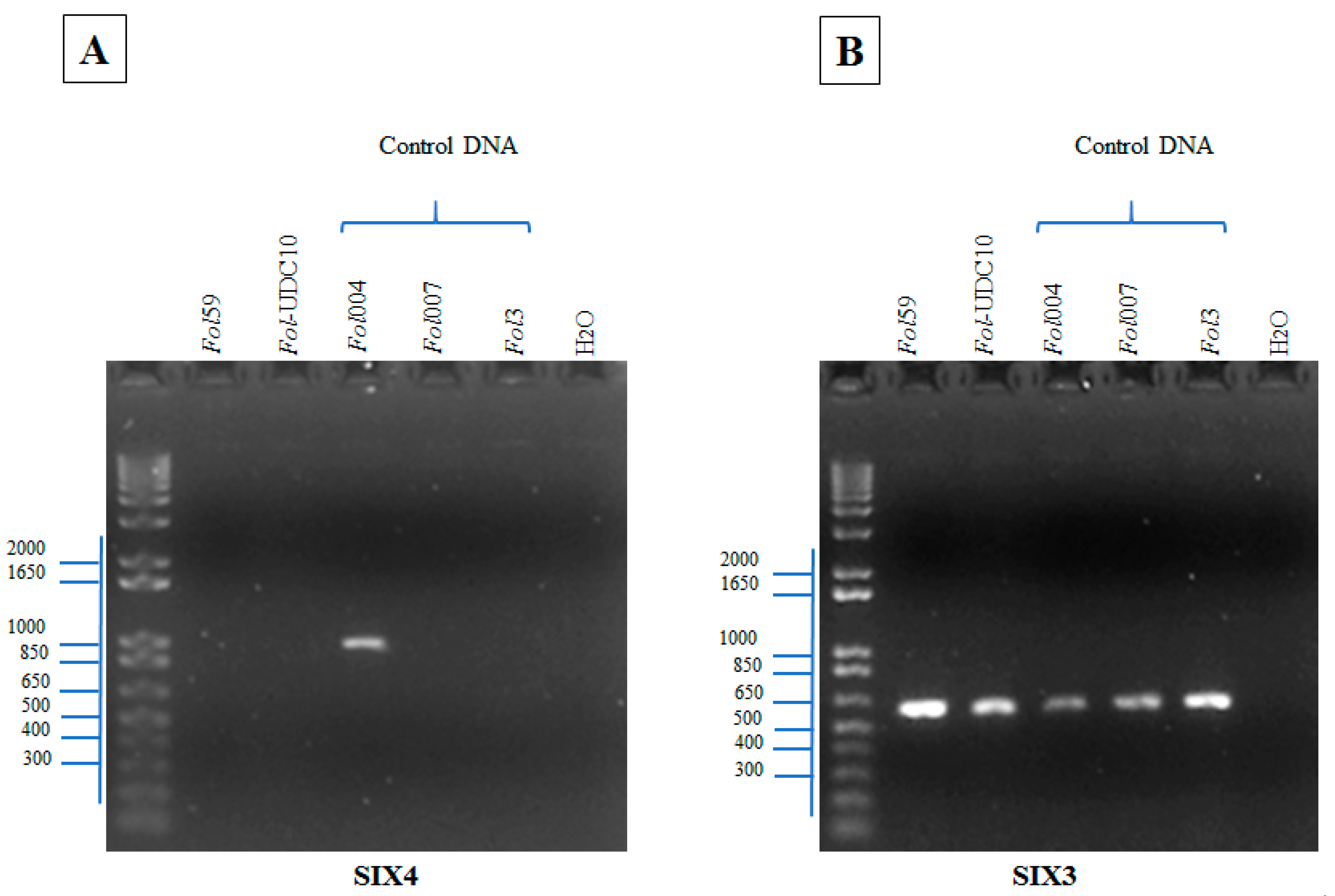
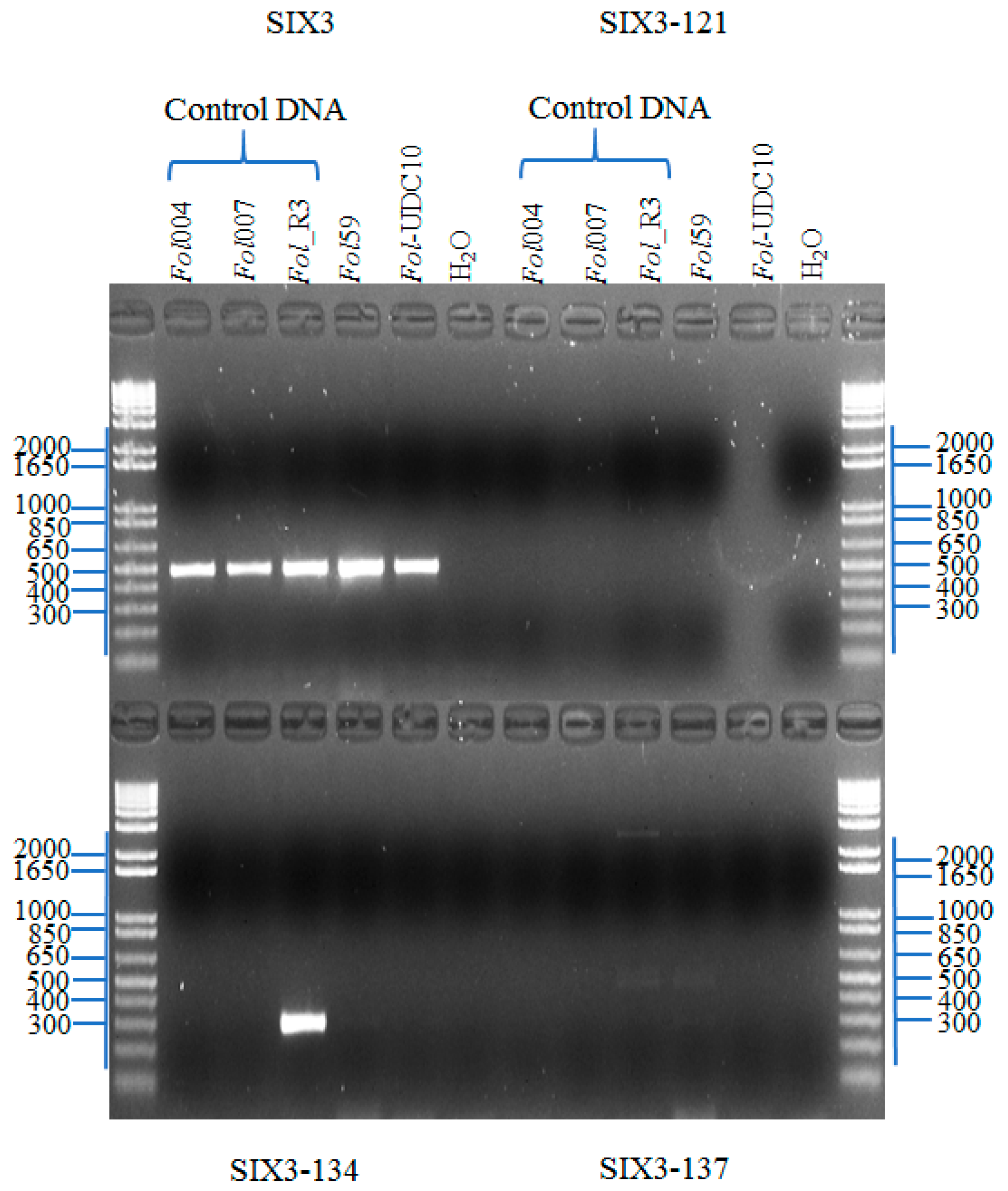
2.2.3. Identification of Fusarium oxysporum f. sp. lycopersici Candidate Isolates

To perform a rapid and unambiguous identification of Fol from the fungal isolates described before, we used the effector genes SIX1, SIX3, and SIX4, which are specific pathogenic strains of the F. oxysporum species complex [18,21,35], as molecular markers. The effector SIX1 is present in the majority of pathogenic strains of Fol [21]. In this study, SIX1 was identified in 2 isolates (94 and 105, named hereafter as Fol59 and Fol-UDC10, respectively) out of the 120 fungal isolates (Table 1, Supplementary Materials, Figure S4A). In Fol, the effector gene SIX4 was only present in strains classified as race 1 [36]. Thus, we used SIX4 to determine whether Fol59 and/or Fol-UDC10 could correspond to race 1. However, no amplification of SIX4 was observed in either Fol59 or Fol-UDC10 (Figure 3A, Supplementary Materials, Figure S4B). To determine whether Fol59 and Fol-UDC10 might correspond to Fol race 2 (lost SIX4, carrying SIX1 and SIX3) or race 3 (lost SIX4, carrying SIX1 and SIX3 with Single Nucleotide Polymorphisms (SNPs) in the nucleotides 121, 134, or 137) [21], a PCR amplification and a sequencing of SIX3 were performed on both Fol candidate isolates (Figure 3B and Figure 4). SIX3 was identified on Fol59 and Fol-UDC10 (Supplementary Materials, Figure S4C) isolates, and the sequencing analysis of the SIX3 Coding Sequence (CDS) suggested that both Fol59 and Fol-UDC10 belonged to Fol race 2, as no polymorphism was found in SIX3 (compared to the corresponding reference sequences of Fol races 2 and 3) (Figure 5).

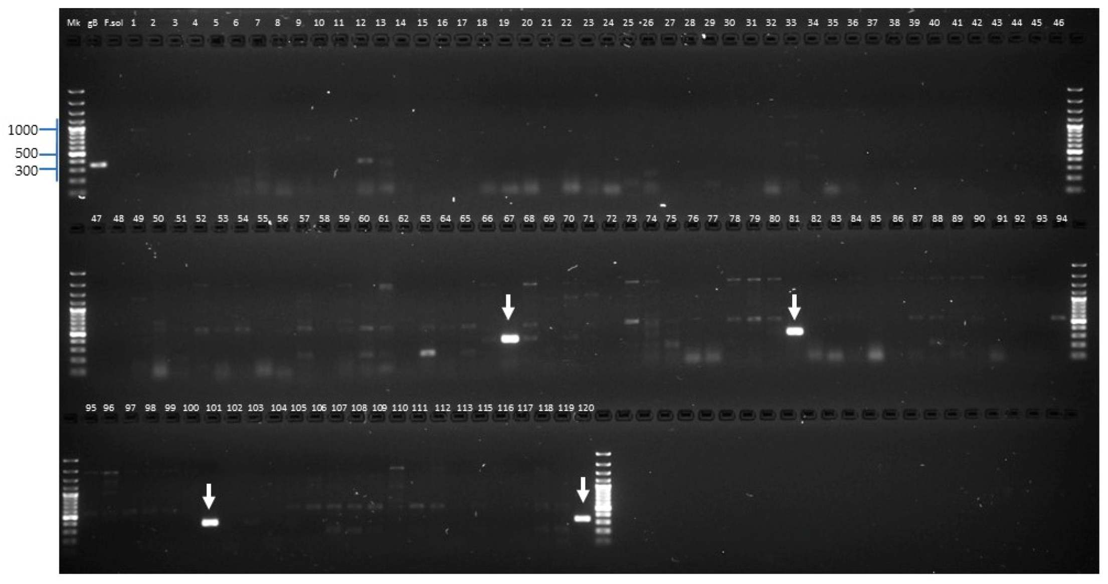
2.2.4. Identification of Forl Candidates

A specific primer pair that could distinguish Forl from Fol was designed. Briefly, the genome sequences of Fol race 2 (GCA_000259975.2), Fol race 3 (GCA_000149955.2), and Forl (GCA_000260155.3) were compared, and a pair of primers (Forl_155.3) specific to a region of 337 bp that is only present in the Forl genome was designed. We used the Forl_155.3 primers in the 118 fungal isolates (with no evidence of the presence of SIX genes) and both Fol59 and FolUDC-10 as a negative control. As a result, the newly designed marker specific for Forl isolates was amplified only in 4 isolates (Fut52, Fur55, Fur38, and Fut64) out of the 120 tested (Table 1 and Figure 6). As shown in Figure 6, a specific and well-defined PCR band was observed for the Forl positive isolates.

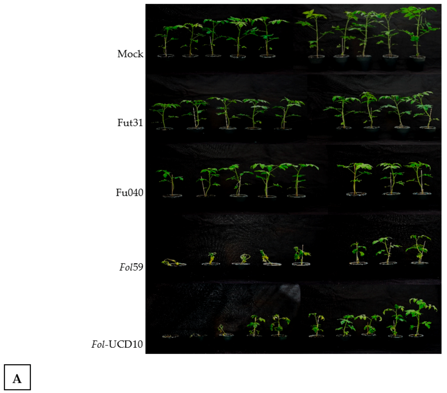
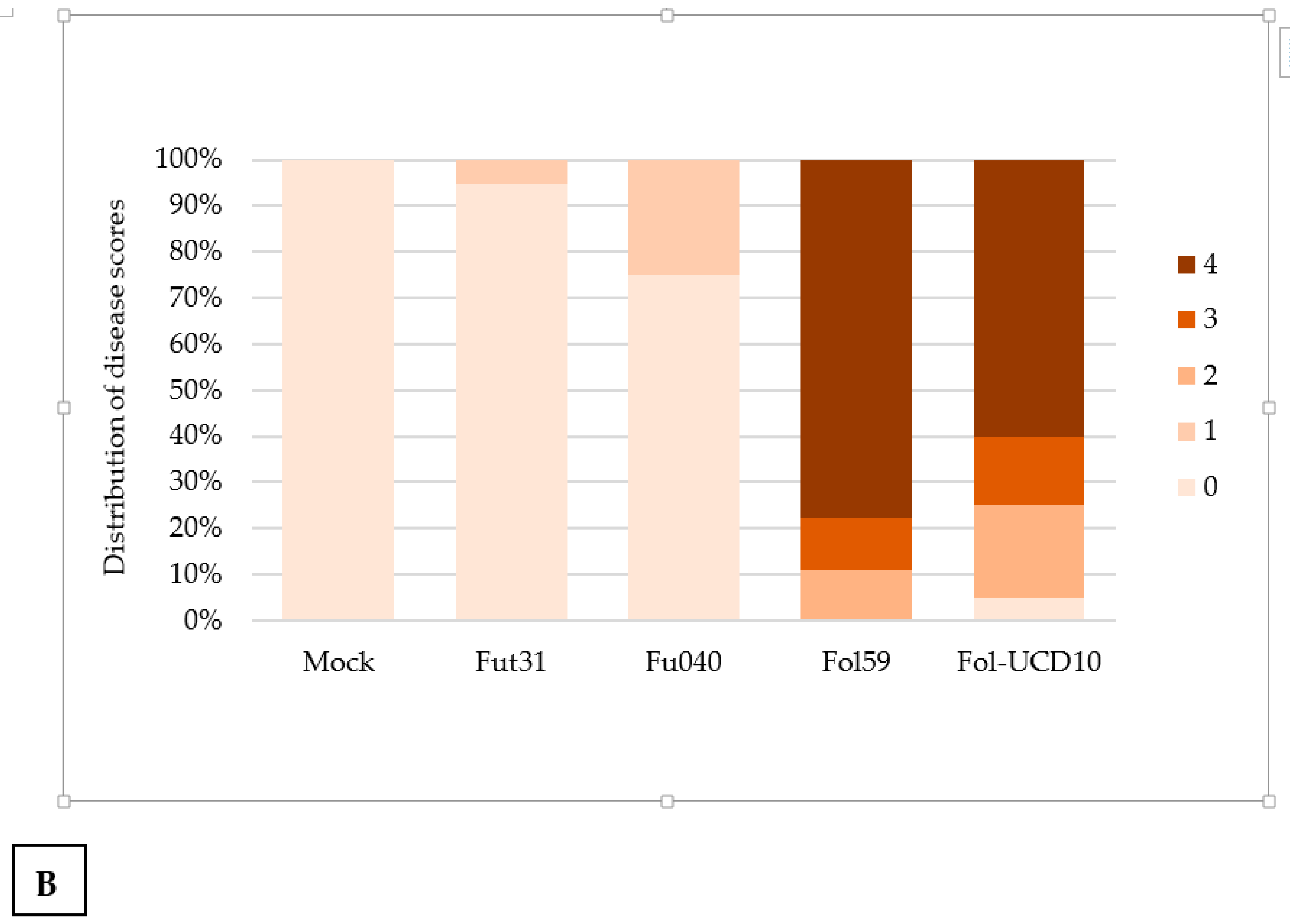
2.3. The Colombian Isolates Fol59 and Fol-UDC10 Were Highly Virulent on Susceptible Tomato Plants

A comparative analysis of the three inoculation methods (soil suspension (SS), root dipping (RD), and an RD + SS combination) was performed in tomato seedlings two, three, and four weeks old of the cultivar Santa Cruz Kada (susceptible to all three races of Fol) with the Colombian isolate Fol-UDC10. Plants inoculated using both the RD and RD + SS methods exhibited typical symptoms of vascular wilt disease (e.g., chlorosis of the lower leaves and wilting) (Supplementary Materials, Figure S5). After 18 dpi, the plants inoculated using both the RD and RD + SS methods showed significantly higher incidence and severity of wilting disease symptoms compared to those inoculated using the SS method (Figure 7). Although there were no significant differences between RD and RD + SS, the RD method was selected for further analysis (using both Fol59 and Fol-UDC10 isolates) due to the development of the typical symptoms of Fusarium wilt (Supplementary Materials, Figure S5).
Once the RD infection protocol was selected, seedlings of three different ages (i.e., two, three, and four weeks old) were inoculated with Fol59 in order to describe the typical wilting symptoms appropriately and to describe the disease progress in a time course experiment. After 14 dpi, plants corresponding to all three ages showed the typical disease symptoms of Fusarium wilt, with an incidence ranging from 95% to 100%. Disease incidence and severity were not significantly different when comparing inoculated plants at different ages. Nevertheless, both disease symptoms and progress were better recorded using four-week-old plants (Supplementary Materials, Figure S5). Therefore, four-week-old seedlings of Santa Cruz Kada were used in further pathogenicity tests.
In further plant disease assays, the RD inoculation method was carried out with a spore suspension of 5 × 10⁶ conidia mL−1 using the preselected isolates Fol59 and Fol-UDC10. To test whether Fol59 and Fol-UDC10 isolates were indeed virulent, four-week-old seedlings were inoculated with both isolates, and severe disease symptoms were observed after 21 dpi (Figure 8A,B). Non-inoculated plants (mock control) as well as Fol isolates Fut31 and Fu040 (which lack the SIX genes and were identified as F. oxysporum in this study, but with no evidence that they are pathogenic in tomato-susceptible plants) served as controls. The distribution of disease scores for susceptible plants to Fol (Figure 8B) showed that plants inoculated with Fut31 and Fu040 presented mostly a score of 0 (indicating healthy plants), whereas Fol59- and Fol-UDC10-inoculated plants were scored as 4, indicating wilted plants with brown coloration in vascular bundles. This result confirmed that Fol59 and Fol-UDC10 were highly virulent strains of Fol.

2.4. Pathogenicity Assays with the Forl Candidate Isolates Showed Root Rot Symptoms in Tomato Plants

In the tomato plants with Fusarium wilt symptoms sampled in the Central Andean departments of Colombia, four isolates (Fut52, Fur55, Fur38, and Fut64) were identified as Forl using the new diagnostic molecular marker (Forl_155.3) reported in this study (Table 1 and Figure 6). To investigate whether these isolates identified as Forl could produce Fusarium crown and root rot (FCRR) symptoms, tomato plants 30 days old were inoculated through root dipping using a microconidia suspension of Forl, and plants non-inoculated with Forl were used as a control. After 60 dpi, external symptoms of FCRR, including brown discoloration and rot in the crown and root, were observed (Supplementary Materials, Figure S6).

3. Discussion and Conclusions

In Colombia, a total of 347,636 tons of tomato were produced during 2017, representing one of the most cultivable vegetable crops in the country. However, this crop is highly susceptible to biotic and abiotic stresses, which result in an increase in production costs [26,28]. Fusarium wilt is currently a disease of major significance throughout the country. Varietal resistance is a highly efficient alternative to control the disease, using a number of commercial tomato hybrid varieties derived from the introgression of resistance genes specific to Fol races 1 and 2 [11]. The seeds of tomato hybrids used in Colombia are imported, and there is no information about Fol races present in the country. Tomato producers located in departments with high yields, such as Cundinamarca, Boyacá, Antioquia, and Caldas, use tomato seeds with no information about resistance to local populations of Fol [28]. The lack of knowledge about the causal agent of the vascular wilting symptoms affecting tomato crops makes the development of adequate management practices difficult. After varietal resistance, the use of chemical fungicides has been the most common management strategy for disease symptoms caused by either Fol or Forl [11]. The survey carried out in this study indicated that Colombian farmers use agrochemicals to control Fusarium wilt, in most cases without knowledge. However, there is no suitable fungicide for controlling the wilt disease caused by Fol [3,6]. The average use of chemical pesticides for the year 2015 in Colombia was 14.7 kg/ha, and Colombia was third in the word in terms of pesticide consumption (compared to a 3.9 kg/ha worldwide average). The control of Fusarium wilt should be based on an integrated disease management (IDM) strategy based on monitoring, economic thresholds, and preventive approaches to determine when and how fungicide application is needed [37]. Despite the fact that appropriate control strategies for wilt disease that could be implemented in Colombian tomato crops exist (reviewed in Reference [38]), a suitable IDM strategy in Colombia to control Fusarium wilt in tomatoes has not been established due to the absence of knowledge, sources, and socioeconomic conditions among producers.
This study presents the current state of pathogenic and nonpathogenic F. oxysporum populations associated with tomato fields in four different regions from the Central Colombian Andean Region. Most of the fungal isolates obtained exhibited a variable range of morphological characteristics of F. oxysporum (e.g., colony color, texture, and sporulation) according to the taxonomic description reported in References [29,39]. Combining all of the analyses performed using the new specific markers for the Forl_155.3, SIX, and EF1a genes in the 120 isolates obtained in this study, 2 isolates were identified as Fol (Fol59 and Fol-UDC10), 4 isolates were identified as Forl (Fur38, Fut64, Fut52, and Fur55, Table 1), 5 isolates belonged to the F. solani clade (49_Boyacá, 58_Boyacá, 69_Antioquia, 71_Boyacá, 93_Antioquia, and 116_Caldas, Table 1), 3 isolates clustered were with other Fusarium species different from Fol and Forl (47_Boyacá, 48_Boyacá, and 89_Cundinamarca, Table 1), and finally, most of the fungal isolates were clustered with reference sequences of the F. oxysporum species complex and could potentially correspond to nonpathogenic strains of F. oxysporum.
To test the potential of SIX genes as pathogenicity markers, we initially assessed the presence of SIX1, SIX3, and SIX4 in a collection of 120 F. oxysporum isolates. Only two isolates, Fol59 and Fol-UDC10, were positive for SIX1 and SIX3 amplification. Then, the absence of SIX4 and SNPs in the coding sequence of SIX3 on Fol59 and Fol-UDC10 (compared to the corresponding reference sequences in Fol races 1, 2, and 3) indicated that these two isolates belonged to Fol race 2 [11,21]. Pathogenicity assays showed that Fol59 and Fol-UDC10 were both highly virulent in tomato-susceptible plants compared to the isolate Fut31 (25_Antioquia, Table 1) obtained in this study and to the local reference strain Fu040 (16_Cundinamarca, Table 1) of Fusarium oxysporum. As a result, no disease symptoms were observed in plants inoculated with Fu040, contrary to the previous evidence described by Moreno et al. [40]; however, different inoculation methods, experimental conditions, and plant material were used compared to the results obtained by Moreno et al. [40].
In addition, the SIX genes evaluated were found to be absent in the other 118 nonpathogenic isolates (including Fut31 and Fu040), thus showing a direct correlation between the presence of SIX genes and the ability of Fol isolates to cause severe wilting symptoms (Figure 7). Previous studies have demonstrated the usefulness of SIX genes for the discrimination of Fol races [7,21]. These results propose a basis for an understanding of the races of Fol present in Colombia and for establishing a linkage between races and commercial tomato hybrid varieties. As mentioned before, more than 90% of the commercial tomato seeds growing in Colombia are introduced from other countries without adequate resistance tests for local Fol and Forl populations, and there is deficient technological appropriation in terms of the conditions of specific microenvironments.
During the isolation process of the causal agent of the wilting symptoms from infected plant tissues, it is common to recover more than one species of Fusarium (as they are common inhabitants of soil and the rhizosphere), but some species or strains may be pathogens, while others are saprophytes or endophytes and have no role in the disease process whatsoever [29,41,42,43].
A phylogenetic analysis of the 95 isolates using the EF1a gene showed that 7 of them could be related to other Fusarium species (F. solani, F. falciforme, and F. fujikuroi), while 82 corresponded to F. oxysporum (possibly nonpathogenic isolates due to the absence of any of the SIX effector genes analyzed). This evidence agrees with other studies where F. oxysporum nonpathogenic isolates were collected from infected plant tissues from tomato crops in India and Algeria and from soil samples from Florida (the United States) [41,42,43].
Therefore, nonpathogenic strains of F. oxysporum are commonly isolated from tissue samples of wilted tomato plants. The F. oxysporum species complex also includes numerous nonpathogenic strains, some of which have been shown to be effective in plant protection or biocontrol [44,45]. According to our results, we hypothesize that when the tissue samples were collected to isolate pathogenic strains of F. oxysporum, there was a high proportion of nonpathogenic F. oxysporum strains, and we obtained more nonpathogenic isolates during the purification of the fungal cultures. The nonpathogenic isolates collected in this work may therefore represent interesting candidates for further analyses on alternative biocontrol approaches to control Fusarium wilt.
It is important to note that after the phylogenetic analysis of EF1a sequences, Forl isolates were not clustered alone or with any Fusarium pathogenic strains in tomatoes. They were scattered within the FOSC group, without evidence of a phylogenetic structure with respect to pathogenicity (Figure 2). This suggests that EF1a might not be useful in resolving the phylogenetic positions of the Forl isolates studied. Thus, further studies are needed using the polymorphisms in DNA sequences for other markers, such as RNA polymerase II (rpb2) or the Internal Transcribed Spacer (ITS) region, to gain insight into the phylogenetic relationship between Fol and Forl within the FOSC group.
In addition, the results obtained in this study have another practical consequence for Colombian tomato crops: the SIX genes and the specific marker Forl_155.3 may be used as specific markers in epidemiological studies of F. oxysporum pathogenic isolates, for rapid diagnosis, and for discrimination from nonpathogenic isolates. Such tools, which are applicable for both diagnosis and tracking, will help to prevent the spread of pathogenic strains of Fol and Forl to other regions in Colombia and to nearby countries.
Knowledge of the Fol physiological races affecting tomato crops will be useful for the development of better disease management strategies, including plant breeding and tomato grafting. Part of the diversity of tomato genetic resources (more than 1000 accessions) is maintained in the Tomato Germplasm Bank (TGB) located in the Colombian Agricultural Research Corporation (AGROSAVIA). The majority of accessions contained in the TGB have not yet been characterized for their resistance/susceptibility to any phytopathogen, such as F. oxysporum. Therefore, this study provides primary insight into the search for tomato-resistant genotypes and their interactions with local Fol and Forl populations and thus contributes to improving management strategies for this important disease.

4. Materials and Methods

4.1. Fusarium Isolates and Culture Conditions

Samples consisting of roots and stem tissues from wilted tomato plants (Table 1) were collected from the surveyed fields, wrapped in plastic bags, and carried to the Agricultural Microbiology Laboratory at the Tibaitatá Research Center of AGROSAVIA-Colombia for subsequent analysis. The samples were surface-sterilized using 2% sodium hypochlorite (NaOCl) solution for 15 min and 70% ethanol for 1 min, and they were finally rinsed three times in sterile water. For each sample, plant tissue was dried, sectioned in six segments of 0.5 cm, and placed directly on one plate of Potato Dextrose Agar (PDA; Merck, Darmstadt, Germany) amended with 0.1% Triton 100X (Sigma-AldrichTM, Saint Louis, MO, USA) and 0.3% chloramphenicol to avoid bacterial contamination. Plates were incubated at 25 ± 2 °C in 24-h light conditions for four days. The plates were observed routinely, and all colonies with typical Fusarium morphology were checked using optical microscopy for microconidia, macroconidia, and chlamydospores. Then, positive colonies were transferred to fresh PDA until pure fungal colonies were obtained, from which monosporic cultures were obtained. Two long-term fungal culture preservation methods were used: filter paper and 30% (v/v) glycerol–water.

4.2. Morphological Characterization

Fungal isolates were plated onto Carnation Leaf Agar (CLA) medium [39] and incubated at 25 °C for three to five days. Propagative structures of F. oxysporum were observed using an optical microscope (Olympus CX31™, Tokyo, Japan). To perform environmental scanning electron microscopy, samples collected from fields with vascular wilting symptoms were disinfected with a 1% NaOCl solution for 1 min and 70% ethanol for 50 s, triple-washed with distilled sterile water, and transferred onto a sterile paper towel to remove excess moisture. A portion of the infected material was placed on PDA medium and incubated for 5 days at 28 °C until pure fungal colonies were obtained. Visualization of the samples was carried out using a scanning electron microscope (ESEM-FEI QUANTA 250, FEI Co., Hillsboro, OR, USA) located in the Instituto de Estatigrafía de la Universidad de Caldas, Colombia. An untreated mycelium portion was cut, fixed, and placed on aluminum stubs (FEI Co., Hillsboro, OR, USA) double-coated with conductive carbon tape. The samples were examined with the ESEM using a secondary electron detector (SE), an acceleration voltage of 10 kV, a working distance of 10 mm, and a magnification of 5000×. The morphological and microscopic characteristics were described according to Leslie and Summerell [29].

4.3. DNA Extraction and PCR Analysis

The purified isolates were grown on PDA, and DNA was extracted from 100 mg of the mycelia from each isolate using the cetyl trimethylammonium bromide (CTAB) protocol [46] modified for fungal DNA. Additionally, DNA from the reference strains Fol004 (race 1), Fol007 (race 2), and Fol1943 (race 3), kindly provided by Professor David Jones of the Australian National University, were used as a positive control. The sets of primers Uni-F and Uni-R, which amplify a fragment of 670~672 bp (from the endo-polygalacturonase gene (Pg1) [32]), and EF1-F and EF1-R, which amplify a fragment of approximately 770 bp (from the translation elongation factor 1-alpha (EF1a) gene of F. oxysporum (GenBank: MK172058.1), reported by Cobo-Diaz et al. [30] (Table 2)), were used in order to determine whether the isolates could correspond to the F. oxysporum species complex. An identification of the effector gene SIX1 (using the primers P12-F and P12-R (Table 2)) was performed to determine putative Fol isolates. Additionally, primers that amplify the effector genes SIX4 and SIX3 (Table 2) were used to determine the race of the Fol isolates (i.e., isolates positive for SIX4 were classified as race 1, while negatives for SIX4 but positives for SIX3 could be classified as either race 2 or 3) [18,21,35,36]. The primer pairs SIX3-F1/SIX3-R2, SIX3-G121A-F2/SIX3-R2, SIX3-G134A-F2/SIX3-R2, and SIX3-G137C-F1/SIX3-R2 were used to differentiate between Fol races 2 and 3 [21]. DNA from the reference strains Fol067 (FolMM10), Fol029 (5397), and Fol035 (IPO3), which belong to Fol race 3 and contain SNPs on the nucleotides 121, 134, and 137 of SIX3, respectively, were kindly provided by Dr. Martijn Rep, University of Amsterdam, and were used as a control.
To identify putative Forl isolates, a specific pair of primers (named Forl_155.3) was designed based on a comparison of the genome sequences of Fol race 2 (NCBI genome accession GCA_000259975.2), Fol race 3 (GCA_000149955.2), and Forl (GCA_000260155.3). A DNA fragment that contained the Forl_155.3 region was synthesized (gBlock, Integrated DNA Technologies, Coralville IA, USA) and used as a positive control.
For amplification of the SIX genes and the Forl_155.3 fragment, PCR reactions were conducted with Taq DNA Polymerase (Invitrogen™, Carlsbad, CA, USA) in a 25-µL reaction volume. The PCR reaction consisted of 0.25 µL Taq Polymerase, 2.5 µL of 10X buffer (Invitrogen™, Carlsbad, CA, USA), 0.16 µM of each primer, 0.16 mM of dNTP mix, 2 mM MgCl2, and 25 ng of template DNA. PCRs were carried out with an initial denaturing step at 95 °C for 2 min, followed by 30 cycles of denaturing at 95 °C for 45 s, the annealing of primers at 59 °C (62 °C for Forl_155.3) for 45 s, and primer extension at 72 °C for 45 s. The PCR was completed by a final extension at 72 °C for 10 min. The melting temperature for each primer was determined using a temperature gradient. A touchdown PCR [47] was performed to amplify the primer pairs for the SIX3 gene as follows: the first phase consisted of 10 cycles with an annealing temperature of 69 °C for the first PCR cycle, which decreased by 1 °C per cycle until an optimal annealing temperature of 59 °C was reached. The remaining cycling conditions consisted of 20 cycles with an annealing temperature of 59 °C. Amplification was visualized on 1.4% w/v agarose gel stained using SYBR™ Safe DNA Gel Stain (Invitrogen™, Carlsbad, CA, USA).

4.4. Sequencing Analysis of the EF1a Gene among the Fungal Isolates

The EF1a gene was amplified by PCR with the same conditions used for the SIX genes (described above) with modifications of the annealing temperature at 62 °C. The PCR products of the EF1a gene, which were obtained from the 120 fungal isolates, were sequenced by SANGER (Applied Biosystems/Thermo Fisher Scientific, model ABI 3500 Genetic Analyser, Foster City, CA, USA), trimmed for sequence quality using the software Geneious v.2019.2.1 [48], and compared to 12 EF1a reference sequences reported for other Fusarium species and 10 EF1a sequences reported for F. oxysporum isolates associated with tomatoes (Table 3) (using the software MAFFT) [49,50]. Phylogenetic analysis was performed with the multiple sequence alignment of the EF1a gene mentioned above using the software BEAST (Bayesian Evolutionary Analysis Sampling Trees) v2.6.1 [51,52] with default settings. The resulting phylogenetic trees were visualized using Figtree v1.4.3 [53].
Sequences of SIX1, SIX3, and EF1a from the fungal isolates obtained in this study were deposited in the GenBank database under accession numbers MN745203, MN745204, MN745207, MN745208 (SIX1 and SIX3), and MN745105-MN745199 (EF1a). Unique sequence identifiers and annotation information are provided in the Supplementary Materials, Table S1.

4.5. Pathogenicity Assays

Three inoculation methods were evaluated using the Fol59 isolate with the presence of SIX genes on the susceptible tomato cultivar Santa Cruz Kada (Impulsemillas™, Bogotá, Colombia) in order to establish a reliable pathogenicity assay with a virulent strain of Fol and susceptible tomato plants. Seeds were disinfected using 2% NaOCl solution for 10 min and then 70% ethanol for 1 min, rinsed three times in sterile water, and finally sown in sterile peat. Fifteen-day-old seedlings were inoculated with Fol microconidia using three methods, as follows: (i) Soil suspension (SS) consisted of transplanting the seedlings to 0.5-L pots with a sterile vermiculite/soil (2:8) mixture containing 1 × 10⁶ microconidia mL−1 of Fol suspension. Seedlings transplanted to pots with a sterile mixture and mock-inoculated plants with water were used as a control. (ii) Root dipping (RD), the second method, which has been described by Mes et al. [54] and adapted by Rep et al. [55], Gonzalez-Cendales et al., [15] and Simbaqueba et al. [56], with modifications, consisted of the uprooting of the seedlings, preserving the root integrity. The roots were submerged for 15 min in a 5 × 10⁶ microconidia mL−1 suspension of Fol. Seedlings dipped in sterile water served as a mock for inoculation. (iii) The third method was a combination of the first two methods mentioned. Additionally, seedlings from three developmental times (i.e., 15 days old, 25 days old, and 30 days old, or the seedling stage for transplanting) were tested using the most effective inoculation method. The plants were maintained in a controlled-environment growth room with a 12 h/30 °C day (100 mmol m−2 s−1) and a 12 h/30 °C night cycle for the first 6 days post-inoculation (dpi). They were transferred to greenhouse conditions until 14 to 18 dpi. Plants were sampled according to external disease symptoms every four days during the infection process.
The pathogenicity assays of Fol consisted of a randomized complete-block design, where four different treatments were compared: SS, RD, RD + SS, and mock-inoculated plants. To maximize the statistical reliability of the data, three biological replicates were carried out, and for each biological replicate, four technical replicates were performed, where the experimental unit consisted of 5 plants. The number of wilted leaves per plant was recorded as a Fol symptom, according to the disease severity scale reported by Cakir et al. [57], Akhter et al. [58], and Rongai et al. [59] and modified for this study (Supplementary Materials, Table S2 and Figure S5). The severity disease index was calculated using the Equation (1) described by Chiang et al. [60]:
D S I ( % ) = ( d i s e a s e   c l a s s   f r e q u e n c y × d i s e a s e   c l a s s ) ( #   t o t a l   o b s e r v a t i o n s ) × ( h i g h e s t   d i s e a s e   l e v e l ) × 100
Differences in the disease incidence and disease severity between treatments were tested for statistical significance with ANOVA and Kruskal–Wallis tests using the software Statistix version 8.0 with a probability value of p = 0.05. Additional plant wilting symptoms and vascular browning were recorded and used to calculate disease scores according to the following criteria (described by Rep et al. [61], Gonzalez-Cendales et al. [15], and Simbaqueba et al. [56]): 0, healthy plant; 1, slightly swollen or bent hypocotyl; 2, one or two brown vascular bundles in the hypocotyl; 3, at least two brown vascular bundles and growth distortion; and 4, all vascular bundles brown and plant either dead or very small and wilted. Differences in the distributions of disease scores between treatments were tested for statistical significance through pairwise two-tailed Mann–Whitney tests [62].
For the pathogenicity test with Forl isolates, we performed the same inoculation procedure used for Fol with modifications in the controlled-environment growth room (with a 12 h/25 °C day cycle and a 12 h/25 °C night cycle for 60 dpi). Plants were considered diseased when they exhibited brown necrosis on the crown and vascular discoloration in the lower stem (Supplementary Materials, Figure S6).

Supplementary Materials

The following are available online at https://0-www-mdpi-com.brum.beds.ac.uk/2076-0817/9/1/70/s1, Figure S1: Morphological propagative structures of the isolate Fol-UDC10, observed on CDA plates; Figure S2: Polymerase chain reaction (PCR) analysis showing the presence of the fragment of the pg1 gene specific to Fusarium oxysporum; Figure S3: PCR analysis showing the presence of the EF1a gene on 113 from the 120 isolates obtained; Figure S4: PCR results for SIX1, SIX4, and SIX3 for the 120 fungal isolates; Figure S5: Disease scale implemented in this study; Figure S6: Symptom development of Fusarium oxysporum f. sp. radicis-lycopersici (Forl)-infected tomato cultivar (Santa Cruz Kada); Table S1: GenBank sequence unique identifiers and annotation information for the EF1a gene; Table S2: Disease scale implemented in this study.

Author Contributions

Conceptualization, M.S.-S. and J.S.; methodology, S.L.C., D.B.-D., M.R.G., W.L., and J.S.; formal analysis, J.S., S.L.C., D.B.-D., M.R.G., N.C., J.C.-Z., and M.S.-S.; writing—original draft preparation, M.S.-S., S.L.C., D.B.-D., M.R.G., and J.S.; project administration, M.S.-S. All authors have read and agreed to the published version of the manuscript.

Funding

This research was funded by the Ministerio de Agricultura y Desarrollo Rural (MADR), Gobierno de Colombia.

Acknowledgments

This work was supported by the Corporación Colombiana de Investigación Agropecuaria, AGROSAVIA, and the Ministerio de Agricultura y Desarrollo Rural (MADR). We thank Martijn Rep from the University of Amsterdam for kindly providing us with DNA from Fol067 (FolMM10), Fol029 (5397), and Fol035 (IPO3), which are Fol race 3-containing SNPs in the positions 121, 134, and 137, respectively. The authors would like to thank the anonymous reviewers for their constructive comments on this article.

Conflicts of Interest

The authors declare no conflicts of interest.

References

  1. Ma, L.-J.; Shea, T.; Young, S.; Zeng, Q.; Kistler, H.C. Genome sequence of Fusarium oxysporum f. sp. melonis strain NRRL 26406, a fungus Causing wilt disease on melon. Genome Announc. 2014, 2, e00730-14. [Google Scholar] [CrossRef] [PubMed] [Green Version]
  2. Ma, L.-J.; Geiser, D.M.; Proctor, R.H.; Rooney, A.P.; O’Donnell, K.; Trail, F.; Gardiner, D.M.; Manners, J.M.; Kazan, K. Fusarium Pathogenomics. Annu. Rev. Microbiol. 2013, 67, 399–416. [Google Scholar] [CrossRef] [PubMed] [Green Version]
  3. Michielse, C.B.; Rep, M. Pathogen profile update: Fusarium oxysporum. Mol. Plant Pathol. 2009, 10, 311–324. [Google Scholar] [CrossRef] [PubMed]
  4. O’Donnell, K.; Gueidan, C.; Sink, S.; Johnston, P.R.; Crous, P.W.; Glenn, A.; Riley, R.; Zitomer, N.C.; Colyer, P.; Waalwijk, C.; et al. A two-locus DNA sequence database for typing plant and human pathogens within the Fusarium oxysporum species complex. Fungal Genet. Biol. 2009, 46, 936–948. [Google Scholar] [CrossRef]
  5. Dean, R.; Van Kan, J.A.L.; Pretorius, Z.A.; Hammond-Kosack, K.E.; Di Pietro, A.; Spanu, P.D.; Rudd, J.J.; Dickman, M.; Kahmann, R.; Ellis, J.; et al. The Top 10 fungal pathogens in molecular plant pathology. Mol. Plant Pathol. 2012, 13, 414–430. [Google Scholar] [CrossRef] [Green Version]
  6. Di Pietro, A.; Madrid, M.P.; Caracuel, Z.; Delgado-Jarana, J.; Roncero, M.I.G. Fusarium oxysporum: Exploring the molecular arsenal of a vascular wilt fungus. Mol. Plant Pathol. 2003, 4, 315–325. [Google Scholar] [CrossRef]
  7. Van Der Does, H.C.; Lievens, B.; Claes, L.; Houterman, P.M.; Cornelissen, B.J.C.; Rep, M. The presence of a virulence locus discriminates Fusarium oxysporum isolates causing tomato wilt from other isolates. Environ. Microbiol. 2008, 10, 1475–1485. [Google Scholar] [CrossRef]
  8. FAOSTAT. Production—Crops—Area Harvested/Production Quantity—Tomatoes. Available online: www.fao.org/faostat/en (accessed on 1 September 2018).
  9. Menzies, J.G. Additions to the host range of Fusarium oxysporum f. sp. radicis-lycopersici. Plant Dis. 1990, 74, 569. [Google Scholar] [CrossRef]
  10. Lagopodi, A.L.; Ram, A.F.J.; Lamers, G.E.M.; Punt, P.J.; Van den Hondel, C.A.M.J.J.; Lugtenberg, B.J.J.; Bloemberg, G.V. Novel aspects of tomato root colonization and infection by Fusarium oxysporum f. sp. radicis-lycopersici revealed by confocal laser scanning microscopic analysis using the green fluorescent protein as a marker. Mol. Plant-Microbe Interact. 2002, 15, 172–179. [Google Scholar] [CrossRef] [Green Version]
  11. McGovern, R.J. Management of tomato diseases caused by Fusarium oxysporum. Crop Prot. 2015, 73, 78–92. [Google Scholar] [CrossRef]
  12. Brayford, D. IMI descriptions of fungi and bacteria. Mycopathologia 1996, 133, 35–63. [Google Scholar] [CrossRef]
  13. Catanzariti, A.-M.; Lim, G.T.T.; Jones, D.A. The tomato I-3 gene: A novel gene for resistance to Fusarium wilt disease. New Phytol. 2015, 207, 106–118. [Google Scholar] [CrossRef] [PubMed]
  14. Catanzariti, A.-M.; Do, H.T.T.; Bru, P.; de Sain, M.; Thatcher, L.F.; Rep, M.; Jones, D.A. The tomato I gene for Fusarium wilt resistance encodes an atypical leucine-rich repeat receptor-like protein whose function is nevertheless dependent on SOBIR1 and SERK3/BAK1. Plant J. 2017, 89, 1195–1209. [Google Scholar] [CrossRef] [PubMed] [Green Version]
  15. Gonzalez-Cendales, Y.; Catanzariti, A.-M.; Baker, B.; Mcgrath, D.J.; Jones, D.A. Identification of I -7 expands the repertoire of genes for resistance to Fusarium wilt in tomato to three resistance gene classes. Mol. Plant Pathol. 2016, 17, 448–463. [Google Scholar] [CrossRef]
  16. Ori, N.; Eshed, Y.; Paran, I.; Presting, G.; Aviv, D.; Tanksley, S.; Zamir, D.; Fluhr, R. The I2C family from the wilt disease resistance locus I2 belongs to the nucleotide binding, leucine-rich repeat superfamily of plant resistance genes. Plant Cell 1997, 9, 521–532. [Google Scholar]
  17. Houterman, P.M.; Speijer, D.; Dekker, H.L.; De Koster, C.G.; Cornelissen, B.J.C.; Rep, M. The mixed xylem sap proteome of Fusarium oxysporum-infected tomato plants. Mol. Plant Pathol. 2007, 8, 215–221. [Google Scholar] [CrossRef]
  18. Houterman, P.M.; Cornelissen, B.J.C.; Rep, M. Suppression of plant resistance gene-based immunity by a fungal effector. PLoS Pathog. 2008, 4, 1–6. [Google Scholar] [CrossRef] [Green Version]
  19. Ma, L.; Houterman, P.M.; Gawehns, F.; Cao, L.; Sillo, F.; Richter, H.; Clavijo-Ortiz, M.J.; Schmidt, S.M.; Boeren, S.; Vervoort, J.; et al. The AVR2-SIX5 gene pair is required to activate I-2 -mediated immunity in tomato. New Phytol. 2015, 208, 507–518. [Google Scholar] [CrossRef]
  20. Carrillo-Fasio, J.A.; Rodríguez, T.D.J.M.; Estrada, I.M.; Ortega, J.E.C.; Zequera, I.M.; Barajas, A.J.S. Razas de Fusarium oxysporum f. sp lycopersici Snyder y Hansen, en tomate (Lycopersicon esculentum Mill.) en el Valle de Culiacán, Sinaloa, México. Rev. Mex. Fitopatol. 2003, 21, 123–127. [Google Scholar]
  21. Lievens, B.; Houterman, P.M.; Rep, M. Effector gene screening allows unambiguous identification of Fusarium oxysporum f. sp. lycopersici races and discrimination from other formae speciales. FEMS Microbiol. Lett. 2009, 300, 201–215. [Google Scholar]
  22. Buriticá, P. Las Enfermedades de las Plantas y su Ciencia en Colombia; Universidad Nacional de Colombia: Medellín, Colombia, 1999; Available online: http://bdigital.unal.edu.co/9340/1/17081124.%201999%20Parte1.pdf (accessed on 17 December 2019).
  23. Garcia-Bastidas, F.; Quintero-Vargas, C.; Ayala-Vasquez, M.; Seidl, M.; Schermer, T.; Santos-Paiva, M.; Noguera, A.M.; Aguilera-Galvez, C.; Wittenberg, A.; Sørensen, A.; et al. First report of Fusarium wilt tropical race 4 in Cavendish bananas caused by Fusarium odoratissimum in Colombia. Plant Dis. 2019, 1–5. [Google Scholar] [CrossRef]
  24. Arbeláez, G.; de Granada, E.G.; de Amézquita, M.O.; Calderón, O.L. Respuesta de algunas variedades de clavel estándar a cuatro razas fisiológicas de Fusarium oxysporum f. sp. Dianthi. Agron. Colomb. 1996, 13, 117–127. [Google Scholar]
  25. Forero-Reyes, C.M.; Alvarado-Fernández, A.M.; Ceballos-Rojas, A.M.; González-Carmona, L.C.; Linares-Linares, M.Y.; Castañeda-Salazar, R.; Pulido-Villamarín, A.; Góngora-Medina, M.E.; Cortés-Vecino, J.A.; Rodríguez-Bocanegra, M.X. Evaluación de la capacidad patogénica de Fusarium spp. en modelos vegetal y murino. Rev. Argent. Microbiol. 2018, 50, 90–96. [Google Scholar] [CrossRef] [PubMed]
  26. AGRONET, Red de Información y Comunicación del Sector Agropecuario Colombiano. Available online: http://www.agronet.gov.co/estadistica/Paginas/default.aspx (accessed on 14 May 2018).
  27. Marín-Serna, S.; González-Guzmán, J.; Castaño-Zapata, J.; Ceballos-Aguirre, N. Respuesta de quince introducciones de tomate tipo cereza (Solanum spp.) a la marchitez vascular (Fusarium oxysporum f. sp. lycopersici Snyder & Hansen). Agron. Colomb. 2015, 22, 48–59. [Google Scholar]
  28. Jaramillo, J.; Rodriguez, V.P.; Gil, L.F.; García, M.C.; Hio, J.C.; Quevedo, D.; Guzmán, M.; Sánchez, G.D.; Aguilar, P.; Pinzón, L.; et al. Tecnologías del Cultivo del Tomate Bajo Condiciones Protegidas, 1st ed.; Corpoica: Bogotá, Colombia, 2012; Volume 1, ISBN 978-958-740-120-2. [Google Scholar]
  29. Leslie, J.; Summerel, B. The Fusarium Laboratory Manual, 1st ed.; Blackwell: Ames, IA, USA, 2006; ISBN 978-0-8138-1919-8. [Google Scholar]
  30. Cobo-Díaz, J.F.; Baroncelli, R.; Le Floch, G.; Picot, A. A novel metabarcoding approach to investigate Fusarium species composition in soil and plant samples. FEMS Microbiol. Ecol. 2019, 95, 1–13. [Google Scholar] [CrossRef]
  31. Imazaki, I.; Kadota, I. Molecular phylogeny and diversity of Fusarium endophytes isolated from tomato stems. FEMS Microbiol. Ecol. 2015, 91, 1–16. [Google Scholar] [CrossRef] [Green Version]
  32. Hirano, Y.; Arie, T. PCR-based differentiation of Fusarium oxysporum ff. sp. lycopersici and radicis-lycopersici and races of F. oxysporum f. sp. lycopersici. J. Gen. Plant Pathol. 2006, 72, 273–283. [Google Scholar] [CrossRef]
  33. Manikandan, R.; Harish, S.; Karthikeyan, G.; Raguchander, T. Comparative proteomic analysis of different isolates of Fusarium oxysporum f.sp. lycopersici to exploit the differentially expressed proteins responsible for virulence on tomato plants. Front. Microbiol. 2018, 9, 420. [Google Scholar] [CrossRef] [Green Version]
  34. van Dam, P.; Fokkens, L.; Schmidt, S.M.; Linmans, J.H.J.; Kistler, H.C.; Ma, L.-J.; Rep, M. Effector profiles distinguish formae speciales of Fusarium oxysporum. Environ. Microbiol. 2016, 18, 4087–4102. [Google Scholar] [CrossRef]
  35. Ma, L.-J.; van der Does, H.C.; Borkovich, K.A.; Coleman, J.J.; Daboussi, M.-J.; Di Pietro, A.; Dufresne, M.; Freitag, M.; Grabherr, M.; Henrissat, B.; et al. Comparative genomics reveals mobile pathogenicity chromosomes in Fusarium. Nature 2010, 464, 367–373. [Google Scholar] [CrossRef]
  36. Houterman, P.M.; Ma, L.; van Ooijen, G.; de Vroomen, M.J.; Cornelissen, B.J.C.; Takken, F.L.W.; Rep, M. The effector protein Avr2 of the xylem-colonizing fungus Fusarium oxysporum activates the tomato resistance protein I-2 intracellularly. Plant J. 2009, 58, 970–978. [Google Scholar] [CrossRef] [PubMed]
  37. Duarte-Carvajalino, J.; Alzate, D.; Ramirez, A.; Santa-Sepulveda, J.; Fajardo-Rojas, A.; Soto-Suárez, M. Evaluating late blight severity in potato crops using unmanned aerial vehicles and machine learning algorithms. Remote Sens. 2018, 10, 1513. [Google Scholar] [CrossRef] [Green Version]
  38. Vásquez Ramírez, L.M.; Castaño Zapata, J. Manejo integrado de la marchitez vascular del tomate [Fusarium oxysporum f. sp. lycopersici (sacc.) W.C. Snyder & H.N. Hansen]: Una revisión. Rev. U.D.C.A Act. Div. Cient. 2017, 20, 363–374. [Google Scholar]
  39. Nelson, P.E.; Toussoun, T.A.; Marasas, W.F.O. Fusarium Species: An Illustrated Manual for Identification; Pennsylvania State University Press: University Park, PA, USA, 1983; ISBN 978-0-271-00349-8. [Google Scholar]
  40. Moreno, C.A.; Castillo, F.; González, A.; Bernal, D.; Jaimes, Y.; Chaparro, M.; González, C.; Rodriguez, F.; Restrepo, S.; Cotes, A.M. Biological and molecular characterization of the response of tomato plants treated with Trichoderma koningiopsis. Physiol. Mol. Plant Pathol. 2009, 74, 111–120. [Google Scholar] [CrossRef]
  41. Nirmaladevi, D.; Venkataramana, M.; Srivastava, R.K.; Uppalapati, S.R.; Gupta, V.K.; Yli-Mattila, T.; Clement Tsui, K.M.; Srinivas, C.; Niranjana, S.R.; Chandra, N.S. Molecular phylogeny, pathogenicity and toxigenicity of Fusarium oxysporum f. sp. lycopersici. Sci. Rep. 2016, 6, 21367. [Google Scholar] [CrossRef] [Green Version]
  42. Jelinski, N.A.; Broz, K.; Jonkers, W.; Ma, L.-J.; Kistler, H.C. Effector gene suites in some soil isolates of Fusarium oxysporum are not sufficient predictors of vascular wilt in tomato. Phytopathology 2017, 107, 842–851. [Google Scholar] [CrossRef]
  43. Debbi, A.; Boureghda, H.; Monte, E.; Hermosa, R. Distribution and genetic variability of Fusarium oxysporum associated with tomato diseases in Algeria and a biocontrol strategy with indigenous Trichoderma spp. Front. Microbiol. 2018, 9, 282. [Google Scholar] [CrossRef] [Green Version]
  44. Alabouvette, C.; Olivain, C.; Migheli, Q.; Steinberg, C. Microbiological control of soil-borne phytopathogenic fungi with special emphasis on wilt-inducing Fusarium oxysporum. New Phytol. 2009, 184, 529–544. [Google Scholar] [CrossRef]
  45. Aimé, S.; Alabouvette, C.; Steinberg, C.; Olivain, C. The Endophytic strain Fusarium oxysporum Fo47: A good candidate for priming the defense responses in tomato roots. Mol. Plant-Microbe Interact. 2013, 26, 918–926. [Google Scholar]
  46. Griffith, G.W.; Shaw, D.S. Polymorphisms in Phytophthora infestans: Four mitochondrial haplotypes are detected after PCR amplification of DNA from pure cultures or from host lesions. Appl. Environ. Microbiol. 1998, 64, 4007–4014. [Google Scholar] [CrossRef] [Green Version]
  47. Korbie, D.J.; Mattick, J.S. Touchdown PCR for increased specificity and sensitivity in PCR amplification. Nat. Protoc. 2008, 3, 1452–1456. [Google Scholar] [CrossRef] [PubMed]
  48. Geneious Biologics. Available online: Geneious.com (accessed on 8 May 2018).
  49. Katoh, K.; Rozewicki, J.; Yamada, K.D. MAFFT online service: Multiple sequence alignment, interactive sequence choice and visualization. Brief. Bioinform. 2019, 20, 1160–1166. [Google Scholar] [CrossRef] [PubMed] [Green Version]
  50. MAFFT (Multiple Alignment Using Fast Fourier Transform). Available online: https://www.ebi.ac.uk/Tools/msa/mafft/ (accessed on 9 February 2018).
  51. Drummond, A.J.; Suchard, M.A.; Xie, D.; Rambaut, A. Bayesian phylogenetics with BEAUti and the BEAST 1.7. Mol. Biol. Evol. 2012, 29, 1969–1973. [Google Scholar] [CrossRef] [PubMed] [Green Version]
  52. BEAST. Bayesian Evolutionary Analysis Sampling Trees. Available online: https://beast.community/ (accessed on 9 August 2018).
  53. Molecular Evolution, Phylogenetics and Epidemiology. Available online: http://tree.bio.ed.ac.uk/software/figtree/ (accessed on 15 October 2018).
  54. Mes, J.J.; Weststeijn, E.A.; Herlaar, F.; Lambalk, J.J.M.; Wijbrandi, J.; Haring, M.A.; Cornelissen, B.J.C. Biological and molecular characterization of Fusarium oxysporum f. sp. lycopersici divides race 1 isolates into separate virulence groups. Phytopathology 1999, 89, 156–160. [Google Scholar] [CrossRef] [Green Version]
  55. Rep, M.; Van Der Does, H.C.; Meijer, M.; Van Wijk, R.; Houterman, P.M.; Dekker, H.L.; De Koster, C.G.; Cornelissen, B.J.C. A small, cysteine-rich protein secreted by Fusarium oxysporum during colonization of xylem vessels is required for I-3-mediated resistance in tomato. Mol. Microbiol. 2004, 53, 1373–1383. [Google Scholar] [CrossRef]
  56. Simbaqueba, J.; Catanzariti, A.-M.; González, C.; Jones, D.A. Evidence for horizontal gene transfer and separation of effector recognition from effector function revealed by analysis of effector genes shared between cape gooseberry- and tomato-infecting formae speciales of Fusarium oxysporum. Mol. Plant Pathol. 2018, 19, 2302–2318. [Google Scholar] [CrossRef] [Green Version]
  57. Çakır, B.; Gül, A.; Yolageldi, L.; Özaktan, H. Response to Fusarium oxysporum f.sp. radicis-lycopersici in tomato roots involves regulation of SA- and ET-responsive gene expressions. Eur. J. Plant Pathol. 2014, 139, 379–391. [Google Scholar]
  58. Akhter, A.; Hage-Ahmed, K.; Soja, G.; Steinkellner, S. Compost and biochar alter mycorrhization, tomato root exudation, and development of Fusarium oxysporum f. sp. lycopersici. Front. Plant Sci. 2015, 6, 529. [Google Scholar] [CrossRef] [Green Version]
  59. Rongai, D.; Pulcini, P.; Pesce, B.; Milano, F. Antifungal activity of pomegranate peel extract against Fusarium wilt of tomato. Eur. J. Plant Pathol. 2017, 147, 229–238. [Google Scholar] [CrossRef]
  60. Chiang, K.S.; Liu, H.I.; Tsai, J.W.; Tsai, J.R.; Bock, C.H. A discussion on disease severity index values. Part II: Using the disease severity index for null hypothesis testing. Ann. Appl. Biol. 2017, 171, 490–505. [Google Scholar] [CrossRef]
  61. Rep, M.; Meijer, M.; Houterman, P.M.; van der Does, H.C.; Cornelissen, B.J.C. Fusarium oxysporum evades I-3 -mediated resistance without altering the matching avirulence gene. Mol. Plant-Microbe Interact. 2005, 18, 15–23. [Google Scholar] [CrossRef] [PubMed] [Green Version]
  62. VassarStats: Website for Statistical Computation. Available online: http://vassarstats.net/ (accessed on 21 October 2018).
Figure 1. Location of the Central Colombian Andean Region, where 32 tomato fields with vascular wilting symptoms were visited. (A) Tomato fields visited in four different departments of the Central Colombian Andean Region (Cundinamarca, Boyacá, Antioquia, and Caldas). (B) Photographs of tomato plants showing symptoms of vascular wilt disease taken from the field locations visited in the Central Andean Region of Colombia. Upper part: plants showing wilting symptoms. Bottom: horizontal cortex of the stem, showing vascular browning of the xylem vessels. Pictures from Magda R. Gómez and Sandra L. Carmona. Photographic records from the Agricultural Microbiology Group, AGROSAVIA.
Pathogens 09 00070 g001aPathogens 09 00070 g001b
Figure 2. Phylogenetic tree of a partial sequence of the EF1a gene from 32 reference sequences of the Fusarium clade (Table 3) and the 95 fungal isolates obtained in this study. The node labels are the bootstrapping support, indicated as Bayesian probabilities. A key to the labels used for each taxa is provided in Table 1 and Table 3. The scale bar indicates time in millions of years. The pathogenic strains of Fol identified in this study (Fol59 and Fol-UDC10) are highlighted in dark blue to show their close phylogenetic relationship with reference strains of F. oxysporum f. sp. radicis-lycopersici (GenBank accession FOCG_09093) and Fol races 1, 2, and 3 (GenBank accessions MALH01000103.1, MALV01000081.1, and RBXW01000019.1, respectively). Strains with one asterisk indicate those positive for Forl molecular identification. Strains with two asterisks indicate ones used for pathogenicity tests. FOSC = Fusarium oxysporum species complex.
Pathogens 09 00070 g002
Figure 3. Polymerase chain reaction (PCR) analysis showing the presence of Fusarium oxysporum f. sp. lycopersici (Fol) SIX4 and SIX3 on the positive isolates and the presence of SIX1 (Supplementary Materials, Figure S4A). (A) PCR amplification of SIX4, showing a band of the expected size (967 bp) only in the DNA from the strain Fol004, which was used as a positive control for Fol race 1. (B) PCR amplification of SIX3, showing a band of the expected size (608 bp) in the DNA of the Colombian isolates Fol59 and Fol-UDC10, compared to the control DNA from the three races. Colombian isolates Fol59 = 94 and Fol-UDC10 = 105 (Table 1, Supplementary Materials, Figure S4). Control DNA: Fol004 = race 1, Fol007 = race 2, and Fol3 = race 3; bp: base pair. A fragment of approximately 608 and 967 bp was expected for SIX3 and SIX4, respectively. PCR product visualization was carried out following electrophoresis in a 1% agarose gel.
Pathogens 09 00070 g003
Figure 4. Polymerase chain reaction (PCR) analysis showing a comparison between the presence of Fol SIX3 gene and the possible Single Nucleotide Polymorphisms (SNPs) on the coding sequence (CDS) of five isolates of Fol. It has been already reported that Fol race 3 isolates differing in a single nucleotide from race 1 and race 2 isolates (within the CDS: G121 > A, G134 > A and G137 > C [32]). The upper left part of the gel shows the bands corresponding to the SIX3 CDS using SIX3-F1/SIX3-R2 primers. Based on these three SNPs differences on SIX3 gene, three primer pairs were developed by Lievens et al. [32] (SIX3-G121A-F2/SIX3-R2, SIX3-G134A-F2/SIX3-R2 and SIX3-G137C-F1/SIX3-R2) enabling unambiguous PCR differentiation of race 2 and race 3 isolates when using stringent PCR conditions. The lower left part of the figure shows an amplification of the SIX3 gene using primers SIX3-G134A-F2/SIX3-R2 in the Fol_R3 control strain for Fol race 3, indicating that Fol_R3 carry the point mutation G134 > A on SIX3 gene. The right part shows no PCR products using primers SIX3-G121A-F2/SIX3-R2 or SIX3-G137C-F1/SIX3-R2 in any of the five strains analyzed. Fol004 = Fol race 1, Fol007 = Fol race 2, and Fol_R3 = Fol race 3 (control strains). Fol59 and Fol-UDC10 = the Colombian isolates analyzed in this study. A fragment of approximately 429, 414, or 412 bp was generated when using SIX3-G121A-F2/SIX3-R2, SIX3-G134A-F2/SIX3-R2, or SIX3-G137C-F1/SIX3-R2, respectively. PCR product visualization was carried out following electrophoresis in a 1% agarose gel.
Pathogens 09 00070 g004
Figure 5. Schematic representation of the nucleotide sequence alignment of the SIX3 gene, comparing the sequences of five different isolates. The brown highlighted nucleotide in the alignment and the bullet point in the chromatograms indicate the presence of an SNP, A/G, in nucleotide 134, which was only observed in the sequencing products of the Fol1943 and Fol029 strains, which were used as a control and reference sequence of Fol race 3, respectively. No SNPs were identified in the rest of the sequencing products of the SIX3 gene from Fol races 1 and 2 and the Colombian isolates. Fol029_R3 = the reference sequence for SIX3 used in this analysis. Variable nucleotides are indicated with *. Fol 004_R1, Fol007_R2, and Fol1943_R3 = the sequencing products of SIX3 used as a control for Fol races 1, 2, and 3, respectively. Fol_59 and Fol_UDC10 = the sequencing products of the SIX3 gene from the Fol Colombian isolates. The alignment and SNP identification analyses were performed with the software SequencherTM v4.9.
Pathogens 09 00070 g005
Figure 6. PCR analysis showing the amplification of a new specific marker (Forl_155.3) developed for Forl in this study. The four positive bands are indicated by white arrows (expected size 337 bp). A DNA fragment contained in the Forl_155.3 region was synthesized (gBlock, Integrated DNA Technologies, Coralville, IA, USA) and used as a positive control (gB). The DNA of Fusarium solani was used as a negative control (F.sol). Mk: Gene Ruler 100 bp Plus DNA Ladder, Thermo Scientific Waltham, MA, USA. PCR product visualization was carried out following electrophoresis in a 1% agarose gel.
Pathogens 09 00070 g006
Figure 7. Inoculation methods tested in this study. Disease incidence and severity were evaluated at four different time points (0, 10, 14, and 18 days post-inoculation) with Fol-UDC10. The percentage of disease incidence (A) and severity (B) were significantly higher in plants inoculated with Fol-UDC10 using the root-dipping method compared to those inoculated using the soil-suspension method. RD = root-dipping inoculation method, SS = soil-suspension inoculation method, RD + SS = mix of root-dipping and soil-suspension inoculation methods. Treatments with different letters are significantly different at p = 0.05.
Pathogens 09 00070 g007
Figure 8. Pathogenicity tests on Santa Cruz Kada tomato plants with the F. oxysporum isolates Fut31, Fu040 (reference strain), Fol59, and Fol-UDC10. (A) Photographs taken at 21 days post-inoculation (dpi) of infected Santa Cruz Kada plants from one experiment. (B) Distribution of disease scores for plants shown in (A), pooled from two replicate experiments (n = 18–20). Probability values were obtained using the nonparametric Mann–Whitney test to determine significant differences in disease scores between plants infected with different fungal isolates. Treatments with different letters are significantly different at p = 0.05. Photographs taken by Jaime Simbaqueba. Photographic records are from the Agricultural Microbiology Group, AGROSAVIA.
Pathogens 09 00070 g008aPathogens 09 00070 g008b
Table 1. Endophyte fungal isolates found in 32 tomato fields in the Colombian Andean Region.
DepartmentField LocationCoordinatesTomato CultivarFol ResistanceIsolates and Tissue of Sample Collection
TownLatitudeLongitudeIsolates from StemIsolates from Roots
CundinamarcaCáquezaN.A.N.A.Libertador aR1, R2 36_Cundinamarca
CundinamarcaCáquezaN.A.N.A.Nicolas bR1, R2 37_Cundinamarca
CundinamarcaCáqueza4°39′84.46″73°94′90.32″Nicolas bR1, R21_Cundinamarca38_Cundinamarca
CundinamarcaCáquezaN.A.N.A.Libertador aR1, R22_Cundinamarca39_Cundinamarca
CundinamarcaCáquezaN.A.N.A.Libertador aR1, R23_Cundinamarca40_Cundinamarca
CundinamarcaQuetameN.A.N.A.Nicolas bR1, R24_Cundinamarca41_Cundinamarca
CundinamarcaQuetameN.A.N.A.Nicolas bR1, R25_Cundinamarca42_Cundinamarca
CundinamarcaQuetameN.A.N.A.Nicolas bR1, R26_Cundinamarca43_Cundinamarca
CundinamarcaQuetameN.A.N.A.Nicolas bR1, R27_Cundinamarca44_Cundinamarca
CundinamarcaQuetameN.A.N.A.Nicolas bR1, R28_Cundinamarca45_Cundinamarca
BoyacáVilla de LeyvaN.A.N.A.Aslam aR1, R2, R3 46_Boyacá
BoyacáVilla de LeyvaN.A.N.A.Aslam aR1, R2, R49_Boyacá58_Boyacá
BoyacáVilla de LeyvaN.A.N.A.Aslam aR1, R2, R310_Boyacá47_Boyacá
BoyacáVilla de LeyvaN.A.N.A.Libertador aR1, R211_Boyacá48_Boyacá
BoyacáVilla de LeyvaN.A.N.A.Roble aR1, R2, R312_Boyacá49_Boyacá
BoyacáVilla de LeyvaN.A.N.A.Roble aR1, R2, R313_Boyacá50_Boyacá
BoyacáVilla de LeyvaN.A.N.A.Roblea aR1, R2, R314_Boyacá71_Boyacá
BoyacáVilla de LeyvaN.A.N.A.Libertador aR1, R2 117_Boyacá
BoyacáSutamarchánN.A.N.A.Libertador aR1, R215_Boyacá51_Boyacá
CundinamarcaMosquera4°41′44″74°12′12″Santa Clara aN.D.16_Cundinamarca (Fu40) e
BoyacáSanta Sofía5°43′2.36″73°36′7.38″Conquistador aR1, R217_Boyacá52_Boyacá
BoyacáSanta Sofía5°43′15.36″73°35′28.9″Monterone aR1, R218_Boyacá53_Boyacá
BoyacáSanta Sofía5°43′10.57″73°35′41.05″Nicolás bR219_Boyacá54_Boyacá
BoyacáSanta Sofía5°41′42.72″73°35′19.73″Libertador aR1, R220_Boyacá55_Boyacá
BoyacáSanta Sofía5°41′42.72″73°35′19.73″Aslam aR1, R221_Boyacá56_Boyacá
AntioquiaEl Peñol6°15′25.39″75°16′28.18″Aslam aR1, R2, R322_Antioquia57_Antioquia
AntioquiaEl Peñol6°15′25.39″75°16′28.18″Aslam aR1, R2, R323_Antioquia59_Antioquia
AntioquiaEl PeñolN.A.N.A.Aslam aR1, R2, R324_Antioquia72_Antioquia
AntioquiaEl Peñol6°15′25.39″75°16′28.18″Gem 604 aR1, R2, R325_Antioquia (Fut31)60_Antioquia
AntioquiaEl Peñol6°16′04.98″75°14′54.27″Aslam aR1, R226_Antioquia61_Antioquia
AntioquiaEl Peñol6°16′04.98″75°14′54.27″Aslam aR1, R2, R327_Antioquia62_Antioquia
AntioquiaUrrao6°15′8.41″75°14′48″Venanzio aR1, R2, R328_Antioquia63_Antioquia
AntioquiaUrrao6°15′48.78″76°07′39.20″Venanzio aN.D.29_Antioquia64_Antioquia
AntioquiaUrrao6°15′48.78″76°07′39.20″Torrano aN.D.30_Antioquia65_Antioquia
AntioquiaUrrao6°16′41.74″76°05′46.18″Torrano aR1, R231_Antioquia66_Antioquia
AntioquiaUrrao6°16′41.74″76°05′46.18″Torrano aR1, R232_Antioquia67_Antioquia (Fur38) g
AntioquiaUrrao6°20′38.56″76°05′30.03″Torrano aR1, R233_Antioquia68_Antioquia
AntioquiaUrrao6°20′38.56″76°05′30.03″Torrano aR1, R234_Antioquia69_Antioquia
AntioquiaUrrao6°20′38.56″76°05′30.03″Torrano aR1, R235_Antioquia70_Antioquia
AntioquiaUrrao6°20′29.3″76°07′62.1″Torrano aR1, R2 118_Antioquia
AntioquiaUrrao6°20′11.5″76°08′30.50″Roble aR1, R2, R3 119_Antioquia
AntioquiaMedellín6°18′33.3″75°67′54.54″Roble aR1, R2, R3120_Antioquia (Fut64) g
CundinamarcaFómeque4°28.094′73°31.868′Roble aR1, R2, R373_Cundinamarca88_Cundinamarca
CundinamarcaFómeque4°28.094′73°31.868′Roble aR1, R2, R374_Cundinamarca89_Cundinamarca
CundinamarcaFómeque4°28.053′33°51.980′Libertador aR1, R2 90_Cundinamarca
CundinamarcaFómequeN.A.N.A.Nicolás bR1, R275_Cundinamarca91_Cundinamarca
CundinamarcaFómequeN.A.N.A.Nicolás bR1, R276_Cundinamarca92_Cundinamarca
AntioquiaRionegro6°9.735′75°24.307′Tropical cR1, R277_Antioquia93_Antioquia
AntioquiaRionegro6°9.735′75°24.307′Tropical cR1, R278_Antioquia95_Antioquia
AntioquiaRionegro6°9.735′75°24.307′Tropical cR1, R279_Antioquia96_Antioquia
AntioquiaRionegro6°9.735′75°24.307′Tropical cR1, R280_Antioquia97_Antioquia
AntioquiaRionegro6°9.735′75°24.307′Tropical cR1, R281_Antioquia (Fut52) g98_Antioquia
AntioquiaSan Vicente6°16.222′75°22.361′N.A.N.D.82_Antioquia99_Antioquia
AntioquiaSan Vicente6°16.222′75°22.361′N.A.N.D.83_Antioquia100_Antioquia
AntioquiaSan Vicente6°16.222′75°22.361′N.A.N.D.84_Antioquia101_Antioquia (Fur55) g
AntioquiaSan Vicente6° 16.222′75°22.361′N.A.N.D.85_Antioquia102_Antioquia
AntioquiaGuarneN.A.N.A.N.A.N.D.86_Antioquia103_Antioquia
AntioquiaGuarneN.A.N.A.N.A.N.D.87_Antioquia104_Antioquia
CaldasChinchiná4° 56′33″75°37′10″Calima aR1, R2116_Antioquia94_Caldas (Fol59) f
CaldasManizales5°3′14″75°29′30″N.A.N.D. 105_Caldas (Fol-UDC10) f
CaldasManizalesN.A.N.A.N.A.N.D. 106_Caldas
CaldasChinchiná4°59′7″75°39′58″Armada Carguero dR1, R2 107_Caldas
CaldasChinchiná4°59′7″75°39′58″Armada Carguero dR1, R2 108_Caldas
CaldasPalestina4°56′29″75°39′25″Roble aR1, R2, R3 109_Caldas
CaldasManizales5°5′55″75°37′13″Venanzio aN.D. 110_Caldas
CaldasVillamaría4°57′297″75°31.371′Roble aR1, R2, R3 111_Caldas
CaldasChinchiná05°00.447′75°35.831′Calima aR1, R2 112_Caldas
CaldasVillamaría4° 57′397′75° 31.377′Roble aR1, R2, R3 113_Caldas
CaldasChinchiná04°56′ 34″75°37′7″Calima aR1, R2 114_Caldas
CaldasPalestina05°1′ 6″75°36′40″Ciénaga aR1, R2 115_Caldas
a Hybrids derived from the cultivar Chonto. b Hybrids derived from the variety Milano. c Hybrids derived from the cultivar Cherry (Solanum lycopersicum var. cerasiforme). d Hybrids obtained by grafting. e Fusarium oxysporum reference strain (Fu40) obtained from the microorganism germplasm bank of AGROSAVIA. f 94_Caldas = Fol59 and 105_Caldas = Fol-UDC10 (F. oxysporum f. sp. lycopersici). g Fungal isolates identified as Forl using the new marker (Forl_155.3). N.A.: not available. N.D.: not determined. R1 = Fol race 1; R2 = Fol race 2; R3 = Fol race 3.
Table 2. Primers used to identify Fusarium oxysporum isolates.
PrimerSequenceRegion AmplifiedPredicted Size
Uni FATCATCTTGTGCCAACTTCAGPg1a670 bp a
Uni RGTTTGTGATCTTTGAGTTGCCA
EF1-FATGGGTAAGGAGGACAAGACTCAEF1Ab776 bp b
EF1-RTGGAGATACCAGCCTCGAAC
P12-F2BGTATCCCTCCGGATTTTGAGCSIX1c992 bp
P12-R1AATAGAGCCTGCAAAGCATG
SIX3-F1 CCAGCCAGAAGGCCAGTTT SIX3c608 bp c
SIX3-R2 GGCAATTAACCACTCTGCC
G121A-F2 eACGGGGTAACCCATATTGCA429 bp d
G134A-F2 eTTGCGTGTTTCCCGGCCA414 bp d
G137C-F1 eGCGTGTTTCCCGGCCGCCC412 bp d
SIX4-F1TCAGGCTTCACTTAGCATACSIX4c967 bp
SIX4-R1GCCGACCGAAAAACCCTAA
Forl_155.3FGGTGAGGTTGCCACATTTCTGCA_000260155.3 f337 bp
Forl_155.3RTCTTTGTTCATTCCCCAAGC
aEndo-polyglacturonase gene (Pg1) (Ensembl fungi ID: FOXG_14695), reported by Hirano and Arie [32]. b Translation elongation factor 1 alpha gene (EF1a) (GenBank: MK172058.1), reported by Cobo-Diaz et al. [30]. c Reported by van der Does et al. [7]. d Reported by Lievens et al. [21]. e Amplicon generated through combination with the SIX3-R2 primer, according to Lievens et al. [21]. f National Center for Biotechnology Information (NCBI) genome accession number of Fusarium oxysporum f. sp. radicis-lycopersici.
Table 3. Homologous EF1a genes in the Fusarium species used in the phylogenetic analysis.
GenBank AccessionFusarium SpeciesIsolate/CodeReference
KY365602.1avenaceumUBOCC-A-109 096[30]
MG550947.1verticillioidesUBOCC-A-109 122
MH496628.1proliferatumSL0018[31]
MK604519.1fujikuroiSL0089
KY283870.1graminearum17aGenBank
KX034250.1falciformeRRK20[33]
MG857253.1solaniSL0002[31]
MG707130.1solaniSL0016
MH716809.1communeSL0021
JF740825.1foetensFOSC-21
AB917000.1Fusarium sp.SL0584GenBank
AB817202.1Fusarium sp.FOSC-1065
Fusarium oxysporum Species Complex
MALH01000103.1f. sp. lycopersici race 1Fol004[34]
MALV01000081.1f. sp. lycopersici race 2Fol4287
RBXW01000019.1f. sp. lycopersici race 3D11GenBank
FOCG_09093f. sp. radicis-lycopersici 26381Ensembl fungi
FOZG_08502oxysporumFo047
JX258849.1oxysporumBra2GenBank
MK172058.1oxysporumFR3
MK226334.1oxysporumSL0001[31]
MK414772.1oxysporumSL0035
MK799835.1oxysporumSL0019

