Article

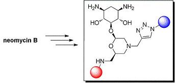
2-Deoxystreptamine Conjugates by Truncation–Derivatization of Neomycin

by
M. Waqar Aslam
1,
Leandro C. Tabares
2,
Alessio Andreoni
2,
Gerard W. Canters
2,
Floris P.J.T. Rutjes
1 and
Floris L. Van Delft
1,*
1
Radboud University Nijmegen, IMM Organic Chemistry, Heyendaalseweg 135, 6525 AJ, Nijmegen, the Netherlands
2
Leiden Institute of Chemistry, Leiden University, Einsteinweg 55, 2300 RA, Leiden, the Netherlands
*
Author to whom correspondence should be addressed.
Pharmaceuticals 2010, 3(3), 679-701; https://0-doi-org.brum.beds.ac.uk/10.3390/ph3030679
Submission received: 18 December 2009 / Revised: 8 March 2010 / Accepted: 10 March 2010 / Published: 15 March 2010
(This article belongs to the Special Issue Antibiotics)

Abstract

:
A small library of truncated neomycin-conjugates is prepared by consecutive removal of 2,6-diaminoglucose rings, oxidation-reductive amination of ribose, oxidation-conjugation of aminopyridine/aminoquinoline and finally dimerization. The dimeric conjugates were evaluated for antibacterial activity with a unique hemocyanin-based biosensor. Based on the outcome of these results, a second-generation set of monomeric conjugates was prepared and found to display significant antibacterial activity, in particular with respect to kanamycin-resistant E. coli.

Graphical Abstract

Introduction

Aminoglycosides, a group of naturally occurring compounds obtained from actinomycetes of the genus Streptomyces or Micromonospora [1] are in clinical use as antibiotics as a result of their broad antimicrobial spectrum and rapid bactericidal effects [2]. Aminoglycosides bind to 16S ribosomal RNA at the tRNA acceptor A-site (aminoacyl site), and affect the ability of the ribosome to decode mRNA correctly during protein synthesis [3,4,5]. Unfortunately, toxic side effects and growing bacterial resistance [6,7] have narrowed the significance of aminoglycosides as antibiotics. The most common mechanism of resistance is the enzymatic modification of one or more functional groups of the aminoglycoside drug by bacterial enzymes [8,9,10]. Due to these limitations, aminoglycosides are the focus of attention of research groups around the world and numerous structural analogues of the aminoglycosides have been synthesized over the years [11,12,13]. The main objective of the synthetic modifications of the aminoglycosides is to circumvent the bacterial resistance without loss in binding affinity of these drugs. In the majority of studies, naturally occurring aminoglycosides are modified by regioselective diversifications of the appropriate functional groups while keeping the whole structure intact [14,15,16,17,18,19,20,21]. However, it is clear that structures with a high resemblance to the natural compounds are most likely to undergo modification by bacterial resistance enzymes. Therefore, unlike this strategy we intended to utilize a minimal core element for the development of new structural analogues. Because bacterial enzymes have evolved to modify the structures of naturally occurring aminoglycosides, stripping off the targeted alcohol and amino functions evades the problem of bacterial resistance. On the other hand, such a strategy will concomitantly also reduce antibacterial activity because the same heteroatoms are responsible for RNA binding. Therefore, in order to restore affinity, lost by functional group removal, we envisaged conjugation of such a truncated aminoglycoside with a non-aminoglycoside type RNA ligand. Such a strategy has earlier proven successful for conjugation of native aminoglycosides to acridines [22,23], nucleobases [24], nucleotides [25], peptides [26,27], and other antibiotics [28]. Also, diversification of neamine as a structural motif for the synthesis of RNA ligands has been explored by several research groups [29,30,31,32,33]. However, neamine still contains the diaminosugar ring I of aminoglycosides, and is therefore a substrate for several resistance enzymes. Dimeric aminoglycoside have also divulged an improved RNA binding than individual aminoglycosides [34,35,36,37,38,39,40,41,42,43], therefore we made the dimers of our conjugates with conformationally adaptable linkers to further enhance binding affinity. These compounds were then tested for antibacterial activity against E. coli with a fluorescence-based assay.

Results and Discussion

Synthesis of 5-O-Morpholino-2-Deoxystreptamine

Our strategy is based on the fundamental observation that the key structural feature of (nearly) all aminoglycosides is not a carbohydrate but a diaminocyclohexitol ring termed 2-deoxystreptamine [44]. It was hypothesized that 2-deoxystreptamine is a crucial scaffold to build aminoglycoside libraries and that the all-equatorial substitution pattern is highly favorable to position other pharmacophores in the proper orientation. Although a large number of synthetic routes to 2-deoxystreptamine have been developed over the years [44], including contributions from our own lab [45,46,47,48], we realized that the most straightforward and cheapest route to 2-deoxystreptamine commences from natural neomycin. Apart from that, we surmised that partial degradation of neomycin would leave the ribofuranoside as a suitable substituent at the 5-position of 2-deoxystreptamine, as in structure 2 (Scheme 1). Thus, N-Boc-protected neomycin 1 [49] was reacted with 12 equivalents of sodium metaperiodate resulting in the oxidative cleavage of vicinal diols on both 2,6-diaminosugars. The intermediate tetraaldehyde was not purified due to its instability, but upon base treatment underwent smooth β-elimination under the influence of an amine base, to give 5-O-ribosyl-2-deoxystreptamine 2. Use of different bases like n-butyl amine, ammonium hydroxide and triethylamine gave the same results in terms of yield and reaction time. Importantly, 2 was purified by direct recrystallization from dichloromethane leading to highly pure product 2 in a 48% average yield over the two steps. The resulting pseudodisaccharide 2 was once again subjected to sodium metaperiodate cleavage of the diol to give a dialdehyde, this time followed by reductive amination with propargylamine, to afford morpholine 3 substituted with an acetylene moiety. Unfortunately, the formation of open chain diamine side products could not be fully avoided, thus significantly suppressing the yield.
Scheme 1. Synthesis of acetylene-substituted morpholino-2-deoxystreptamine conjugate.
Pharmaceuticals 03 00679 g001

Synthesis and Conjugation of Aminopyridines and Aminoquinolines

As aminoglycoside antibiotics function by selective recognition and binding to a specific RNA sequence, the removal of two of the aminoglycopyranosides is detrimental for RNA affinity. Therefore, we opted to restore the binding affinity by conjugation with other RNA ligands. To this end, we surmised that aminopyridines and aminoquinolines [50] could nicely serve our purpose because these molecules are of low molecular weight and contain no aliphatic amine functionality, but nevertheless have been reported to bind with E. coli A-site RNA in micromolar range. We selected the two tightest binders from the series of aminopyridines, e.g., 2-(2-aminoethylamino)-4-methylpyridine and 2-(2-aminoethylamino)-5-methylpyridine, and the best aminoquinoline ligand 2-(2-aminoethylamino)-4-methylquinoline (Scheme 2). In order to be able to conjugate the arylamine ligands to our morpholine compound, we designed a route involving reductive amination via the primary alcohol of 3. Therefore, we first prepared derivatives of the arylamines by treating the commercially available chloropyridines and a chloroquinoline with 1,2-ethylenediamine at 150 °C for 18 hours as shown in the Scheme 2, to afford the desired 2-aminoethyl modified arylamines 57 in reasonable yield.
Scheme 2. Synthesis of substituted aminopyridines and aminoquinolines.
Pharmaceuticals 03 00679 g002
Having the primary amines at hand, the next step involved the diversification of morpholine 3 by conjugation with the aminoethyl derivatives 57. For this purpose the primary hydroxyl of the compound 3 appeared most appropriate since it is most accessible and leaves the chiral secondary alcohols of 2-deoxystreptamine intact. Since reductive amination is such a robust conjugation technology, we aimed to selectively oxidize the primary alcohol of 3. Because it is known that Swern oxidations can be selectively executed at O-TES protected primary alcohols [51], we treated compound 3 with TESOTf and triethylamine in dichloromethane to protect all hydroxyls with the TES group. Subsequently, the triply TES-protected derivative was subjected to Swern oxidation conditions (oxalyl chloride, DMSO, -78 °C→0 °C), in order to selectively oxidize the primary hydroxyl. However, an inseparable mixture of compounds was obtained (Table 1, entry 1).
Scheme 3. Conjugation of morpholineto aminopyridines and aminoquinolines.
Pharmaceuticals 03 00679 g003
Because Swern oxidation of TES ethers was not successful, we opted for a selective oxidation of the primary alcohol without protective groups. However, attempts to oxidize the primary hydroxyl with TCCA/TEMPO [52] (entry 2) or IBX [53] (entry 3) gave no or less than 10% conversion, respectively. Finally, we succeeded (entry 4) in oxidizing the primary hydroxyl group with Dess-Martin periodinane [54] to give selectively the desired aldehyde 4b. However, also in this case the reaction did not go to completion, reaching a maximum of about 60% conversion. Further attempts to optimize the reaction conditions, e.g., variation in temperature, solvents or stoichiometry of the oxidant did not improve the outcome.
Table 1. Conditions for selective oxidation of primary alcohal.
EntryConditionsProductYield
1i) TESOTf, Et3N, CH2Cl2, r.t., 3 h
ii) (COCl)2, DMSO, CH2Cl2, -78 °C→0°C
4amixture
2TCCA, TEMPO, CH3CN, r.t., 16 h4bno reaction
3IBX, DMF, r.t.→80 °C, 16 h4b<10% conversion
4DMP, DMF, r.t., 16 h4b60% conversion
Reagents and conditions: a) 5/6/7, NaBH3CN, AcOH, MeOH, 3 h. b) Boc2O, DMAP, CH2Cl2, 4 h (9a: 34%, 9b: 37%, 9c: 32%, for two steps).
The resulting aldehyde 4b was subjected to the next reaction without purification on account of its instability. We therefore proceeded to couple crude aldehyde 4b with aminopyridine and aminoquinoline derivatives 5–7 (Scheme 3). Therefore, compounds 5–7 were condensed with aldehyde 4b employing standard reductive amination conditions, e.g., NaBH3CN and AcOH in methanol. The relatively low yields of the resulting compounds 8a-c can be ascribed to incomplete oxidation of the compound 3 in the proceeding step, apart from the fact that accompanying dialkylation of amines was observed during the reductive amination. However, in this case the desired conjugates 9a-c could be readily purified by silica gel column chromatography. Both for this purpose and for follow-up chemistry, it was found most convenient to convert the resulting secondary amino functions of the initial products 8a-c into Boc-protected carbamates under the influence of Boc2O and DMAP in CH2Cl2. Thus, heteroconjugates 9a-c of truncated neomycin with non-aminoglycoside type RNA ligands were successfully prepared.

Dimerization and Deprotection of the Conjugates

Obviously, the obtained arylamine-morpholine-2-deoxystreptamine conjugates contain a functional handle at the morpholine ring in the form of the propargyl moiety. Based on the popular copper(I)-catalyzed azide acetylene cycloaddition (CuAAC) [55,56], it is clear that the propargyl moiety provides an excellent handle for the preparation of further conjugates. Therefore, we next utilized the copper-catalyzed (3+2) cycloaddition chemistry to make immers with a range of mono- and bisazido-functionalized linkers. This approach provided maximum synthetic flexibility and proved effective for the synthesis of various monomers and immers from the building blocks 9a-c. Thus, the synthetic heteroconjugates were treated with a monoazide (benzyl azide, 1 equiv.) or bisazides A-E (0.5 equiv.) in a mixture of water and acetonitrile in the presence of copper-wire [57] to make the respective monomers 9a-cE and dimers 9a-cA to 9a-cD in reasonable to good yields (Table 2). Finally, acidic deprotection of the Boc protective groups was performed in a 1:2 mixture of TFA and DCM to afford the respective monomers and the dimers in quantitative yields, giving a final set of 15 arylamine-aminoglycoside conjugates (3 monomers, 12 dimers). For biological evaluation, all of the compounds were purified by reversed phase HPLC, giving the pure conjugates in 5–20 mg quantitities.
Scheme 4. Dimerization and deprotection of the conjugates.
Pharmaceuticals 03 00679 g004
Table 2. Linkers used for synthesis of dimers 9aA-9cE.
EntryLinker (L)StartingProductYield
material
1 Pharmaceuticals 03 00679 i0019a9aA63%
29b9bA73%
39c9cA60%
4 Pharmaceuticals 03 00679 i0029a9aB57%
59b9bB65%
69c9cB58%
7 Pharmaceuticals 03 00679 i0039a9aC61%
89b9bC66%
99c9cC69%
10 Pharmaceuticals 03 00679 i0049a9aD60%
119b9bD65%
129c9cD66%
13 Pharmaceuticals 03 00679 i0059a9aE87%
149b9bE92%
159c9cE90%

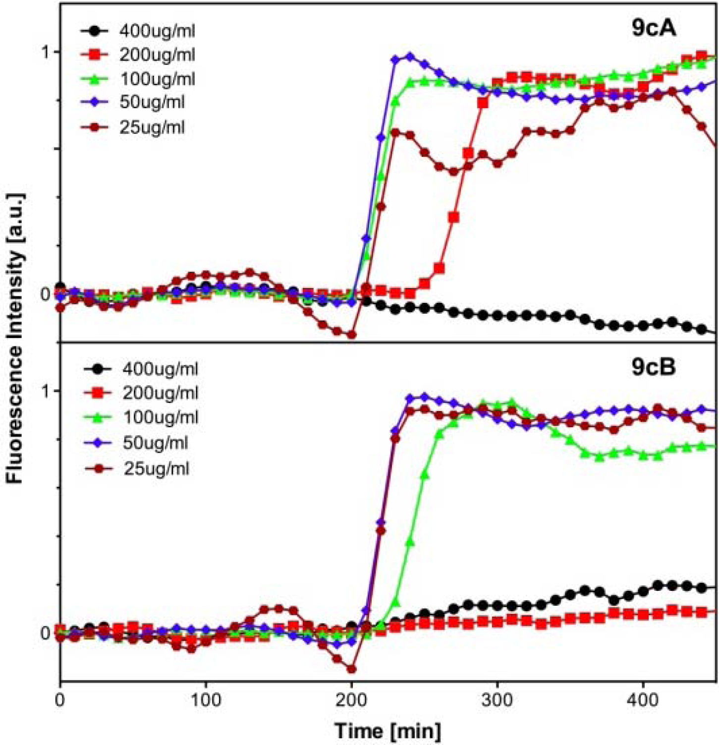
Evaluation of Antibacterial Activity

Having prepared the desired conjugates, we were interested to investigate the antibacterial activity of these compounds. Determination of antibacterial activity is normally expressed in MIC (minimal inhibitory concentration) by growing bacteria in medium containing increasing amounts of the presumed antibiotic. Such an assay typically requires substantial amounts of compounds (30–70 mg), clearly exceeding the amount of compound that we had prepared. With small amounts of substrate, antibacterial activity can also be determined with Kirby-Bauer disc test, but that provides only a qualitative measure of activity. Alternatively, a plethora of RNA-binding assays have been developed [58,59,60,61,62,63,64,65,66,67,68,69], but a strong drawback of such assays is that they do not include cell-wall penetration or resistance mechanisms, obviously key in determining the overall activity of an antibiotic [70]. An alternative approach, recently reported by some of us [71], determines MICs on whole bacteria in a simple and rapid assay and moreover requires only minimal amounts (< 2 mg) of substrate. The technique relies on a fluorescence-based cell viability assay and involves a hemocyanin-based oxygen biosensor to monitor oxygen consumption of bacteria and subsequently bacterial cell growth (Figure 1).
Figure 1. (A) Representation of Cy3-labeled hemocyanin. The Cy3 dye (cyan) is showed attached to the end terminus of a monomer hemocyanin. (B) Detail of hemocyanin active site showing the active site histidines (blue), copper ions (orange) and the binding oxygen molecule (red). (C) The absorption spectra of the deoxygenated (red) and oxygenated (blue) forms of hemocyanin together with the emission spectrum of Cy3 (green).
Pharmaceuticals 03 00679 g005
Deoxygenated hemocyanin has no absorption in the visible spectrum but upon oxygenation a strong band centered at 340 nm and a weak band around 570 nm appear (Figure 1B). If a fluorescent label is attached to the N-terminus of hemocyanin, this change in absorption can be translated into a change of fluorescence intensity of an attached label (Cy3, Cyanine 3 fluorescent dye) through a Förster resonance energy transfer (FRET) mechanism (Figure 1A) [72]. The efficiency of this energy transfer depends on the distance and orientation between the donor and the acceptor and also on the overlap integral between the donor emission and acceptor absorption spectra (Figure 1C). In this way, when the protein is in its deoxygenated state (Cy3-Deoxy-Hemocyanin), all the energy absorbed by the label is emitted normally as fluorescence. Upon oxygenation of hemocyanin (Cy3-Oxy-Hemocyanin), energy is transferred from Cy3 by radiationless decay to the copper center of hemocyanin, resulting in a decrease in the fluorescence intensity. Thus, when a bacterial culture grows aerobically, oxygen in the medium is gradually consumed. This results in a drop of the oxygen concentration, eventually resulting in an anaerobic medium. When hemocyanin-based oxygen biosensor is present, this oxygen depletion results in an increase of the fluorescence intensity emitted by the Cy3 label attached to protein [71]. Tests were carried out as described [71] with an initial cell concentration of 105 cfu/mL.
Figure 2. Fluorescence intensity time traces at 570 nm (excitation 550 nm) of cultures of E. coli containing Cy3-labeled hemocyanin as a function of antibiotic concentration. Antibiotic concentrations of 9cA and 9cB are stated.
Pharmaceuticals 03 00679 g006
We determined the MICs of our heteroconjugates with the fluorescence-based cell viability assay against several bacterial strains (E. coli, E. coli kan.r e.g., kanamycin-resistant E. coli) using kanamycin as a positive control. As can be seen from Figure 2, using compound 9cA and 9cB as an example, the MIC is determined by performing the bacterial growth in medium containing increasing amounts of our compounds. The MIC values of our synthetic analogues can be determined from Figure 2 and are depicted in Table 3.
Table 3. MIC values (µg/ mL) as determind by the fluorescence-based cell growth assay.
Entrybacterial strainMIC (μg/ mL)
kanamycins.m.linker Alinker Blinker Clinker Dlinker E
1E. coli109a200–400200–400400–800400–800400–800
2-do-109b400–800400–800400–800400–800400–800
3-do-109c200–400100–200200–400400–800200–400

Second Generation Monomeric Ligands

Unfortunately, our set of dimers of heteroconjugates exhibited lower antibacterial activity than that of natural kanamycin when tested against E. coli. In fact, only compound 9cB demonstrated reasonable antibacterial activity. Moreover, and in contrast to expectation, it is clear that the dimers do not show improved activity compared to the monomeric substrates 9a-cE.
Scheme 5. Synthesis and deprotection of monomeric ligands.
Pharmaceuticals 03 00679 g007
The lack of improved activity is in contrast to findings by other groups [34,35,36,37,38,39,40,41,42,43], which may be explained in our case by the relatively short linkers employed. It is not excluded that a short linker is precluding cooperative binding of the other end of the molecule to a second binding pocket. Whatever the reason, the fact that the MIC value of the monomeric compound 9cE was comparable to that of different dimers stimulated us to explore whether antibacterial activity of the more simple, monomeric heteroconjugates could be further raised by modification of the azide partner for cycloaddition to the acetylene. Thus, we selected a range of aromatic and aliphatic azides and coupled these to acetylene derivatives 9a-c with CuAAC to obtain a set of monomeric compounds 10a-g with different substituents (Scheme 5). Thus, compounds 10a–e were synthesized by simply treating 9c with different monoazides in a mixture of water and acetonitrile in the presence of copper wire. For the synthesis of amino-terminated compounds 10f and 10g, acetylene 9c was treated with an excess of 1,4-bis(azidomethyl)benzene or 1,5-diazidopentane, to give the respective monosubstituted triazole adducts, followed by Staudinger reduction of the unreacted azide group at the other chain terminus under the action of PMe3. Again, all compounds 10a-g were purified to homogeneity by reversed-phased HPLC before evaluation of antibacterial activity. Next, we tested the antibacterial activity of this set of compounds employing the same fluorescence-based cell viability assay and the results are summarized in Table 3. As evident from MIC values, para-bromo substitution of the aromatic ring enhanced the antibacterial activity of compounds 10a and 10b. The rest of the monomeric compounds, with a methoxy- or methylamino-substituent on the aromatic ring 10c-10f, or with the aliphatic chain 10g showed a reduced antibacterial activity compared to that of unsubstituted parent compound 9cE. Finally we tested the MIC of the most active compounds against the kanamycin resistant strains of E. coli (Table 4, entry 2). We were happy to find that compounds 10a and 10b have distinct antibacterial activity against E. coli. More importantly, compound 10b displayed an improved activity against the kanamycin-resistant E. coli strain revealing that omission of the diaminoglycosides indeed fulfills our desired aim to reduce susceptibility to resistance mechanisms. The search for further synthetic analogues of aminoglycosides with such improved traits is currently ongoing in our laboratory.
Table 4. MIC values (μg/mL) as determined by fluorescence-based cell growth assay.
Entrybacterial strainsMIC (μg/mL)
kanamycin10a10b10c10d10e10f10g9cB9cE
1E. coli10100-200100-200400-800400-800400-800400-800400-800--
2E. coli kanr400–800400-800200-400>800---->800>800

Experimental Section

General

TLC analyses are performed on silica gel-coated plates (Merck 60 F254) with the indicated solvent mixture. Compounds were detected with ammonium molybdate or potassium permanganate staining, or UV light. Flash column chromatography was carried out using ACROS silica gel (0.035–0.070 mm, ca 6 nm pore diameter). Solvents were distilled from appropriate drying agents prior to use. Unless stated otherwise, all chemicals were purchased and used as such. IR spectra were recorded on an ATI Mattson Genesis Series FTIR spectrometer; absorption reported in cm-1. NMR spectra were recorded on a Bruker DMX 300 (75 MHz), and a Varian 400 (400 MHz) spectrometer. 1H-NMR and 13C-NMR spectra are reported in ppm units on the δ scale. Coupling constants are reported as J-values in Hz. Semipreparative RP-HPLC was performed using a C18 column (10 × 250 mm, 5 µm) with HPLC grade MilliQ containing 0.1% TFA (eluant A) and acetonitrile containing 0.1% TFA (eluant B) with a gradient of 5 → 50% of B in 30 min. Compounds were detected by UV absorption at 215 and 254 nm.

Hexa-N-(tert-butoxycarbonyl) neomycin B (1) [49]

To a solution of neomycin B (2 g, 2.2 mmol) in a mixture of H2O/1,4-dioxane (30 mL, 1:1 v/v) was added Na2CO3 and di-tert-butyl dicarbonate. The reaction mixture was stirred at room temperature for 6 h. Dioxane was removed by evaporation and the residue was partitioned between EtOAc (100 mL) and H2O (100 mL). The aqueous layer was extracted with EtOAc (2 ×50 mL) and the combined organic layers werewashed with brine (100 mL), dried (Na2SO4) and concentrated in vacuo. Purification by column chromatography (MeOH/CH2Cl2, 1/9) afforded 1 (2.2 g, 82%) as an amorphous white solid. RF 0.49 (MeOH/CH2Cl2, 1/9). HRMS (ESI) m/z calcd. for C53H94N6O25 (M+Na)+: 1237.6166, found: 1237.6141.

5-O-(β-d-ribofuranosyl)-2-deoxystreptamine (2)

To a solution of 1 (1 g, 0.82 mmol) in 25 mL of MeOH, was added 25 mL of a H2O solution of NaIO4 (2.12 g, 9.8 mmol). The reaction mixture was stirred at room temperature for 16 h. Methanol was removed by evaporation and the residue was partitioned between EtOAc (100 mL) and H2O (50 mL). The aqueous layer was extracted with EtOAc (2 × 50 mL) and the combined organic layers werewashed with brine (100 mL), dried (Na2SO4) and concentrated in vacuo. The residue was dissolved in MeOH (30 mL), and Et3N (2 mL) was added. The reaction mixture was stirred at room temperature for 16 h and then concentrated in vacuo. Purification by crystallization from CH2Cl2 (100 mL) afforded compound 2 (212 mg, 52%) as a white amorphous solid. RF 0.12 (MeOH/CH2Cl2, 1/9); 1H-NMR (MeOD, 400 MHz) δ 5.21 (s, 1H), 4.32 (dd, J =4.5, 7.7 Hz, 1H), 3.99 (d, J =4.5 Hz, 1H), 3.90 (dt, J =2.6, 7.7 Hz, 1H), 3.78 (dd, J =2.3, 12.1 Hz, 1H), 3.60 (dd, J =2.6, 12.1 Hz, 1H), 3.40–3.15 (m, 10H), 2.02 (dt, J =4.2, 7.9 Hz, 1H), 1.41 (s, 18H), 1.21 (m, 1H); 13C-NMR (MeOD, 75 MHz): δ 156.3, 107.8, 83.8, 82.3, 78.2, 74.7, 72.7, 68.6, 59.7, 50.6, 33.8,26.8; HRMS (ESI) m/z calcd. for C21H38N2O11 (M+Na)+: 517.2373, found: 517.2375.

5-O-(N-propyn-1-yl -morpholino)-2-deoxystreptamine (3)

To a solution of 2 (830 mg, 1.68 mmol) in 20 mL of MeOH, was added 20 mL of a H2O solution of NaIO4 (431 mg, 2.016 mmol). The reaction mixture was stirred at room temperature for 2 h. The methanol was removed by evaporation and the residue was partitioned between EtOAc (100 mL) and H2O (50 mL). The aqueous layer was extracted with EtOAc (2 ×50 mL) and the combined organic layers werewashed with brine (100 mL), dried (Na2SO4) and concentrated in vacuo. The residue was dissolved in 15 mL of MeOH, and NaBH3CN (422 mg, 6.72 mmol) was added. To the resulting mixture was added drop-wise a solution of propargylamine (138 μL, 2.52 mmol) and AcOH (403 μL, 6.72 mmol) in MeOH (1 mL). The reaction mixture was stirred at room temperature for 2 h. The reaction was then quenched with 0.5 mL of Et3N and concentrated in vacuo. The residue was taken up in EtOAc (100 mL), washed with saturated NaHCO3 (2 × 50 mL) and brine (50 mL). The organic layer was dried (Na2SO4), filtered and concentrated in vacuo.Purification by column chromatography (MeOH/CH2Cl2, 1/9) afforded 1 (358 mg, 41%) as an amorphous white solid. RF 0.34 (MeOH/CH2Cl2, 1/9); 1H-NMR (MeOD, 400 MHz) δ 4.85 (dd, J = 2.3, 8.6 Hz, 1H), 3.70 (m, 1H), 3.57 (dd, J = 1.7, 5.1Hz, 2H), 3.42–3.19 (m, 11H), 3.01 (d, J = 10.5 Hz, 1H), 2.70 (d, J = 11.1 Hz, 1H), 2.65 (t, J = 2.3 Hz, 1H), 2.13 (m, 2H), 2.02 (dt, J = 3.9, 13.0 Hz, 1H), 1.41 (s, 18H), 1.21 (dd, J = 12.5 Hz, 1H). 13C-NMR (MeOD, 75 MHz): δ 156.2, 99.7, 85.2, 78.1, 76.7, 74.1, 73.5, 72.5, 61.9, 54.1, 51.3, 50.6, 50.4, 45.1, 33.6, 26.8; HRMS (ESI) m/z calcd. for C24H41N3O9 (M+H)+: 516.2921, found: 516.2925.

2-(2-Aminoethylamino)-4-methylpyridine (5)

A solution of 2-chloro-4-methylpyridine (0.876 mL, 10 mmol) in 1,2-diaminoethane (6 mL) was heated at 150°C for 18 h. The volatiles were removed in vacuo. Purification by column chromatography (10% MeOH/CH2Cl2 to 20% MeOH/CH2Cl2) afforded 5 (700 mg, 46%) as a yellow oil; 1H-NMR (MeOD, 300 MHz) δ 7.76 (d, J = 4.8 Hz, 1H), 6.36 (m, 2H), 3.31 (t, J = 6.2 Hz, 2H), 2.79 (t, J = 6.2 Hz, 2H), 2.17 (s, 3H); 13C-NMR (MeOD, 75 MHz): δ 156.5, 145.0, 136.2, 121.1, 106.6, 40.1, 39.9, 15.5.HRMS (ESI) m/z calcd. for C8H13N3 (M+H)+: 152.1187, found: 152.1199.

2-(2-Aminoethylamino)-5-methylpyridine (6)

A solution of 2-chloro-5-methylpyridine (0.434 mL, 5 mmol) in 1,2-diaminoethane (3 mL) was heated at 150°C for 18 h. The volatiles were removed in vacuo. Purification by column chromatography (10% MeOH/CH2Cl2 to 20% MeOH/CH2Cl2) afforded 6 (386 mg, 51%) as a yellow oil; 1H-NMR (MeOD, 300 MHz) δ 7.72 (m, 1H), 7.27 (dd, J = 2.5, 8.5 Hz, 1H), 6.46 (d, J = 8.5 Hz, 1H), 3.30 (t, J = 6.2 Hz, 2H), 2.79 (t, J = 6.2 Hz, 2H), 2.13 (s, 3H). 13C-NMR (MeOD, 75 MHz): δ 156.6, 145.3, 138.0, 120.3, 107.7, 43.5, 40.1, 15.5; HRMS (ESI) m/z calcd. For C8H13N3 (M+H)+: 152.1187, found: 152.1199.

2-(2-Aminoethylamino)-4-methylquinoline (7)

A solution of 2-chloro-4-methylquinoline (1.7 g, 10 mmol) in 1,2-diaminoethane (6 mL) was heated at 150°C for 18 h. The volatiles were removed in vacuo. Purification by column chromatography (10% MeOH/CH2Cl2 to 20% MeOH/CH2Cl2) afforded 7 (963 mg, 48%) as a yellow oil; 1H-NMR (MeOD, 300 MHz) δ 7.72 (dd, J = 1.2, 8.1 Hz, 1H), 7.59 (dq, J = 0.5, 1.2, 8.4 Hz, 1H), 7.45 (ddd, J = 1.4, 6.9, 8.4 Hz, 1H), 7.17 (ddd, J = 1.2, 6.9, 8.1 Hz, 1H), 6.59 (d, J = 1.0 Hz, 1H), 3.50 (t, J = 6.2 Hz, 2H), 2.86 (t, J = 6.2 Hz, 2H), 2.48 (d, J = 1.0 Hz, 3H). 13C-NMR (MeOD, 75 MHz): δ 156.9, 147.0, 144.2, 128.4, 124.5, 123.0, 122.8, 120.9, 111.9, 42.6, 40.3, 16.9; HRMS (ESI) m/z calcd. for C12H15N3 (M+H)+: 202.1344, found: 202.1335.

5-O-(N-propyn-1-yl-2-(methylamino-N-ethylamino-N -4-methylpyridin-2-yl)-morpholino)-2-deoxystreptamine 8a

To a solution of 3 (65 mg, 0.126 mmol) in DMF (1 mL), was added Dess-Martin periodinane (64 mg, 0.151mmol). The reaction mixture was stirred overnight at room temperature. The reaction was quenched with a saturated solution of NaHCO3 (20 mL) and extracted with EtOAc (3 ×25 mL). The combined organic layers were washed with H2O (2 × 25 mL) and brine (25 mL), dried (Na2SO4) and concentrated in vacuo. The residue was dissolved in 2 mL of MeOH, and NaBH3CN (12 mg, 0.189 mmol) was added. To the reaction mixture was added a solution of 5 (57 mg, 0.378 mmol) and AcOH (11 μL, 0.189 mmol) in MeOH (0.5 mL). The reaction mixture was stirred at room temperature for 4 h. The reaction was quenched with 0.1 mL of Et3N and concentrated in vacuo. The residue was taken up in EtOAc (50 mL), washed with saturated NaHCO3 (2 × 25 mL) and brine (25 mL). The organic layer was dried (Na2SO4), filtered and concentrated in vacuo. Purification by column chromatography (MeOH/CH2Cl2, 1/9) afforded 8a (32 mg, 39%) as a white solid. RF 0.22 (MeOH/CH2Cl2, 1/9); 1H- NMR (MeOD, 400 MHz) δ 7.89 (d, J = 5.4 Hz, 1H), 6.52 (d, J = 5.4 Hz, 1H), 6.47 (s, 1H), 5.03 (d, J = 7.5 Hz, 1H), 3.96 (m, 1H), 3.57 (q, J = 5.6, 10.3 Hz, 2H), 3.41–3.15 (m, 12H), 2.94 (d, J = 10.3 Hz, 1H), 2.70 (m, 2H), 2.29 (dd, J = 7.7, 11.0 Hz, 1H), 2.22 (s, 3H), 1.99 (m. 1H), 1.41 (d, J = 8.4 Hz, 18H), 1.24 (m, 1H); 13C-NMR (MeOD, 75 MHz): δ 156.3, 156.2, 149.1, 144.9, 126.9, 114.3, 109.2, 99.5, 83.2, 78.2, 76.4, 74.8, 73.7, 72.8, 69.1, 54.0, 51.5, 48.7, 45.0, 38.2, 33.8, 26.8, 19.2; HRMS (ESI) m/z calcd. for C32H52N6O8 (M+H)+: 649.3924, found: 649.3923.

5-O-(N-propyn-1-yl-2-(methylamino-N-ethylamino-N -5-methylpyridin-2-yl)-morpholino)-2-deoxystreptamine 8b

To a solution of 3 (80 mg, 0.155 mmol) in DMF (1 mL), was added Dess-Martin periodinane (79 mg, 0.186 mmol). The reaction mixture was stirred overnight at room temperature. The reaction was quenched with a saturated solution of NaHCO3 (20 mL) and extracted with EtOAc (3 × 25 mL). The combined organic layers were washed with H2O (2 × 25 mL) and brine (25 mL), dried (Na2SO4) and concentrated in vacuo. The residue was dissolved in 3 mL of MeOH, and NaBH3CN (14 mg, 0.232 mmol) was added. To the resulting mixture was added a solution of 6 (70 mg, 0.465 mmol) and AcOH (14 μL, 0.232 mmol) in MeOH (0.5 mL). The reaction mixture was stirred at room temperature for 4 h. The reaction was quenched with 0.1 mL of Et3N and concentrated in vacuo. The residue was taken up in EtOAc (50 mL), washed with saturated NaHCO3 (2 × 25 mL) and brine (25 mL). The organic layer was dried (Na2SO4), filtered and concentrated in vacuo. Purification by column chromatography (MeOH/CH2Cl2, 1/9) afforded 8b (41 mg, 41%) as a white solid. RF 0.22 (MeOH/CH2Cl2, 1/9); 1H- NMR (MeOD, 400 MHz) δ 7.74 (s, 1Η),7.28 (dd, J = 2.3, 8.5 Hz, 1H), 6.5 (d, J = 8.5 Hz, 1H), 4.85 (dd, J = 2.3, 8.6 Hz, 1H), 3.80 (m, 1H), 3.42–3.19 (m, 11H), 2.99 (d, J = 10.3 Hz, 1H), 2.18–2.00 (m, 5H), 1.40 (d, J = 7.2 Hz, 18H), 1.26 (m, 1H); 13C-NMR (MeOD, 75 MHz): δ 156.6, 156.3, 156.1, 145.1, 138.2, 120.5, 108.1, 99.8, 85.2, 78.2, 76.6, 76.2, 74.2, 73.5, 72.6, 71.7, 54.1, 52.5, 50.5, 50.0, 45.0, 39.9, 33.8, 26.8, 15.4; HRMS (ESI) m/z calcd. for C32H52N6O8 (M+H)+: 649.3924, found: 649.3923.

5-O-(N-propyn-1-yl-2-(methylamino-N-ethylamino-N-4-methylquinolin-2-yl)-morpholino)-2-deoxystreptamine 8c

To a solution of 3 (100 mg, 0.194 mmol) in DMF (1 mL), was added Dess-Martin periodinane (99 mg, 0.232 mmol). The reaction mixture was stirred overnight at room temperature. The reaction was quenched with saturated solution of NaHCO3 (20 mL) and extracted with EtOAc (3 × 25 mL). The combined organic layers were washed with H2O (2 × 25 mL) and brine (25 mL), dried (Na2SO4) and concentrated in vacuo. The residue was dissolved in 4 mL of MeOH, and NaBH3CN (18 mg, 0.291 mmol) was added. To the resulting mixture was added a solution of 7 (116 mg, 0.582 mmol) and AcOH (17.5 μL, 0.291 mmol) in MeOH (0.5 mL). The reaction mixture was stirred at room temperature for 4 h. The reaction was quenched with 0.1 mL of Et3N and concentrated in vacuo. The residue was taken up in EtOAc (50 mL), washed with saturated NaHCO3 (2 × 25 mL) and brine (25 mL). The organic layer was dried (Na2SO4), filtered and concentrated in vacuo. Purification by column chromatography (MeOH/CH2Cl2, 1/9) afforded 8c (52 mg, 38%) as a white solid. RF 0.21 (MeOH/CH2Cl2, 1/9); 1H-NMR (MeOD, 400 MHz) δ 7.84 (d, J = 8.1 Hz, 1H), 7.62 (d, J = 3.4 Hz, 1H), 7.30 (q, J = 4.1, 8.1 Hz, 1H), 6.72 (d, J = 1.0 Hz, 1H), 5.06 (d, J = 6.3 Hz, 1H), 3.97 (m, 1H), 3.75 (t, J = 5.0 Hz, 2H), 3.44–3.18 (m, 13H), 2.95 (d, J = 11.0 Hz, 1H), 2.72–2.65 (m, 2H), 2.57 (d, J = 1.0 Hz, 3H), 2.30 (dd, J = 7.7, 11.0 Hz, 1H), 2.17 (dd, J = 9.3, 11.1 Hz, 1H), 2.03 (dt, J = 3.8, 12.5 Hz, 1H), 1.42 (d, J = 9.1 Hz, 18H), 1.32 (m, 1H); 13C-NMR (MeOD, 75 MHz): δ 156.7, 145.9, 145.2, 129.4, 123.7, 123.3, 123.1, 122.0, 112.0, 99.6, 83.5, 78.2, 76.4, 74.7, 73.7, 72.8, 69.2, 54.0, 51.6, 49.1, 48.5, 45.0, 38.4, 26.9, 17.0; HRMS (ESI) m/z calcd. for C36H54N6O8 (M+H)+: 699.4081, found: 699.4062.

General procedure for Boc-protection of 9a-c

To a solution of 8a-c (0.15 mmol) in CH2Cl2 (2 mL) was added di-tert-butyl dicarbonate (0.18 mmol, 1.2 equiv.) and DMAP (5 mg). The reaction mixture was stirred at room temperature for 5 h. The volatiles were removed in vacuo. Purification by column chromatography (MeOH/CH2Cl2, 1/9) gave the desired product.
5-O-(N-propyn-1-yl-2-(methylamino-N-(Tert-Butoxycarbonyl)ethylamino-N -4-methylpyridin-2-yl)-morpholino)-2-deoxystreptamine 9a: Yield 87%, white solid. RF 0.43 (MeOH/CH2Cl2, 1/9); HRMS (ESI) m/z calcd. for C37H60N6O10 (M+H)+: 749.4449, found: 749.4434.
5-O-(N-propyn-1-yl-2-(methylamino-N-(Tert-Butoxycarbonyl)ethylamino-N -5-methylpyridin-2-yl)-orpholino)-2-deoxystreptamine 9b: Yield 90%, white solid. RF 0.43 (MeOH/CH2Cl2, 1/9); HRMS (ESI) m/z calcd. for C37H60N6O10 (M+H)+: 749.4449, found: 749.4434.
5-O-(N-propyn-1-yl-2-(methylamino-N-(Tert-Butoxycarbonyl)ethylamino-N -4-methylquinolin-2-yl)-morpholino)-2-deoxystreptamine 9c: Yield 84%, white solid. RF 0.46 (MeOH/CH2Cl2, 1/9); HRMS (ESI) m/z calcd. for C41H62N6O10 (M+H)+: 799.4605, found: 799.4587.

3.3. General procedure for the synthesis of bis-azides A-D

To a solution of a commercially obtained dibromide (2.5 mmol) in 10 mL of DMF was added NaN3 (6.0 mmol). The reaction mixture was stirred at 60 °C for 5 h. The reaction was then quenched with H2O and extracted with Et2O. The organic layers were washed with H2O and brine, dried (Na2SO4) and concentrated in vacuo. Purification by column chromatography (EtOAc/heptane, 1/20) gave the pure product.
1,4-bis(Azidomethyl)benzene (A): Yield 86%, oil. RF 0.34 (EtOAc/heptane, 1/8); 1H-NMR (CDCl3, 300 MHz) δ 7.34 (s, 4H), 4.35 (s, 4H); 13C-NMR (CDCl3, 75 MHz): δ 135.0, 128.1, 53.9.
1,2-bis(Azidomethyl)benzene (B): Yield 82%, oil. RF 0.36 (EtOAc/heptane, 1/8); 1H-NMR (CDCl3, 300 MHz) δ 7.38 (s, 4H), 4.43 (s, 4H); 13C-NMR (CDCl3, 75 MHz): δ 133.4, 129.6, 128.5, 51.7.
1,5-Diazidopentane (C): Yield 91%, liquid. RF 0.49 (EtOAc/heptane, 1/8); 1H-NMR (CDCl3, 300 MHz) δ3.25 (t, J = 6.7 Hz, 4H), 1.60 (m, 4H), 1.42 (m, 2H); 13C-NMR (CDCl3, 75 MHz) δ 50.7, 27.9, 23.4.
1,4-Diazidobutane (D): Yield 86%, liquid. RF 0.51 (EtOAc/heptane, 1/8); 1H-NMR (CDCl3, 300 MHz) δ 3.27(t, J = 5.7 Hz, 4H), 1.61 (m, 4H); 13C-NMR (CDCl3, 75 MHz) δ 50.4, 25.6.

1-Bromo-4-(azidomethyl)bromobenzene

To a solution of 1-bromo-4-(bromomethyl)benzene (200 mg, 0.80 mmol) in 1 mL of DMF was added NaN3 (52 mg, 0.80 mmol). The reaction mixture was stirred at 60 °Cfor 5 h. The reaction was then quenched with H2O and extracted with Et2O. The organic layers were washed with H2O and brine, dried (Na2SO4)and concentrated in vacuo. Purification by column chromatography (EtOAc/heptane, 1/20) gave 1-bromo-4-(azidomethyl)benzene (156 mg, 92%) as a colorless liquid. RF 0.51 (EtOAc/heptane, 1/8); 1H-NMR (CDCl3, 400 MHz) δ 7.47 (d, J = 8.4 Hz, 2H), 7.15 (d, J = 8.4 Hz, 2H), 4.24 (s, 2H); 13C-NMR (CDCl3, 75 MHz) δ 133.9, 131.5, 129.3, 121.8, 53.6.

1-Azido-4-bromobenzene

To a solution of NaN3 (1.5 g, 24.3mmol) in a mixture of H2O/CH2Cl2 (7.5 mL, 1:1 v/v) at 0 °C, was added Tf2O (2.01 mL, 12.15mmol). The reaction mixture was stirred at room temperature for 2 h. After quenching with aqueous NaHCO3, the layers were separated and the aqueous layer was extracted with CH2Cl2 (5.75 mL). The organic layers were combined to afford 9.5 mL of TfN3 solution. Then, to a solution of para-bromoaniline (700mg, 4.06mmol) and CuSO4 (5 mg) in H2O (9.5 mL) was added the TfN3 solution, MeOH (31 mL) and Et3N (1.6 mL). The reaction mixture was stirred overnight at room temperature. Then solid NaHCO3 (1.0 g) was added carefully and the organic solvents were evaporated. The aqueous residue was extracted with EtOAc (3 × 100 mL), and the organic layers were combined, dried (Na2SO4), and concentrated in vacuo to give a yellow liquid. Purification by column chromatography (EtOAc/heptane, 1/20) afforded 1-azido-4-bromobenzene (630mg, 78%) as a colorless liquid. RF 0.61 (EtOAc/heptane, 1/8); 1H-NMR (CDCl3, 400 MHz) δ 7.44 (d, J = 8.9 Hz, 2H), 6.89 (d, J = 8.9 Hz, 2H); 13C-NMR (CDCl3, 75 MHz) δ 138.7, 132.3, 120.1, 117.2.

1-(Azidomethyl)-4-methoxybenzene

To a solution of 1-(chloromethyl)-4-methoxybenzene (400 μL, 2.9 mmol) in 2 mL of DMF was added NaN3 (188 mg, 2.9 mmol). The reaction mixture was stirred at 60 °C for 5 h. The reaction was then quenched with H2O and extracted with Et2O. The organic layers were washed with H2O and brine, dried (Na2SO4) and concentrated in vacuo. Purification by column chromatography (EtOAc/heptane, 1/20) gave 1-(azidomethyl)-4-methoxybenzene (410 mg, 87%) as a colorless liquid. RF 0.36 (EtOAc/heptane, 1/8); 1H-NMR (CDCl3, 400 MHz) δ 7.23(d, J = 8.8 Hz, 2H), 6.90 (d, J = 8.8 Hz, 2H), 4.24 (s, 2H), 3.78 (s, 3H); 13C-NMR (CDCl3, 75 MHz) δ 159.2, 129.2, 126.9, 113.7, 54.7, 53.9.

1-Azido-4-methoxybenzene

To a solution of NaN3 (1.5 g, 24.3mmol) in a mixture of H2O/CH2Cl2 (7.5 mL, 1:1 v/v) at 0 °C, was added Tf2O (2.01 mL, 12.15mmol). The reaction mixture was stirred at room temperature for 2 h. After quenching with aqueous NaHCO3, the layers were separated and the aqueous layer was extracted with CH2Cl2 (5.75 mL). The organic layers were combined to afford 9.5 mL of TfN3 solution. Then, to a solution of para-methoxyaniline (500mg, 4.06mmol) and CuSO4 (5 mg) in H2O (9.5 mL) was added the TfN3 solution, MeOH (31 mL) and Et3N (1.6 mL). The reaction mixture was stirred overnight at room temperature. Then solid NaHCO3 (1.0 g) was added carefully and the organic solvents were evaporated. The aqueous residue was extracted with EtOAc (3 × 100 mL), and the organic layers were combined, dried (Na2SO4), and concentrated in vacuo to give yellow liqued. Purification by column chromatography (EtOAc/heptane, 1/20) afforded 1-azido-4-methoxybenzene (490mg, 81%) as a colorless liquid. RF 0.42 (EtOAc/heptane, 1/8); 1H-NMR (CDCl3, 400 MHz) δ 6.94 (m, 2H), 6.87 (m, 2H), 3.78 (s, 3H); 13C-NMR (CDCl3, 75 MHz) δ156.5, 131.8, 119.5, 114.6, 55.0.

1-(Azidomethyl)-2-methoxybenzene

To a solution of 1-(chloromethyl)-2-methoxybenzene (400 μL, 2.9 mmol) in 2 mL of DMF was added NaN3 (188 mg, 2.9 mmol). The reaction mixture was stirred at 60 °C for 5 h. The reaction was then quenched with H2O and extracted with Et2O. The organic layers were washed with H2O and brine, dried (Na2SO4) and concentrated in vacuo. Purification by column chromatography (EtOAc/heptane, 1/20) gave 1-(azidomethyl)-2-methoxybenzene (406 mg, 86%) as a colorless liquid. RF 0.44 (EtOAc/heptane, 1/8). 1H-NMR (CDCl3, 400 MHz) δ 7.29(m, 2H), 6.94 (m, 2H), 4.35 (s, 2H), 3.84 (s, 3H).13C-NMR (CDCl3, 75 MHz) δ 157.2, 129.6, 129.3, 123.4, 120.1, 110.1, 54.8, 49.7.

General Procedure for Copper (I)-Catalyzed Azido-Alkyne Cycloaddition (CuAAC) (Compounds 9aA-10g), exemplified for compound 9aA

To a solution of 9a (15 mg, 0.020 mmol) in a mixture of MeCN/H2O (2 mL, 20:1 v/v), was added linker A (1.8 mg, .010 mmol). The reactionmixture was stirred at room temperature in the presence of a Cu-wired stirringbar. After 3 h, the Cu-wire was removed and stirring wasprolonged until all starting material was consumed. The volatiles were removed in vacuo. Purification by column chromatography (MeOH/CH2Cl2, 1/9) afforded 9aA (11 mg, 63%) as a white solid. HRMS (ESI) m/z calcd. for C52H80N18O8 (M+H)+: 1085.6484, found: 1085.6464; RP-HPLC: tR = 15.42 min.
Dimer 9aB: Yield 57%, white solid. HRMS (ESI) m/z calcd. for C52H80N18O8 (M+H)+: 1085.6484, found: 1085.6482; RP-HPLC: tR = 17.23 min.
Dimer 9aC: Yield 61%, white solid. 1H-NMR (MeOD, 400 MHz): δ 7.95 (s, 2H), 7.78 (d, J = 6.4 Hz, 2H), 6.91 (s, 2H), 6.81 (dd, J = 6.6, 1.4 Hz, 2H), 5.09 (d, J = 7.7 Hz, 2H), 4.36 (t, J = 7.0 Hz, 4H), 4.10 (m, 2H), 3.89 (s, 2H), 3.77 (t, J = 6.0 Hz, 4H), 3.65 (t, J = 9.2 Hz, 2H), 3.48 (m, 6H), 3.35 (m, 4H), 3.21 (m, 6H), 2.39 (s, 6H), 2.36 (dt, J = 8.2, 3.8 Hz, 2H), 1.91 (m, 4H), 1.72 (q, J = 12.4 Hz, 2H), 1.27 (m, 2H). HRMS (ESI) m/z calcd for C49H82N18O8 (M+H)+: 1051.6641, found: 1051.6577; RP-HPLC: tR = 16.33 min.
Dimer 9aD: Yield 60%, white solid. HRMS (ESI) m/z calcd. for C48H80N18O8 (M+H)+: 1037.6484, found: 1037.6448; RP-HPLC: tR = 16.28 min.
Dimer 9aE: Yield 87%, white solid. HRMS (ESI) m/z calcd. for C29H43N9O4 (M+H)+: 582.3516, found: 582.3516; RP-HPLC: tR = 15.12 min.
Dimer 9bA: Yield 73%, white solid. HRMS (ESI) m/z calcd. for C52H80N18O8 (M+H)+: 1085.6412, found: 1085.6419; RP-HPLC: tR = 16.72 min.
Dimer9bB: Yield 65%, white solid. HRMS (ESI) m/z calcd. for C52H80N18O8 (M+H)+: 1085.6484, found: 1085.6442; RP-HPLC: tR = 16.44 min.
Dimer9bC: Yield 66%, white solid. HRMS (ESI) m/z calcd. for C49H82N18O8 (M+H)+: 1051.6641, found: 1051.6641; RP-HPLC: tR = 17.11 min.
Dimer 9bD: Yield 65%, white solid. HRMS (ESI) m/z calcd. for C48H80N18O8 (M+H)+: 1037.6484, found: 1037.6460; RP-HPLC: tR = 16.81 min.
Dimer 9bE: Yield 92%, white solid. HRMS (ESI) m/z calcd. for C29H43N9O4 (M+H)+: 582.3516, found: 582.3516; RP-HPLC: tR = 16.19 min.
Dimer 9cA: Yield 60%, white solid. HRMS (ESI) m/z calcd. for C60H84N18O8 (M+H)+: 1185.6797, found: 1185.6878; RP-HPLC: tR = 22.72 min.
Dimer 9cB: Yield 58%, white solid; HRMS (ESI) m/z calcd. for C60H84N18O8 (M+H)+: 1185.6797, found: 1185.6781; RP-HPLC: tR = 24.13 min.
Dimer 9cC: Yield 69%, white solid. 1H-NMR (MeOD, 400 MHz): δ 8.09 (s, 2H), 7.96 (d, J = 8.1 Hz, 2H), 7.82 (s, 2H), 7.71 (t, J = 7.2 Hz, 2H), 7.49 (t, J = 8.1 Hz, 2H), 6.99 (s, 2H), 5.24 (d, J = 7.4 Hz, 2H), 4.44 (s, 2H), 4.34 (t, J = 7.0 Hz, 4H), 3.95 (s, 4H), 3.65 (t, J = 9.1 Hz, 4H), 3.38–3.60 (m, 8H), 3.27 (t, J = 6.6 Hz, 4H), 3.16 (m, 4H), 2.95 (s, 2H), 2.63 (s, 4H), 2.32 (dt, J = 12.2, 4.1 Hz, 2H), 1.87 (m, 4H), 1.69 (q, J = 12.2 Hz, 2H), 1.26 (m, 2H); HRMS (ESI) m/z calcd. for C57H86N18O8 (M+H)+: 1151.6954, found: 1151.6945; RP-HPLC: tR = 21.61 min.
Dimer 9cD: Yield 66%, white solid; HRMS (ESI) m/z calcd. for C56H84N18O8 (M+H)+: 1137.6797, found: 1137.6793; RP-HPLC: tR = 23.12 min.
Dimer 9cE: Yield 90%, white solid; HRMS (ESI) m/z calcd. for C33H45N9O4 (M+H)+: 632.3672, found: 632.3669; RP-HPLC: tR = 19.87 min.
Compound 10a: Yield 79%, white solid; 1H-NMR (MeOD, 400 MHz): δ 8.04 (s, 1H), 8.02 (d, J = 8.4 Hz, 1H), 7.77 (t, J = 7.7 Hz, 1H), 7.55 (t, J = 7.7 Hz, 1H), 7.50 (d, J = 8.2 Hz, 2H), 7.24 (d, J = 8.2 Hz, 2H), 5.55 (s, 2H), 5.16 (d, J = 7.2 Hz, 1H), 4.17 (s, 3H), 3.98 (t, J = 6.0 Hz, 2H), 3.65 (t, J = 9.0 Hz, 1H), 3.56 (m, 1H), 3.48 (t, J = 9.7 Hz, 4H), 3.37 (dd, J = 12.8, 2.3 Hz, 1H), 3.20 (m, 4H), 2.69 (s, 3H), 2.60 (m, 2H), 2.36 (dt, J = 8.1, 3.7 Hz, 1H), 1.71 (q, J = 12.4 Hz, 1H); HRMS (ESI) m/z calcd. for C33H44N9O4Br (M+H)+: 710.2777, found: 710.2761; RP-HPLC: tR = 20.43 min.
Compound 10b: Yield 71%, white solid; HRMS (ESI) m/z calcd. for C32H42N9O4Br (M+H)+: 696.2621, found: 696.2664; RP-HPLC: tR = 21.18 min.
Compound 10c: Yield 77%, white solid; 1H-NMR (MeOD, 400 MHz): δ 8.03 (s, 1H), 8.01 (d, J = 8.1 Hz, 1H), 7.76 (t, J = 7.7 Hz, 1H), 7.54 (t, J = 7.7 Hz, 1H), 7.28 (d, J = 8.7 Hz, 2H), 6.88 (d, J = 8.7 Hz, 2H), 5.49 (s, 2H), 5.23 (d, J = 7.0 Hz, 1H), 4.36 (s, 2H), 4.28 (t, J = 9.5 Hz, 1H), 3.99 (t, J = 5.5 Hz, 2H), 3.74 (s, 3H), 3.66 (t, J = 9.1 Hz, 1H), 3.63 (d, J = 11.5 Hz, 1H), 3.57 (dd, J = 12.8, 6.4 Hz, 1H), 3.36–3.55 (m, 4H), 3.31 (s, 3H), 2.84 (m, 2H), 2.68 (s, 2H), 2.37 (dt, J = 12.0, 4.0 Hz, 1H), 1.73 (q, J = 12.5 Hz, 1H); HRMS (ESI) m/z calcd. for C34H47N9O5 (M+H)+: 662.3778, found: 662.3758; RP-HPLC: tR = 21.34 min.
Compound 10d: Yield 72%, white solid; HRMS (ESI) m/z calcd. for C33H45N9O5 (M+H)+: 648.3621, found: 648.3600; RP-HPLC: tR = 20.63 min.
Compound 10e: Yield 81%, white solid; 1H-NMR (MeOD, 400 MHz): δ 8.02 (d, J = 8.2 Hz, 1H), 7.97 (s, 1H), 7.77 (dd, J = 8.2, 7.2 Hz, 1H), 7.55 (dd, J = 8.2, 7.2 Hz, 1H), 7.33 (m, 1H), 7.23 (dd, J = 7.4, 1.6 Hz, 1H), 7.00 (d, J = 8.3 Hz, 1H), 6.92 (dt, J = 7.5, 1.0 Hz, 1H), 5.55 (s, 2H), 5.19 (d, J = 8.3 Hz, 1H), 4.24 (s, 3H), 3.99 (t, J = 5.9 Hz, 2H), 3.83 (s, 3H), 3.68 (t, J = 9.2 Hz, 1H), 3.35–3.60 (m, 5H), 2.84 (m, 3H), 2.69 (s, 4H), 2.36 (dt, J = 12.1, 4.0 Hz, 1H), 1.71 (q, J =12.6 Hz, 1H); HRMS (ESI) m/z calcd for C34H47N9O5 (M+H)+: 662.3778, found: 662.3769; RP-HPLC: tR = 21.15 min.
Compound 10f: Yield 70%, white solid; HRMS (ESI) m/z calcd. for C34H48N10O4 (M+H)+: 661.3938, found: 661.3914; RP-HPLC: tR = 21.32 min.
Compound10g: Yield 68%, white solid; HRMS (ESI) m/z calcd. for C31H50N10O4 (M+H)+: 627.4094, found: 627.4084; RP-HPLC: tR = 21.12 min.

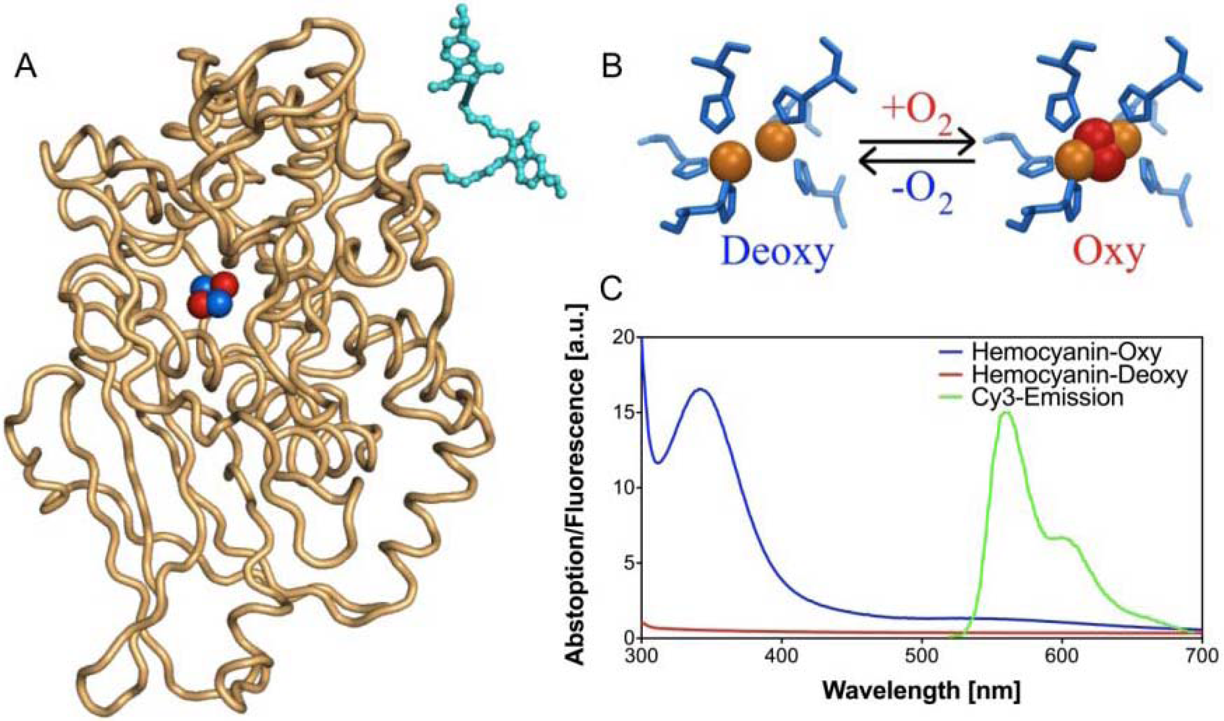
FRET-based oxygen-sensitive assay [71]

A typical experiment using a 96-well U-bottom plate was performed as follows. Serial dilutions of a freshly grown cell culture were made in LB to achieve 109 to 102 colony-forming units (cfu, confirmed by colony plate counting). Wells were prepared in triplicate by filling each well with 180 μl of the cell dilution plus 20 μL of Cy3-labeled hemocyanin solution (0.5–1 μM end-concentration). To avoid evaporation and rapid oxygen diffusion, 50 μL of mineral oil was added on top of each well. For antibiotic resistance tests, the initial number of cells was 107 cfu/well. Kanamycin was added to a final concentration of 0.05 to 5000 μg/mL. Controls were performed by adding Cy3 or Cy3-labeled BSA instead of Cy3-labeled hemocyanin to the cell culture as well as by using unlabeled hemocyanin. The fluorescence of Cy3-labeled hemocyanin in LB without cells was also monitored as a control in the absence of oxygen consumption.

Conclusions

A set of dimers of aminoglycoside analogues has been synthesized from natural neomycin via a straightforward route. Cleavage of both diaminoglucose pyranoses was followed by installation of an N-propargylated morpholine. Subsequent selective oxidation of the primary alcohol and immediate conjugation to arylamine by reductive amination led to three different 2-deoxystreptamine conjugates. Dimerization of the resulting conjugates with a range of bisazides afforded a small library of potential RNA-binding ligands in a straightforward synthetic route involving minimal protecting group transformations. All compounds were evaluated for antibacterial activity employing a novel fluorescence-based cell viability essay and compared to kanamycin. Several of the compounds, dimers and monomers, showed antibacterial activity, with values lying in the high micromolar range. Further optimization of the conjugates is therefore required, e.g., by introduction of longer spacers between the arylamines and the morpholine to allow cooperative binding. Similar reasoning may be applied to the dimers, as well as to the monofunctionalized morpholines.

Acknowledgements

Luigi Bubacco (Padua University) is kindly acknowledged for supplying us with hemocyanin protein. This work is financially supported by the Higher Education Commission (HEC) of Pakistan, by providing a PhD-fellowship (M.W.A.).

References and Notes

  1. Zembower, T.R.; Noskin, G.A.; Postelnick, M.J.; Nguyen, C.; Peterson, L.R. The Utility of Aminoglycosides in an Era of Emerging During of Resistance. Int. J. Antimicrob. Agents 1998, 10, 95–105. [Google Scholar]
  2. Recht, M.I.; Douthwaite, S.; Puglisi, J.D. Basis for Prokaryotic Specificity of Action of Aminoglycoside Antibiotics. EMBO J. 1999, 18, 3133–3138. [Google Scholar] [PubMed]
  3. Beaucaire, G. The role of aminoglycosides in Modern Therapy. J. Chemother. 1995, 7, 111–123. [Google Scholar] [PubMed]
  4. Zhao, F.; Zhao, Q.; Blount, K.F.; Han, Q.; Tor, Y.; Hermann, T. Molecular Recognition of RNA by Neomycin and a Restricted Neomycin Derivative. Angew. Chem. Int. Ed. 2005, 44, 5329–5334. [Google Scholar]
  5. Magnet, S.; Blanchard, J.S. Molecular Insights into Aminoglycoside Action and Resistance. Chem. Rev. 2005, 105, 477–497. [Google Scholar]
  6. Zembower, T.R.; Noskin, G.A.; Postelnick, M.J.; Nguyen, C.; Peterson, L.R. The Utility of Aminoglycosides in an Era of Emerging During of Resistance. Int. J. Antimicrob. Agents 1998, 10, 95–105. [Google Scholar] [PubMed]
  7. Nagai, J.; Takano, M. Molecular Aspects of Renal Handling of Aminoglycoside and Strategies for Preventing the Nephrotoxicity. Drug Metab. Pharmacokinet. 2004, 19, 159–170. [Google Scholar] [PubMed]
  8. Hayashi, S.F.; Norcia, L.J.; Seibel, S.B.; Silvia, A.M. Structure-activity Relationships of Hygromycin A and its Analogs: Protein Synthesis Inhibition Activity in a Cell-Free System. J. Antibiot. 1997, 50, 514–521. [Google Scholar] [PubMed]
  9. Hotta, K.; Zhu, C.B.; Ogata, T.; Sunada, A.; Ishikawa, J.; Mizuno, S.; Kondo, S. Enzymatic 2'-N-Acetylation of Arbekacin and Antibiotic Activity of its Product. J. Antibiot. 1996, 49, 458–464. [Google Scholar] [PubMed]
  10. Shaw, K.J.; Rather, P.N.; Hare, R.S.; Miller, G.H. Molecular Genetics of Aminoglycoside Resistance Genes and familial Relationships of the Aminoglycoside-Modifying Enzymes. Microbiol. Rev. 1993, 57, 138–163. [Google Scholar] [PubMed]
  11. Rai, R.; Chen, H.N.; Czyryca, P.G.; Li, J.; Chang, C.W.T. Design and Synthesis of Pyrankacin: A Pyranmycin Class of Broad-Spectrum Aminoglycoside Antibiotic. Org. Lett. 2006, 8, 887–889. [Google Scholar] [PubMed]
  12. Wang, J.; Li, J.; Chen, H.N.; Chang, H.; Tanifum, C.T.; Liu, H.H.; Czyryca, P.G.; Chang, C.W.T. Glycodiversification for the Optimization of the Kanamycin Class Aminoglycosides. J. Med. Chem. 2005, 48, 6271–6285. [Google Scholar] [PubMed]
  13. Ding, Y.; Hofstadler, S.A.; Swayze, E.E.; Risen, L.; Griffey, R.H. Design and Synthesis of Paromomycin-Related Heterocycle-Substituted Aminoglycoside Mimetics Based on a Mass Spectrometry RNA-Binding Assay. Angew. Chem. Int. Ed. 2003, 42, 3409–3412. [Google Scholar]
  14. Kudyba, I.; Fernandez, D.P.; Bottger, E.C.; Vasella, A. Synthesis of Paromomycin Derivatives Modified at C(5'') to Selectively Target Bacterial rRNA. Carbohydr. Res. 2007, 342, 499–519. [Google Scholar] [PubMed]
  15. Francois, B.; Szychowski, J.; Adhikari, S.S.; Pachamuthu, K.; Swayze, E.E.; Griffey, R.H.; Migawa, M.T.; Westhof, E.; Hanessian, S. Antibacterial Aminoglycosides with a Modified Mode of Binding to the Ribosomal-RNA Decoding Site. Angew. Chem. Int. Ed. 2004, 43, 6735–6738. [Google Scholar]
  16. Fridman, M.; Blakhov, V.; Yaron, S.; Baasov, T. A New Class of Branched Aminoglycosides: Pseudo-Pentasaccharide Derivatives of Neomycin B. Org. Lett. 2003, 5, 3575–3578. [Google Scholar] [PubMed]
  17. Roestamadji, J.; Grapsas, I.; Mobashery, S. Mechanism-based Inactivation of Bacterial Aminoglycoside 3' Phosphotransferases. J. Am. Chem. Soc. 1995, 117, 80–84. [Google Scholar]
  18. Hanessian, S.; Tremblay, M.; Swayze, E.E. Tobramycin Analouges with C-5 Aminoalkyl Ether Chains Intended to Mimic Rings III and IV of Paromomycin. Tetrahedron 2003, 59, 983–993. [Google Scholar]
  19. Fridman, M.; Blakhov, V.; Lee, L.V.; Liang, F.S.; Wong, C.H.; Baasov, T. Dual Effect of Synthetic Aminoglycosides: Antibacterial Activity Against Bacillus Anthracis and Inhibition of Anthrax Lethal Factor. Angew. Chem. Int. Ed. 2005, 44, 447–452. [Google Scholar]
  20. Ye, X.S.; Zhang, L.H. Aminoglycoside Mimetics as Small-molecule Drugs Targeting RNA. Curr. Med. Chem. 2002, 9, 929–939. [Google Scholar] [PubMed]
  21. Arya, D.P. Design, Chemical Synthesis and Antibacterial Activity of Kanamycin and Neomycin Class Aminoglycoside Antibiotics. In Aminoglycoside Antibiotics: From Chemical Biology to Drug Discovery; Wiley: New Jersey, USA, 2007. [Google Scholar]
  22. Kirk, S.R.; Luedtke, N.W.; Tor, Y. Neomycin-Acridine Conjugate: A Potent Inhibitor of Rev-RRE Binding. J. Am. Chem. Soc. 2000, 122, 980–981. [Google Scholar]
  23. Kaiser, M.; Sainlos, M.; Lehn, J.; Bombard, S.; Teulade-Fichou, M. Aminoglycoside-quinacridine Conjugates: Towards Recognition of the P6.1 Element of Telomerase RNA. Chem. Bio. Chem. 2006, 7, 321–329. [Google Scholar]
  24. Blount, K.F.; Tor, Y. A Tale of Two Targets: Differential RNA Selectivity of Nucleobase-Aminoglycoside Conjugates. Chem. Bio. Chem. 2006, 7, 1612–1621. [Google Scholar]
  25. Charles, I.; Xi, H.; Arya, D.P. Sequence-specific Targeting of RNA with an Oligonuleotide-Neomycin Conjugate. Bioconjug. Chem. 2007, 18, 160–169. [Google Scholar] [PubMed]
  26. Ahn, D.; Yu, J. Library Construction of Neomycin-dipeptide Heteroconjugates and Selection Against RRE RNA. Bioorg. Med. Chem. 2005, 13, 1177–1183. [Google Scholar] [PubMed]
  27. Hyun, S.; Lee, K.H.; Yu, J. A Strategy for the Design of Selective RNA Binding Agents, Preparations and RRE RNA Binding Affinities of a Neomycin-peptide Nucleic Acid Heteroconjugate Library. Bioorg. Med. Chem. Lett. 2006, 16, 4757–4759. [Google Scholar] [PubMed]
  28. Lee, J.; Kwon, M.; Lee, K.H.; Jeong, S.; Hyun, S.; Shin, K.J.; Yu, J. An Approach to Enhance Specificity Against RNA Targets Using Heteroconjugates of Aminoglycosides and Chloramphenicol (or Linezolid). J. Am. Chem. Soc. 2004, 126, 1956–1957. [Google Scholar] [PubMed]
  29. Park, W.K.C.; Auer, M.; Jaksche, H.; Wong, C.H. Rapid Combinatorial Synthesis of Aminoglycoside Antibiotics Mimetics: Use of a Polyethylene glycol-linked Amine and Neamine Derived Aldehyde in Multicomponent Condensation as a Strategy for the Discovery of New Inhibitors of the HIV RNA Rev Response Element. J. Am. Chem. Soc. 1996, 118, 10150–10155. [Google Scholar]
  30. Greenberg, W.A.; Priestley, E.S.; Sears, P.S.; Alper, P.B.; Rosenbohn, C.; Hendrix, M.; Hung, S.C.; Wong, C.H. Design and Synthesis of New Aminoglycoside Antibiotics Containing Neamine as an Optimal Core: Correlation of Antibiotic Activity with in Vitro Inhibition of Translation. J. Am. Chem. Soc. 1999, 121, 6527–6541. [Google Scholar]
  31. Nunns, C.L.; Spence, L.A.; Slater, M.J.; Berrisford, D.J. Synthesis of Neamine Libraries for RNA Recognition Using Solution Phase Chemistry. Tetrahedron Lett. 1999, 40, 9341–9345. [Google Scholar]
  32. Ryu, D.H.; Tan, C.H.; Rando, R.R. Synthesis of (+), (-) Neamine and Their Positional Isomers as Potential Antibiotics. Bioorg. Med. Chem. Lett. 2003, 13, 901–903. [Google Scholar] [PubMed]
  33. Minowa, N.; Akiyama, Y.; Hiraiwa, Y.; Maebashi, K.; Usui, T.; Ikeda, D. Synthesis and Antibacterial Activity of Novel Neamine Derivatives. Bioorg. Med. Chem. Lett. 2006, 16, 6351–6354. [Google Scholar] [PubMed]
  34. Michael, K.; Wang, H.; Tor, Y. Enhanced RNA Binding of Dimerized Aminoglycosides. Bioorg. Med. Chem. 1999, 7, 1361–1371. [Google Scholar] [PubMed]
  35. Wang, H.; Tor, Y. Dimeric Aminoglycosides: Design, Synthesis and RNA Binding. Bioorg. Med. Chem. Lett. 1997, 7, 1951–1956. [Google Scholar]
  36. Sucheck, S.J.; Wong, A.L.; Koeller, K.M.; Boehr, D.D.; Draker, K.A.; Sears, P.; Wright, G.D.; Wong, C.H. Design of Bifunctional Antibiotics that Target Bacterial rRNA and Inhibit Resistance-causing Enzymes. J. Am. Chem. Soc. 2000, 122, 5230–5231. [Google Scholar]
  37. Tok, J.B.H.; Huffman, G.R. Enhanced Binding of Aminoglycoside Dimer to a ”Dimerized” A-site rRNA Construct. Bioorg. Med. Chem. Lett. 2000, 10, 1593–1595. [Google Scholar] [PubMed]
  38. Tok, J.B.H.; Dunn, L.J.; Des Jean, R.C. Binding of Dimeric Aminoglycosides to the HIV-1 Rev Response Element (RRE) RNA Construct. Bioorg. Med. Chem. Lett. 2001, 11, 1127–1131. [Google Scholar] [PubMed]
  39. Tok, J.B.H.; Fenker, J. Novel Synthesis and RNA-binding Properties of Aminoglycoside Dimers Conjugated via a Naphthalene diimide-based Intercalator. Bioorg. Med. Chem. Lett. 2001, 11, 2987–2991. [Google Scholar] [CrossRef] [PubMed]
  40. Leudtke, N.W.; Liu, Q.; Tor, Y. RNA-ligand Interactions: Affinity and Specificity of Aminoglycoside Dimers and Acridine Conjugates to the HIV-1 Rev Response Element. Biochemistry 2003, 42, 11391–11403. [Google Scholar] [PubMed]
  41. Riguet, E.; Desire, J.; Boden, O.; Ludwig, V.; Gobel, M.; Bailly, C.; Decout, J.L. Neamine Dimers Targeting the HIV-1 TAR RNA. Bioorg. Med. Chem. Lett. 2005, 15, 4651–4655. [Google Scholar] [PubMed]
  42. Liang, C.H.; Romero, A.; Rabuka, D.; Sgarbi, P.W.M.; Marby, K.A.; Duffield, J.; Yao, S.; Cheng, M.L.; Ichikawa, Y.; Sears, P.; Hu, C.; Hwang, S.B.; Shue, Y.K.; Sucheck, S.J. Structure-activity Releationship of Bivalent Aminoglycosides and Evaluation of Their Microbiological Activities. Bioorg. Med. Chem. Lett. 2005, 15, 2123–2128. [Google Scholar] [PubMed]
  43. Chen, G.H.; Pan, P.; Chen, Y.; Meng, X.B.; Li, Z.J. Selective Deprotection of the Cbz Amine Protecting Group for the Facile Synthesis of Kanamycin A Dimers Linked at N-3'' Position. Tetrahedron 2009, 65, 5922–5927. [Google Scholar]
  44. Busscher, G.F.; Rutjes, F.P.J.T.; van Delft, F.L. 2-Deoxystreptamine: Central Scaffold of Aminoglycoside Antibiotics. Chem. Rev. 2005, 205, 775–792. [Google Scholar]
  45. Busscher, G.F.; Rutjes, F.P.J.T.; van Delft, F.L. Synthesis of a Protected Enantiomerically Pure 2-Deoxystreptamine Derivative from d-Allylglycine. Tetrahedron Lett. 2004, 69, 3629–3632. [Google Scholar]
  46. Busscher, G.F.; Groothuys, S.; de Gelder, R.; Rutjes, F.P.J.T.; van Delft, F.L. Efficient Preparation of a 1,3-Diazidocyclitol as a Versatile 2-Deoxystreptamine Precursor. J. Org. Chem. 2004, 69, 4477–4481. [Google Scholar] [PubMed]
  47. van den Broek, S.A.M.W.; Gruijters, B.W.T.; Rutjes, F.P.J.T.; van Delft, F.L.; Blaauw, R.H. A Short and Scalable Route to Orthogonally O-Protected 2-Deoxystreptamine. J. Org. Chem. 2007, 72, 3577–3580. [Google Scholar] [PubMed]
  48. Aslam, M.W.; Busscher, G.F.; Weiner, D.P.; de Gelder, R.; Rutjes, F.P.J.T.; van Delft, F.L. Fully Orthogonally Protected 2-Deoxystreptamine from Kanamycin. J. Org. Chem. 2008, 73, 5131–5134. [Google Scholar] [PubMed]
  49. Michael, K.; Wang, H.; Tor, Y. Enhanced RNA-Binding of Dimerized Aminoglycosides. Bioorg. Med. Chem. 1999, 7, 1361–1371. [Google Scholar] [PubMed]
  50. Yu, L.; Oost, T.K.; Schkeryantz, J.M.; Yang, J.; Janowick, D.; Fesik, S.W. Discovery of Aminoglycoside Mimetics by NMR-based Screening of Escherichia Coli A-site RNA. J. Am. Chem. Soc. 2003, 125, 4444–4450. [Google Scholar] [PubMed]
  51. Rodriguez, A.; Nomen, M.; Spur, B.W.; Godfroid, J.J. Selective Oxidation of Primary Silyl Ethers and its Application to the Synthesis of Natural Products. Tetrahedron Lett. 1999, 40, 5161–5164. [Google Scholar]
  52. Luca, L.D.; Giacomelli, G.; Masala, S.; Porcheddu, A. Trichloroisocynauric/TEMPO Oxidation of Alcohols under Mild Conditions: A Close Investigation. J. Org. Chem. 2003, 68, 4999–5001. [Google Scholar] [PubMed]
  53. More, J.D.; Finney, N.S. A Simple and Advantageous Protocol for the Oxidation of Alcohols with O-iodoxybenzoic Acid (IBX). Org. Lett. 2002, 4, 3001–3003. [Google Scholar] [PubMed]
  54. Dess, D.B.; Martin, J.C. Readially Accessible12-1-5 Oxidant for the Conversion of Primary and Secondary Alcohols to Aldehydes and Ketones. J. Org. Chem. 1983, 48, 4155–4156. [Google Scholar]
  55. Tornøe, C.W.; Christensen, C.; Meldal, M. Peptidotriazoles on Solid Phase: [1,2,3]-triazoles by Regiospecific Copper (I)-catalyzed 1,3-dipolar Cycloadditions of Terminal Alkynes to Azides. J. Org. Chem. 2002, 67, 3057–3064. [Google Scholar] [PubMed]
  56. Rostovtsev, V.V.; Green, L.G.; Fokin, V.V.; Sharpless, K.B. A Stepwise Huisgen Cycloaddition Process: Copper (I)-catalyzed Regioselective Ligation of Azides and Terminal Alkynes. Angew. Chem. Int. Ed. 2002, 41, 2596–2599. [Google Scholar]
  57. Jawalekar, A.M.; Cremers, N.M.J.G.O.; van der Marel, H.S.O.G.A.; Rutjes, F.P.J.T.; van Delft, F.L. Conjugation of Nucleosides and Oligonucleotides by [3+2] Cycloaddition. J. Org. Chem. 2008, 73, 287–290. [Google Scholar] [PubMed]
  58. Wang, Y.; Killian, J.; Hamasaki, K.; Rando, R.R. RNA Molecules That Specifically and Stoichiometrically Bind Aminoglycoside Antibiotics with High Affinities. Biochemistry 1996, 35, 12338–12346. [Google Scholar] [PubMed]
  59. Wang, Y.; Hamasaki, K.; Rando, R.R. Specificity of Aminoglycoside Binding to RNA Construct Derived from the 16S rRNA Decoding Region and the HIV-RRE Activator Region. Biochemistry 1997, 36, 768–779. [Google Scholar] [PubMed]
  60. Wang, Y.; Hamasaki, K.; Rando, R.R. Specificity in the Binding of Aminoglycoside to HIV-RRE RNA. Biochemistry 1999, 38, 8548–8554. [Google Scholar] [PubMed]
  61. Ryu, D.H.; Rando, R.R. Aminoglycoside Binding to Human and Bacterial A-site rRNA Decoding Region Construct. Bioorg. Med. Chem. 2001, 9, 2601–2608. [Google Scholar] [PubMed]
  62. Ryu, D.H.; Litovchick, A.; Rando, R.R. Stereospecificity of Aminoglycoside-Ribosomal Interactions. Biochemistry 2002, 41, 10499–10509. [Google Scholar] [PubMed]
  63. Luedtke, N.W.; Liu, Q.; Tor, Y. RNA-ligand Interactions: Affinity and Specificity of Aminoglycoside Dimers and Acridine Conjugates to the HIV-1 Rev Response Element. Biochemistry 2003, 42, 11391–11403. [Google Scholar] [PubMed]
  64. Xiao, G.; Kumar, A.; Li, K.; Rigl, C.T.; Bajic, M.; Davis, T.M.; Boykin, D.W.; Wilson, W.D. Inhibition of the HIV-1 Rev-RRE Complex Formation by Unfused Aromatic Cations. Bioorg. Med. Chem. 2001, 9, 1097–1113. [Google Scholar] [PubMed]
  65. Arya, D.P.; Coffee, R.L.; Xue, L. From Triplex to B-form Duplex Stabalization: Reversal of Target Selectivity by Aminoglycoside Dimers. Bioorg. Med. Chem. Lett. 2004, 14, 4643–4646. [Google Scholar] [PubMed]
  66. Kaul, M.; Barbieri, C.M.; Pilch, D.S. Fluorescence-based Approach for Detecting and Characterizing Antibiotic-induced Conformational Changes in Ribosomal RNA: Comparing Aminoglycoside Binding to Prokaryotic and Eukaryotic Ribosomal RNA Sequences. J. Am. Chem. Soc. 2004, 126, 3447–3453. [Google Scholar] [PubMed]
  67. Verhelst, S.H.; Michiels, P.J.; van der Marel, G.A.; van Boeckel, C.A.; van Boom, J.H. Surface Plasmon Resonance Evaluation of Various Aminoglycoside-RNA Hairpin Interactions Reveal Low Degree of Selectivity. Chem. Bio. Chem. 2004, 5, 937–942. [Google Scholar]
  68. Agnelli, F.; Sucheck, S.J.; Marby, K.A.; Rabuka, D.; Yao, S.L.; Sears, P.S.; Liang, F.S.; Wong, C.H. Dimeric Aminoglycosides as Antibiotics. Angew. Chem. Int. Ed. 2004, 43, 1562–1566. [Google Scholar]
  69. Swayze, E.E.; Jefferson, E.A.; Sannes-Lowery, K.A.; Blyn, L.B.; Risen, L.M.; Arakawa, S.; Osgood, S.A.; Hofstadler, S.A.; Griffey, R.H. SAR by MS: A Ligand Based Technique for Drug Lead Discovery Against Structured RNA Targets. J. Med. Chem. 2002, 45, 3816–3819. [Google Scholar] [PubMed]
  70. Kaul, M.; Barbieri, C.M.; Pilch, D.S. Aminoglycoside-induced Reduction in Nucleotide Mobility at the Ribosomal RNA A-site as a Potentially Key Determinant of Antibacterial Activity. J. Am. Chem. Soc. 2006, 128, 1261–1271. [Google Scholar] [PubMed]
  71. Strianese, M.; Zauner, G.; Tepper, A.W.J.W.; Bubacco, L.; Breukink, E.; Aartsma, T.J.; Canters, G.W.; Tabares, L.C. A Protein-based Oxygen Biosensor for High-throughput Mointoring of Cell Growth and Cell Viability. Anal. Biochem. 2009, 385, 242–248. [Google Scholar] [PubMed]
  72. Zauner, G.; Strianes, M.; Bubacco, L.; Aartsma, T.J.; Tepper, A.W.J.W.; Canters, G.W. Tryptophan-to-dye Fluorescence Energy Transfer Applied to Oxygen Sensing by Using Type-3 Copper Proteins. Chem. Eur. J. 2007, 13, 7085–7090. [Google Scholar]

