Article

Cell-Penetrating Fragments of the Cdk5 Regulatory Subunit Are Protective in Models of Neurodegeneration

1
Department of Neurology, University of Göttingen, Robert-Koch Str. 40 37075 Göttingen, Germany
2
DFG-Research Center for Molecular Physiology of the Brain (CMPB), Humboldtallee 23, 37075 Göttingen, Germany
3
Department 828, Molecular Neurobiology, H. Lundbeck A/S, Ottiliavej 9, 2500 Valby, Denmark
*
Author to whom correspondence should be addressed.
Pharmaceuticals 2010, 3(4), 1232-1240; https://0-doi-org.brum.beds.ac.uk/10.3390/ph3041232
Submission received: 11 February 2010 / Revised: 16 April 2010 / Accepted: 21 April 2010 / Published: 23 April 2010
(This article belongs to the Special Issue Cell-penetrating Peptides 2012)

Abstract

:
Cdk5 is essential for neuronal differentiation processes in the brain. Activation of Cdk5 requires the association with the mostly neuron-specific p35 or p39. Overactivation of CDK5 by cleavage of p35 into p25 is thought to be involved in neurodegenerative processes. Here, we have tested an approach to inhibit pathological Cdk5 activation with a Tat-linked dominant-negative fragment of p25. It reduced cell death induced by staurosporine and showed a tendency to alleviate manganese-induced cell death, while it did not protect against 6-OHDA toxicity. Our results suggest that the Tat technique is a suitable tool to inhibit dysregulated CDK5.

Graphical Abstract

1. Introduction

Cdk5 belongs to the family of cyclin-dependent kinases, which are essential for cell cycle and differentiation processes. In contrast to other Cdks, Cdk5 does not require the association with a specific cyclin, but the neuron specific protein p35 for activation. This Cdk5/p35 complex is essential for neurite outgrowth, cortical development, and other differentiation processes [1]. Cellular stress can lead to a calcium influx into the cell with consecutive activation of calpain, which cleaves p35 into p25, resulting in an aberrant activation of Cdk5. This mechanism is thought to be involved in neurodegenerative diseases such as Parkinson’s disease, Amyotrophic Lateral Sclerosis or Alzheimer’s dementia [2,3,4]. In post mortem brains of Alzheimer’s disease patients, enhanced processing of p35 to the p25 fragment, resulting in an increased p25/35 ratio, has been found [5]. p25/Cdk5 can also induce intraneuronal accumulation of beta Amyloid in vitro and vivo [6]. Moreover, it has been shown that pathological activation of Cdk5 by p25 can lead to hyperphosphorylation of Tau in various models [7]. Vice versa it has been shown that inhibition of p25 formation can abolish Tau hyperphosphorylation [8]. The cleavage of p35 to p25 is also found in models for Parkinson’s disease and Huntington’s disease, underlining its general importance as a pathological process in neurodegenerative diseases [9]. p25 is cleaved by calpain after different apoptotic stimuli [10]. To date, different approaches have been made to inhibit pathological Cdk5 activation, e.g. Cdk5 inhibition by the small molecule pan-CDK inhibitor roscovitine, which did not block pathological Tau phosphorylation [6], or application of a calpain inhibitor, which could decrease p25 levels and abrogate tau hyperphoshporylation [8]. In our study we have addressed a challenge of previous experiments, which is to specifically inhibit pathological Cdk5 activation, and to deliver such an inhibitor to its target site. In recent years, strategies have been developed to overcome the blood brain barrier and cellular membranes [11,12]. One approach is to link the protein of interest to basic peptides, such as the cell-penetrating peptide (CPP) derived from the HIV Tat protein [13]. Here, we have generated a Tat-linked dominant negative p25 fusion protein and tested it in cellular models for oxidative stress and apoptosis, as oxidative stress is thought to be one factor in the development of various neurodegenerative diseases.

2. Experimental Section

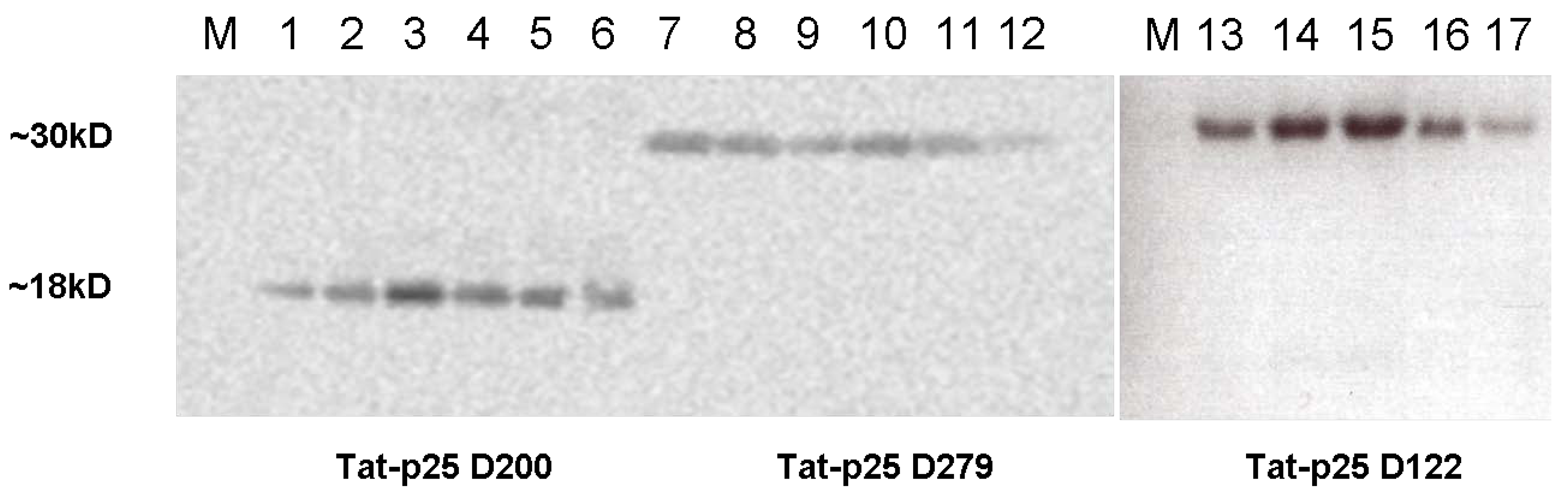
2.1. Cloning of constructs and purification of Tat fusion proteins

Tat-p25 constructs were generated from expression vectors in E. coli and purified via Ni-affinity chromatography as described [14,15]. S.F. Dowdy (San Diego, CA) kindly provided the pTAT-HA vector that we used to generate the Tat-p25 expression constructs [13]. 3 different p25 cDNA fragments were selected according to their previously described functions [16]. Amino acid residues 122–291 (N122) served as a positive control, residues 200–291 (N200) as a negative control, residues 150–279 (N279) as the dominant negative p25 fragment.
For expression of the p25 mutants, p25 full length cDNA was generated by RT PCR amplification from whole mouse brain using the primer GCTACAGGGGGC3′ (MWG Biotech AG Ebersberg, Germany) followed by PCR amplification with the primers AGTCATCCATGGTCAAGAAGGCCCC GCACCC/GTCATGAATTCATTCTTCAAGTCAGAGAACA (for N122); AGTCATCCATGGCCA ATGTGGTCTTCCTCTACAT/GTCATGAATTCATTCTTCAAGTCAGAGAACA (for N200) and AGTCATCCATGGCTCAGCCTCCCGCCCCCCC/GTCATGAATTCTGGGTCGGCATTGATCTGCA (for N279) to add XhoI and NcoI restriction sites and cloning into the vector pRSET A (Invitrogen, Carlsbad, CA). The vectors used to isolate Tat-fusion proteins are derivatives of this vector. Tat-p25 (N122, 200 and 279) were expressed in E. coli strain BL21(DE3) pLysS (Novagen, Madison, WI). After sonication in 8M urea, we pelleted the bacterial debris and purified the supernatant by metal-affinity chromatography using a Ni-NTA matrix (Qiagen, Hilden, Germany). We removed salt by gel filtration on Sephadex G-25M (Amersham Biosciences, Uppsala, Sweden) The desalting buffer consisted of Tris 10mM pH6.0; 50% Glycerol, 0,02% Tween-80 and 0,1% Pluronic. We confirmed the identity of proteins by Western blotting (Figure 1). The calculated molecular weights of Tat-p25 D122, D200, and D279 are 27 kDa, 19 kDa, or 28 kDa, respectively. Anti-hemagglutinin (HA) antibodies were purchased from BAbCO (Richmond, CA, USA).
Figure 1. Integrity of fusion proteins confirmed by Western analysis. After purification of recombinant protein by nickel affinity chromatography, column fractions 4–9 (lane 1–6: Tat-p25 D 200; lane 7–12: Tat-p25 D279, lane 13–17: Tat-p25 D122) were analysed by western blotting using an antibody against the hemagglutinin tag included in the recombinant protein.
Pharmaceuticals 03 01232 g001

2.2. Cell culture and cell death assays

SH-SY5Y cells were plated at a density of 20,000 cells/well of a 96 well dish. Cells were maintained in Dulbecco’s modified Eagle’s medium (PAA, Pasching, Austria) supplemented with 10% fetal bovine serum (PAA), 1 mM L-glutamine, 100 U penicillin/mL, and 100 µg of streptomycin sulfate/mL at 37 °C. 24 h after plating, cells were treated with 10 µM retinoic acid.
Exactly 24 h after retinoid acid treatment, in all toxicity assays, Tat constructs were applied. Exactly 1 h after protein application, toxic agents were added. 24 h after toxin application, cell survival was assessed by WST viability assay according to the manufacturer’s protocol (Roche, Basel, Switzerland). MnCl2 was used at 100 µM concentration. Western blots revealing CDK5 expression after MnCl2 treatment were analysed by densitomety (Alpha Innotech Software Version 3.1.2.). Values measured for CDK5 band intensities were normalized by dividing them by the corresponding intensities of the same blot probed with an anti-actin antibody. To induce apoptotic cell death, cells were treated with 400 nM staurosporine for 24 h. 6-OHDA toxicity assays were performed as described [17], with 80 µM 6-OHDA used here. Statistics were performed using the two-tailed student’s t-test.

3. Results and Discussion

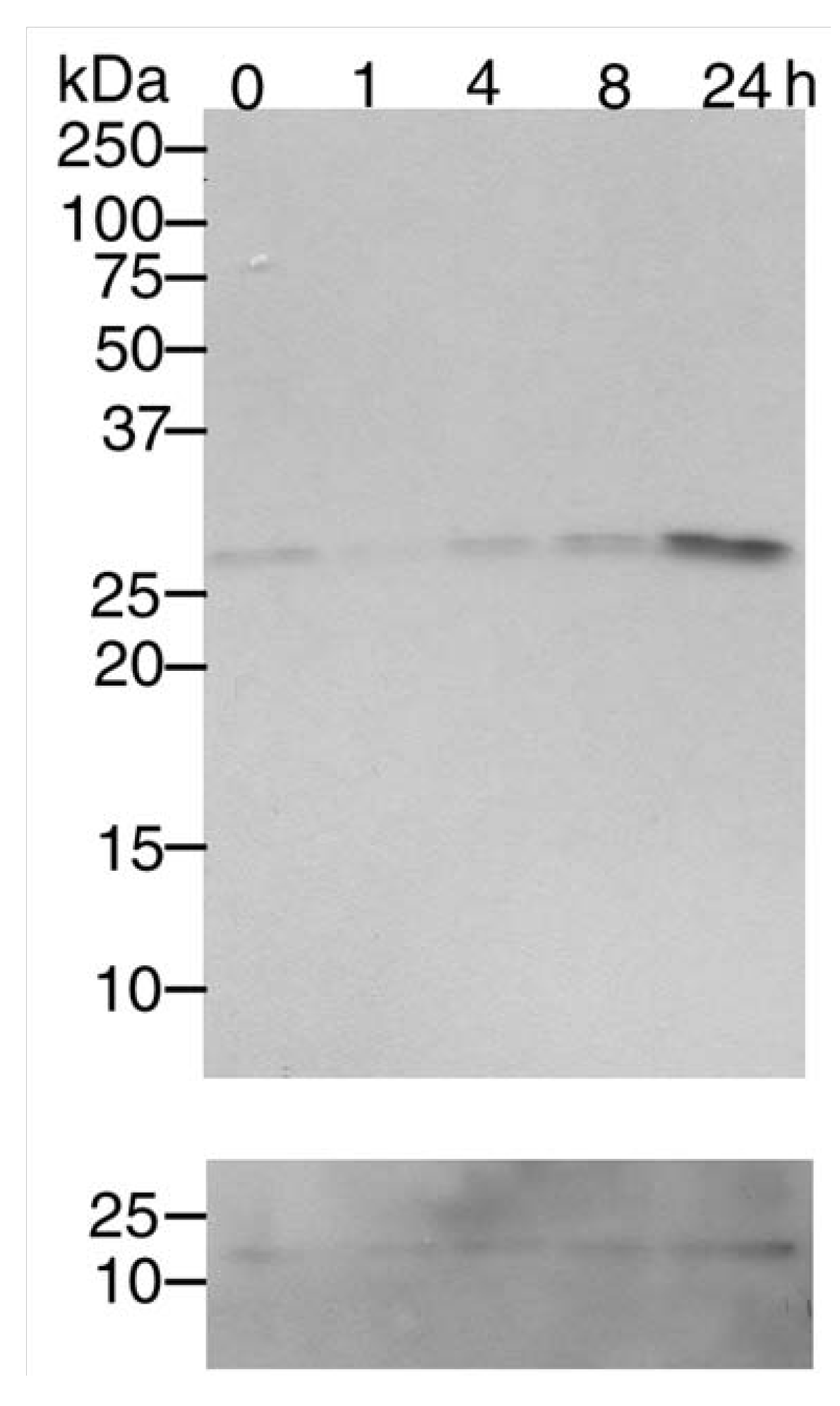
3.1. Mn2+ induces Cdk5 expression

We treated SH-SY5Y cells with MnCl2, as a paradigm for oxidative stress and found that Cdk5 expression was increased up to 24 h after treatment (Figure 2). By densitometric analysis, at 24 h, CDK5 was upregulated 7-fold compared to the 1 h time point. To examine whether cell death induced by oxidative stress was partially mediated by Cdk5 activation, we tested how cell death was modified by dominant-negative p25.
Figure 2. Mn2+ treatment induces Cdk5 expression. Differentiated SH-SY5Y neuroblastoma cells were treated with 100µM MnCl2 and lysed up to 24 h later as indicated above the lanes. Lysates were examined by Western analysis using an antibody against Cdk5. Anti-Actin staining is shown in the bottom panel.
Pharmaceuticals 03 01232 g002

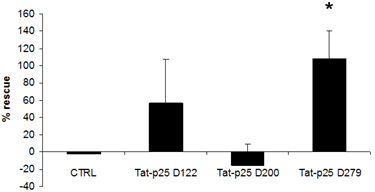
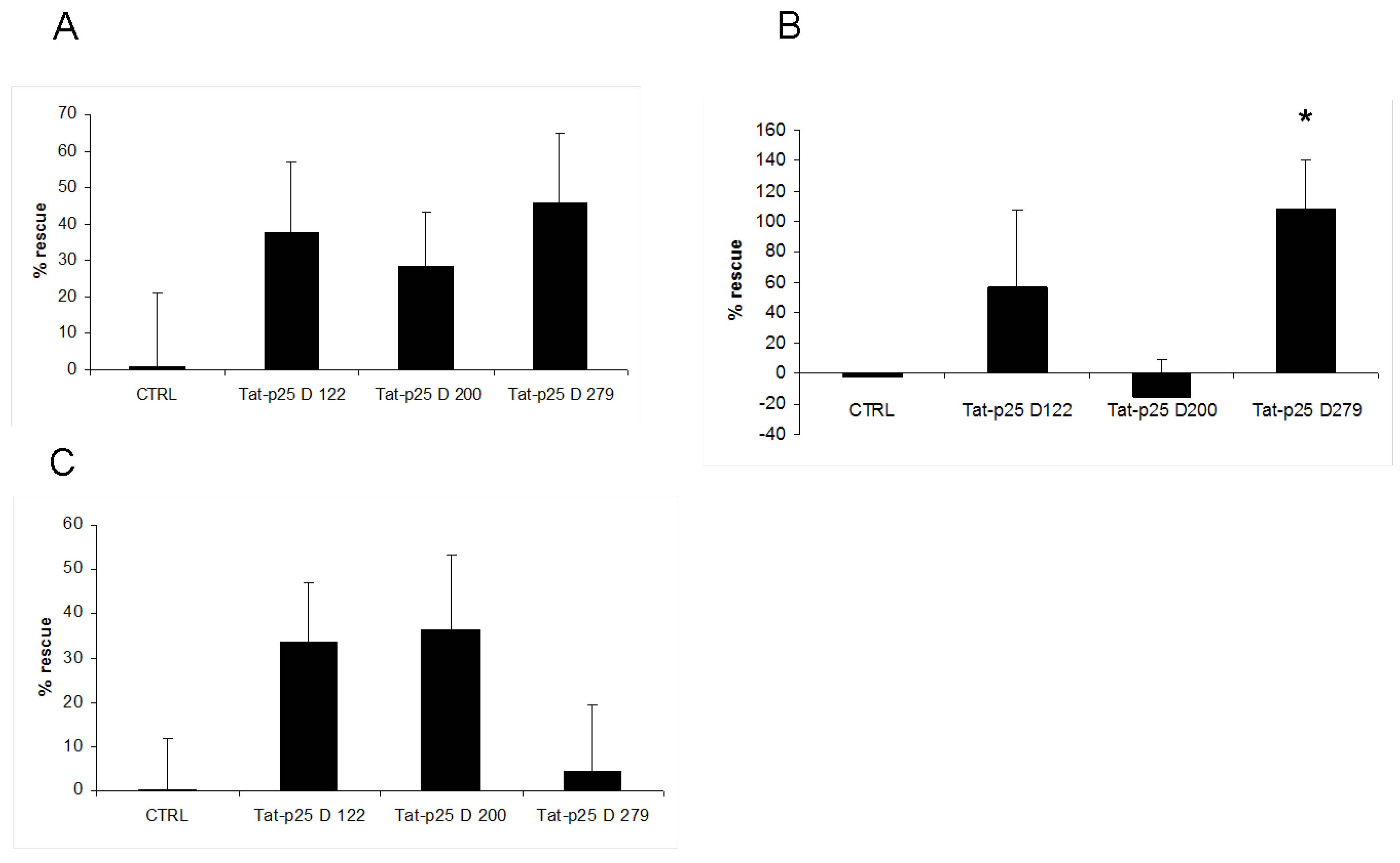
3.2. p25 fragments protect SH-SY5Y cells against cell death stimuli

Treatment of SH-SY5Y cells with cell-penetrating p25 fragments resulted in statistically not significant trend towards rescue from MnCl2-induced cell death. Surprisingly, this was not only observed for the p25 fragment previously reported to act as a dominant-negative [16], but also for other fragments derived from p25 that were reported to bind and still activate, or not to bind to CDK5 (Figure 3a).
We then were interested how the TAT-p25 constructs would influence cell survival in other cell death assays, which have been shown to be influenced by the Cdk5/p25 pathway. Zhang et al. were able to show previously, that staurosporine induces cleavage of p35 to p25 with a maximum at 12 and 24 hours, which they suggested to be the cause for staurosporine (STS) induced apoptosis in their paradigm [18]. Moreover, Meuer et al. demonstrated CDK5-mediated mitochondrial fragmentation upon staurosporine-treatment of neuronal cells [20]. We observed that a dominant-negative Tat-p25 fragment rescued neuroblastoma cells from cell death induced with 0.4 µM STS (Figure 3b). At higher or lower STS concentrations, we did not observe such an effect (not shown).
Figure 3. Tat-p25 inhibits apoptosis in neuroblastoma cells. In all paradigms, SH-SY5Y cells were incubated with 100nM Tat peptides before a 24h toxin application. Control cells (CTRL) were treated with a peptide containing only the Tat cell-penetrating peptide without p25. D279 indicates the dominant-negative p25 fragment; D122 and D200 are p25 control fragments. % cells rescued from demise by recombinant protein application after treatment with 100 µM MnCl2 (A); 400 nM Staurosporine {B, * indicates significance (p: 0.02)}; or with 80 µM 6-OHDA (C).
Pharmaceuticals 03 01232 g003
To test whether inhibition of Cdk5 would be beneficial in another model for oxidative stress, we tested the p25 mutants in a 6-OHDA toxicity assay; this drug is often applied in models for Parkinson’s disease [20]. None of the applied fragments had a significant effect on cell survival (Figure 3c).

4. Conclusions

In our approach we addressed two challenges of therapeutic Cdk5 inhibition: first, we aimed to block its pathological activation, rather than is normal function, by blocking the binding site for p25, and possibly other off-site targets. Secondly, we addressed the problem of drug delivery by using Tat-linked proteins.
Using in vitro studies we addressed different paradigms of cellular stress believed to play a role in neurodegenerative diseases. We applied manganese, which can induce neurodegeneration, including Parkinsonism [21,22]. Manganese is therefore widely used in models for Parkinson’s disease, which includes induction of cell death in SH-SY5Y neuroblastoma cells [23,24,25]. One of its pro-apoptotic actions is mediated by the ERK pathway, by phosphorylating ERK 1 and 2 [26]. It has also been shown to act directly via the NMDA receptor, thereby leading to increased calcium release with subsequent activation of calpains, which cleave p35 to its pathological residuum p25 [27,28]. Our results indicate that p25 fragments, including domains known to bind Cdk5, show a tendency to prevent cell death in this model. Surprisingly, not only a supposedly dominant-negative fragment, but also control p25 fragments that either did not bind CDK5 in previous assays or still did activate it [16] showed a tendency towards a protective effect. We amplified the p25 fragments from whole mouse brains, rather than a bovine source as reported by Poon et al. [16]. The cell lines we used originate from human (SH-SH5Y). As we did not test the individual action of each fragment in cell free in vitro binding studies or in assays directly assessing the effect on CDK5 enzymatic activity, the previously described functions may not be alienable from bovine to mouse. Moreover, the results obtained using viral transduction and Tat-mediated transduction may not be immediately comparable; the gene expression from different viral constructs may be variable and different from intracellular protein concentrations reached after Tat-mediated transduction [29,30]. Moreover, activation by an E.coli-expressed p25 fusion protein may be different from p25 directly expressed and folded in the cell to be examined. In future studies, assays to test the impact of the different Tat-p25 fragments on Cdk5 enzymatic activity could address that issue.
Neither overexpression of a dominant-negative CDK5, nor the CDK5 inhibitors roscovitine and butyrolactone-1 prevented staurosporine-induced cell death [31,32]. Consistent with the results shown here, we have shown earlier that another CDK5 inhibitor protected CSM cells against staurosporine-induced cell death [19]. The discrepancy between those results and the protection we observed might be due to the fact that we applied a dominant-negative form of the CDK5 activator protein p25, rather than directly targeting CDK5, or due to the different cellular system used. The interpretation of the data is further complicated by the fact that staurosporine itself inhibits CDK5 [33].
In a third paradigm, 6-OHDA, which is often used in models for PD, different p25 constructs did not have a significant effect on cell survival; however, there was a trend towards protection with the control constructs. The main apoptotic stimulus by 6-OHDA is believed to be delivered via activation of the ERK pathway [34,35]. Cdk5 in its normal state has been shown to inhibit ERK activation, by phosphorylating MEK1/2. Thus, a dominant-negative p25, as used in our model, may be expected to decrease that inhibition, therefore not protecting against 6-OHDA-induced cell death. On the contrary, some activation of Cdk5 might be expected to alleviate cell death induction.
In conclusion, the effects of virally or cell-penetrating peptide-mediated delivery of proteins may be different. Moreover, the effect of dominant-negative p25 delivery depends on the experimental model examined: For instance, while it blocks staurosporine-induced cell death, it does not protect against 6-OHDA-induced toxicity.

Acknowledgements

We thank Birgit Dietz for excellent technical assistance. S.F. Dowdy (San Diego, CA) kindly provided the pTat-HA vector. Supported by the Research Center for Molecular Physiology of the Brain (CMPB) of the Deutsche Forschungsgemeinschaft (DFG).

References

  1. Dhavan, R.; Tsai, L.H. A decade of Cdk5. Nat. Rev. Mol. Cell. Biol. 2001, 2749–2759. [Google Scholar]
  2. Lee, M.S.; Kwon, Y.T.; Mingwei, L.; Peng, J.; Friedlander, R.M.; Tsai, H.T. Neurotoxicity induces cleavage of p35 to p25 by calpain. Nature 2000, 18, 360–364. [Google Scholar]
  3. Patrick, G.N.; Zukerberg, L.; Nikolic, M.; de la Monte, S.; Dikkes, P.; Tsai, L.H. Conversion of p35 to p25 de-regulates cdk5 activity and promotes neurodegeneration. Nature 1999, 9615–9622. [Google Scholar]
  4. Nguyen, M.D.; Lariviere, R.C.; Juien, J.P. Deregulation of Cdk5 in a mouse model of ALS: toxicity alleviated by perikaryal neurofilament inclusions. Neuron 2001, 30, 135–147. [Google Scholar]
  5. Tseng, H.C.; Zhou, Y.; Shen, Y.; Tsai, L.H. A survey of Cdk5 activator p35 and p25 levels in Alzheimer’s disease brains. FEBS Lett. 2002, 523, 58–62. [Google Scholar] [PubMed]
  6. Cruz, J.C.; Kim, D.; Moy, L.Y.; Dobbin, M.M.; Sun, X.; Bronson, R.T.; Tsai, L.H. p25/cyclin-dependent kinase 5 induces production and intraneuronal accumulation of amyloid beta in vivo. J. Neurosci. 2006, 26, 10536–10541. [Google Scholar] [PubMed]
  7. Ahlijanian, M.K.; Barrezueta, N.X.; Williams, R.D.; Jakowski, A.; Kowsz, K.P.; McCarthy, S.; Coskran, T.; Carlo, A.; Seymour, P.A.; Burkhardt, J.E.; Nelson, R.B.; McNeish, J.D. Hyperphosphorylated tau and neurofilament and cytoskeletal disruptions in mice overexpressing human p25, an activator of cdk5. Proc. Nat. Acad. Sci. USA 2000, 97, 2910–2915. [Google Scholar]
  8. Hung, K.S.; Hwang, S.L.; Liang, C.L.; Chen, Y.J.; Lee, T.H.; Liu, J.K.; Howng, S.L.; Wang, C.H. Calpain inhibitor inhibits p35-p25-Cdk5 activation, decreases tau hyperphosphorylation, and improves neurological function after spinal cord hemisection in rats. J. Neuropathol. Exp. Neuro. 2005, 64, 15–26. [Google Scholar]
  9. Smith, P.D.; Mount, M.P.; Shree, R.; Callaghan, S.; Slack, R.S.; Ansiman, H.; Vincent, I.; Wang, X.; Mao, Z.; Park, D.S. Calpain regulated p35/cdk5 plays a central role in dopaminergic neuron death through modulation of the transcription factor myocyte enhancer factor 2. J. Neurosci. 2006, 26, 440–447. [Google Scholar] [PubMed]
  10. Saito, T.; Konno, T.; Hosokawa, T.; Asada, A.; Ishiguro, K.; Hisanaga, S. p25/cyclin-dependent kinase 5 promotes the progression of cell death in nucleus of endoplasmic reticulum-stressed neurons. J. Neurochem. 2007, 102, 133–140. [Google Scholar]
  11. Dietz, G.P.; Bähr, M. Synthesis of cell-penetrating peptides and their application in neurobiology. Methods Mol. Biol. 2007, 399, 181–198. [Google Scholar]
  12. Dietz, G.P.; Bähr, M. Peptide-enhanced cellular internalization of proteins in neuroscience. Brain Res. Bull. 2005, 68, 103–114. [Google Scholar]
  13. Schwarze, S.R.; Ho, A.; Vocero-Akbani, A.; Dowdy, S.F. In vivo protein transduction: delivery of a biologically active protein into the mouse. Science 1999, 285, 1569–1572. [Google Scholar] [PubMed]
  14. Kilic, U.; Kilic, E.; Dietz, G.P.; Bähr, M. Intravenous TAT-GDNF is protective after focal cerebral ischemia in mice. Stroke 2003, 34, 1304–1310. [Google Scholar]
  15. Nagahara, H.; Vocero-Akbani, A.M.; Snyder, E.L.; Ho, A.; Latham, D.G.; Lissy, N.A.; Becker-Hapak, M.; Ezhevsky, S.A.; Dowdy, S.F. Transduction of full-length TAT fusion proteins into mammalian cells: TAT-p27Kip1 induces cell migration. Nat. Med. 1998, 4, 1449–1452. [Google Scholar]
  16. Poon, R.Y.C.; Lew, J.; Hunter, T. Identification of Functional Domains in the Neuronal Cdk5 Activator Protein. J. Biol. Chem. 1997, 272, 5703–5708. [Google Scholar] [PubMed]
  17. Nagel, F.; Dohm, C.P.; Bähr, M.; Wouters, F.S.; Dietz, G.P. Quantitative evaluation of chaperone activity by different preparations of a cell-penetrating Hsp70. J. Neurosci. Methods 2008, 171, 226–232. [Google Scholar]
  18. Zhang, B.F.; Peng, F.F.; Zhang, W.; Shen, H.; Wu, S.B.; Wu, D.C. Involvement of cyclin dependent kinase 5 and its activator p35 in staurosporine-induced apoptosis of cortical neurons. Acta Pharmacol. Sin. 2004, 25, 1105–1111. [Google Scholar]
  19. Meuer, K.; Suppanz, I.E.; Lingor, P.; Planchamp, V.; Göricke, B.; Fichtner, L.; Braus, G.H.; Dietz, G.P.; Jakobs, S.; Bähr, M.; Weishaupt, J.H. Cyclin-dependent kinase 5 is an upstream regulator of mitochondrial fission during neuronal apoptosis. Cell Death Differ. 2007, 14, 651–661. [Google Scholar]
  20. Alvira, D.; Tajes, M.; Verdaguer, E.; Acuña-Castroviejo, D.; Folch, J.; Camins, A.; Pallas, M. Inhibition of the cdk5/p25 fragment formation may explain the antiapoptotic effects of melatonin in an experimental model of Parkinson's disease. J. Pineal. Res. 2006, 40, 251–258. [Google Scholar]
  21. Calne, D.B.; Chu, N.S.; Luz, C.C.; Huang, C.S.; Olanow, W. Manganism and idiopathic parkinsonism: Similarities and differences. Neurology 1994, 44, 1583–1586. [Google Scholar]
  22. Cook, D.G.; Fahn, S.; Brait, K.A. Chronic manganese intoxication. Arch. Neurol. 1974, 30, 59–64. [Google Scholar]
  23. Higashi, Y.; Asanuma, M.; Miyazaki, I.; Hattori, N.; Mizuno, Y.; Ogawa, N. Parkin attenuates manganese-induced dopaminergic cell death. J. Neurochem. 2004, 89, 1490–1497. [Google Scholar]
  24. Roth, J.A.; Singleton, S.; Feng, J.; Garrick, M.; Paradkar, P.N. Parkin regulates metal transport via proteasomal degradation of the 1B isoforms of divalent metal transporter 1. J. Neurochem. 2010, 113, 454–464. [Google Scholar]
  25. Li, Y.; Sun, L.; Cai, T.; Zhang, Y.; Lv, S.; Wang, Y.; Ye, L. Alpha-Synuclein overexpression during manganese-induced apoptosis in SH-SY5Y neuroblastoma cells. Brain Res. Bull. 2010, 81, 428–433. [Google Scholar]
  26. Hirata, Y. Manganese-induced apoptosis in PC12 cells. Neurotoxicol. Teratol. 2002, 24, 639–653. [Google Scholar]
  27. Paoletti, P.; Vila, I.; Rife, M.; Lizcano, J.M.; Alberch, J.; Gines, S. Dopaminergic and Glutaminergic Signaling Crosstalk in Huntington’s Disease Neurodegeneration: The Role of p25/Cyclin-Dependent Kinase 5. Neurobiol. Dis. 2008, 28, 10090–10101. [Google Scholar]
  28. Brouilet, E.P.; Shinobu, L.; McGarvey, U.; Hochberg, F.; Beal, M.F. Manganese injection into the Rat Striatum Produces Excitotoxic Lesions by Impairing Energy Metabolism. Exp. Neurol. 1993, 120, 89–94. [Google Scholar]
  29. Kojima, H.; Abiru, Y.; Sakajiri, K.; Watabe, K.; Ohishi, N.; Takamori, M.; Hatanaka, H.; Yagi, K. Adenovirus-mediated transduction with human glial cell line-derived neurotrophic factor gene prevents 1-methyl-4-phenyl-1,2,3,6-tetrahydropyridine-induced dopamine depletion in striatum of mouse brain. Biochem. Biophys. Res. Commun. 1997, 238, 569–573. [Google Scholar]
  30. Dietz, G.P.; Valbuena, P.C.; Dietz, B.; Meuer, K.; Mueller, P.; Weishaupt, J.H.; Bähr, M. Application of a blood-brain-barrier-penetrating form of GDNF in a mouse model for Parkinson's disease. Brain Res. 2006, 1082, 61–66. [Google Scholar] [CrossRef] [PubMed]
  31. Meikrantz, W.; Schlegel, R. Suppression of apoptosis by dominant negative mutants of cyclin- dependent protein kinases. J. Biol. Chem. 1996, 271, 10205–102059. [Google Scholar]
  32. Sandal, T.; Stapnes, C.; Kleivdal, H.; Hedin, L.; Doskeland, S.O. A novel, extraneuronal role for cyclin-dependent protein kinase 5 (CDK5): Modulation of cAMP-induced apoptosis in rat leukemia cells. J. Biol. Chem. 2002, 277, 20783–20793. [Google Scholar]
  33. Veeranna, Shetty, K.T.; Amin, N.; Grant, P.; Albers, R.W.; Pant, H.C. Inhibition of neuronal cyclin-dependent kinase-5 by staurosporine and purine analogs is independent of activation by Munc-18. Neurochem. Res. 1996, 21, 629–636. [Google Scholar] [CrossRef] [PubMed]
  34. Kesavapany, S.; Li, B.S.; Amin, N.; Zheng, Y.L.; Grant, P.; Pant, H.C. Neuronal cyclin-dependent kinase 5: Role in nervous system function and its specific inhibition by the Cdk5 inhibitory peptide. Biochemica et Biophysica Acta 2004, 1697, 143–153. [Google Scholar]
  35. Kulich, S.M.; Horbinski, C.; Patel, M.; Chu, C.T. 6-Hydroxydopamine induces mitochondrial ERK activation. Free Radic. Biol. Med. 2007, 43, 372–383. [Google Scholar]

