Review

Cell Wall Amine Oxidases: New Players in Root Xylem Differentiation under Stress Conditions

by
Sandip A. Ghuge
1,
Alessandra Tisi
2,
Andrea Carucci
2,
Renato A. Rodrigues-Pousada
3,
Stefano Franchi
2,
Paraskevi Tavladoraki
2,4,
Riccardo Angelini
2,4 and
Alessandra Cona
2,4,*
1
Institute of Crystallography, Consiglio Nazionale delle Ricerche (CNR), Monterotondo 00015, Italy
2
Department of Sciences, Università Roma Tre, Roma 00146, Italy
3
Department of Life, Health, and Environmental Sciences, Università dell'Aquila, L'Aquila 67100, Italy
4
Istituto Nazionale Biostrutture e Biosistemi (INBB), Rome 00136, Italy
*
Author to whom correspondence should be addressed.
Submission received: 18 May 2015 / Revised: 12 June 2015 / Accepted: 9 July 2015 / Published: 14 July 2015
(This article belongs to the Special Issue Plant Root Development)

Abstract

:
Polyamines (PAs) are aliphatic polycations present in all living organisms. A growing body of evidence reveals their involvement as regulators in a variety of physiological and pathological events. They are oxidatively deaminated by amine oxidases (AOs), including copper amine oxidases (CuAOs) and flavin adenine dinucleotide (FAD)-dependent polyamine oxidases (PAOs). The biologically-active hydrogen peroxide (H2O2) is a shared compound in all of the AO-catalyzed reactions, and it has been reported to play important roles in PA-mediated developmental and stress-induced processes. In particular, the AO-driven H2O2 biosynthesis in the cell wall is well known to be involved in plant wound healing and pathogen attack responses by both triggering peroxidase-mediated wall-stiffening events and signaling modulation of defense gene expression. Extensive investigation by a variety of methodological approaches revealed high levels of expression of cell wall-localized AOs in root xylem tissues and vascular parenchyma of different plant species. Here, the recent progresses in understanding the role of cell wall-localized AOs as mediators of root xylem differentiation during development and/or under stress conditions are reviewed. A number of experimental pieces of evidence supports the involvement of apoplastic H2O2 derived from PA oxidation in xylem tissue maturation under stress-simulated conditions.

Graphical Abstract

1. Introduction

In growing root tissues, meristematic cells deal with the need to coordinate sequential transitions from division to expansion and differentiation, which occur as separate phases at distinct developmental zones. In the Arabidopsis (Arabidopsis thaliana) root apex, four zones can be defined based on cell activity: (1) the meristematic zone up to 200 µm from the root cap; (2) the transition zone from about 200 up to 520 µm from the root cap; (3) the elongation zone from about 520 up to 850 µm from the root cap; and (4) the growth terminating zone from about 850 up to about 1500 µm from the root cap. In the latter zone, cell elongation slows down, and cells reach their final length [1]. In the proximal region beyond the zone of maximum elongation growth, protoxylem cells mature, undertaking the deposition of secondary walls [2]. The boundaries defining the division, elongation and maturation zones of the root are developmentally regulated. Of note, changes in their positions occur in a coordinated fashion during development, as reflected by the correlation observed in pea roots among root length, meristem size and protoxylem element position [3]. In Arabidopsis roots, the final meristem size is reached at five days post germination, when the rate of cell differentiation is balanced with the rate of cell division [4]. During development, the growth rate of the meristem is maintained stably during the plant’s lifespan by a complex cross-regulatory circuit relying on the antagonistic interaction between auxin and cytokinin, promoting respectively cell division in the proximal meristem and cell differentiation at the transition zone [4,5]. In this regard, it has been recently proposed that auxin defines the developmental zonation of division, expansion and differentiation activities by cooperating with the auxin-induced PLETHORA (PLT) transcription factor through different mechanisms [6]. Likewise, vascular patterning is specified through a mutually-inhibitory feedback between auxin and cytokinin, the former promoting and the latter inhibiting protoxylem identity [5,7,8]. However, the strong correlation among root length, meristem size and protoxylem element position observed in pea roots might cease under stress conditions, interfering with cell division, elongation or maturation events [3]. Of note, the stress signaling hormone jasmonic acid (JA) interferes with the auxin pathway involved in the maintenance of the root zonation by repressing PLT expression [9]. Furthermore, reactive oxygen species have also been involved in meristem size specification by controlling the transition between cell proliferation and differentiation, independently from the cytokinin/auxin pathway [10]. The present review illustrates recent insights into the role played in root development and xylem differentiation by hydrogen peroxide (H2O2) derived from the oxidation of polyamines (PAs) in the cell wall. In particular, we focused our attention on the involvement of cell wall-localized amine oxidases (AOs) in root xylem maturation under stress-simulated conditions.

2. Polyamines as Signaling Compounds and/or Hydrogen Peroxide Sources

2.1. Polyamines in Plants

PAs are low molecular weight aliphatic amines involved in various physiological and pathological events in plants, including growth, development, stress tolerance and defense responses [11,12,13]. The most common PAs are the diamine putrescine (Put), the triamine spermidine (Spd) and the tetramine spermine (Spm). Additionally, thermospermine (T-Spm), an isomer of Spm, which has not as yet been detected in mammalian cells, has been found to be widely distributed throughout the plant kingdom [14,15]. Put and Spd are essential for life, as Arabidopsis mutants defective in their biosynthetic pathways are embryo-lethal, whereas Spm and T-Spm have been specifically linked to stress responses and development, respectively [16,17]. Owing to the presence of regularly-spaced positive charges, PAs may act through stabilization of negatively-charged intracellular macromolecules, such as proteins, nucleic acids and phospholipids. However, besides their biophysical effects, these molecules may be involved in signal transduction pathways during developmentally-controlled programs or stress-induced responses and/or may exert their action as sources of biologically-active compounds, such as H2O2 [18,19]. In plants, a key signaling role in determining cell fate has been ascribed to the PA/H2O2 balance, especially in cell death associated with both defense and developmental processes [20,21].

2.2. Polyamine Signaling in Xylem Development

Alteration of PA homeostasis strongly affects higher plant architecture. An interplay between the PA and the cytokinin/auxin pathways has been revealed to occur in Arabidopsis. Perturbation of higher PA biosynthesis in the Arabidopsis loss-of-function bud2 mutant, a knock-out of the S-adenosylmethionine decarboxylase 4 (SAMDC4) gene necessary for Spd, Spm and T-Spm biosynthesis, results in a bushy and dwarf phenotype with altered vascularization due to an increase in the number of tracheary vessels along with a decrease in their size [22]. Considering that the bud2 mutant displays hyposensitivity to auxin and hypersensitivity to cytokinin together with the findings that BUD2 is inducible by the auxin signaling pathway, it has been hypothesized that PAs may affect plant architecture by both increasing sensitivity to auxin perception and repressing cytokinin biosynthesis and/or signaling [23]. Mutation of the ACAULIS5 (ACL5) gene encoding the aminopropyl transferase driving T-Spm biosynthesis severely affects xylem specification in Arabidopsis hypocotyl. Bearing in mind that (1) ACL5 is strongly expressed in provascular/procambial cells, (2) the acl5 mutant displays over-proliferation and altered phenotype of xylem vessels, with the preponderance of very small and spiral-type xylem vessel elements and the absence of pitted vessels and xylem fibers, and (3) cell death occurs before the onset of secondary cell wall formation in acl5 xylem vessels, a key role for T-Spm in preventing premature maturation and death of xylem elements has been suggested to allow complete expansion and correct secondary cell wall patterning [24,25,26]. It has also been proposed that T-Spm slows down xylem differentiation by antagonizing auxin signaling [14]. Indeed, in acl5 seedlings, a number of genes related to auxin signaling have been shown to be upregulated, among which MONOPTEROS (MP) and its target genes, such as the homeodomain-leucine zipper (HD-ZIP) III encoding for the ARABIDOPSIS THALIANA HOMEOBOX8 (ATHB8) transcription factor. Of note, ATHB8 promotes the formation and differentiation of procambial cells into vascular cells [27,28,29]. An interplay between T-Spm and ATHB8 leading to a negative feedback loop has been supposed to occur in differentiating xylem cells as follows: in established xylem precursor cells in which patterning and differentiation triggered by auxin flow are taking place, ATHB8 induces the expression of BUD2 and ACL5, leading to the biosynthesis of T-Spm, which, in turn, negatively affects the expression of MP, HD-ZIP III and key auxin signaling genes, thus counteracting the auxin-mediated differentiation of xylem precursor cells [27].
PAs may also play a role in the environmentally-induced plasticity of root architecture, by affecting primary root growth and lateral and adventitious root formation [30]. It has been suggested that exogenous Spd may act as a stress signal in maize (Zea mays) roots [31]. Indeed, coherent with the observation that stressed roots show a reduced rate of growth and altered architecture [30], exogenous Spd has been shown to inhibit primary root growth by affecting both the mitotic index and cell elongation in maize [32]. Furthermore, in Spd-treated maize roots, an earlier differentiation of xylem tissues has been revealed concurrently with an increase of cell wall phenolic autofluorescence in vascular tissues and rhizodermis [31], the latter event also being indicative of responses to stress, such as wounding in maize and tobacco (Nicotiana tabacum) [33,34]. Consistently, Spd supply has been shown to inhibit K+ uptake in maize roots, analogous to the effect of cutting roots into segments [35].

3. Terminal Polyamine Oxidation in the Cell Wall Is Triggered at Specific Developmental Stages or under Stress Conditions

3.1. Polyamines Are Oxidized by Copper and FAD-Dependent Amine Oxidases

AOs oxidize PAs to amino aldehydes, with the production of an amine moiety and H2O2 [15,19]. The reaction products vary depending on both the substrates and enzymes involved (Figure 1). The cell wall copper amine oxidases (CuAOs) purified from Fabaceae preferentially oxidize Put at the carbon next to the primary amino group with the production of 4-aminobutanal, ammonia and H2O2 (Figure 1) [36]. However, out of ten annotated Arabidopsis thaliana CuAOs (AtCuAOs), the apoplastic AtAO1 (At4g14940) and AtCuAO1 (At1g62810) and the peroxisomal AtCuAO2 (At1g31710) and AtCuAO3 (At2g42490) have been shown to additionally oxidize Spd at the primary amino group with an affinity comparable to that for Put, producing N-(3-aminopropyl)-4-aminobutanal, ammonia and H2O2 (Figure 1) [19,37,38,39,40,41].
Figure 1. Polyamine (PA) oxidation in plants. The compartment with the violet background highlights PA terminal oxidations occurring in the cell wall. Scissors indicate cleavage sites. The peroxisomal Put and Spd oxidation and the extracellular monoamine oxidation carried out by Arabidopsis thaliana and Malus domestica CuAOs, as well as the vacuolar Hordeum vulgare HvPAO2-mediated oxidation of Spm and Spd are not shown for simplicity. TC, terminal catabolism; BC, back-conversion; PAO, polyamine oxidase.
Plants 04 00489 g001
On the other hand, a Malus domestica peroxisomal CuAO (MdAO1), highly expressed in fruits, predominantly oxidizes the diamines Put, cadaverine and 1,3-diaminopropane, but not Spd and Spm [42], while the extracellular MdAO2 represents the first plant CuAO so far described able to exclusively utilize long chain aliphatic and aromatic monoamines as substrates [42]. In tobacco, an apoplastic CuAO activity responsible for the extracellular oxidation of Put has been described [43], along with two peroxisome-resident CuAOs, the N-methylputrescine oxidase 1 (MPO1) and the Nicotiana tabacum diamine oxidase 1 (NtDAO1, formerly known as MPO2), respectively involved in nicotine biosynthesis and peroxisomal Put oxidation [39]. FAD-dependent polyamine oxidases (PAOs) oxidize Spd and Spm at the carbon located at the endo- or exo-side of the N4 atom by either a terminal catabolism (TC-PAOs) or a back-conversion (BC-PAOs) pathway, the latter being responsible for recycling Spd and Put, respectively from Spm and Spd. In particular, TC-PAOs oxidize the carbon at the endo-side of the N4 of Spd and Spm, producing 4-aminobutanal and N-(3-aminopropyl)-4-aminobutanal, respectively, in addition to 1,3-diaminopropane and H2O2, whereas BC-PAOs oxidize the carbon at the exo-side of the N4 of Spd and Spm to produce Put and Spd, respectively, in addition to 3-aminopropanal and H2O2 [15,19,44,45]. Some BC-PAOs from Arabidopsis and rice (Oryza sativa), including AtPAO1, OsPAO1, OsPAO4 and OsPAO5, are able to recycle Spd by oxidation of T-Spm, with the production of 3-aminopropanal and H2O2 (Figure 1) [19,44,45,46,47]. Furthermore, different from other TC- and BC-PAOs so far characterized, AtPAO5, which catalyzes the back-conversion of N1-acetyl-Spm, T-Spm and Spm to Spd, has been shown to have a 180-fold higher activity as a dehydrogenase than as an oxidase [48,49], thus suggesting that this enzyme does not have a role in H2O2 production, but rather plays a role in PA homeostasis. The TC-PAOs so far described in Poaceae and Nicotiana tabacum are secretory and mostly targeted to the cell wall, with only one exception represented by the barley (Hordeum vulgare) PAO isoform HvPAO2, which is targeted to the vacuole [15,19,43,50]. On the contrary, the BC-PAOs currently described in Arabidopsis and rice have an intracellular localization [44,46,47,48,49].

3.2. Features and Roles of Polyamine Oxidation in the Apoplast

A specific feature of apoplastic PA oxidation is the absence of the PA inter-conversion pathway in this compartment [44,48]. As a consequence, the availability of free soluble PAs in the cell wall would mainly depend on events of PA secretion, which may occur under environmental stresses or at specific developmental stages. Indeed, while in the absence of any internal or external stimulus, PAs are present in the cell wall at very low or even undetectable levels [51], under specific adverse conditions, such as pathogen infection or salt stress, they can be transported in the cell wall [20,43,52,53,54], thus becoming available for the apoplastic AOs. Likewise, AOs have been shown to undergo redistribution from cytoplasm towards the cell wall depending on developmentally-regulated or light-induced tissue maturation [55,56]. Hence, it can be stated that a complex interplay of events modulating both AO and PA levels and the rate of their secretion in the cell wall governs the spatio-temporal features of the AO-dependent biosynthesis of extracellular H2O2, which has been shown to play the dual role of triggering peroxidase-mediated wall stiffening events and signaling the modulation of defense and hypersensitive response (HR)-cell death gene expression [57,58].
Apoplastic CuAOs and TC-PAOs have been found at very high levels in several species belonging to Fabaceae and Poaceae families, respectively, especially in tissues fated to undertake extensive wall stiffening events, such as xylem, xylem parenchyma, endodermis and epidermis, and/or in cells undergoing programmed cell death (PCD), namely tracheary elements and root cap cells [57,59]. Consistently, in Arabidopsis, the cell wall-localized AtAO1 has been reported to be expressed in root cap cells and protoxylem precursors at early stages of vascular tissue differentiation [38,60].
In the cell wall, CuAOs and PAOs share overlapping roles as H2O2 sources in developmentally- or light-regulated cell wall maturation events [55,56,57,59], as well as in the oxidative bursts occurring during defense responses against biotic and abiotic stresses [57,61], especially during pathogen attack [16,19,38,43,54,57,62,63], salt stress and wound healing [20,33,52,57,61,64,65,66]. Consistently, the expression of cell wall-localized AOs is induced by stress signaling hormones. Indeed, the expression of TC-PAO from Zea mays (ZmPAO), CuAO from Cicer arietinum, AtAO1 and AtCuAO1 is strongly induced by JA and/or methyl-jasmonate (MeJA) treatments [33,40,60,64]. AtCuAO1 expression is also induced by abscisic acid, salicylic acid and flagellin 22 treatments [40]. In recent years, growing evidence has suggested the involvement of H2O2 derived from the apoplastic AO-mediated PA-oxidation in root xylem differentiation under stress conditions, as discussed in detail in the following paragraphs [19,34,60,67].

4. Apoplastic Spermidine Oxidation Mediates Early Xylem Differentiation in the Maize Primary Root

In maize, three genes (ZmPAO1, ZmPAO2 and ZmPAO3) have been identified as encoding identical secretory proteins, overall reported as ZmPAO, which are involved in the terminal catabolism of Spd and Spm in the cell wall [15]. ZmPAO has been detected mainly in epidermis, rhizodermis, endodermis and vascular tissues by histochemical and immunocytochemical techniques [55,56,57,58,59,60,61,62,63,64,65,66,67].

4.1. ZmPAO Expression and Sub-Cellular Distribution Are Developmentally Regulated in Maize Primary Root

In maize primary roots, ZmPAO expression and its subcellular localization are finely regulated during development [56]. It has been demonstrated that differentiating sub-apical tissues show higher ZmPAO levels as compared to mature basal tissues, independently of the root age [56]. Moreover, a subcellular redistribution from cytoplasm towards cell wall depending on tissue maturation stages has been revealed. In this regard, the percentage of insolubilized tightly wall-bound ZmPAO enzyme activity remarkably increases in mature basal root portion vs. differentiating sub-apical root tissues, especially in older as compared to younger seedlings [56]. A comprehensive analysis of ZmPAO expression has revealed that in apical meristematic and sub-apical differentiating root tissues, this protein is mostly present in the cytoplasm of early and late metaxylem precursors, regardless of the root age. In particular, the ZmPAO enzyme activity appears early in procambium and sloughed root cap cells and greatly increases in stelar tissues up to 1000 µm from the apical meristem [67]. In more mature tissues, an intense enrichment of ZmPAO has been reported to occur in the primary and secondary walls of early and late metaxylem precursors as compared to the apical zones. This difference is strengthened in an age-dependent manner, appearing higher in older roots [56].
Interestingly, an analogous sub-cellular redistribution of ZmPAO has been revealed in epidermal cells and xylem precursors of de-etiolated maize mesocotyls, as a consequence of light-induced tissues differentiation [55,56]. In detail, exposure of dark-grown maize seedlings to intense white light induces a marked increase of ZmPAO expression associated with its secretion in the cell wall [55], suggesting that under this condition, an enhanced catabolism of PAs in the apoplast may occur by modulation of both AO expression and secretion of the encoded proteins to the apoplast.
Overall, redistribution of ZmPAO from cytoplasm toward cell wall has been shown to be concomitant with tissue differentiation, such as during light-induced and developmentally-regulated maturation of mesocotyl and root tissues, respectively [55,56].

4.2. ZmPAO-Driven Oxidation of Spermidine in the Apoplast Mediates Early Xylem Differentiation in Maize Primary Root

Exogenous Spd affects maize root development by inhibiting elongation and altering cell cycle phase distribution in the root apex [32,67]. Concerning this, the key role in root growth inhibition and xylem differentiation of H2O2 derived from Spd oxidation (Spd-derived H2O2) has been demonstrated in vivo by exploiting a specific and selective inhibitor of the ZmPAO enzyme activity, as well as a H2O2 (N,N1-dimethylthiourea (DMTU)) trap, which allows fast removal of this compound soon after its production [67,68]. Furthermore, the increase in cell wall phenolic auto-fluorescence occurring in rhizodermis, xylem elements and vascular parenchyma has been shown to be mediated by the Spd-derived H2O2. Moreover, widespread in root tissues, Spd treatment induces nuclear condensation and DNA fragmentation, the latter being mediated by the Spd-derived H2O2. Likewise, precocious differentiation of early and late metaxylem occurring upon oxidation of exogenous Spd, whose position is closer to the tip compared to untreated roots, has been ascribed to the enhanced production of H2O2, which is clearly visible in xylem parenchyma and in differentiating xylem elements. However, the precocious cell death occurring in early differentiating xylem elements hinders full differentiation of the secondary wall [67]. The phenotype shown by Spd-treated roots is consistent with the hypothesis that exogenous PAs may contribute to create a stressing environment by simulating apoplastic accumulation of PAs reported to occur in stressed plants. Considering this, it could be reasonable to suppose that Spd-derived H2O2 mediates early xylem differentiation under stress conditions.

5. Cell Wall PAO and CuAO Signal Early Xylem Differentiation in Roots of Tobacco Plants

In tobacco, PAO and CuAO are highly expressed in cells destined to undergo lignification, especially in vascular tissues [69]. Both CuAO and PAO enzyme activities have been detected in the intercellular washing fluids of tobacco leaves [43]. These enzymes, which catalyze the terminal apoplastic oxidation of Put and Spm, are respectively inhibited in vivo by the well-known CuAO and PAO inhibitors, 2-bromoethylamine (2-BrEt) and N,N′-diaminoguanidine and guazatine [34,43].

5.1. Overexpression of ZmPAO in Tobacco Plants Induces Early Differentiation of Root Vascular Tissues

Ectopic overexpression of ZmPAO in the cell wall of tobacco plants drives early xylem differentiation associated with enhanced H2O2 production in root apex and induces PCD in root cap cells [67]. In these plants, oxidation of developmentally-secreted PAs may be accelerated by the overexpressed ZmPAO, simulating the high level of PA oxidation occurring in stressed plants and, thus, leading to an altered phenotype [67].

5.2. CuAO Mediates Early Xylem Differentiation in Transgenic Tobacco Plants with Constitutively-Activated Defense Responses

Nicotiana tabacum plants overexpressing a fungal endopolygalacturonase (PG plants) show constitutively-activated defense responses, owing to the production of an excess of oligogalacturonides (OGs) that are perceived by the plant as damage-associated molecular patterns [70]. H2O2 accumulation is a specific feature of PG plants [70], which also display higher CuAO activity and lower PA levels in leaves, especially Put, with respect to wild-type (WT) plants [34]. Histochemical analysis has shown a tissue-specific expression of CuAO in xylem cells, vascular cambium and neighboring derivative cells of petioles and stems of PG plants [34]. Moreover, phenotypic analysis of root apices from PG plants has revealed an early xylem differentiation associated with an enhanced accumulation of extracellular H2O2. Of note, in the roots of PG plants, Put level sharply decreases as compared to the value detected in roots of WT plants [34]. Overall, these results suggest that the root xylem phenotype could be associated with a perturbation of PA metabolism. The key role of the CuAO activity in early differentiation of root xylem precursors and extracellular H2O2 accumulation in PG plants has been established by exploiting in vivo the specific CuAO inhibitor 2-BrEt. Concerning this, upon 2-BrEt treatments, the position of xylem precursors with secondary cell wall thickenings partially recover to that observed in the WT, while inhibitor treatments are ineffective in WT roots. Furthermore, H2O2 accumulation detected in PG plants is strongly reversed by treatments with 2-BrEt. As a whole, these results suggest a role for the CuAO-driven Put oxidation in xylem differentiation under stress conditions, such as those signaled by pectin integrity alteration [34].

6. Hydrogen Peroxide Produced by the Apoplastic Copper Amine Oxidase 1 (AtAO1) Signals the Methyl-Jasmonate-Mediated Protoxylem Differentiation in Arabidopsis Roots

A comprehensive analysis of AtAO1 expression pattern in Arabidopsis root apex has revealed a vascular tissue localization, especially in the transition, elongation and maturation zones, as well as in the root cap cells. Proceeding basipetally along the root length, from the transition zone up to the elongation and maturation zone, AtAO1 promoter activity has been detected in protoxylem precursors, whole vascular cylinder and metaxylem precursors [60]. MeJA strongly induces AtAO1 expression in the vascular cylinder, concurrently with a spatial anticipation of its pattern, which appears closer to the transition zone upon the hormone treatment. However, ATAO1 loss-of-function mutants display no altered phenotype in root xylem tissues in comparison to WT plants [60], suggesting an irrelevant or non-prevalent role of AtAO1 in vascular development under physiological growth conditions or, alternatively, the occurrence of a redundancy effect caused by another member of the CuAO gene family. Conversely, the evidence that MeJA treatments are effective in inducing early protoxylem differentiation in WT seedlings without affecting it in AtAO1 mutants reveals the involvement of AtAO1 in the MeJA-signaled differentiation of root vascular tissues. The hypothesis that MeJA’s effect on protoxylem differentiation can be mediated by the H2O2 produced via the AtAO1-driven Put oxidation is supported by: (1) the reduction of Put levels in roots of MeJA-treated WT plants; (2) the reversion effect exerted by DMTU on MeJA-induced early protoxylem differentiation; and (3) the increased accumulation of H2O2 upon the same treatment at the site of appearance of protoxylem cells with fully-developed secondary wall thickenings [60]. Of note, Put-derived H2O2 modulates the protoxylem position under MeJA-signaled stress conditions independently of changes in the whole root elongation rate and meristem size [60]. Furthermore, Put supply and AtAO1 overexpression also induce early protoxylem differentiation [60]. Overall, these data suggest that AtAO1-driven production of H2O2 may play a role in xylem differentiation under stress conditions, such as those signaled by MeJA or simulated by either Put treatments or AtAO1 overexpression.

7. Conclusions and Future Perspective

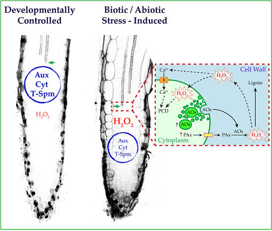
In response to environmental stresses, plants may respond by accumulating PAs in the cell wall. Induction of apoplastic AO expression is also a feature of stressed plants. Considering this, treatments with exogenous PAs or apoplastic overexpression of AOs may simulate environmental stressing conditions by driving a PA-dependent oxidative burst in the cell wall. H2O2 derived from the AO-driven terminal oxidation of apoplastic PAs behaves as a co-substrate in the peroxidase-mediated wall stiffening events and as a signal for defense responses, such as HR-cell death and defense gene expression (Figure 2) [58].
Figure 2. Schematic representation of the hypothetical stress-induced signaling triggered by PA oxidation and leading to root xylem differentiation. (A) Developmentally-controlled xylem differentiation is governed by the auxin/cytokinin/T-Spm loop. Apoplastic PA oxidation-derived H2O2 may contribute to the oxidative burst needed for full differentiation of the secondary wall. (B) Under stress conditions, an early root xylem differentiation occurs, and the first xylem precursor with secondary walls appears positioned closer to the root tip. This event is prevalently signaled by the H2O2 derived from the AO-driven terminal oxidation of PAs in the apoplast of differentiating xylem elements, independent of the auxin/cytokinin/T-Spm loop. (Inset in (B)) The square on the right illustrates a hypothetical scheme of the events at the level of differentiating xylem tissue under biotic/abiotic stress. Under these conditions, the expression of PA metabolic genes and apoplastic vascular-expressed AOs is induced along with PA and AO secretion in the cell wall. Terminal oxidation of PAs accumulated in the cell wall triggers an extracellular oxidative burst. H2O2 in the cell wall signals developmental cell death and acts as co-substrate in peroxidase-mediated lignin polymerization. PCD, programmed cell death.
Plants 04 00489 g002
Plants take advantage of the developmental plasticity of roots, whose architecture is deeply affected under changing environmental conditions. An early differentiation of xylem vessels has been revealed to occur in roots of plants under artificial stress conditions, such as those simulated upon exogenous PA treatments or apoplastic AO overexpression [60,67], or signaled by both MeJA treatment and pectin integrity alteration in transgenic PG plants [34,60]. The key role of the cell wall-localized AOs in the stress-induced xylem differentiation has been demonstrated by genetic and pharmacological approaches. Concerning this, PA-derived H2O2 has been shown to be involved as a mediator in the final events needed to accomplish full xylem differentiation, namely secondary wall deposition and developmental PCD (Figure 2). Overall, the reviewed evidence supports the hypothesis that PA oxidation mediated by apoplastic vascular-expressed AOs could assume a prevalent role in H2O2-mediated xylem differentiation under stress conditions, autonomously from the auxin/cytokinin/T-Spm loop.
In roots undertaking the early xylem differentiation process, in which fully-differentiated xylem elements appear positioned closer to the root tip, the stress-induced architecture of the water conducting system could be functional in improving the efficiency of water absorption under adverse conditions. The involvement of the apoplastic vascular-expressed AOs in the optimization of water uptake deserves to be carefully explored.

Acknowledgments

This work was implemented in the frame of the European COST Action (COSTFA0605) and was supported by the Italian Ministry of Education, University and Research (Project PRIN “Progetti di Ricerca di Interesse Nazionale” 2009WTCJL8_003) and Università degli Studi “Roma Tre”, Italy.

Author Contributions

All authors contributed to this review. Alessandra Tisi, Andrea Carucci and Stefano Franchi generated images and prepared figures for the manuscript. Sandip A. Ghuge, Alessandra Cona and Renato A. Rodrigues-Pousada reviewed the literature, and wrote and edited the manuscript. Paraskevi Tavladoraki and Riccardo Angelini performed critical review and gave corrections to improve the manuscript. All authors have reviewed and approved the manuscript submitted.

Conflicts of Interest

The authors declare no conflict of interest.

References

  1. Verbelen, J.P.; de Cnodder, T.; Le, J.; Vissenberg, K.; Baluska, F. The root apex of Arabidopsis thaliana consists of four distinct zones of growth activities: Meristematic zone, transition zone, fast elongation zone and growth terminating zone. Plant Signal. Behav. 2006, 1, 296–304. [Google Scholar] [CrossRef] [PubMed]
  2. Kobayashi, K.; Takahashi, F.; Suzuki, M.; Suzuki, H. Examination of morphological changes in the first formed protoxylem in Arabidopsis seedlings. J. Plant Res. 2002, 115, 107–112. [Google Scholar] [CrossRef] [PubMed]
  3. Rost, T.L.; Baum, S. On the correlation of primary root length, meristem size and protoxylem tracheary element position in pea seedlings. Am. J. Bot. 1988, 75, 414–424. [Google Scholar] [CrossRef]
  4. Perilli, S.; di Mambro, R.; Sabatini, S. Growth and development of the root apical meristem. Curr. Opin. Plant Biol. 2012, 15, 17–23. [Google Scholar] [CrossRef] [PubMed]
  5. Petricka, J.J.; Winter, C.M.; Benfey, P.N. Control of Arabidopsis root development. Annu. Rev. Plant Biol. 2012, 63, 563–590. [Google Scholar] [CrossRef] [PubMed]
  6. Mähönen, A.P.; ten Tusscher, K.; Siligato, R.; Smetana, O.; Díaz-Triviño, S.; Salojärvi, J.; Wachsman, G.; Prasad, K.; Heidstra, R.; Scheres, B. PLETHORA gradient formation mechanism separates auxin responses. Nature 2014, 515, 125–129. [Google Scholar] [CrossRef] [PubMed]
  7. Bishopp, A.; Help, H.; El-Showk, S.; Weijers, D.; Scheres, B.; Friml, J.; Benková, E.; Mähönen, A.P.; Helariutta, Y. A mutually inhibitory interaction between auxin and cytokinin specifies vascular pattern in roots. Curr. Biol. 2011, 21, 917–926. [Google Scholar] [CrossRef] [PubMed]
  8. Mähönen, A.P.; Bishopp, A.; Higuchi, M.; Nieminen, K.M.; Kinoshita, K.; Törmäkangas, K.; Ikeda, Y.; Oka, A.; Kakimoto, T.; Helariutta, Y. Cytokinin signaling and its inhibitor AHP6 regulate cell fate during vascular development. Science 2006, 311, 94–98. [Google Scholar] [CrossRef] [PubMed]
  9. Chen, Q.; Sun, J.; Zhai, Q.; Zhou, W.; Qi, L.; Xu, L.; Wang, B.; Chen, R.; Jiang, H.; Qi, J.; et al. The basic helix-loop-helix transcription factor MYC2 directly represses PLETHORA expression during jasmonate-mediated modulation of the root stem cell niche in Arabidopsis. Plant Cell 2011, 23, 3335–3352. [Google Scholar] [CrossRef] [PubMed]
  10. Tsukagoshi, H.; Busch, W.; Benfey, P.N. Transcriptional regulation of ROS controls transition from proliferation to differentiation in the root. Cell 2010, 143, 606–616. [Google Scholar] [CrossRef] [PubMed]
  11. Handa, A.K.; Mattoo, A.K. Differential and functional interactions emphasize the multiple roles of polyamines in plants. Plant Physiol. Biochem. 2010, 48, 540–546. [Google Scholar] [CrossRef] [PubMed]
  12. Minocha, R.; Majumdar, R.; Minocha, S.C. Polyamines and abiotic stress in plants: A complex relationship. Front. Plant Sci. 2014, 5, e175. [Google Scholar] [CrossRef] [PubMed]
  13. Tiburcio, A.F.; Altabella, T.; Bitrián, M.; Alcázar, R. The roles of polyamines during the lifespan of plants: From development to stress. Planta 2014, 240, 1–18. [Google Scholar] [CrossRef] [PubMed]
  14. Takano, A.; Kakehi, J.; Takahashi, T. Thermospermine is not a minor polyamine in the plant kingdom. Plant Cell Physiol. 2012, 53, 606–616. [Google Scholar] [CrossRef] [PubMed]
  15. Tavladoraki, P.; Cona, A.; Federico, R.; Tempera, G.; Viceconte, N.; Saccoccio, S.; Battaglia, V.; Toninello, A.; Agostinelli, E. Polyamine catabolism: Target for antiproliferative therapies in animals and stress tolerance strategies in plants. Amino Acids 2012, 42, 411–426. [Google Scholar] [CrossRef] [PubMed]
  16. Kusano, T.; Berberich, T.; Tateda, C.; Takahashi, Y. Polyamines: Essential factors for growth and survival. Planta 2008, 228, 367–381. [Google Scholar] [CrossRef] [PubMed]
  17. Takahashi, T.; Kakehi, J. Polyamines: Ubiquitous polycations with unique roles in growth and stress responses. Ann. Bot. 2010, 105, 1–6. [Google Scholar] [CrossRef] [PubMed]
  18. Angelini, R.; Cona, A.; Federico, R.; Fincato, P.; Tavladoraki, P.; Tisi, A. Plant amine oxidases “on the move”: An update. Plant Physiol. Biochem. 2010, 48, 560–564. [Google Scholar] [CrossRef] [PubMed]
  19. Moschou, P.N.; Wu, J.; Cona, A.; Tavladoraki, P.; Angelini, R.; Roubelakis-Angelakis, K.A. The polyamines and their catabolic products are significant players in the turnover of nitrogenous molecules in plants. J. Exp. Bot. 2012, 63, 5003–5015. [Google Scholar] [CrossRef] [PubMed]
  20. Moschou, P.N.; Paschalidis, K.A.; Delis, I.D.; Andriopoulou, A.H.; Lagiotis, G.D.; Yakoumakis, D.I.; Roubelakis-Angelakis, K.A. Spermidine exodus and oxidation in the apoplast induced by abiotic stress is responsible for H2O2 signatures that direct tolerance responses in tobacco. Plant Cell 2008, 20, 1708–1724. [Google Scholar] [CrossRef] [PubMed]
  21. Tisi, A.; Angelini, R.; Cona, A. Does polyamine catabolism influence root development and xylem differentiation under stress conditions? Plant Signal. Behav. 2011, 11, 1844–1847. [Google Scholar] [CrossRef] [PubMed]
  22. Ge, C.; Cui, X.; Wang, Y.; Hu, Y.; Fu, Z.; Zhang, D.; Cheng, Z.; Li, J. BUD2, encoding an S-adenosylmethionine decarboxylase, is required for Arabidopsis growth and development. Cell Res. 2006, 16, 446–456. [Google Scholar] [CrossRef] [PubMed]
  23. Cui, X.; Ge, C.; Wang, R.; Wang, H.; Chen, W.; Fu, Z.; Jiang, X.; Li, J.; Wang, Y. The BUD2 mutation affects plant architecture through altering cytokinin and auxin responses in Arabidopsis. Cell Res. 2010, 20, 576–586. [Google Scholar] [CrossRef] [PubMed]
  24. Clay, N.K.; Nelson, T. Arabidopsis thick vein mutation affects vein thickness and organ vascularization, and resides in a provascular cell-specific spermine synthase involved in vein definition and in polar auxin transport. Plant Physiol. 2005, 138, 767–777. [Google Scholar] [CrossRef] [PubMed]
  25. Muñiz, L.; Minguet, E.G.; Singh, S.K.; Pesquet, E.; Vera-Sirera, F.; Moreau-Courtois, C.L.; Carbonell, J.; Blázquez, M.A.; Tuominen, H. ACAULIS5 controls Arabidopsis xylem specification through the prevention of premature cell death. Development 2008, 135, 2573–2582. [Google Scholar] [CrossRef] [PubMed]
  26. Vera-Sirera, F.; Minguet, E.G.; Singh, S.K.; Ljung, K.; Tuominen, H.; Blázquez, M.A.; Carbonell, J. Role of polyamines in plant vascular development. Plant Physiol. Biochem. 2010, 48, 534–539. [Google Scholar] [CrossRef] [PubMed]
  27. Baima, S.; Forte, V.; Possenti, M.; Peñalosa, A.; Leoni, G.; Salvi, S.; Felici, B.; Ruberti, I.; Morelli, G. Negative feedback regulation of auxin signaling by ATHB8/ACL5-BUD2 transcription module. Mol. Plant 2014, 7, 1006–1025. [Google Scholar] [CrossRef] [PubMed]
  28. Donner, T.J.; Sherr, I.; Scarpella, E. Regulation of preprocambial cell state acquisition by auxin signaling in Arabidopsis leaves. Development 2009, 136, 3235–3246. [Google Scholar] [CrossRef] [PubMed]
  29. Tong, W.; Yoshimoto, K.; Kakehi, J.; Motose, H.; Niitsu, M.; Takahashi, T. Thermospermine modulates expression of auxin-related genes in Arabidopsis. Front. Plant Sci. 2014, 5, e94. [Google Scholar] [CrossRef] [PubMed]
  30. Couée, I.; Hummel, I.; Sulmon, C.; Gouesbet, G.; Amrani, A.E.I. Involvement of polyamines in root development. Plant Cell Tissue Organ. Cult. 2004, 76, 1–10. [Google Scholar] [CrossRef]
  31. De Agazio, M.; Federico, R.; Angelini, R.; de Cesare, F.; Grego, S. Spermidine pretreatment or root tip removal in maize seedlings: Effects on K+ uptake and tissue modifications. J. Plant Physiol. 1992, 140, 741–746. [Google Scholar] [CrossRef]
  32. De Agazio, M.; Grego, S.; Ciofi-Luzzatto, A.; Rea, E.; Zaccaria, M.L.; Federico, R. Inhibition of maize primary root elongation by spermidine: Effect on cell shape and mitotic index. Plant Growth Regul. 1995, 14, 85–89. [Google Scholar] [CrossRef]
  33. Angelini, R.; Tisi, A.; Rea, G.; Chen, M.M.; Botta, M.; Federico, R.; Cona, A. Involvement of polyamine oxidase in wound healing. Plant Physiol. 2008, 146, 162–177. [Google Scholar] [CrossRef] [PubMed]
  34. Cona, A.; Tisi, A.; Ghuge, S.A.; Franchi, S.; de Lorenzo, G.; Angelini, R. Wound healing response and xylem differentiation in tobacco plants over-expressing a fungal endopolygalacturonase is mediated by copper amine oxidase activity. Plant Physiol. Biochem. 2014, 82, 54–65. [Google Scholar] [CrossRef] [PubMed]
  35. De Agazio, M.; Federico, R.; Grego, S. Involvement of polyamines in the inhibiting effect of injury caused by cutting on K(+) uptake through the plasma membrane. Planta 1989, 177, 388–392. [Google Scholar] [CrossRef] [PubMed]
  36. Pietrangeli, P.; Morpurgo, L.; Mondovì, B.; di Paolo, M.L.; Rigo, A. Soluble copper amine oxidases from mammals. In Copper Amine Oxidases: Structure, Catalytic Mechanism and Role in Pathophysiology; Floris, G., Mondovì, B., Eds.; Taylor and Francis group, C.R.C. Press: Boca Raton, FL, USA, 2009; Volume 5, pp. 51–68. [Google Scholar]
  37. Boudart, G.; Jamet, E.; Rossignol, M.; Lafitte, C.; Borderies, G.; Jauneau, A.; Esquerré-Tugayé, M.T.; Pont-Lezica, R. Cell wall proteins in apoplastic fluids of Arabidopsis thaliana rosettes: Identification by mass spectrometry and bioinformatics. Proteomics 2005, 5, 212–221. [Google Scholar] [CrossRef] [PubMed]
  38. Møller, S.G.; McPherson, M.J. Developmental expression and biochemical analysis of the Arabidopsis atao1 gene encoding an H2O2-generating diamine oxidase. Plant J. 1998, 13, 781–791. [Google Scholar] [CrossRef] [PubMed]
  39. Naconsie, M.; Kato, K.; Shoji, T.; Hashimoto, T. Molecular evolution of N-methylputrescine oxidase in tobacco. Plant Cell Physiol. 2014, 55, 436–444. [Google Scholar] [CrossRef] [PubMed]
  40. Planas-Portell, J.; Gallart, M.; Tiburcio, A.F.; Altabella, T. Copper containing amine oxidases contribute to terminal polyamine oxidation in peroxisomes and apoplast of Arabidopsis thaliana. BMC Plant Biol. 2013, 13. [Google Scholar] [CrossRef] [PubMed] [Green Version]
  41. Reumann, S.; Quan, S.; Aung, K.; Yang, P.; Manandhar-Shrestha, K.; Holbrook, D.; Linka, N.; Switzenberg, R.; Wilkerson, C.G.; Weber, A.P.; et al. In-depth proteome analysis of Arabidopsis leaf peroxisomes combined with in vivo subcellular targeting verification indicates novel metabolic and regulatory functions of peroxisomes. Plant Physiol. 2009, 150, 125–143. [Google Scholar] [CrossRef] [PubMed]
  42. Zarei, A.; Trobacher, C.P.; Cooke, A.R.; Meyers, A.J.; Hall, J.C.; Shelp, B.J. Apple fruit copper amine oxidase isoforms: Peroxisomal MdAO1 prefers diamines as substrates, whereas extracellular MdAO2 exclusively utilizes monoamines. Plant Cell Physiol. 2015, 56, 137–147. [Google Scholar] [CrossRef] [PubMed]
  43. Marina, M.; Maiale, S.J.; Rossi, F.R.; Romero, M.F.; Rivas, E.I.; Gárriz, A.; Ruiz, O.A.; Pieckenstain, F.L. Apoplastic polyamine oxidation plays different roles in local responses of tobacco to infection by the necrotrophic fungus Sclerotinia sclerotiorum and the biotrophic bacterium Pseudomonas viridiflava. Plant Physiol. 2008, 147, 2164–2178. [Google Scholar] [CrossRef] [PubMed]
  44. Fincato, P.; Moschou, P.N.; Spedaletti, V.; Tavazza, R.; Angelini, R.; Federico, R.; Roubelakis-Angelakis, K.A.; Tavladoraki, P. Functional diversity inside the Arabidopsis polyamine oxidase gene family. J. Exp. Bot. 2011, 62, 1155–1168. [Google Scholar] [CrossRef] [PubMed]
  45. Tavladoraki, P.; Rossi, M.N.; Saccuti, G.; Perez-Amador, M.A.; Polticelli, F.; Angelini, R.; Federico, R. Heterologous expression and biochemical characterization of a polyamine oxidase from Arabidopsis involved in polyamine back conversion. Plant Physiol. 2006, 141, 1519–1532. [Google Scholar] [CrossRef] [PubMed]
  46. Liu, T.; Kim, D.W.; Niitsu, M.; Berberich, T.; Kusano, T. Oryza sativa polyamine oxidase 1 back-converts tetraamines, spermine and thermospermine, to spermidine. Plant Cell Rep. 2014, 33, 143–151. [Google Scholar] [CrossRef] [PubMed]
  47. Ono, Y.; Kim, D.W.; Watanabe, K.; Sasaki, A.; Niitsu, M.; Berberich, T.; Kusano, T.; Takahashi, Y. Constitutively and highly expressed Oryza sativa polyamine oxidases localize in peroxisomes and catalyze polyamine back conversion. Amino Acids 2012, 42, 867–876. [Google Scholar] [CrossRef] [PubMed]
  48. Ahou, A.; Martignago, D.; Alabdallah, O.; Tavazza, R.; Stano, P.; Macone, A.; Pivato, M.; Masi, A.; Rambla, J.L.; Vera-Sirera, F.; et al. A plant spermine oxidase/dehydrogenase regulated by the proteasome and polyamines. J. Exp. Bot. 2014, 65, 1585–1603. [Google Scholar] [CrossRef] [PubMed]
  49. Kim, D.W.; Watanabe, K.; Murayama, C.; Izawa, S.; Niitsu, M.; Michael, A.J.; Berberich, T.; Kusano, T. Polyamine oxidase 5 regulates Arabidopsis growth through thermospermine oxidase activity. Plant Physiol. 2014, 165, 1575–1590. [Google Scholar] [CrossRef] [PubMed]
  50. Liu, T.; Kim, D.W.; Niitsu, M.; Maeda, S.; Watanabe, M.; Kamio, Y.; Berberich, T.; Kusano, T. Polyamine oxidase 7 is a terminal catabolism-type enzyme in Oryza sativa and is specifically expressed in anthers. Plant Cell Physiol. 2014, 55, 1110–1122. [Google Scholar] [CrossRef] [PubMed]
  51. Rea., G.; de Pinto, M.C.; Tavazza, R.; Biondi, S.; Gobbi, V.; Ferrante, P.; de Gara, L.; Federico, R.; Angelini, R.; Tavladoraki, P. Ectopic expression of maize polyamine oxidase and pea copper amine oxidase in the cell wall of tobacco plants. Plant Physiol. 2004, 134, 1414–1426. [Google Scholar] [CrossRef] [PubMed]
  52. Rodríguez, A.A.; Maiale, S.J.; Menéndez, A.B.; Ruiz, O.A. Polyamine oxidase activity contributes to sustain maize leaf elongation under saline stress. J. Exp. Bot. 2009, 60, 4249–4262. [Google Scholar] [CrossRef] [PubMed]
  53. Yamakawa, H.; Kamada, H.; Satoh, M.; Ohashi, Y. Spermine is a salicylate-independent endogenous inducer for both tobacco acidic pathogenesis-related proteins and resistance against tobacco mosaic virus infection. Plant Physiol. 1998, 118, 1213–1222. [Google Scholar] [CrossRef] [PubMed]
  54. Yoda, H.; Fujimura, K.; Takahashi, H.; Munemura, I.; Uchimiya, H.; Sano, H. Polyamines as a common source of hydrogen peroxide in host- and nonhost hypersensitive response during pathogen infection. Plant Mol. Biol. 2009, 70, 103–112. [Google Scholar] [CrossRef] [PubMed]
  55. Cona, A.; Cenci, F.; Cervelli, M.; Federico, R.; Mariottini, P.; Moreno, S.; Angelini, R. Polyamine oxidase, a hydrogen peroxide-producing enzyme, is up-regulated by light and down-regulated by auxin in the outer tissues of the maize mesocotyl. Plant Physiol. 2003, 131, 803–813. [Google Scholar] [CrossRef] [PubMed]
  56. Cona, A.; Moreno, S.; Cenci, F.; Federico, R.; Angelini, R. Cellular redistribution of flavin-containing polyamine oxidase in differentiating root and mesocotyl of Zea mays L. seedlings. Planta 2005, 221, 265–276. [Google Scholar] [CrossRef] [PubMed]
  57. Cona, A.; Rea, G.; Angelini, R.; Federico, R.; Tavladoraki, P. Functions of amine oxidases in plant development and defence. TRENDS Plant Sci. 2006, 11, 80–88. [Google Scholar] [CrossRef] [PubMed]
  58. Kärkönen, A.; Kuchitsu, K. Reactive oxygen species in cell wall metabolism and development in plants. Phytochemistry 2015, 112, 22–32. [Google Scholar] [CrossRef] [PubMed]
  59. Medda, R.; Bellelli, A.; Peč, P.; Federico, R.; Cona, A.; Floris, G. Copper Amine oxidases from plants. In Copper Amine Oxidases: Structure, Catalytic Mechanism and Role in Pathophysiology; Floris, G., Mondovì, B., Eds.; Taylor and Francis Group, C.R.C. Press: Boca Raton, FL, USA, 2009; Volume 4, pp. 39–50. [Google Scholar]
  60. Ghuge, S.A.; Carucci, A.; Rodrigues Pousada, R.A.; Tisi, A.; Franchi, S.; Tavladoraki, P.; Angelini, R.; Cona, A. The apoplastic copper AMINE OXIDASE1 mediates jasmonic acid-induced protoxylem differentiation in Arabidopsis roots. Plant Physiol. 2015, 168, 690–707. [Google Scholar] [CrossRef] [PubMed]
  61. Savatin, D.V.; Gramegna, G.; Modesti, V.; Cervone, F. Wounding in the plant tissue: The defense of a dangerous passage. Front. Plant Sci. 2014, 5, e470. [Google Scholar] [CrossRef] [PubMed]
  62. Mitsuya, Y.; Takahashi, Y.; Berberich, T.; Miyazaki, A.; Matsumura, H.; Takahashi, H.; Terauchi, R.; Kusano, T. Spermine signaling plays a significant role in the defense response of Arabidopsis thaliana to cucumber mosaic virus. J. Plant Physiol. 2009, 166, 626–643. [Google Scholar] [CrossRef] [PubMed]
  63. Yoda, H.; Hiro, Y.; Sano, H. Polyamine oxidase is one of the key elements for oxidative burst to induce programmed cell death in tobacco cultured cells. Plant Physiol. 2006, 142, 193–206. [Google Scholar] [CrossRef] [PubMed]
  64. Rea, G.; Metoui, O.; Infantino, A.; Federico, R.; Angelini, R. Copper amine oxidase expression in defense responses to wounding and Ascochyta rabiei invasion. Plant Physiol. 2002, 128, 865–875. [Google Scholar] [CrossRef] [PubMed]
  65. Roach, T.; Colville, L.; Beckett, R.P.; Minibayeva, F.V.; Havaux, M.; Kranner, I. A proposed interplay between peroxidase, amine oxidase and lipoxygenase in the wounding-induced oxidative burst in Pisum sativum seedlings. Phytochemistry 2015, 112, 130–138. [Google Scholar] [CrossRef] [PubMed]
  66. Tisi, A.; Angelini, R.; Cona, A. Wound healing in plants: Cooperation of copper amine oxidase and flavin-containing polyamine oxidase. Plant Signal. Behav. 2008, 3, 204–206. [Google Scholar] [CrossRef] [PubMed]
  67. Tisi, A.; Federico, R.; Moreno, S.; Lucretti, S.; Moschou, P.N.; Roubelakis-Angelakis, K.A.; Angelini, R.; Cona, A. Perturbation of polyamine catabolism can strongly affect root development and xylem differentiation. Plant Physiol. 2011, 157, 200–215. [Google Scholar] [CrossRef] [PubMed]
  68. Cona., A.; Rea, G.; Botta, M.; Corelli, F.; Federico, R.; Angelini, R. Flavin-containing polyamine oxidase is a hydrogen peroxide source in the oxidative response to the protein phosphatase inhibitor cantharidin in Zea mays L. J. Exp. Bot. 2006, 57, 2277–2289. [Google Scholar] [CrossRef] [PubMed]
  69. Paschalidis, K.A.; Roubelakis-Angelakis, K.A. Spatial and temporal distribution of polyamine levels and polyamine anabolism in different organs/tissues of the tobacco plant. Correlations with age, cell division/expansion, and differentiation. Plant Physiol. 2005, 138, 142–152. [Google Scholar] [CrossRef] [PubMed]
  70. Ferrari, S.; Galletti, R.; Pontiggia, D.; Manfredini, C.; Lionetti, V.; Bellincampi, D.; Cervone, F.; de Lorenzo, G. Transgenic expression of a fungal endo-polygalacturonase increases plant resistance to pathogens and reduces auxin sensitivity. Plant Physiol. 2008, 146, 669–681. [Google Scholar] [CrossRef] [PubMed]