
Fabrication of Nanofibers Based on Hydroxypropyl Starch/Polyurethane Loaded with the Biosynthesized Silver Nanoparticles for the Treatment of Pathogenic Microbes in Wounds

 , and
, and 
Abstract
:1. Introduction
2. Materials and Methods
2.1. Materials
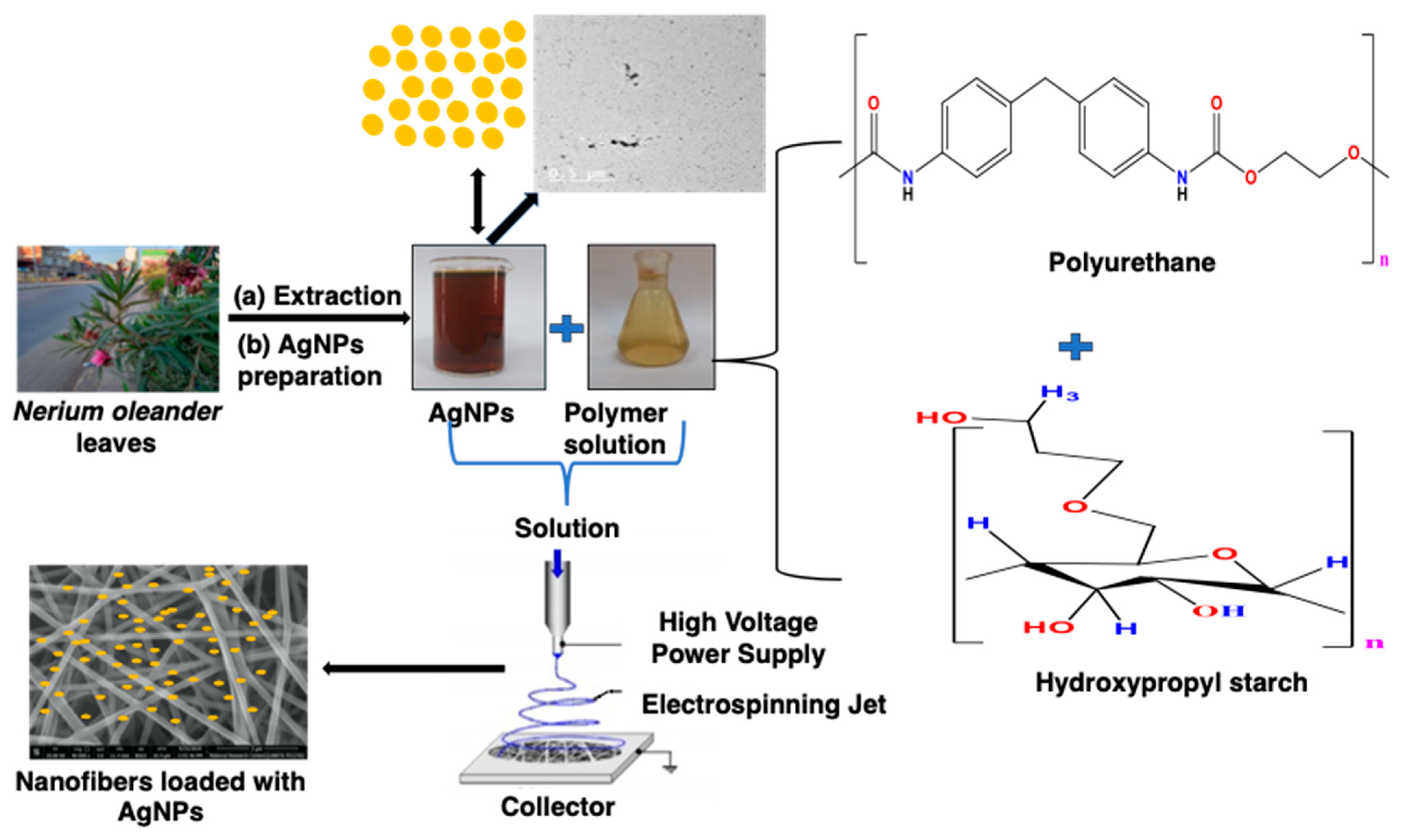
2.2. Biosynthesis of AgNPs
2.3. Electrospinning of HPS/PU Nanofibers Blend Lioaded with AgNPs (AgNPs@NFs)
2.4. Physicochemical Characterization of the Extract, AgNPs and Nanofibers
2.5. Biological Characterization of the Prepared Nanofibers
2.5.1. Tested Microorganisms
2.5.2. Antimicrobial Test
2.5.3. Toxicological Screening
2.6. Statistical Analyses
3. Results and Discussion
3.1. GC-MS Investigation of the Plant Extract
3.2. Characterization of the Biosynthesized AgNPs
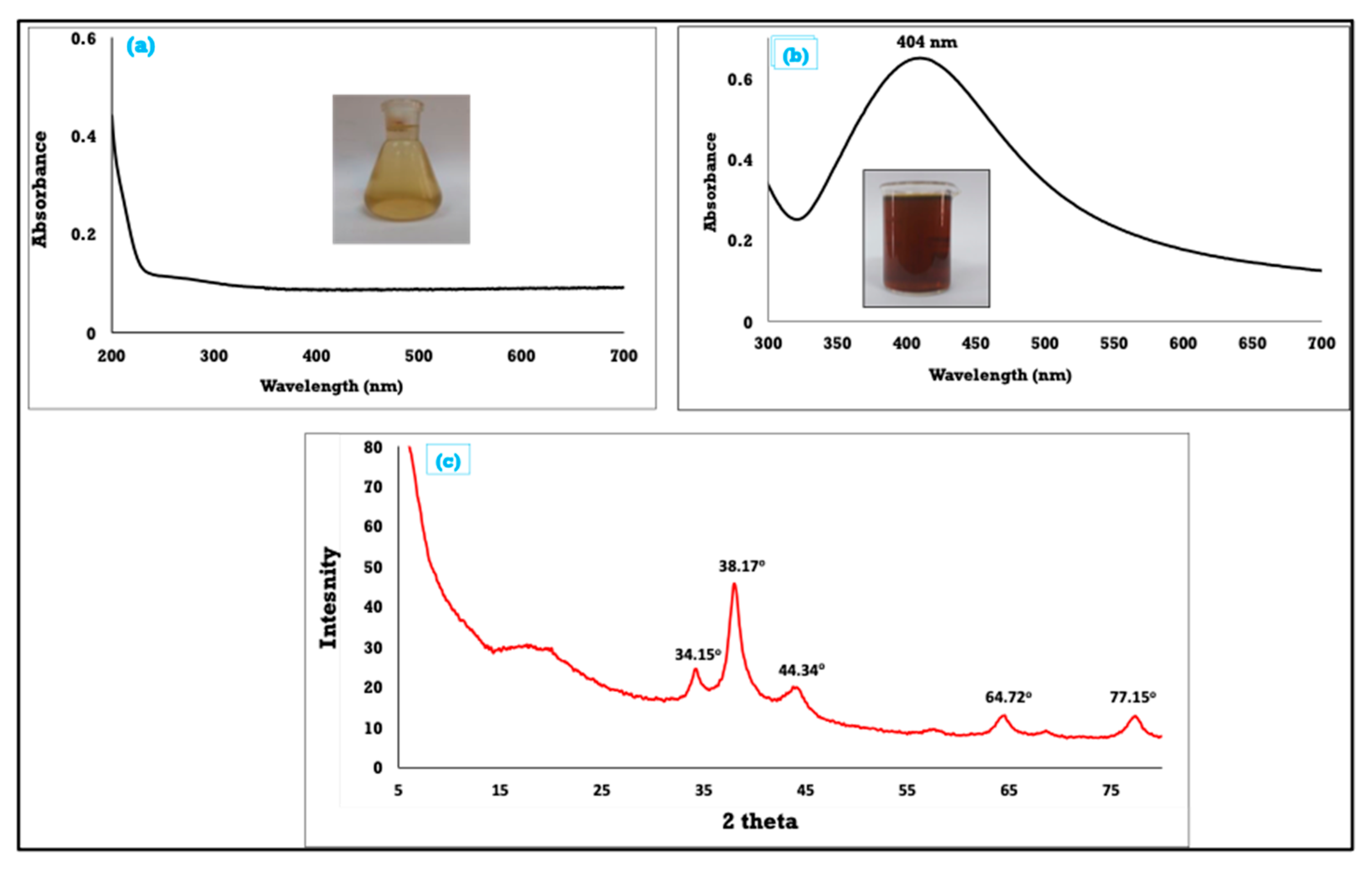
3.2.1. UV–Vis Spectroscopy Study
3.2.2. XRD Analysis
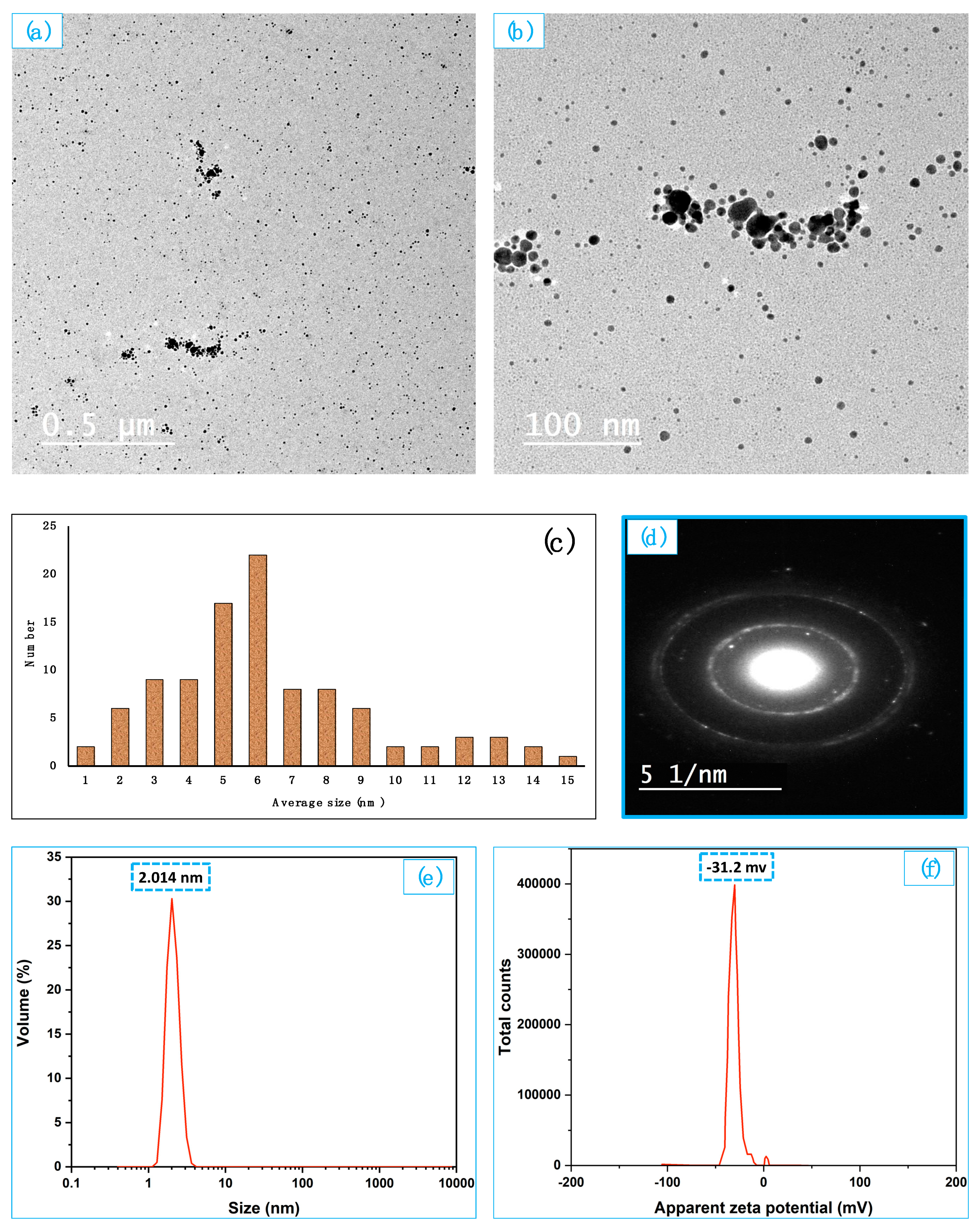
3.2.3. TEM
3.2.4. Dynamic Light Scattering (DLS)
3.2.5. Zeta Potential
3.3. Characterization of AgNPs Loaded Nanofibers (AgNPs@NFs)
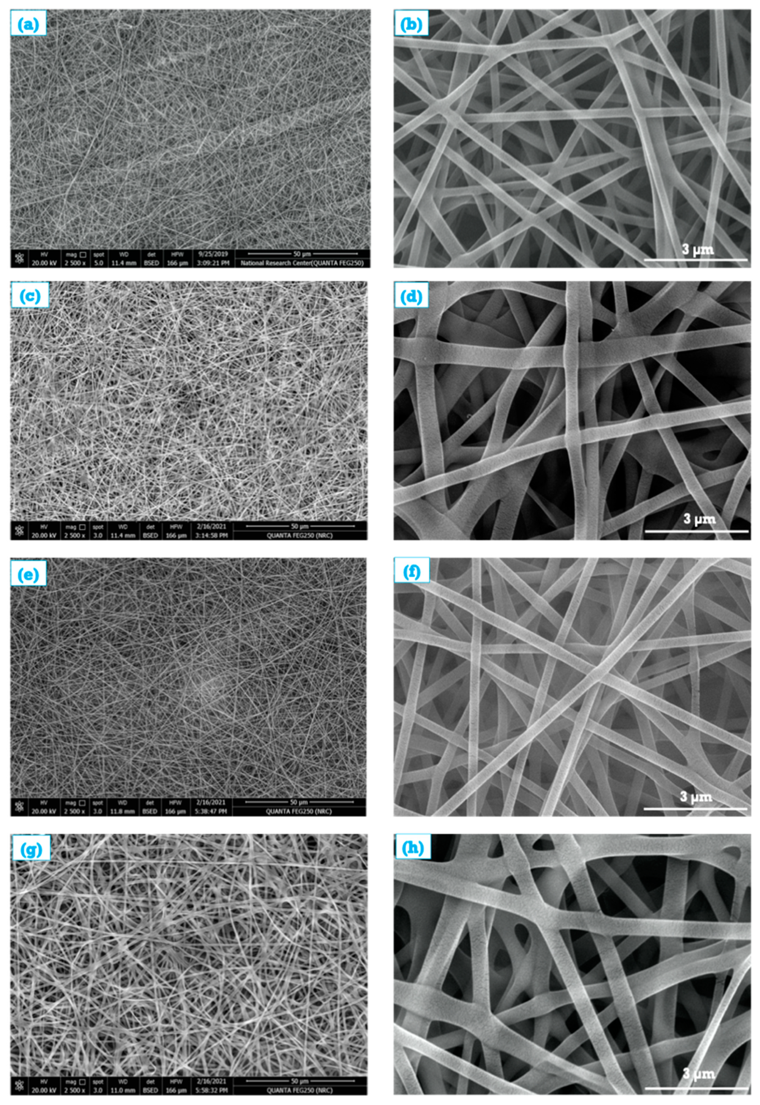
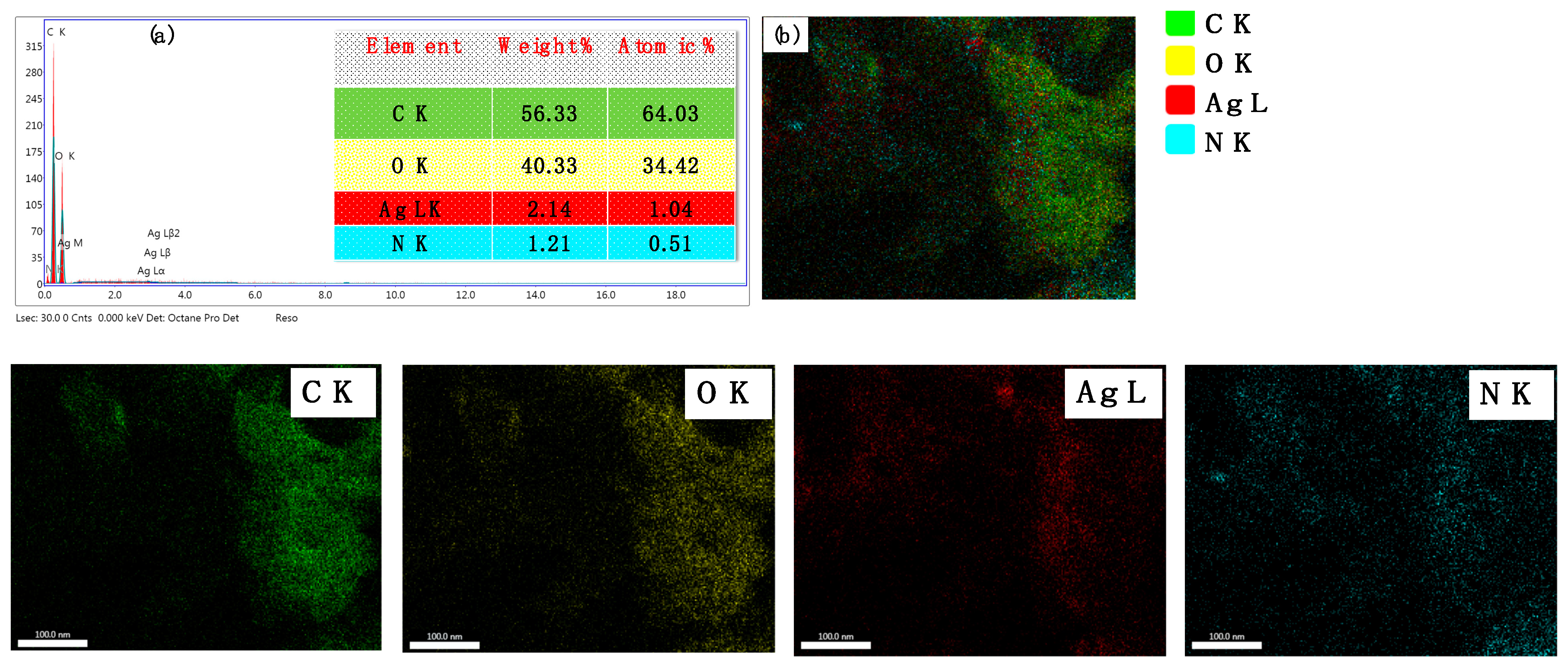
3.3.1. SEM, EDX, Roughness and Contact Angle of AgNPs@NFs
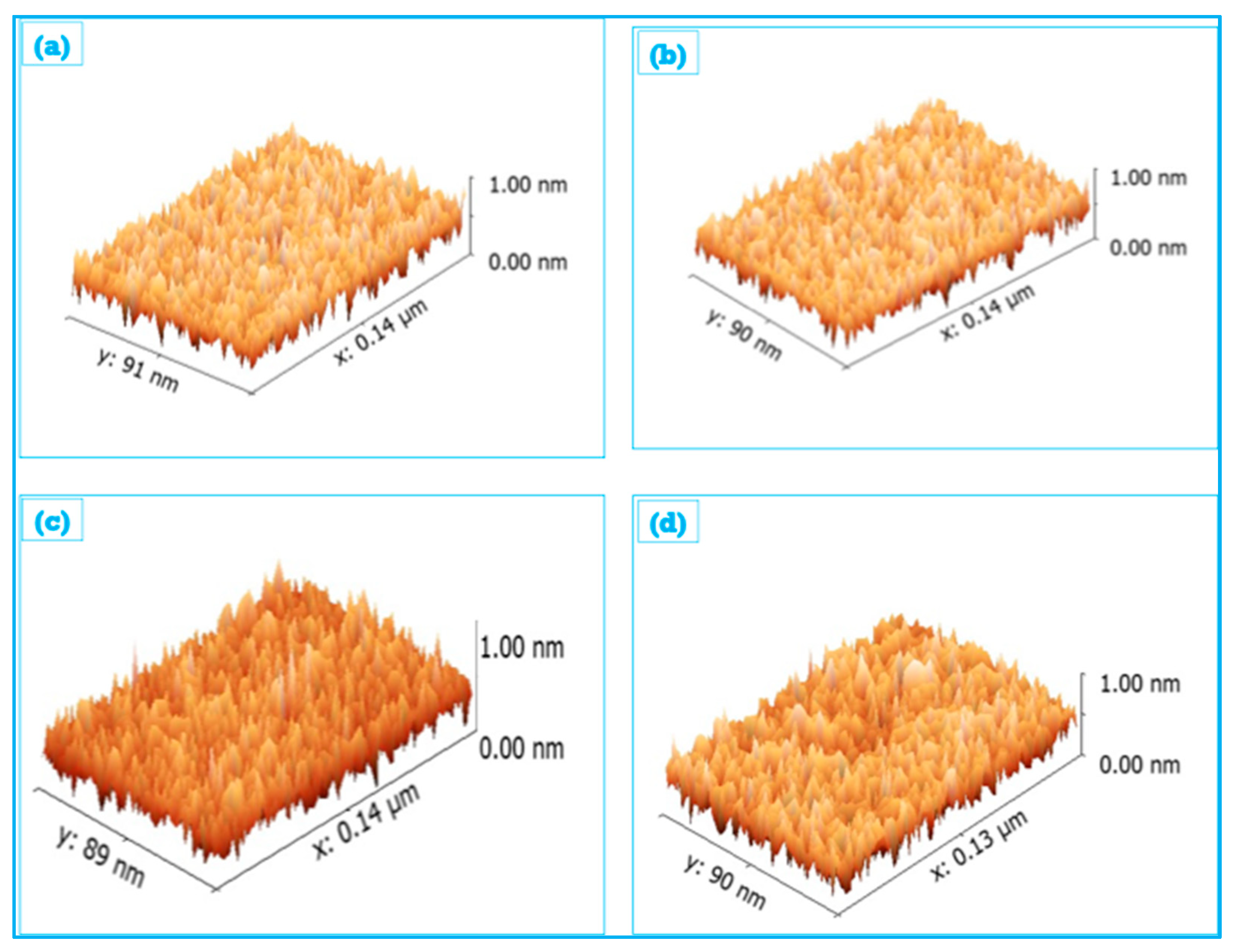
3.3.2. Surface Roughness of AgNPs@NFs
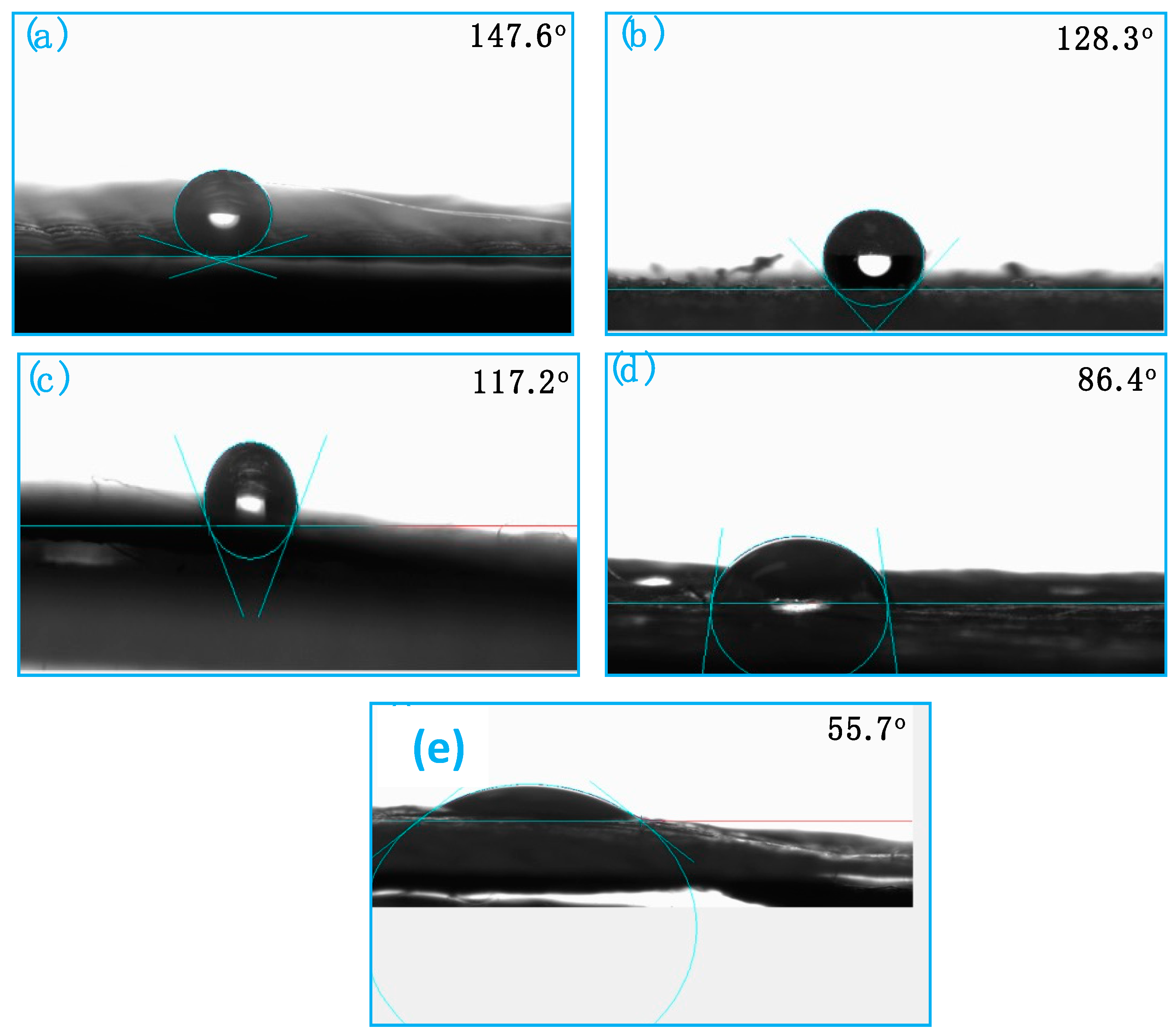
3.3.3. Contact Angle Measurements of AgNPs@NFs
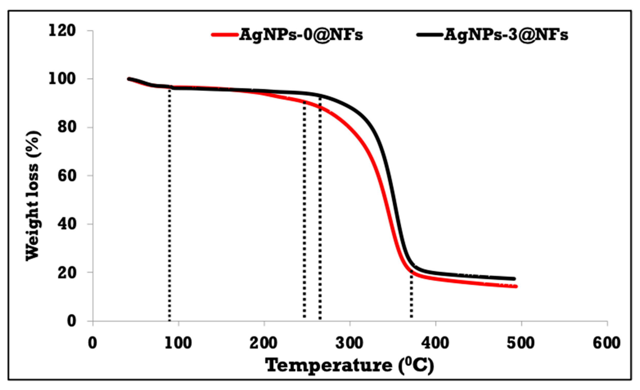
3.3.4. FTIR/ATR Spectra and TGA of AgNPs@NFs
3.4. Evaluation of the Antimicrobial Activity of AgNPs@NFs
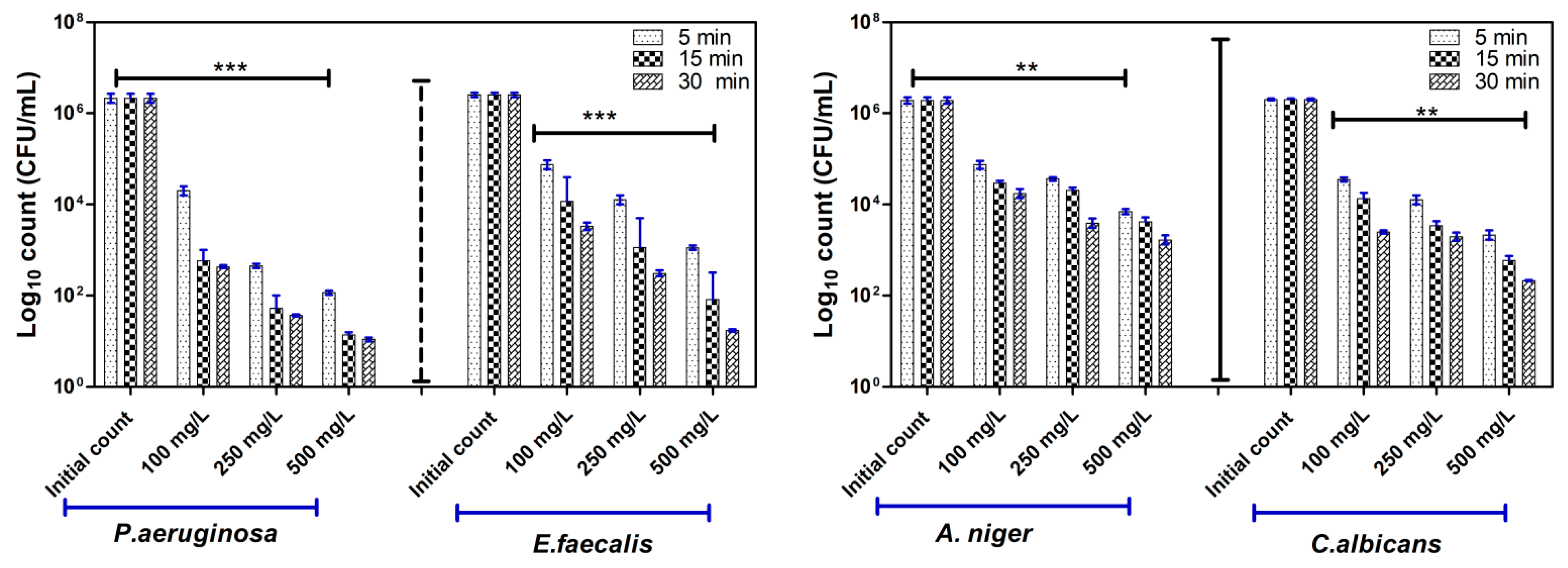
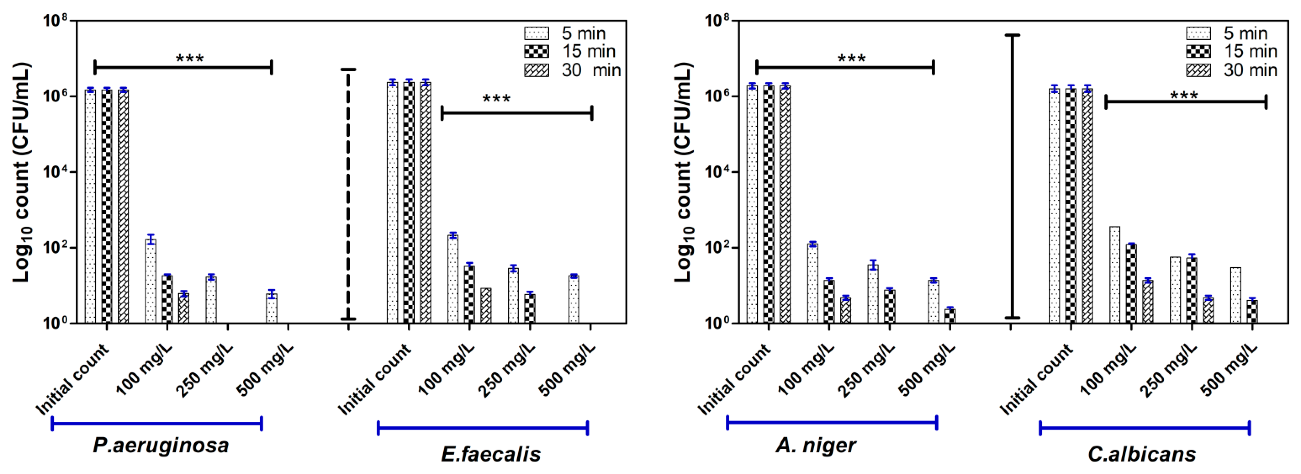
3.4.1. Antimicrobial Activity Assessment
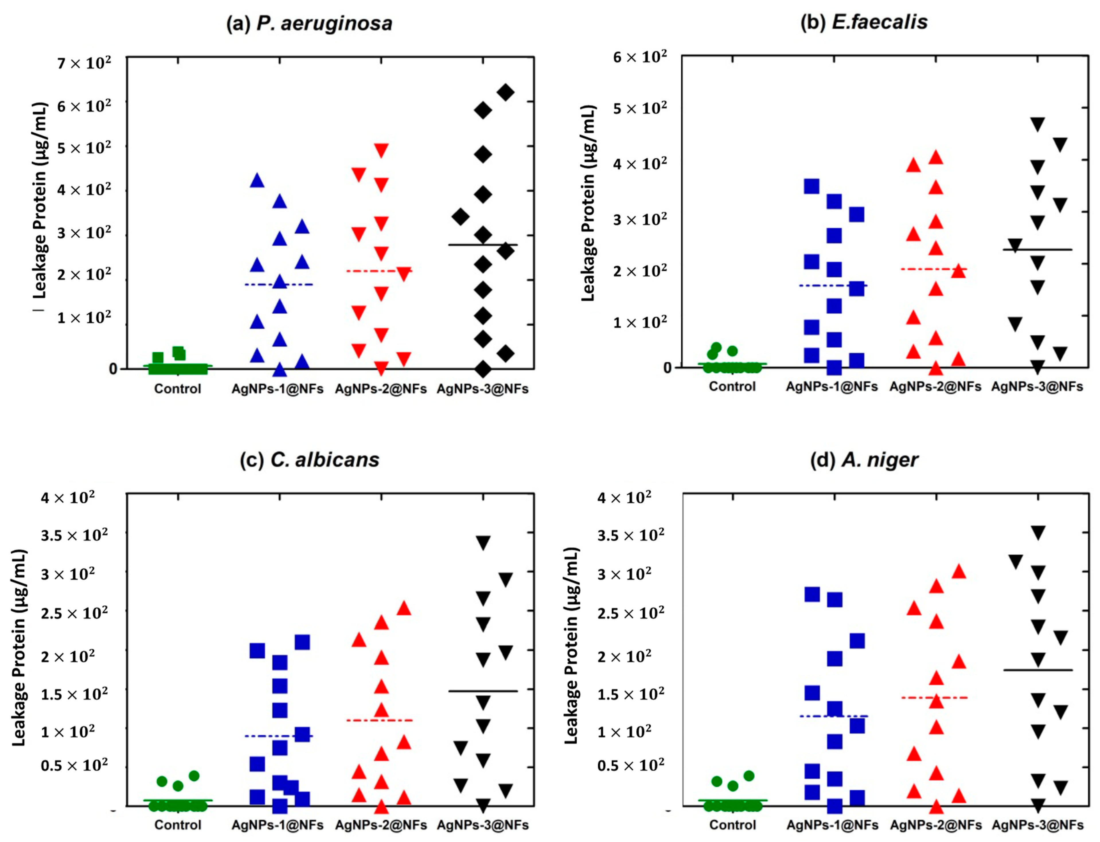
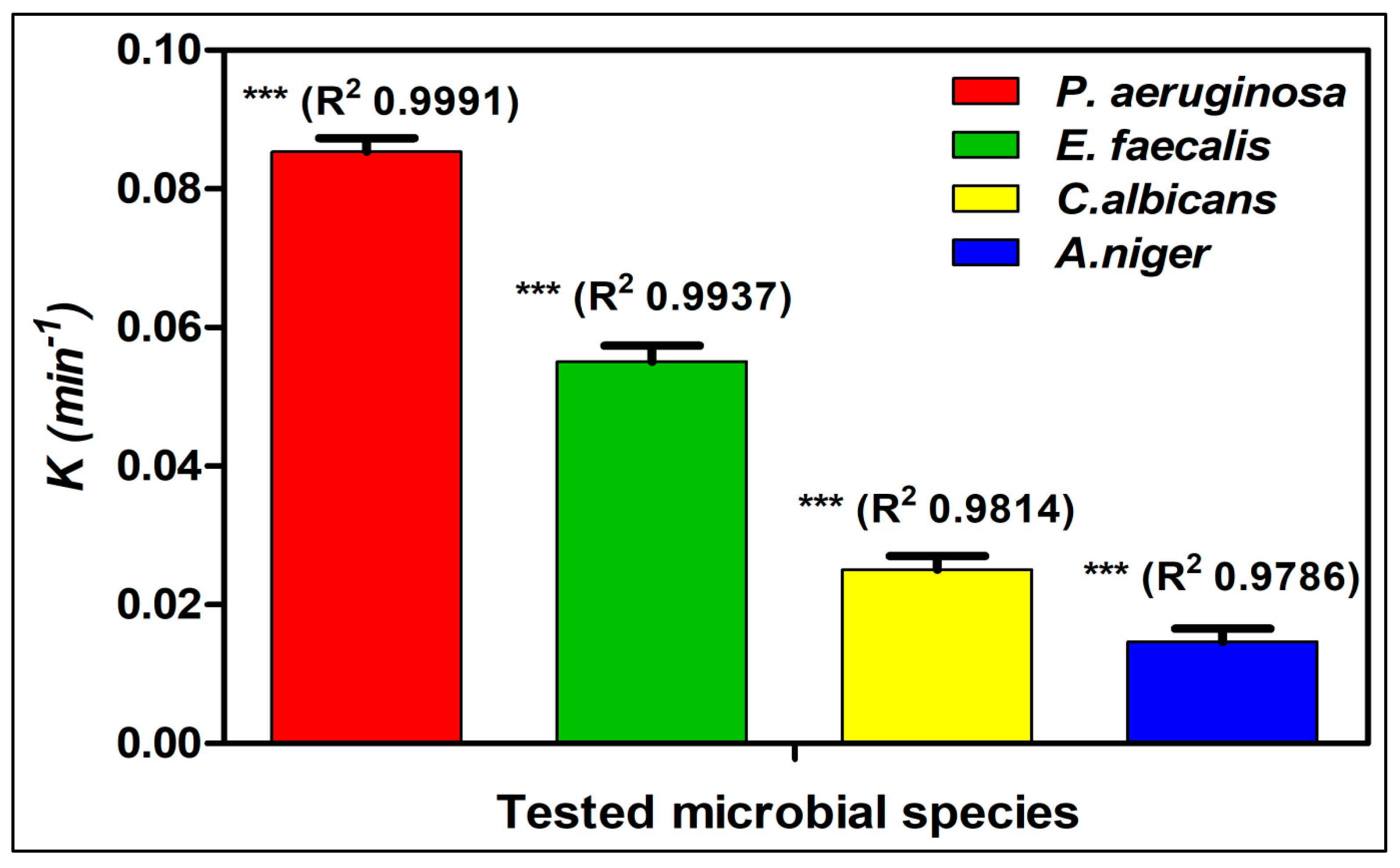
3.4.2. Mode of Action
3.5. Toxicological Performance Assay
4. Conclusions
Supplementary Materials
Author Contributions
Funding
Institutional Review Board Statement
Informed Consent Statement
Data Availability Statement
Acknowledgments
Conflicts of Interest
References
- Samadian, H.; Khastar, H.; Ehterami, A.; Salehi, M. Bioengineered 3D nanocomposite based on gold nanoparticles and gelatin nanofibers for bone regeneration: In vitro and in vivo study. Sci. Rep. 2021, 11, 13877. [Google Scholar] [CrossRef] [PubMed]
- Sohrabi, M.; Abbasi, M.; Ansar, M.M.; Soltani Tehrani, B. Evaluation of electrospun nanofibers fabricated using PCL/PVP and PVA/β-TCP as potential scaffolds for bone tissue engineering. Polym. Bull. 2021, 1–17. [Google Scholar] [CrossRef]
- Halawani, E.M.; Hassan, A.M.; El-Rab, S.M.F.G. Nanoformulation of biogenic cefotaxime-conjugated-silver nanoparticles for enhanced antibacterial efficacy against multidrug-resistant bacteria and anticancer studies. Int. J. Nanomed. 2020, 15, 1889–1901. [Google Scholar] [CrossRef] [Green Version]
- Sharma, D.; Gulati, S.S.; Sharma, N.; Chaudhary, A. Sustainable synthesis of silver nanoparticles using various biological sources and waste materials: A review. Emergent Mater. 2021, 1–30. [Google Scholar] [CrossRef]
- Mangindaan, D.; Lin, G.-Y.; Kuo, C.-J.; Chien, H.-W. Biosynthesis of silver nanoparticles as catalyst by spent coffee ground/recycled poly (ethylene terephthalate) composites. Food Bioprod. Process. 2020, 121, 193–201. [Google Scholar] [CrossRef]
- Chien, H.-W.; Kuo, C.-J.; Kao, L.-H.; Lin, G.-Y.; Chen, P.-Y. Polysaccharidic spent coffee grounds for silver nanoparticle immobilization as a green and highly efficient biocide. Int. J. Biol. Macromol. 2019, 140, 168–176. [Google Scholar] [CrossRef]
- Lo, S.; Fauzi, M.B. Current Update of Collagen Nanomaterials—Fabrication, Characterisation and Its Applications: A Review. Pharmaceutics 2021, 13, 316. [Google Scholar] [CrossRef]
- Wang, Q.; Liu, W.; Tian, B.; Li, D.; Liu, C.; Jiang, B.; Feng, Z. Preparation and characterization of coating based on protein nanofibers and polyphenol and application for salted duck egg yolks. Foods 2020, 9, 449. [Google Scholar] [CrossRef] [PubMed] [Green Version]
- Zhang, Y.; Liang, S.; Zhang, J.; Chi, Y.; Tian, B.; Li, L.; Jiang, B.; Li, D.; Feng, Z.; Liu, C. Preparation of whey protein isolate nanofibrils by microwave heating and its application as carriers of lipophilic bioactive substances. LWT 2020, 125, 109213. [Google Scholar] [CrossRef]
- Celebioglu, A.; Uyar, T. Antioxidant vitamin E/cyclodextrin inclusion complex electrospun nanofibers: Enhanced water solubility, prolonged shelf life, and photostability of vitamin E. J. Agric. Food Chem. 2017, 65, 5404–5412. [Google Scholar] [CrossRef]
- Hebeish, A.; El-Rafie, M.H.; EL-Sheikh, M.A.; Seleem, A.A.; El-Naggar, M.E. Antimicrobial wound dressing and anti-inflammatory efficacy of silver nanoparticles. Int. J. Biol. Macromol. 2014, 65, 509–515. [Google Scholar] [CrossRef]
- Hebeish, A.; El-Naggar, M.E.; Fouda, M.M.G.; Ramadan, M.A.; Al-Deyab, S.S.; El-Rafie, M.H. Highly effective antibacterial textiles containing green synthesized silver nanoparticles. Carbohydr. Polym. 2011, 86, 936–940. [Google Scholar] [CrossRef]
- Radwan, E.K.; El-Naggar, M.E.; Abdel-Karim, A.; Wassel, A.R. Multifunctional cationic starch/nanofibrillated cellulose/silver nanoparticles nanocomposite cryogel: Synthesis, adsorption, and antibacterial characteristics. Int. J. Biol. Macromol. 2021, 189, 420–431. [Google Scholar] [CrossRef] [PubMed]
- Krishnan, P.D.; Banas, D.; Durai, R.D.; Kabanov, D.; Hosnedlova, B.; Kepinska, M.; Fernandez, C.; Ruttkay-Nedecky, B.; Nguyen, H.V.; Farid, A.; et al. Silver nanomaterials for wound dressing applications. Pharmaceutics 2020, 12, 821. [Google Scholar] [CrossRef]
- Siadat, S.A.; Mokhtari, J. Fabrication of Novel Antimicrobial Bio-Fibers Using Silk Wastage, Study of Poly (hexamethylene) Biguanide, and Silver Nanoparticles Interaction. J. Nat. Fibers 2017, 14, 707–717. [Google Scholar] [CrossRef]
- Li, H.; Williams, G.R.; Wu, J.; Lv, Y.; Sun, X.; Wu, H.; Zhu, L.-M. Thermosensitive nanofibers loaded with ciprofloxacin as antibacterial wound dressing materials. Int. J. Pharm. 2017, 517, 135–147. [Google Scholar] [CrossRef]
- Schulte-Werning, L.V.; Murugaiah, A.; Singh, B.; Johannessen, M.; Engstad, R.E.; Škalko-Basnet, N.; Holsæter, A.M. Multifunctional Nanofibrous Dressing with Antimicrobial and Anti-Inflammatory Properties Prepared by Needle-Free Electrospinning. Pharmaceutics 2021, 13, 1527. [Google Scholar] [CrossRef] [PubMed]
- Sousa, M.G.C.; Rezende, T.M.B.; Franco, O.L. Nanofibers as drug-delivery systems for antimicrobial peptides. Drug Discov. Today 2021, 26, 2064–2074. [Google Scholar] [CrossRef]
- Subbaiya, R.; Shiyamala, M.; Revathi, K.; Pushpalatha, R.; Selvam, M.M. Biological synthesis of silver nanoparticles from Nerium oleander and its antibacterial and antioxidant property. Int. J. Curr. Microbiol. Appl. Sci 2014, 3, 83–87. [Google Scholar]
- Gopinath, M.; Subbaiya, R.; Selvam, M.M.; Suresh, D. Synthesis of copper nanoparticles from Nerium oleander leaf aqueous extract and its antibacterial activity. Int. J. Curr. Microbiol. Appl. Sci. 2014, 3, 814–818. [Google Scholar]
- El-Naggar, M.E.; Abdelgawad, A.M.; Tripathi, A.; Rojas, O.J. Curdlan cryogels reinforced with cellulose nanofibrils for controlled release. J. Environ. Chem. Eng. 2017, 5, 5754–5761. [Google Scholar] [CrossRef]
- El-Newehy, M.H.; El-Naggar, M.E.; Alotaiby, S.; El-Hamshary, H.; Moydeen, M.; Al-Deyab, S. Green Electrospining of Hydroxypropyl Cellulose Nanofibres for Drug Delivery Applications. J. Nanosci. Nanotechnol. 2018, 18, 805–814. [Google Scholar] [CrossRef]
- El-Naggar, M.E.; El-Newehy, M.H.; Aldalbahi, A.; Salem, W.M.; Khattab, T.A. Immobilization of anthocyanin extract from red-cabbage into electrospun polyvinyl alcohol nanofibers for colorimetric selective detection of ferric ions. J. Environ. Chem. Eng. 2021, 9, 105072. [Google Scholar] [CrossRef]
- El-Naggar, M.E.; Alharthi, S.; Saleh, D.I.; El-Sayed, W.A.; Abu-Saied, M.A.; Ahmed, M.K. Thallium/vanadate co-substitutions through hydroxyapatite/polycaprolactone nanofibrous scaffolds for biomedical domains. Mater. Chem. Phys. 2021, 271, 124879. [Google Scholar] [CrossRef]
- Ahmed, M.K.; El-Naggar, M.E.; Mahmoud, K.H.; Abdel-Rahim, F.M.; Menazea, A.A. Electrospun membranes of cellulose acetate/polyvinylidene difluoride containing Au/Se nanoparticles via laser ablation technique for methylene blue degradation. J. Polym. Res. 2021, 28, 324. [Google Scholar] [CrossRef]
- Sharaf, S.; El-Naggar, M.E. Eco-friendly technology for preparation, characterization and promotion of honey bee propolis extract loaded cellulose acetate nanofibers in medical domains. Cellulose 2018, 25, 5195–5204. [Google Scholar] [CrossRef]
- El-Naggar, M.E.; Samhan, F.A.; Salama, A.A.A.; Hamdy, R.M.; Ali, G.H. Cationic starch: Safe and economic harvesting flocculant for microalgal biomass and inhibiting E. coli growth. Int. J. Biol. Macromol. 2018, 116, 1296–1303. [Google Scholar] [CrossRef]
- Soares, R.M.D.; Siqueira, N.M.; Prabhakaram, M.P.; Ramakrishna, S. Electrospinning and electrospray of bio-based and natural polymers for biomaterials development. Mater. Sci. Eng. C 2018, 92, 969–982. [Google Scholar] [CrossRef] [PubMed]
- Shogren, R.L.; Bagley, E.B. Natural Polymers as Advanced Materials: Some Research Needs and Directions; ACS Publications: Washington, DC, USA, 1999; ISBN 1947-5918. [Google Scholar]
- Rogovina, S.Z.; Prut, E.V.; Berlin, A.A. Composite materials based on synthetic polymers reinforced with natural fibers. Polym. Sci. Ser. A 2019, 61, 417–438. [Google Scholar] [CrossRef]
- El-Rafie, M.H.; El-Naggar, M.E.; Ramadan, M.A.; Fouda, M.M.G.; Al-Deyab, S.S.; Hebeish, A. Environmental synthesis of silver nanoparticles using hydroxypropyl starch and their characterization. Carbohydr. Polym. 2011, 86, 630–635. [Google Scholar] [CrossRef]
- Mohamed, S.A.; Elaraby, N.M.; Abdel-Aty, A.M.; Shaban, E.; Abu-Saied, M.A.; Kenawy, E.-R.; El-Naggar, M.E. Improvement of enzymatic properties and decolorization of azo dye: Immobilization of horseradish peroxidase on cationic maize starch. Biocatal. Agric. Biotechnol. 2021, 38, 102208. [Google Scholar] [CrossRef]
- Abdo, S.M.; Mahmoud, R.H.; Youssef, M.; El-Naggar, M.E. Cationic Starch and Polyaluminum Chloride as Coagulants for River Nile Water Treatment. Groundw. Sustain. Dev. 2020, 10, 100331. [Google Scholar] [CrossRef]
- Vincent, P.; Ham-Pichavant, F.; Michaud, C.; Mignani, G.; Mastroianni, S.; Cramail, H.; Grelier, S. Extraction and Characterization of Hemicelluloses from A Softwood Acid Sulfite Pulp. Polymers 2021, 13, 2044. [Google Scholar] [CrossRef] [PubMed]
- Wendels, S.; Avérous, L. Biobased polyurethanes for biomedical applications. Bioact. Mater. 2021, 6, 1083–1106. [Google Scholar] [CrossRef]
- Guo, B.; Glavas, L.; Albertsson, A.-C. Biodegradable and electrically conducting polymers for biomedical applications. Prog. Polym. Sci. 2013, 38, 1263–1286. [Google Scholar] [CrossRef]
- Madkour, H.M.F.; Ghareeb, M.A.; Abdel-Aziz, M.S.; Khalaf, O.M.; Saad, A.M.; El-Ziaty, A.K.; Abdel-Mogib, M. Gas chromatography-mass spectrometry analysis, antimicrobial, anticancer and antioxidant activities of n-hexane and methylene chloride extracts of Senna italica. J. Appl. Pharm. Sci. 2017, 7, 23–32. [Google Scholar]
- Khalil, W.A.; Sherif, H.H.A.; Hemdan, B.A.; Khalil, S.K.H.; El Hotaby, W. Biocompatibility enhancement of graphene oxide-silver nanocomposite by functionalisation with polyvinylpyrrolidone. IET Nanobiotechnol. 2019, 13, 816–823. [Google Scholar] [CrossRef]
- He, M.; Wu, T.; Pan, S.; Xu, X. Antimicrobial mechanism of flavonoids against Escherichia coli ATCC 25922 by model membrane study. Appl. Surf. Sci. 2014, 305, 515–521. [Google Scholar] [CrossRef]
- Niemirycz, E.; Nichthauser, J.; Staniszewska, M.; Nałȩcz-Jawecki, G.; Bolałek, J. The Microtox® biological test: Application in toxicity evaluation of surface waters and sediments in Poland. Oceanol. Hydrobiol. Stud. 2007, 36, 151–163. [Google Scholar] [CrossRef]
- Abdel-Wareth, M.T.A.; El-Hagrassi, A.M.; Abdel-Aziz, M.S.; Nasr, S.M.; Ghareeb, M.A. Biological activities of endozoic fungi isolated from Biomphalaria alexandrina snails maintained in different environmental conditions. Int. J. Environ. Stud. 2019, 76, 780–799. [Google Scholar] [CrossRef]
- Shawky, B.T.; Nagah, M.; Ghareeb, M.A.; El-Sherbiny, G.M.; Moghannem, S.A.M.; Abdel-Aziz, M.S. Evaluation of Antioxidants, Total Phenolics and Antimicrobial Activities of Ethyl Acetate Extracts From Fungi Grown on Rice Straw. J. Renew. Mater. 2019, 7, 662–677. [Google Scholar] [CrossRef] [Green Version]
- Sathiya, C.K.; Akilandeswari, S. Fabrication and characterization of silver nanoparticles using Delonix elata leaf broth. Spectrochim. Acta Part A Mol. Biomol. Spectrosc. 2014, 128, 337–341. [Google Scholar] [CrossRef]
- Chen, J.; Liu, Y.; Xiong, Y.; Wei, D.; Peng, J.; Mahmud, S.; Liu, H. Konjac glucomannan reduced-stabilized silver nanoparticles for mono-azo and di-azo contained wastewater treatment. Inorg. Chim. Acta 2021, 515, 120058. [Google Scholar] [CrossRef]
- Rajendran, R.K.; Lin, C.-C. Stability and Microbial Toxicity of Silver Nanoparticles under Denitrifying Conditions. ACS Appl. Mater. Interfaces 2021, 13, 46233–46246. [Google Scholar] [CrossRef]
- Dairi, N.; Ferfera-Harrar, H.; Ramos, M.; Garrigós, M.C. Cellulose acetate/AgNPs-organoclay and/or thymol nano-biocomposite films with combined antimicrobial/antioxidant properties for active food packaging use. Int. J. Biol. Macromol. 2019, 121, 508–523. [Google Scholar] [CrossRef] [PubMed] [Green Version]
- Abd Ali, M.A.; Shareef, A.A. Green Synthesis of Silver Nanoparticles by Enterobacter Aerogenes Bacteria in Combination with Antibiotics Against Multidrug Resistance Streptococcus Mitis Isolated from Oral Cavity of Some Dental Caries Patients in Misan City. Ann. Rom. Soc. Cell Biol. 2021, 25, 13768–13789. [Google Scholar]
- Mythili, R.; Srinivasan, P.; Praburaman, L.; Al-Ansari, M.M.; Al-Humaid, L.; Vijayalakshmi, S.; Selvankumar, T. Biogenic production of silver nanoparticles from milk of Capra aegagrus hircus and mechanism of antibacterial activity on different bacteria. Appl. Nanosci. 2021, 1–8. [Google Scholar] [CrossRef]
- Ubgade, S.; Bapat, A.; Kilor, V. Effect of various stabilizers on the stability of lansoprazole nanosuspension prepared using high shear homogenization: Preliminary investigation. J. Appl. Pharm. Sci. 2021, 11, 85–92. [Google Scholar]
- Varadavenkatesan, T.; Selvaraj, R.; Vinayagam, R. Green synthesis of silver nanoparticles using Thunbergia grandiflora flower extract and its catalytic action in reduction of Congo red dye. Mater. Today Proc. 2020, 23, 39–42. [Google Scholar] [CrossRef]
- Asefnejad, A.; Khorasani, M.T.; Behnamghader, A.; Farsadzadeh, B.; Bonakdar, S. Manufacturing of biodegradable polyurethane scaffolds based on polycaprolactone using a phase separation method: Physical properties and in vitro assay. Int. J. Nanomed. 2011, 6, 2375. [Google Scholar] [CrossRef] [Green Version]
- Mura, P. Analytical techniques for characterization of cyclodextrin complexes in aqueous solution: A review. J. Pharm. Biomed. Anal. 2014, 101, 238–250. [Google Scholar] [CrossRef]
- Hussein, E.A.M.; Mohammad, A.A.-H.; Harraz, F.A.; Ahsan, M.F. Biologically synthesized silver nanoparticles for enhancing tetracycline activity against staphylococcus aureus and klebsiella pneumoniae. Braz. Arch. Biol. Technol. 2019, 62. [Google Scholar] [CrossRef]
- Tripathi, S.; Mehrotra, G.K.; Dutta, P.K. Chitosan–silver oxide nanocomposite film: Preparation and antimicrobial activity. Bull. Mater. Sci. 2011, 34, 29–35. [Google Scholar] [CrossRef] [Green Version]
- Gour, A.; Jain, N.K. Advances in green synthesis of nanoparticles. Artif. Cells Nanomed. Biotechnol. 2019, 47, 844–851. [Google Scholar] [CrossRef] [PubMed] [Green Version]
- Ansar, S.; Tabassum, H.; Aladwan, N.S.M.; Naiman Ali, M.; Almaarik, B.; AlMahrouqi, S.; Abudawood, M.; Banu, N.; Alsubki, R. Eco friendly silver nanoparticles synthesis by Brassica oleracea and its antibacterial, anticancer and antioxidant properties. Sci. Rep. 2020, 10, 18564. [Google Scholar] [CrossRef]
- Du, J.; Singh, H.; Yi, T.H. Biosynthesis of silver nanoparticles by Novosphingobium sp. THG-C3 and their antimicrobial potential. Artif. Cells Nanomed. Biotechnol. 2017, 45, 211–217. [Google Scholar] [CrossRef] [PubMed] [Green Version]
- Ali, G.W.; El-Hotaby, W.; Hemdan, B.; Abdel-Fattah, W.I. Thermosensitive chitosan/phosphate hydrogel-composites fortified with Ag versus Ag@Pd for biomedical applications. Life Sci. 2018, 194, 185–195. [Google Scholar] [CrossRef] [PubMed]
- Jiang, B.; Wang, L.; Zhu, M.; Wu, S.; Wang, X.; Li, D.; Liu, C.; Feng, Z.; Tian, B. Separation, structural characteristics and biological activity of lactic acid bacteria exopolysaccharides separated by aqueous two-phase system. LWT 2021, 147, 111617. [Google Scholar] [CrossRef]
- Akter, S.; Huq, M.A. Biologically rapid synthesis of silver nanoparticles by Sphingobium sp. MAH-11T and their antibacterial activity and mechanisms investigation against drug-resistant pathogenic microbes. Artif. Cells Nanomed. Biotechnol. 2020, 48, 672–682. [Google Scholar] [CrossRef] [Green Version]
- Salem, S.S.; Fouda, M.M.G.; Fouda, A.; Awad, M.A.; Al-Olayan, E.M.; Allam, A.A.; Shaheen, T.I. Antibacterial, Cytotoxicity and Larvicidal Activity of Green Synthesized Selenium Nanoparticles Using Penicillium corylophilum. J. Clust. Sci. 2021, 32, 351–361. [Google Scholar] [CrossRef]
- Kumar, P.; Senthamil Selvi, S.; Lakshmi Prabha, A.; Prem Kumar, K.; Ganeshkumar, R.S.; Govindaraju, M. Synthesis of silver nanoparticles from Sargassum tenerrimum and screening phytochemicals for its antibacterial activity. Nano Biomed. Eng. 2012, 4, 12–16. [Google Scholar] [CrossRef]
- Jiang, K.; Guo, S.; Yang, J.; Liu, J.; Shaukat, A.; Zhao, G.; Wu, H.; Deng, G. Matrine alleviates Staphylococcus aureus lipoteichoic acid-induced endometritis via suppression of TLR2-mediated NF-κB activation. Int. Immunopharmacol. 2019, 70, 201–207. [Google Scholar] [CrossRef] [PubMed]
- Xie, Y.; He, Y.; Irwin, P.L.; Jin, T.; Shi, X. Antibacterial activity and mechanism of action of zinc oxide nanoparticles against Campylobacter jejuni. Appl. Environ. Microbiol. 2011, 77, 2325–2331. [Google Scholar] [CrossRef] [Green Version]
- Vollmer, W.; Blanot, D.; De Pedro, M.A. Peptidoglycan structure and architecture. FEMS Microbiol. Rev. 2008, 32, 149–167. [Google Scholar] [CrossRef] [PubMed] [Green Version]
- Fatimah, I.; Aftrid, Z.H.V.I. Characteristics and antibacterial activity of green synthesized silver nanoparticles using red spinach (Amaranthus Tricolor L.) leaf extract. Green Chem. Lett. Rev. 2019, 12, 25–30. [Google Scholar] [CrossRef] [Green Version]
- Niazvand, F.; Orazizadeh, M.; Khorsandi, L.; Abbaspour, M.; Mansouri, E.; Khodadadi, A. Effects of Quercetin-loaded nanoparticles on MCF-7 human breast cancer cells. Medicina 2019, 55, 114. [Google Scholar] [CrossRef] [Green Version]


| Composition Code | PU Volume (mL) | HPS Volume (mL) | AgNPs (mL) | Total Volume (mL) |
|---|---|---|---|---|
| AgNPs-0@NFs | 10 | 5 | 0 | 15 |
| AgNPs-1@NFs | 9 | 5 | 1 | 15 |
| AgNPs-2@NFs | 8 | 5 | 2 | 15 |
| AgNPs-3@NFs | 7 | 5 | 3 | 15 |
| Composition Code | Ra (nm) | Rq (nm) | Rt% (nm) | Rv (nm) |
|---|---|---|---|---|
| AgNPs-0@NFs | 0.0745 | 0.0915 | 0.5123 | 0.2327 |
| AgNPs-1@NFs | 0.1061 | 0.1353 | 0.7353 | 0.3345 |
| AgNPs-2@NFs | 0.1129 | 0.1449 | 0.8628 | 0.4030 |
| AgNPs-3@NFs | 0.1271 | 0.1557 | 0.7865 | 0.4149 |
| Composition Code | ZOI Diameters (mm) | |||
|---|---|---|---|---|
| P. aeruginosa | E. faecalis | C. albicans | A. niger | |
| AgNPs-0@NFs | 0 ± 0 | 0 ± 0 | 0 ± 0 | 0 ± 0 |
| AgNPs-1@NFs | 15 ± 0.20 b | 13 ± 0.18 c | 10 ± 0.16 c | 11 ± 0.23 c |
| AgNPs-2@NFs | 21 ± 0.17 a | 19 ± 0.12 b | 17 ± 0.20 b | 15 ± 0.15 b |
| AgNPs-3@NFs | 26 ± 0.23 a | 24 ± 0.25 a | 23 ± 0.23 a | 21 ± 0.17 a |
| Ciprofloxacin | 12 ± 0.19 b | 11 ± 0.25 b | 8 ± 0.21 c | 7 ± 0.14 c |
| Studied Nanofibers | EC50 % Concentrations | References of Toxic Ranges | |||
|---|---|---|---|---|---|
| 5 min | 10 min | 15 min | EC50 % Degree | Toxicity Level | |
| AgNPs-0@NFs | 302 | 292 | 286 | 0–19 | Extremely toxic |
| AgNPs-1@NFs | 289 | 272 | 268 | 20–39 | Very toxic |
| AgNPs-2@NFs | 271 | 268 | 265 | 40–59 | Toxic |
| AgNPs-3@NFs | 268 | 254 | 251 | 60–79 | Mild toxic |
| ≥100 | Non-toxic and safe | ||||
Publisher’s Note: MDPI stays neutral with regard to jurisdictional claims in published maps and institutional affiliations. |
© 2022 by the authors. Licensee MDPI, Basel, Switzerland. This article is an open access article distributed under the terms and conditions of the Creative Commons Attribution (CC BY) license (https://creativecommons.org/licenses/by/4.0/).
Share and Cite
El-Hefnawy, M.E.; Alhayyani, S.; El-Sherbiny, M.M.; Sakran, M.I.; El-Newehy, M.H. Fabrication of Nanofibers Based on Hydroxypropyl Starch/Polyurethane Loaded with the Biosynthesized Silver Nanoparticles for the Treatment of Pathogenic Microbes in Wounds. Polymers 2022, 14, 318. https://0-doi-org.brum.beds.ac.uk/10.3390/polym14020318
El-Hefnawy ME, Alhayyani S, El-Sherbiny MM, Sakran MI, El-Newehy MH. Fabrication of Nanofibers Based on Hydroxypropyl Starch/Polyurethane Loaded with the Biosynthesized Silver Nanoparticles for the Treatment of Pathogenic Microbes in Wounds. Polymers. 2022; 14(2):318. https://0-doi-org.brum.beds.ac.uk/10.3390/polym14020318
Chicago/Turabian StyleEl-Hefnawy, Mohamed E., Sultan Alhayyani, Mohsen M. El-Sherbiny, Mohamed I. Sakran, and Mohamed H. El-Newehy. 2022. "Fabrication of Nanofibers Based on Hydroxypropyl Starch/Polyurethane Loaded with the Biosynthesized Silver Nanoparticles for the Treatment of Pathogenic Microbes in Wounds" Polymers 14, no. 2: 318. https://0-doi-org.brum.beds.ac.uk/10.3390/polym14020318