Article

Low-Denaturazing Glucose Oxidase Immobilization onto Graphite Electrodes by Incubation in Chitosan Solutions

by
Mireia Buaki-Sogó
1,*,
Laura García-Carmona
1,*,
Mayte Gil-Agustí
1,
Marta García-Pellicer
1 and
Alfredo Quijano-López
1,2
1
Instituto Tecnológico de la Energía (ITE), Avenida Juan de la Cierva 24, 46980 Paterna, Spain
2
Instituto de Tecnología Eléctrica, Universitat Politècnica de València, Camino de Vera s/n Edificio 6C, 46022 Valencia, Spain
*
Authors to whom correspondence should be addressed.
Submission received: 14 February 2022 / Revised: 12 April 2022 / Accepted: 28 April 2022 / Published: 3 May 2022

Abstract

:
In this work, glucose oxidase (GOx) has been immobilized onto graphite rod electrodes through an assisted-chitosan adsorption reaching an enzyme coverage of 4 nmol/cm2. The direct and irreversible single adsorption of the Flavine Adenine Dinucleotide (FAD) cofactor has been minimized by electrode incubation in a chitosan (CH) solution containing the enzyme GOx. Chitosan keeps the enzyme structure and conformation due to electrostatic interactions preventing FAD dissociation from the protein envelope. Using chitosan, both the redox cofactor FAD and the protein envelope remain in the active form as demonstrated by the electrochemistry studies and the enzymatic activity in the electrochemical oxidation of glucose up to a concentration of 20 mM. The application of the modified electrodes for energy harvesting delivered a power density of 119 µW/cm2 with a cell voltage of 0.3 V. Thus, chitosan presents a stabilizing effect for the enzyme conformation promoted by the confinement effect in the chitosan solution by electrostatic interactions. Additionally, it facilitated the electron transfer from the enzyme to the electrode due to the presence of embedded chitosan in the enzyme structure acting as an electrical wiring between the electrode and the enzyme (electron transfer rate constant 2.2 s−1). This method involves advantages compared with previously reported chitosan immobilization methods, not only due to good stability of the enzyme, but also to the simplicity of the procedure that can be carried out even for not qualified technicians which enable their easy implementation in industry.

Graphical Abstract

1. Introduction

The entire field of enzyme-based electrodes can trace its origin back to the enzymatic glucose-based electrode fabricated by Clark and Lyons in 1962 with the publication of the original patent of an amperometric enzyme electrode for glucose sensing [1]. Currently, the importance of the glucose biosensors market is clear, covering about 85% of the entire biosensor market in the world, mainly due to the notable biomedical significance of the rapid and convenient assay of blood glucose in diabetes [2,3,4]. In addition, other relevant applications, such as biofuel cells for energy harvesting using molecules such as glucose as an attractive new power source, take advantage of technological advances related with GOx enzyme for self-powered, wearable, and implantable biomedical devices [5].
The enzyme GOx catalyzes the oxidation of β-D-glucose to gluconic acid with molecular oxygen as an electron acceptor with the production of hydrogen peroxide (H2O2) following reactions 1 and 2. In these reactions the fully oxidized form of Flavine Adenine Dinucleotide (FAD) catalyzes the oxidation of glucose while the reduced form (FADH2) is regenerated in the presence of O2 producing H2O2. Reaction 3 represents another mechanism in which the electronic transference from the reduced form of the enzyme (FADH2) occurs in the absence of mediators or electron acceptors such as oxygen [6,7,8]. However, this mechanism, usually called Direct Electron Transfer (DET), has not been proved to drive the reaction in the absence of mediators or electron acceptors. Currently, reaction 1 and 2 are the most accepted mechanisms to explain GOx redox mechanism since the two flavin active sites are deeply buried within the structure of the homodimer [9]. For this reason, efforts regarding GOx immobilization, traditionally focused on achieving DET, should be focused on stabilization issues related to the loose of the structure instead on the orientation of the enzyme on the electrodic surface.
Glucose + GOx(FAD) → Gluconic acid + GOx(FADH2) (1)
GOx ( FADH 2 ) + O 2     GOx ( FAD ) + H 2 O 2   ( 2 )
GOx ( FADH 2 )     GOx ( FAD ) + 2 e + 2 H +   ( 3 )
Particularly regarding bioelectrodes development, GOx should be immobilized onto suitable conductive support to carry out the enzymatic reaction. For real industrial use of this technology in long-term devices, the enzyme stability should be preserved to keep activity in the working medium and enhance enzyme stability towards denaturation of the enzyme and FAD irreversible adsorption on the electrode in storage and operational conditions [5,6]. Among the different enzyme immobilization strategies [5,10,11] adsorption is one of the preferred techniques due to its simplicity and limited decrease in enzymatic activity despite the disadvantages related to desorption processes, denaturation, and the method’s low specificity [12].
The use of graphite electrodes as supports in the development of bioelectrodes for biodevices has been widely spread due to the exceptional electrical properties of such materials. Additionally, graphite electrodes present a surface able to adsorb biomolecules due to the affinity of graphite surface for organic entities, so it has been used in several works as a support for enzyme immobilization using different strategies [13,14,15,16]. When performing GOx adsorption onto graphite electrodes, one of the main drawbacks comes from enzyme denaturation due to the most preferable and irreversible adsorption of FAD included into GOx structure, losing the quaternary structure, and, therefore, the catalytic activity [17,18,19]. The denaturation process could be mainly related to enzyme concentration or ionic strength of incubation solution, temperature, and/or incubation time [19]. This is a key aspect for the assessment of the enzyme activity since if FAD is irreversibly adsorbed in a denaturized form of GOx, can lead to misunderstanding when trying to assess the direct electrochemistry of the enzyme and to evaluate the bioelectrocatalytic activity in the presence of glucose [9,20], since no response would be detected.
Inactivation due to denaturation is mainly related to changes in quaternary structure due to subunit dissociation [21,22]. Hence, preventing subunit dissociation is an important strategy for multimeric enzyme stabilization [23]. Some methods based on this idea have been proposed, such as protein engineering or crosslinking [24,25,26,27]. However, these methods are quite complex and present limitations dealing with the stability of engineered enzymes or the selection of reaction conditions and reagents for crosslinking, which is usually a sensitive procedure that can lead to irreversible damage in the enzyme structure [28]. Therefore, the stabilization of multimeric enzymes against inactivation is considered a particular challenge.
Biopolymers can be used to immobilize enzymes belonging to various catalytic classes with the retention of good catalytic properties. Moreover, the produced biocatalytic systems offer improved thermal stability and, in general, are noted for their good reusability [29]. In this sense, several biomaterials are frequently used in research and industry due to the biocompatibility characteristics very convenient for their extensive potential in industrial applications. In this sense, biopolymers, such as Polyhydroxyalkanoates, linear polyesters containing 3-hydroxy fatty acid monomers, or polysaccharides, such as chitosan, are commonly employed [30,31]. However, sometimes it is necessary to modify these polymers in certain applications which can lead to modify their mechanical properties.
Specifically, Chitosan is a widely used polysaccharide obtained from the deacetylation of chitin, which is the major component in crustacean shells such as shrimps, lobsters, and crabs. Chitosan properties such as hydrophilicity, low toxicity, biocompatibility, biodegradability, film-forming ability, and bioactivity attract the interest of biologists and chemists to explore novel uses of this biopolymer. Chitosan molecules possess a positive charge in solution due to the amino groups present in their structure, so the low isoelectric point of some multimeric enzymes, as is the case for GOx, leads to a fast interaction between proteins and chitosan molecules [31]. For all these reasons, chitosan is the most frequently used biopolymer for enzyme immobilization [31,32]. Thus, although several methods using chitosan as the main scaffold for the adsorption immobilization strategy have been developed so far, several disadvantages have been detected, hindering their applicability in real-world applications. Specifically, these methods involved multilayer assemblies, crosslinking steps, acid treatments, and deposition of polymeric films containing GOx enzyme on the electrode surface [33,34,35,36].
Hence, the procedure here presented describes a method for GOx immobilization onto graphite rod electrodes avoiding enzyme denaturation through adsorption by incubation in chitosan solutions. The suitability of the method to keep enzyme activity, which is the key aspect regarding immobilization procedures for bioelectrodes development, is evaluated in this paper through cyclic voltammetry in oxygen-free solutions to assess electron transfer between the graphite surface and the active centre of GOx. Moreover, the catalytic response of the enzyme for energy harvesting in a glucose BFC has been evaluated to confirm the proper stability of the enzyme using this immobilization procedure.

2. Materials and Methods

2.1. Reagents and Instrumentation

Chitosan of medium molecular weight with a deacetylation degree > 85% and graphite rod electrodes were purchased from Sigma-Aldrich (St. Louis, MO, USA). Glucose oxidase (GOx) from Aspergillus niger Type VII, (110 U/mg and a ratio GOx/catalase higher or equal to 100, used without extra purification), D-Glucose biotechnology grade standard, and Hydroquinone (HQ) for mediated electron transfer were from VWR LIFE SCIENCE (Radnor, PA, USA). For the preparation of Phosphate Buffer Saline (PBS) solutions, Na2HPO4 and NaH2PO4 from VWR LIFE SCIENCE (Radnor, PA, USA), and KCl from Scharlab S.L. (Barcelona, Spain) were employed. Acetic acid for chitosan solutions was also acquired from Scharlab S.L. (Barcelona, Spain).

2.2. Apparatus and Electrodes

A Zhaner IM6 electrochemical workstation provided with a PP241 module for high currents was the equipment used to perform Cyclic Voltammetry (CV) and BFC polarization curve determination. Electrochemical characterization in a three-electrode cell configuration was performed using the graphite rod as the working electrode, an Ag/AgCl (3M KCl) electrode as the reference electrode, and a Pt wire as the counter electrode. Prior to all the cyclic voltammetry experiments, the electrolyte solution was purged with nitrogen for 30 min and then the inert atmosphere was kept during the recording of the voltammogram.

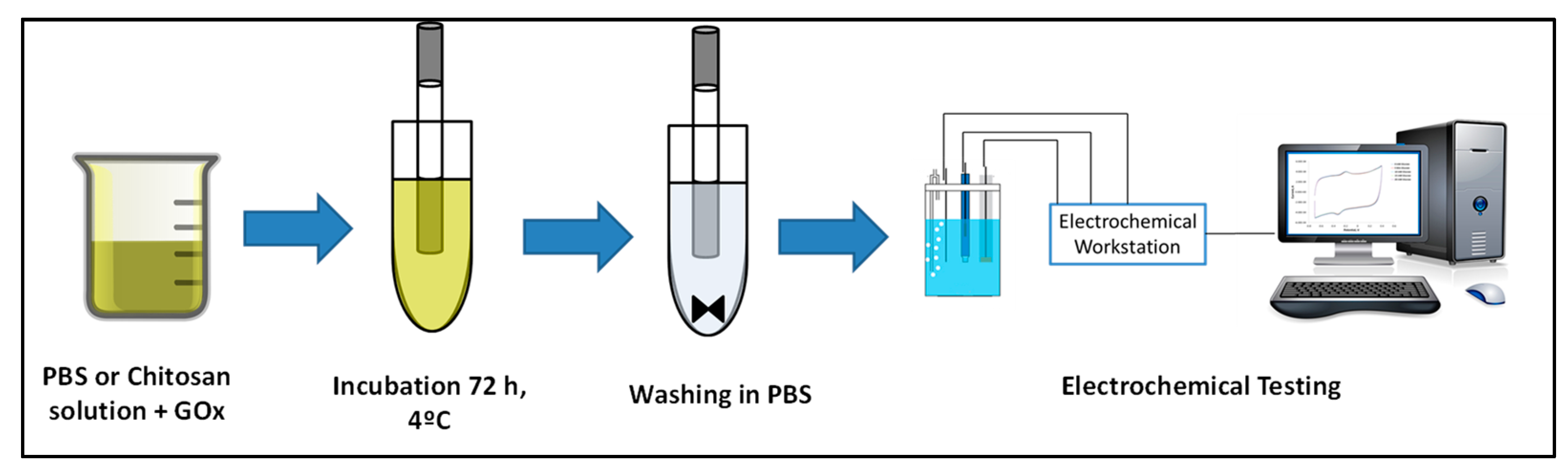
2.3. GOx Immobilization Procedure

For the immobilization of GOx onto graphite rod electrodes, the adsorption method was performed through the incubation of the graphite rods in a chitosan solution of GOx at 4 °C for 72 h. The low-density graphite (LDG) electrodes (Sigma-Aldrich. St. Louis, MO, USA) were first treated with coarse and fine emery paper. Then, the graphite rod was sonicated for 15 min in distilled water. Once the electrode was dried, it was cut into two rods of 7.5 cm and covered with Teflon tape leaving 1 cm in one end for the electrical connection and 2 cm in the other end as the immobilization surface.
For the immobilization procedure of GOx, 250 mg of chitosan were dissolved in 25 mL of a 0.3 M acetic acid solution and filtered through a 0.45 µm Nylon filter (VWR LIFE SCIENCE) before enzyme addition. Then, 5 mL of a solution of 10 mg/mL of GOx in 1% (w/w) chitosan solution was prepared and stirred for 30 min at room temperature to allow the self-assembly of the enzyme in the chitosan solution. Graphite rods were incubated in such solutions for 72 h at 4 °C. After 72 h, graphite rods were removed from the solution and washed in PBS pH 5.5 for 30 min. The washing procedure was repeated three times with fresh PBS solutions. The modified bioelectrode (LDG-CH10) was kept in PBS pH 7.55 at 4 °C until use.
FAD adsorption onto the graphite rod electrodes was performed in PBS pH 7 solutions in the same conditions as reported above. The graphite rod electrode was incubated in a FAD solution of 10 µg/mL during 72 h at 4 °C. After the 72 h, the modified graphite electrode (FAD@LDG) was removed from the solution and washed in PBS pH 5.5 solution for 30 min. This washing procedure was also repeated three times and the FAD-modified graphite electrodes were kept at 4 °C until use.
For the assessment of enzyme denaturation during the adsorption, immobilization was performed in PBS as the incubation medium. A volume of 5 mL of a solution of 10 mg/mL GOx in PBS pH 7.55 was prepared. Graphite rods were incubated in such solutions for 72 h at 4 °C. After 72 h of incubation, the same washing procedure as in the case of immobilization in chitosan was carried out. The bioelectrode thus modified (LDG-PBS10), was kept in PBS pH 7.55 at 4 °C until use.
The evaluation of FAD irreversible adsorption on the graphite electrode surface was evaluated by placing the modified graphite electrode in 5 mL of a KCl 3M solution overnight with magnetic stirring. Later, cyclic voltammetry experiments were performed at 0.1 V/s between −0.7 V and 0.4V under inert atmosphere before and after the KCl treatment to be compared.

2.4. Electrochemical Characterization

The direct electrochemistry of immobilized GOx was evaluated by cyclic voltammetry. The electrolyte for the cyclic voltammetry experiments was PBS KCl 0.1 M pH 7.55 that was purged with nitrogen, at least for 30 min, before cyclic voltammogram recording. Cyclic voltammetry was performed at different scan rates: 25, 50, 100, 150, 200, 250, 300, 400, and 500 mV/s, with a potential window between −0.7 V and 0.4 V.
For activity assays, 1 M glucose solution was prepared before the experiment and left to mutarrotate overnight. Glucose concentration in the electrochemical cell was increased by a factor of 5 mM in each addition. Cyclic voltammograms were recorded between −0.7 V and 0.4 V at 0.1 V/s.
For the polarization curve, a two-electrode configuration cell with a Pt wire as cathode and the GOx-modified graphite electrode as anode were employed. The power density was calculated using the product of voltage and intensity divided by the electroactive surface area obtained from the Randles–Sevcik equation, as previously reported [37].
For the inhibition assays, a solution of AgNO3 0.1M was used. A suitable amount of the AgNO3 solution was poured into the electrochemical cell to obtain a concentration of Ag+ ions of 1 µM. Then, bioelectrocatalytic activity for glucose oxidation was evaluated by cyclic voltammetry following the procedure described above.

3. Results and Discussion

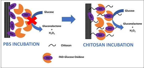
3.1. Bioelectrode Modification with Glucose Oxidase

The modification of the LDG electrodes was carried out using chitosan as an immobilization medium where, apart from hydrophobic interactions, electrostatic interactions between chitosan and the enzyme also act. As a control, the immobilization in PBS, where only hydrophobic interactions between the enzyme and the support were involved, was also performed to assess FAD dissociation and irreversible adsorption. The general procedure for the immobilization procedure is schematized in Figure 1. The electrode surface was first incubated in glucose oxidase (GOx) chitosan-based solution for 72 h at 4 °C for the selected working concentration of 10 mg/mL of GOx. This incubation time allowed the enzyme to be bound to LDG electrodes due to the affinity of carbon-based surfaces for biomolecules with chitosan entities embedded in the enzyme structure being part of the whole system [38]. Later, to remove the loosely bound enzyme, graphite rods were washed using a fresh PBS solution.
The immobilization of the enzyme by incubation in chitosan solutions was confirmed due to the presence of two redox peaks in the cyclic voltammogram corresponding to the oxidation-reduction of the active center of the GOx enzyme FAD at −0.22 V and −0.35 V respectively at 0.2 V/s under N2 atmosphere (solid blue line in Figure 2A). The results indicate that FAD cofactor was undergoing a reversible electron transfer with the graphite electrode as shown in reaction 3.
As mentioned before, bioelectrodes prepared through GOx adsorption on carbon-based materials can suffer enzyme denaturation during the adsorption [18,19,20]. The mechanism for this process starts with FAD dissociation from the protein structure (during the immobilization process or directly from free FAD that commercial GOx can contain as an impurity) where it is embedded and the subsequent migration towards graphite surface, leading to FAD cofactor irreversible adsorption due to the affinity of graphite for organic moieties [39]. The immobilization by incubation of the LDG rod in a biopolymeric solution of chitosan was carried out to avoid the denaturation of the enzyme taking advantage of the stabilizing effect of the biopolymeric medium by electrostatic interactions [29,30,40]. However, before the electrochemical characterization of the GOx-bioelectrode prepared in chitosan solutions, it is necessary to ascertain whether the redox peaks observed in Figure 2 are due or not to denaturized FAD irreversible adsorption.
For this purpose, the bioelectrode prepared by incubation in chitosan solution was submitted to a treatment with concentrated KCl solutions to assess the enzyme denaturation process. It has been reported that treatment with concentrated salt solutions can strip FAD from flavoenzymes, but the same treatment is ineffective in removing adsorbed FAD from graphite surfaces [41,42,43]. Thus, if the specie adsorbed on the graphite surface is the whole GOx enzyme with its protein embedding structure, basic treatment will strip it from the electrode surface and the corresponding redox peaks will disappear from the cyclic voltammogram. In contrast, if the peaks observed come from FAD direct irreversible adsorption due to denaturation, the redox peaks will remain in the cyclic voltammogram after the treatment.
Thus, as can be seen in Figure 2A (blue dashed line), redox peaks of chitosan-based GOx modified electrodes disappear after the treatment with KCl, indicating that signals were originated from non-denaturized GOx and that no dissociation and irreversible adsorption of FAD cofactor has occurred.
On the other hand, in the case of immobilization of GOx in PBS (LDG-PBS10), the redox peaks do not disappear after the treatment with KCl (Figure 2B red solid line), which points to the fact that FAD has been irreversibly adsorbed on the graphite surface. This behavior has been confirmed by an experiment of direct adsorption of FAD on the graphite electrode (FAD@LDG). This modified electrode was also submitted to the KCl treatment and as expected, the redox peaks remained in the cyclic voltammogram after the basic treatment, which confirms that FAD has been irreversibly adsorbed on the graphite surface (see Figure 2B and Figure S1A in Supplementary Materials). Therefore, we can conclude that the incubation in chitosan solutions favors the immobilization of GOx without FAD dissociation.
It is worth highlighting that FAD redox peaks in Figure 2B at −0.22 V and −0.35 V for the GOx enzyme adsorbed on the LDG electrode in PBS, showed a positive shift of about 140 mV with respect to the redox peak potential of FAD directly adsorbed on the LDG electrode also in PBS medium. This result suggests that immobilization conditions and assembly may have distorted the enzyme structure making the flavine center more accessible [9]. Additionally, it is worth mentioning that KCl treatment can also modify the electrode surface, which contributes to an increase in the capacitive current after the KCl treatment, as can be seen in Figure S1B.

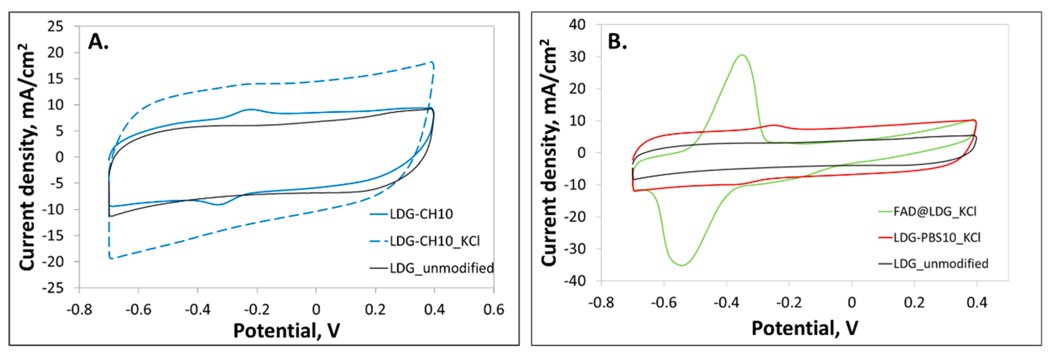
3.2. Electrochemistry of Immobilized GOx in Chitosan Solutions

The electrochemical characterization of the GOx bioelectrodes obtained by incubation in chitosan and PBS solutions was carried out by cyclic voltammetry at different scan rates in an O2 free atmosphere to detect the oxidation-reduction reactions of the FAD cofactor on the graphite surface (Figure S2). The peak current increases with the scan rate demonstrating that the redox reaction of the FAD cofactor is a typical surface-confined process. Enzyme coverage in mol/cm2 was obtained from the Equation (2) derived from Laviron expression (1) [44] for the bioelectrodes prepared by incubating the modified graphite rod electrode in chitosan and PBS solutions at two different concentrations (see Table S1).
I p = n 2 F 2 vA Γ 4 RT = nFQv 4 RT
Q = nFA Γ  
In these equations, Q is the charge obtained from the integration of the cathodic peak in the cyclic voltammograms recorded at different scan rates; n corresponds to the number of transferred electrons (n = 2); F is the Faraday (96,400 C/mol) constant; A is the electrode electroactive area in cm2 obtained from cyclic voltammetry and Randles–Sevcik equation; and Γ is the enzyme coverage in mol/cm2. To explore the efficiency of the bioelectrodes developed, the electron transfer rate constant was also determined from the scan rate dependence of ΔEp using the method of Laviron [44]. Values for the electron transfer rate constant in the LDG electrodes prepared (LDG-CH and LDG-PBS) are presented in Table S1 together with enzyme coverage.
Enzyme coverage was higher for the electrode LDG-CH10 prepared with the higher GOx concentration in the chitosan solution with a value near 4 nmol/cm2. The electron transfer rate constant for the electrodes prepared by incubation in PBS and chitosan solutions of GOx were 1.1 s−1 and 2.2 s−1, respectively. These values are in the same order of magnitude than similar systems that can be found in literature [18]. Moreover, the values for enzyme coverage obtained for the electrodes incubated in PBS are lower than those obtained for the bioelectrodes prepared in chitosan solutions (Table S1). Furthermore, it is worth mentioning that the values of the electron transfer rate constants obtained for electrodes modified in PBS solutions are in the same range than those previously reported for FAD adsorption on graphite electrodes [41].
Several works have been published over the years dealing with Direct Electron Transfer (DET) between GOx and carbon-based electrodes aimed at the development of glucose biosensors and biofuel cells [7,21,45,46] but there is no clear evidence that this phenomenon actually occurs [9,21].
Electron transfer of the bioelectrodes mediated by the flavoprotein FAD embedded into the GOx structure can be evaluated from the analysis of cyclic voltammetry experiments acquired in oxygen-free solutions. According to reaction 3, oxidation and reduction of FAD is a two-electron and two-proton coupled reaction. The cathodic peak obtained in the cyclic voltammetry is attributed to the reduction of GOx(FAD), while the anodic peak current is attributed to oxidation of GOx(FADH2). The pH value of the solution will influence electron transfer of GOx, and both the anodic and cathodic peak potentials will be shifted to more negative values as the solution pH increases as is observed in Figure S3. Moreover, formal potential exhibits a linear dependence of pH ranging from 5 to 9 (Inset of Figure S3) with a slope around to −50 mV/pH (R2 = 0.9964), a value close to the theoretical −59.2 mV/pH for the two-electron and two-proton coupled reaction [11]. In addition, the linear relationship between peak current and scan rate implies that the bioelectrode presents the typical behavior of a surface-confined redox process with anodic and cathodic peak separation characteristic of quasi-reversible redox systems with a mean value lower than 100 mV [47]. In addition, anodic and cathodic peak currents are on the same order of magnitude, this being associated with the stability of the adsorbed redox species (Figure S2 Supplementary Materials).
The redox potential of GOx in solution determined by Vogt and co-workers by UV/vis spectroelectrochemistry was −0.385 V vs Ag/AgCl at pH = 7.4 [48]. According to cyclic voltammograms, a formal potential (E1/2) of −0.307 V is obtained for chitosan-based electrodes. This potential is near the values reported for GOx and out of the range for O2 electrochemical reduction [49]. Moreover, this value is in accordance with the values found in the literature for some GOx-based bioelectrodes where chitosan has been employed as an immobilization matrix [38,50,51]. In particular, Zhao and co-workers [38] stated that chitosan, in a protonated state in acidic solutions, can embed a part in the protein acting as a molecular “wire” that connects the active center of the enzyme with the electrode surface resulting in an enhancement of electron transfer process. Thus, chitosan with hydroxyl groups and positively charged amino groups behaves as a polyelectrolyte and can embed in the protein acting as a molecular wire connecting the graphite electrode and the GOx enzyme. Then, we assume that, in addition to the “wire” effect, chitosan as incubation medium modulates the adsorption allowing the migration of the enzyme towards the electrode surface avoiding unfolding and denaturation due to the electrostatic interactions that would prevent structure distortion [40]. Thus, the behavior detected for the bioelectrodes prepared incubating in chitosan solutions points to the successful immobilization of GOx enzyme onto LDG electrodes surface, and not from FAD irreversible adsorption due to denaturation, demonstrating a favoring effect of the incubation in chitosan solutions in the enzyme structure.

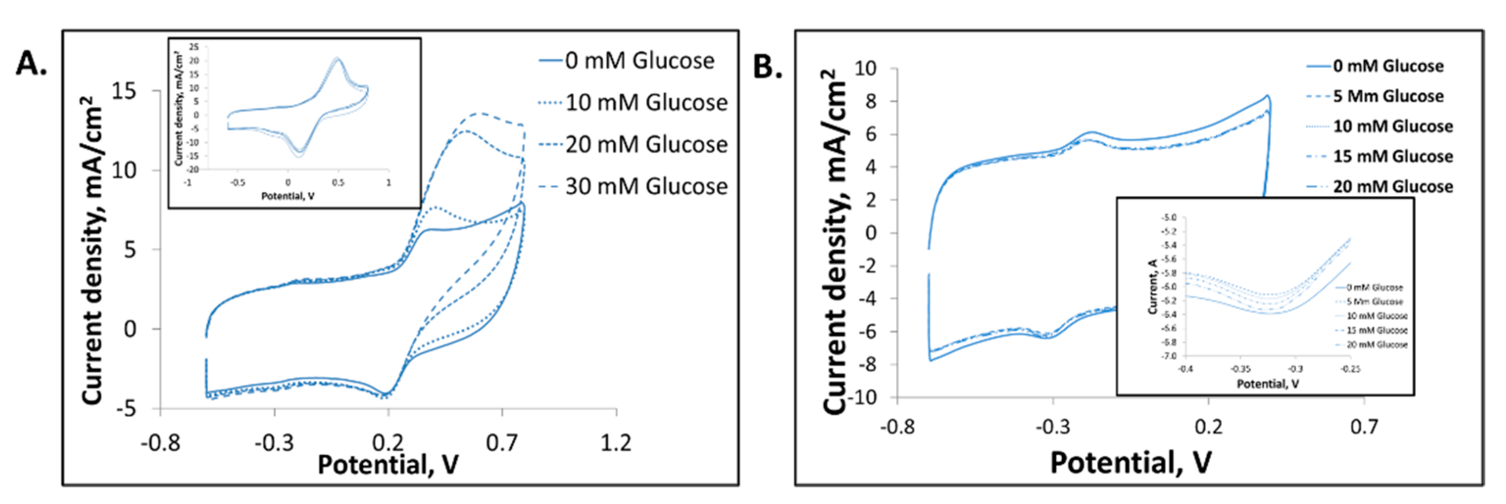
3.3. Evaluation of Immobilized GOx Activity

3.3.1. GOx Activity and Inhibition Assays

To prove the bioelectrocatalytic performance of the GOx adsorbed onto LDG electrodes, the activity of the enzyme for glucose catalysis has been proved. The bioelectrodes prepared by incubation in chitosan solutions have been proved not to be denatured and to retain the quaternary structure of the enzyme and consequently, the bioelectrocatalytic activity as demonstrated from the results of the KCl assays. Cyclic voltammetry was recorded in oxygen-free PBS pH 7.55 solutions with increasing concentrations of glucose (Figure 3A) to check the proper activity of GOx in the electrochemical oxidation of glucose.
For the bioelectrodes prepared starting from a GOx concentration of 10 mg/mL (Figure 3), a reduction in the cathodic current density and a small increase in the anodic current density were observed as a response of the reaction of GOx and glucose. Thus, as explained in reaction 1, GOx(FAD) is reduced by glucose during oxidation. In addition, in KCl treated electrodes, there is little or negligible variation in FAD currents in presence of glucose, which means that there is no active GOx on the electrodic surface (Figure 3B). The effect observed in the voltammograms is more probably related to the modification of the double layer due to the addition of glucose to the working solution.
As observed in Figure 3A, the current corresponding to the reduction of FAD to FADH2 decreases with the increase of glucose concentration up to saturation at a concentration of 15 mM glucose. This phenomenon is due to the simultaneous action of two processes: enzymatic catalysis and electrode reaction [52]. In the absence of glucose, the total amount of FAD that is reduced remains constant, being always reduced the same amount of FAD. However, in the presence of glucose, enzymatic catalysis also occurs; therefore, the amount of FAD that is reduced (monitored by cyclic voltammetry) is decreased, since it is participating in the enzymatic catalysis, thus modifying the current obtained.
The bioactivity of the modified graphite electrode was further investigated by employing mediators to enhance electrochemical currents [7,11]. The cyclic voltammetry in oxygen-free PBS pH 7 solutions containing 2 mM hydroquinone (HQ) as a redox mediator showed, after the addition of glucose, remarkable enzyme activity in presence of the mediator (Figure 4A) while no current increase was observed for the bare graphite rod electrode when increasing amounts of glucose are added in the presence of HQ (Inset in Figure 4A). This increase indicates that the presence of a mediator promotes GOx activity. Specifically, GOx transfers electrons and, thus, regenerates the hydroquinone, which is oxidized to benzoquinone in the electrode. The result can be explained following reactions 1, 4, and 5:
GOx ( FADH 2 ) + 2 HQ +   GOx ( FAD ) + 2 HQ + 2 H + ( 4 )
2 HQ     2 e + 2 HQ +   ( 5 )
On the other hand, in order to ascertain whether the bioelectrocatalytic activity observed comes from the immobilized enzyme, an inhibition assay was performed (Figure 4B). In this way, it can be confirmed that current response obtained is coming from the bioelectrocatalytic activity of the enzyme. One of the inhibitors of GOx enzyme activity is the Ag+ ion [53,54] and its presence in low concentrations prevents, totally or partially, the oxidation of glucose by the enzyme GOx. The chitosan-modified electrodes showing activity towards glucose oxidation were assayed in an inhibition experiment with silver. Thus, when Ag+ in concentration 1 µM is added to the working solution prior to glucose addition, no changes were observed when the glucose concentration in the electrolyte solution is increased. This means that the biocatalytic activity of the enzyme has been inhibited by the Ag+ ions.
In Figure 4B, a partial response is observed up to a glucose concentration of 5 Mm in the presence of silver as inhibitor; further additions return the current density values to the initial value found when the glucose concentration in the working electrolyte was zero. It was determined that this behavior is related to the concentration of GOx in the bioelectrode and its response time, since it is known that as the glucose concentration increases, the response time required in an inhibition assay with silver also does [55]. Moreover, inhibition with silver occurs due to the interaction of Ag+ ions with the functional groups of the enzyme; therefore, the higher the concentration of enzyme in the electrode, the greater the amount of Ag+ ions necessary to totally block the catalytic activity [54]. However, in the case of a bioelectrode prepared with lower concentration of GOx in chitosan (4 mg/mL, LDG-CH4) the effect of the inhibition with silver is clearly observed (Figure S4).

3.3.2. GOx Activity for Energy Harvesting

Finally, the modified graphite electrode by incubation in chitosan solutions was tested as proof of the concept of the suitability of the bioelectrode in a glucose biofuel cell for energy harvesting. For the quantification of the power density achieved in the presence of glucose, the LDG-CH4 electrode was set as anode due to its faster response as observed in the inhibition assay. A Pt wire electrode was used as a cathode and 0.1 M PBS pH 7.4 was used as the working electrolyte. For an electrochemical surface area of 0.07 cm2, obtained from the Randles–Sevcik equation, the power density delivered using GOx chitosan-based modification was 119 µW/cm2 for a concentration of 20 mM glucose with a cell voltage of 0.3 V (Figure 5). This result shows the proper enzyme immobilization by chitosan presence and the potential of the bioelectrode prepared for energy harvesting applications.
The power density of 119 µW/cm2 delivered by the biofuel cell using the modified graphite rod as anode is in the range of the values that can be found in the literature for glucose biofuel cells for similar electrode configurations [5,56]. Thus, these power densities and currents are more appropriate for energy harvesting, where the high OCP and currents are desired for the proper operation of biofuel cells. Nonetheless, this approach for enzyme immobilization would also apply to the development of biosensors, but, in the latter case, it is convenient to work at lower potentials to avoid possible interferences.

4. Conclusions

The proposed method for GOx immobilization using a chitosan solution allows extremely simple and highly versatile enzyme attachment on graphite supports. This strategy allows keeping the stability of the enzyme in terms of quaternary structure, avoiding loss of FAD and enhancing electron transfer kinetics, leading to a good catalytic activity of the enzyme for glucose catalysis. By incubating graphite electrodes in chitosan solutions, FAD adsorption is minimized due to the beneficial effect of the electrostatic interactions in the chitosan medium. Moreover, chitosan is embedded in the enzyme structure promoting electron transfer between the enzyme and the electrode. Bioelectrocatalytic activity towards glucose is retained as demonstrated by the electrochemical response obtained for the electrode when contacted with increasing glucose concentrations in energy harvesting applications.
Thus, the proposed method to immobilize GOx onto graphite electrodes using chitosan as an immobilization medium constitutes a simple and easy-to-use approach for GOx adsorption compared with similar systems reported in the literature so far. To our best knowledge, the most important achievement of the method proposed is based on its extreme simplicity since chitosan does not need any modification for the application of the method but also the ease of the process. These facts allow the use of this strategy in any kind of application related with bioelectrodes development, such as sensing or energy harvesting, and could be further extended to other enzymes of similar features. However, further work will be necessary to evaluate the long-term stability of the immobilization, and deep evaluation of its features for sensing and energy harvesting for power supply.

Supplementary Materials

The following supporting information can be downloaded at: https://0-www-mdpi-com.brum.beds.ac.uk/article/10.3390/polysaccharides3020023/s1. Table S1: Enzyme coverage and electron transfer rate constant obtained for PBS- and Chitosan-incubated bioelectrodes. Figure S1: KCl treatment effects in electrodes. Figure S2: GOx immobilization in PBS for (A) 4mg/mL GOx (LDG-PBS4) and (B) 10 mg/mL GOx, (LDG-PBS10) and in chitosan for (C) 4 mg/mL GOx (LDG-CH4) and (B) 10 mg/mL GOx, (LDG-CH10). Figure S3: pH dependence of redox peaks in LDG_CH10 electrode. Figure S4: Cyclic voltammetry in oxygen-free PBS solution of LDG-CH4 bioelectrode with increasing glucose concentration in the absence (A) and in the presence (B) of the inhibitor Ag+. Voltammograms acquired at 0.1 V/s.

Author Contributions

Conceptualization, M.B.-S.; methodology, M.B.-S.; validation, M.B.-S. and L.G.-C.; formal analysis, M.B.-S. and L.G.-C.; investigation, M.B.-S.; resources, M.G.-A., M.G.-P. and A.Q.-L.; writing—original draft preparation, M.B.-S. and L.G.-C.; writing—review and editing, M.B.-S., L.G.-C., M.G.-A. and A.Q.-L.; visualization, M.B.-S.; L.G.-C.; M.G.-A.; M.G.-P.; and A.Q.-L.; supervision, M.G.-A. and A.Q.-L.; project administration, M.B.-S.; funding acquisition, M.G.-P. All authors have read and agreed to the published version of the manuscript.

Funding

This research was funded by the Spanish Ministry of Science, Innovation, and University, grant number PTQ-14-07145 (Bio2 project) and by the Instituto Valenciano de Competitividad Empresarial-IVACE.

Institutional Review Board Statement

Not applicable.

Informed Consent Statement

Not applicable.

Acknowledgments

The authors acknowledge the Instituto Valenciano de Competitividad Empresarial (IVACE), and M.B.-S. is gratefully acknowledged for a Torres Quevedo research contract through the State Program for Talent and Employability Promotion 2013–2016.

Conflicts of Interest

The authors declare no conflict of interest.

References

  1. Clark, L.C., Jr. Electrochemical Device for Chemical Analysis. U.S. Patent 2913386A, 17 November 1959. [Google Scholar]
  2. Bruen, D.; Delaney, C.; Florea, L.; Diamond, D. Glucose Sensing for Diabetes Monitoring: Recent Developments. Sensors 2017, 17, 1866. [Google Scholar] [CrossRef] [PubMed] [Green Version]
  3. Khor, S.M.; Choi, J.; Won, P.; Ko, S.H. Challenges and Strategies in Developing an Enzymatic Wearable Sweat Glucose Biosensor as a Practical Point-Of-Care Monitoring Tool for Type II Diabetes. Nanomaterials 2022, 12, 221. [Google Scholar] [CrossRef] [PubMed]
  4. Kim, J.; Campbell, A.S.; Wang, J. Wearable non-invasive epidermal glucose sensors: A review. Talanta 2018, 177, 163–170. [Google Scholar] [CrossRef] [PubMed]
  5. Buaki-Sogó, M.; García-Carmona, L.; Gil-Agustí, M.; Zubizarreta, L.; García-Pellicer, M.; Quijano-López, A. Enzymatic Glucose-Based Bio-batteries: Bioenergy to Fuel Next-Generation Devices. Top. Curr. Chem. 2020, 378, 49. [Google Scholar] [CrossRef] [PubMed]
  6. Bankar, S.B.; Bule, M.V.; Singhal, R.S.; Ananthanarayan, L. Glucose oxidase—An overview. Biotechnol. Adv. 2009, 27, 489–501. [Google Scholar] [CrossRef]
  7. Yin, Z.; Ji, Z.; Zhang, W.; Taylor, E.W.; Zeng, X.; Wei, J. The glucose effect on direct electrochemistry and electron transfer reaction of glucose oxidase entrapped in a carbon nanotube-polymer matrix. ChemistrySelect 2020, 5, 12224–12231. [Google Scholar] [CrossRef]
  8. Huang, J.; Zhang, Y.; Deng, X.; Li, J.; Huang, S.; Jin, X.; Zhu, X. Self-encapsulated enzyme through in situ growth of polypyrrole for high-performance enzymatic biofuel cell. Chem. Eng. J. 2021, 429, 132148. [Google Scholar] [CrossRef]
  9. Bartlett, P.N.; Al-Lolage, F.A. There is no evidence to support literature claims of direct electron transfer (DET) for native glucose oxidase (GOx) at carbon nanotubes or graphene. J. Electroanal. Chem. 2018, 819, 26–37. [Google Scholar] [CrossRef] [Green Version]
  10. Homaei, A.A.; Sariri, R.; Vianello, F.; Stevanato, R. Enzyme immobilization: An update. J. Chem. Biol. 2013, 6, 185–205. [Google Scholar] [CrossRef] [Green Version]
  11. You, C.; Li, X.; Zhang, S.; Kong, J.; Zhao, D.; Liu, B. Electrochemistry and biosensing of glucose oxidase immobilized on Pt-dispersed mesoporous carbon. Microchim. Acta 2009, 167, 109–116. [Google Scholar] [CrossRef]
  12. Jesionowski, T.; Zdarta, J.; Krajewska, B. Enzyme immobilization by adsorption: A review. Adsorption 2014, 20, 801–821. [Google Scholar] [CrossRef] [Green Version]
  13. Haghighi, B.; Gorton, L.; Ruzgas, T.; Jönsson, L.J. Characterization of graphite electrodes modified with laccase from Trametes Versicolor and their use for bioelectrochemical monitoring of phenolic compounds in flow injection analysis. Anal. Chim. Acta. 2003, 487, 3–14. [Google Scholar] [CrossRef]
  14. Jarosz-Wilkołazka, A.; Ruzgas, T.; Gorton, L. Use of laccase-modified electrode for amperometric detection of plant flavonoids. Enzyme Microb. Technol. 2004, 35, 238–241. [Google Scholar] [CrossRef]
  15. Onay, A.; Dogan, Ü.; Ciftci, H.; Cetin, D.; Suludere, Z.; Tamer, U. Amperometric glucose sensor based on the glucose oxidase enzyme immobilized on graphite rod electrode modified with Fe3O4-CS-Au magnetic nanoparticles. Ionics 2018, 24, 4015–4022. [Google Scholar] [CrossRef]
  16. Bandapati, M.; Krishnamurthy, B.; Goel, S. Fully Assembled Membraneless Glucose Biofuel Cell With MWCNT Modified Pencil Graphite Leads as Novel Bioelectrodes. IEEE Trans. NanoBioscience 2019, 18, 170–175. [Google Scholar] [CrossRef] [PubMed]
  17. Chi, Q.; Zhang, J.; Dong, S.; Wang, E. Direct electrochemistry and surface characterization of glucose oxidase adsorbed on anodized carbon electrodes. Electrochimica Acta 1994, 39, 2431–2438. [Google Scholar] [CrossRef]
  18. Ianniello, R.M.; Lindsay, T.J.; Yacynych, A.M. Differential pulse voltammetric study of direct electron transfer in glucose oxidase chemically modified graphite electrodes. Anal. Chem. 1982, 54, 1098–1101. [Google Scholar] [CrossRef]
  19. Goran, J.M.; Mantilla, S.M.; Stevenson, K.J. Influence of Surface Adsorption on the Interfacial Electron Transfer of Flavin Adenine Dinucleotide and Glucose Oxidase at Carbon Nanotube and Nitrogen-Doped Carbon Nanotube Electrodes. Anal. Chem. 2013, 85, 1571–1581. [Google Scholar] [CrossRef]
  20. Pereira, S.; Santos, N.; Carvalho, A.; Fernandes, A.; Costa, F. Electrochemical Response of Glucose Oxidase Adsorbed on Laser-Induced Graphene. Nanomaterials 2021, 11, 1893. [Google Scholar] [CrossRef]
  21. Pilipenko, O.S.; Atyaksheva, L.F.; Poltorak, O.M.; Chukhrai, E.S. Dissociation and catalytic activity of oligomer forms of β-galactosidases. Russ. J. Phys. Chem. A 2007, 81, 990–994. [Google Scholar] [CrossRef]
  22. Poltorak, O.M.; Chukhrai, E.S.; Kozlenkov, A.A.; Chaplin, M.F.; Trevan, M.D. The putative common mechanism for inactivation of alkaline phosphatase isoenzymes. J. Mol. Catal. B Enzym. 1999, 7, 157–163. [Google Scholar] [CrossRef]
  23. Fernandez-Lafuente, R. Stabilization of multimeric enzymes: Strategies to prevent subunit dissociation. Enzym. Microb. Technol. 2009, 45, 405–418. [Google Scholar] [CrossRef]
  24. Bolivar, J.M.; Cava, F.; Mateo, C.; Rocha-Martín, J.; Guisán, J.M.; Berenguer, J.; Fernandez-Lafuente, R. Immobilization–stabilization of a new recombinant glutamate dehydrogenase from Thermus thermophilus. Appl. Microbiol. Biotechnol. 2008, 80, 49–58. [Google Scholar] [CrossRef] [PubMed]
  25. Kabashima, T.; Li, Y.; Kanada, N.; Ito, K.; Yoshimoto, T. Enhancement of the thermal stability of pyroglutamyl peptidase I by introduction of an intersubunit disulfide bond. Biochim. Biophys. Acta Protein Struct. Mol. Enzymol. 2001, 1547, 214–220. [Google Scholar] [CrossRef]
  26. Fuentes, M.; Segura, R.L.; Abian, O.; Betancor, L.; Hidalgo, A.; Mateo, C.; Fernandez-Lafuente, R.; Guisan, J.M. Determination of protein-protein interactions through aldehyde-dextran intermolecular cross-linking. Proteomics 2004, 4, 2602–2607. [Google Scholar] [CrossRef] [PubMed]
  27. Yamazaki, T.; Tsugawa, W.; Sode, K. Increased thermal stability of glucose dehydrogenase by cross-linking chemical modification. Biotechnol. Lett. 1999, 21, 199–202. [Google Scholar] [CrossRef]
  28. Tao, Q.; Li, A.; Zhang, Z.; Ma, R.; Shi, L. Stabilization of Multimeric Enzymes against Heat Inactivation by Chitosan-graft-poly(N-isopropylacrylamide) in Confined Spaces. ACS Biomater. Sci. Eng. 2017, 3, 3141–3145. [Google Scholar] [CrossRef]
  29. Dosadina, E.E.; Belov, A.A. Interaction Between Chitosan Solutions, Cellulose Carriers and Some of the Multi-enzyme Complexes. Int. J. Bioorg. Chem. 2017, 2, 51–60. [Google Scholar]
  30. Raza, Z.A.; Riaz, S.; Banat, I.M. Polyhydroxyalkanoates: Properties and chemical modification approaches for their functionalization. Biotechnol. Prog. 2018, 34, 29–41. [Google Scholar] [CrossRef]
  31. Riaz, S.; Rhee, K.Y.; Park, S.J. Polyhydroxyalkanoates (PHAs): Biopolymers for Biofuel and Biorefineries. Polymers 2021, 13, 253. [Google Scholar] [CrossRef]
  32. Verma, M.L.; Kumar, S.; Das, A.; Randhawa, J.S.; Chamundeeswari, M. Chitin and chitosan-based support materials for enzyme immobilization and biotechnological applications. Environ. Chem. Lett. 2020, 18, 315–323. [Google Scholar] [CrossRef]
  33. Dabhade, A.; Jayaraman, S.; Paramasivan, B. Development of glucose oxidase-chitosan immobilized paper biosensor using screen-printed electrode for amperometric detection of Cr(VI) in water. 3 Biotech 2021, 11, 183. [Google Scholar] [CrossRef] [PubMed]
  34. El Ichi, S.; Zebda, A.; Alcaraz, J.-P.; Laaroussi, A.; Boucher, F.; Boutonnat, J.; Reverdy-Bruas, N.; Chaussy, D.; Belgacem, M.N.; Cinquin, P.; et al. Bioelectrodes modified with chitosan for long-term energy supply from the body. Energy Environ. Sci. 2015, 8, 1017–1026. [Google Scholar] [CrossRef]
  35. Nasar, A.; Rahman, M.M. Applications of chitosan (CHI)-reduced graphene oxide (rGO)-polyaniline (PAni) conducting composite electrode for energy generation in glucose biofuel cell. Sci. Rep. 2020, 10, 10428. [Google Scholar]
  36. Rassas, I.; Braiek, M.; Bonhomme, A.; Bessueille, F.; Rafin, G.; Majdoub, H.; Jaffrezic-Renault, N. Voltammetric glucose biosensor based on glucose oxidase encapsulation in a chitosan-kappa-carrageenan polyelectrolyte complex. Mater. Sci. Eng. C 2019, 95, 152–159. [Google Scholar] [CrossRef]
  37. Bai, L.; Yuan, R.; Chai, Y.; Yuan, Y.; Wang, Y.; Xie, S. Direct electrochemistry and electrocatalysis of a glucose oxidase-functionalized bioconjugate as a trace label for ultrasensitive detection of thrombin. Chem. Commun. 2012, 48, 10972–10974. [Google Scholar] [CrossRef]
  38. Zhao, C.; Meng, Y.; Shao, C.; Wan, L.; Jiao, K. Unadulterated Glucose Biosensor Based on Direct Electron Transfer of Glucose Oxidase Encapsulated Chitosan Modified Glassy Carbon Electrode. Electroanalysis 2008, 20, 520–526. [Google Scholar] [CrossRef]
  39. Kanda, M.; Brady, F.O.; Rajagopalan, K.V.; Handler, P. Studies on the Dissociation of Flavin Adenine Dinucleotide from Metalloflavoproteins. J. Biol. Chem. 1972, 247, 765–770. [Google Scholar] [CrossRef]
  40. Xie, Y.; Li, Z.; Zhou, J. Hamiltonian replica exchange simulations of glucose oxidase adsorption on charged surfaces. Phys. Chem. Chem. Phys. 2018, 20, 14587–14596. [Google Scholar] [CrossRef]
  41. Gorton, L.; Johansson, G. Cyclic voltammetry of FAD adsorbed on graphite, glassy carbon, platinum, and gold electrodes. J. Electroanal. Chem. Inter. Electrochem. 1980, 113, 151–158. [Google Scholar] [CrossRef]
  42. Ianniello, R.M.; Lindsay, T.J.; Yacynych, A.M. Direct electron transfer in immobilized flavoenzyme chemically modified graphite electrodes. Anal. Chim. Acta 1982, 141, 23–32. [Google Scholar] [CrossRef]
  43. Coughlan, M.P.; Johnson, D.B. Preparation and properties of immobilised xanthine oxidase. Biochim. Biophys. Acta Enzymol. 1973, 302, 200–204. [Google Scholar] [CrossRef]
  44. Laviron, E. General expression of the linear potential sweep voltammogram in the case of diffusionless electrochemical systems. J. Electroanal. Chem. Inter. Electrochem. 1979, 101, 19–28. [Google Scholar] [CrossRef]
  45. Liang, B.; Guo, X.; Fang, L.; Hu, Y.; Yang, G.; Zhu, Q.; Wei, J.; Ye, X. Study of direct electron transfer and enzyme activity of glucose oxidase on graphene surface. Electrochem. Commun. 2015, 50, 1–5. [Google Scholar] [CrossRef]
  46. Kang, Z.; Jiao, K.; Yu, C.; Dong, J.; Peng, R.; Hu, Z.; Jiao, S. Direct electrochemistry and bioelectrocatalysis of glucose oxidase in CS/CNC film and its application in glucose biosensing and biofuel cells. RSC Adv. 2017, 7, 4572–4579. [Google Scholar] [CrossRef] [Green Version]
  47. Nicholson, R.S. Theory and Application of Cyclic Voltammetry for Measurement of Electrode Reaction Kinetics. Anal. Chem. 1965, 37, 1351–1355. [Google Scholar] [CrossRef]
  48. Vogt, S.; Schneider, M.; Schafer-Eberwin, H.; Noll, G. Determination of the pH dependent redox potential of glucose oxidase by spectroelectrochemistry. Anal. Chem. 2014, 86, 7530–7535. [Google Scholar] [CrossRef]
  49. Wang, Y.; Yao, Y. Direct electron transfer of glucose oxidase promoted by carbon nanotubes is without value in certain mediator-free applications. Microchim. Acta 2012, 176, 271–277. [Google Scholar] [CrossRef]
  50. Ivnitski, D.; Branch, B.; Atanassov, P.; Apblett, C. Glucose oxidase anode for biofuel cell based on direct electron transfer. Electrochem. Commun. 2006, 8, 1204–1210. [Google Scholar] [CrossRef]
  51. Gao, Y.-F.; Yang, T.; Yang, X.-L.; Zhang, Y.-S.; Xiao, B.-L.; Hong, J.; Sheibani, N.; Ghourchian, H.; Hong, T.; Moosavi-Movahedi, A.A. Direct electrochemistry of glucose oxidase and glucose biosensing on a hydroxyl fullerenes modified glassy carbon electrode. Biosens. Bioelectron. 2014, 60, 30–34. [Google Scholar] [CrossRef]
  52. Zhang, D.; Chen, X.; Ma, W.; Yang, T.; Li, D.; Dai, B.; Zhang, Y. Direct electrochemistry of glucose oxidase based on one step electrodeposition of reduced graphene oxide incorporating polymerized l-lysine and its application in glucose sensing. Mater. Sci. Eng. C 2019, 104, 109880. [Google Scholar] [CrossRef] [PubMed]
  53. Dhekra, A.; Khaled, H.; Mina, S.; Mustapha, M.; Ali, O. Immobilization of Glucose Oxidase in Anthracene-Based Semi-Conducting Polymer: Application on Glucose Biosensing. J. Bioeng. Biomed. Sci. 2017, 7, 5. [Google Scholar]
  54. Wang, S.; Su, P.; Yang, Y. Online immobilized enzyme microreactor for the glucose oxidase enzymolysis and enzyme inhibition assay. Anal. Biochem. 2012, 427, 139–143. [Google Scholar] [CrossRef] [PubMed]
  55. Rust, I.M.; Goran, J.M.; Stevenson, K.J. Amperometric Detection of Aqueous Silver Ions by Inhibition of Glucose Oxidase Immobilized on Nitrogen-Doped Carbon Nanotube Electrodes. Anal. Chem. 2015, 87, 7250–7257. [Google Scholar] [CrossRef]
  56. Yu, S.; Myung, N.V. Recent Advances in the Direct Electron Transfer-Enabled Enzymatic Fuel Cells. Front. Chem. 2021, 8, 620153. [Google Scholar] [CrossRef] [PubMed]
Figure 1. GOx immobilization procedure and working operation.
Polysaccharides 03 00023 g001
Figure 2. Bioelectrode evaluation for FAD irreversible adsorption. (A) chitosan-based GOx immobilization (solid blue line), chitosan-based GOx modified electrode treated with KCl (blue dashed line). (B) KCl treatment of the electrodes modified by GOx adsorption using PBS (solid red line) and FAD direct adsorption (solid green line). Unmodified LDG electrodes (solid black line). All the voltammograms were acquired at 0.2 V/s in O2 free atmosphere.
Polysaccharides 03 00023 g002
Figure 3. GOx activity evaluation in the presence of increasing concentrations of glucose using (A) Chitosan-modified bioelectrode (LDG-CH10) and (B) PBS modified bioelectrode (LDG-PBS10) treated with KCl. Voltammograms acquired at 0.1 V/s.
Polysaccharides 03 00023 g003
Figure 4. Cyclic voltammetry in oxygen-free PBS solution of LDG-CH10 bioelectrode using increasing glucose concentrations (A) in the presence of HQ 2 mM (inset: bare graphite rod) and (B) in the presence of the inhibitor Ag+ 1 µM. Voltammograms acquired at 0.1 V/s.
Polysaccharides 03 00023 g004
Figure 5. Polarization and power density curves of the graphite bioelectrode modified with chitosan solutions of GOx (4 mg/mL) in the presence of 20 mM glucose.
Polysaccharides 03 00023 g005
Publisher’s Note: MDPI stays neutral with regard to jurisdictional claims in published maps and institutional affiliations.

