
Toxicological and Nutraceutical Screening Assays of Some Artificial Sweeteners

 ,
,
Abstract
:1. Introduction
2. Materials and Methods
2.1. Samples
2.2. Fly Stocks
2.3. Cell Culture Conditions
2.4. In Vivo Assays
2.4.1. Toxicity and Antitoxicity Assays
2.4.2. Genotoxicity and Antigenotoxicity Assays
2.4.3. Chronic Treatments: Lifespan and Healthspan Assays
2.5. In Vitro Assays
2.5.1. Cytotoxicity Assays
2.5.2. DNA Fragmentation Status
2.5.3. Clastogenicity: SCGE (Comet Assay)
2.5.4. Methylation Status of HL-60 Cells
3. Results
3.1. Toxicity/Antitoxicity
3.2. Genotoxicity/Antigenotoxicity
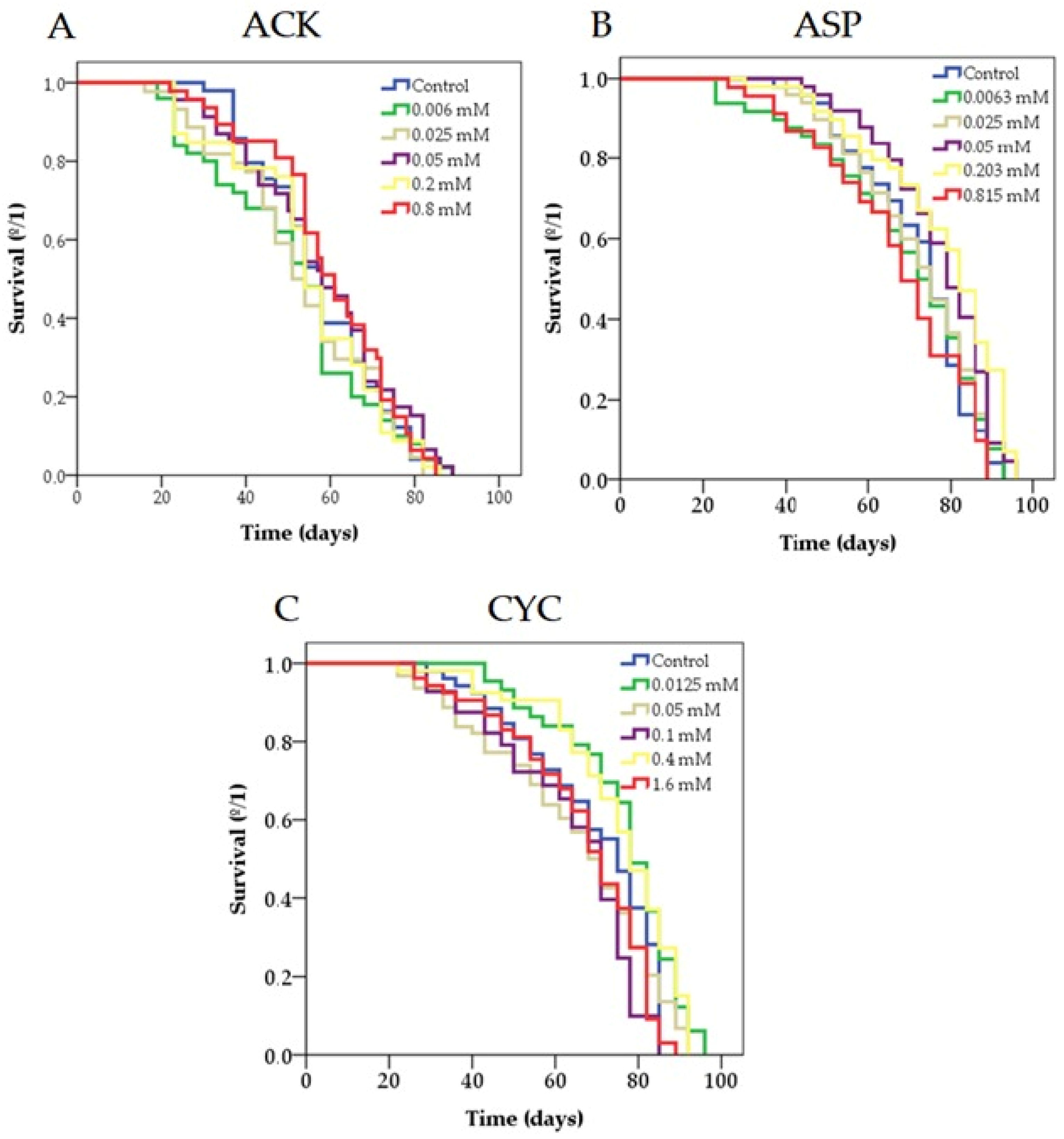
3.3. Lifespan
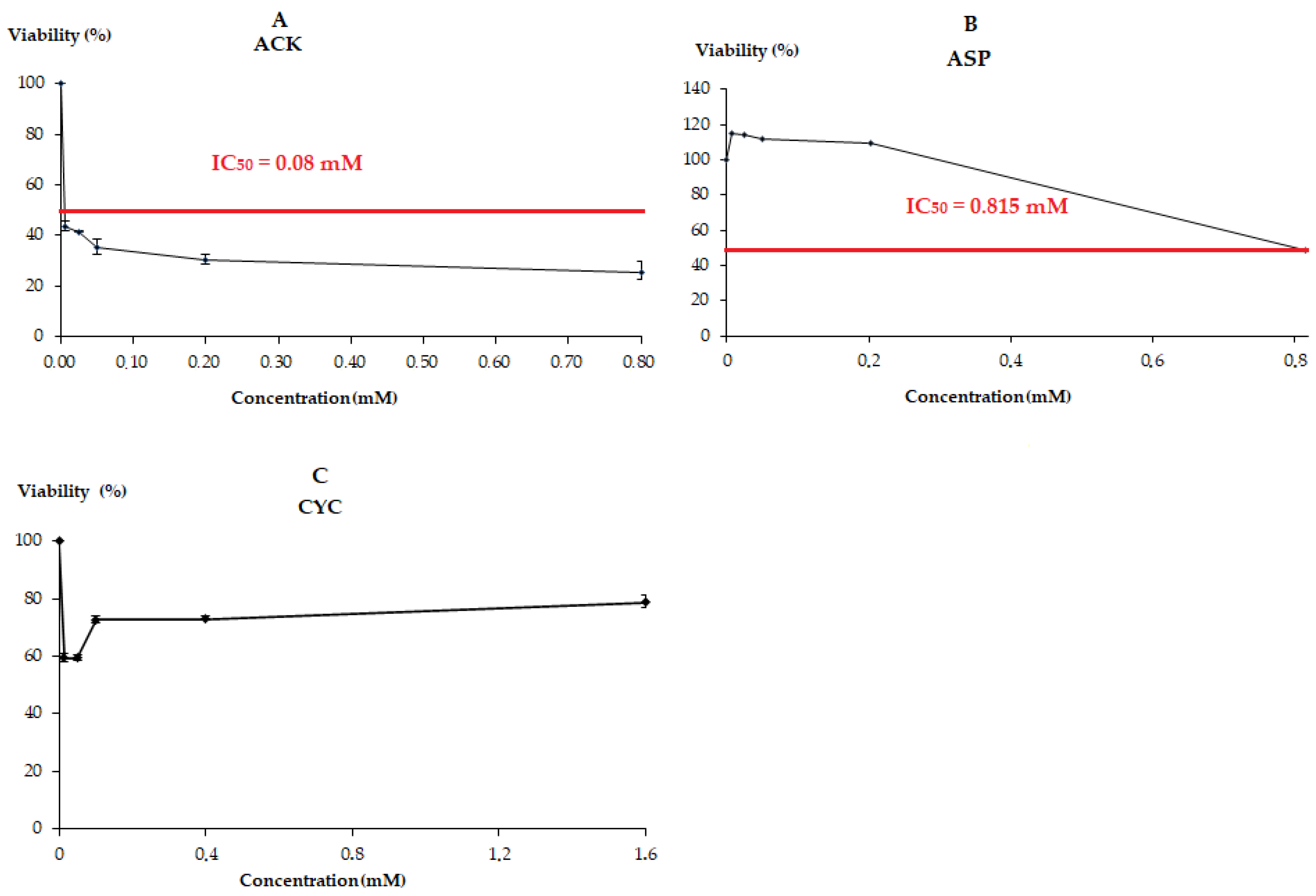
3.4. Cytotoxicity
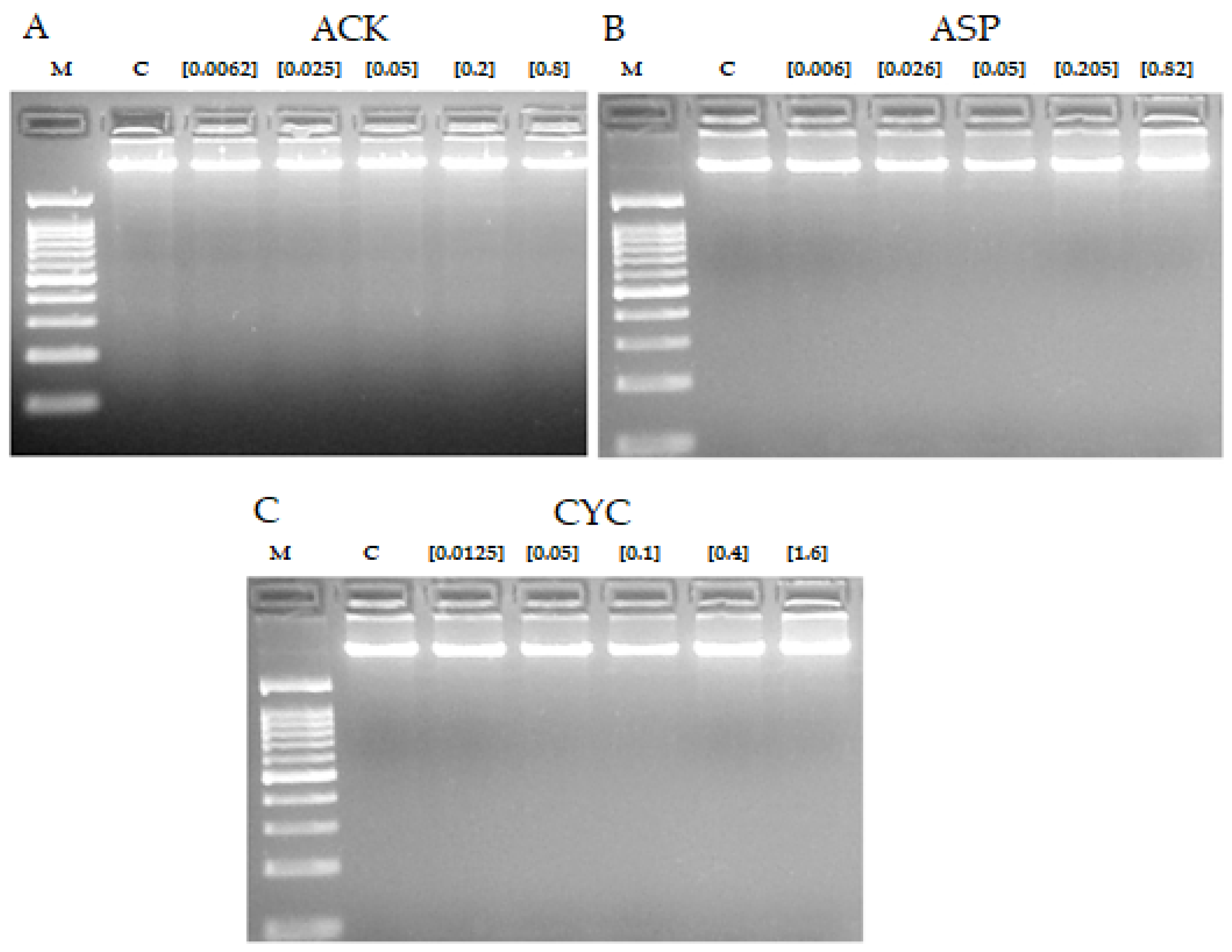
3.5. DNA Fragmentation
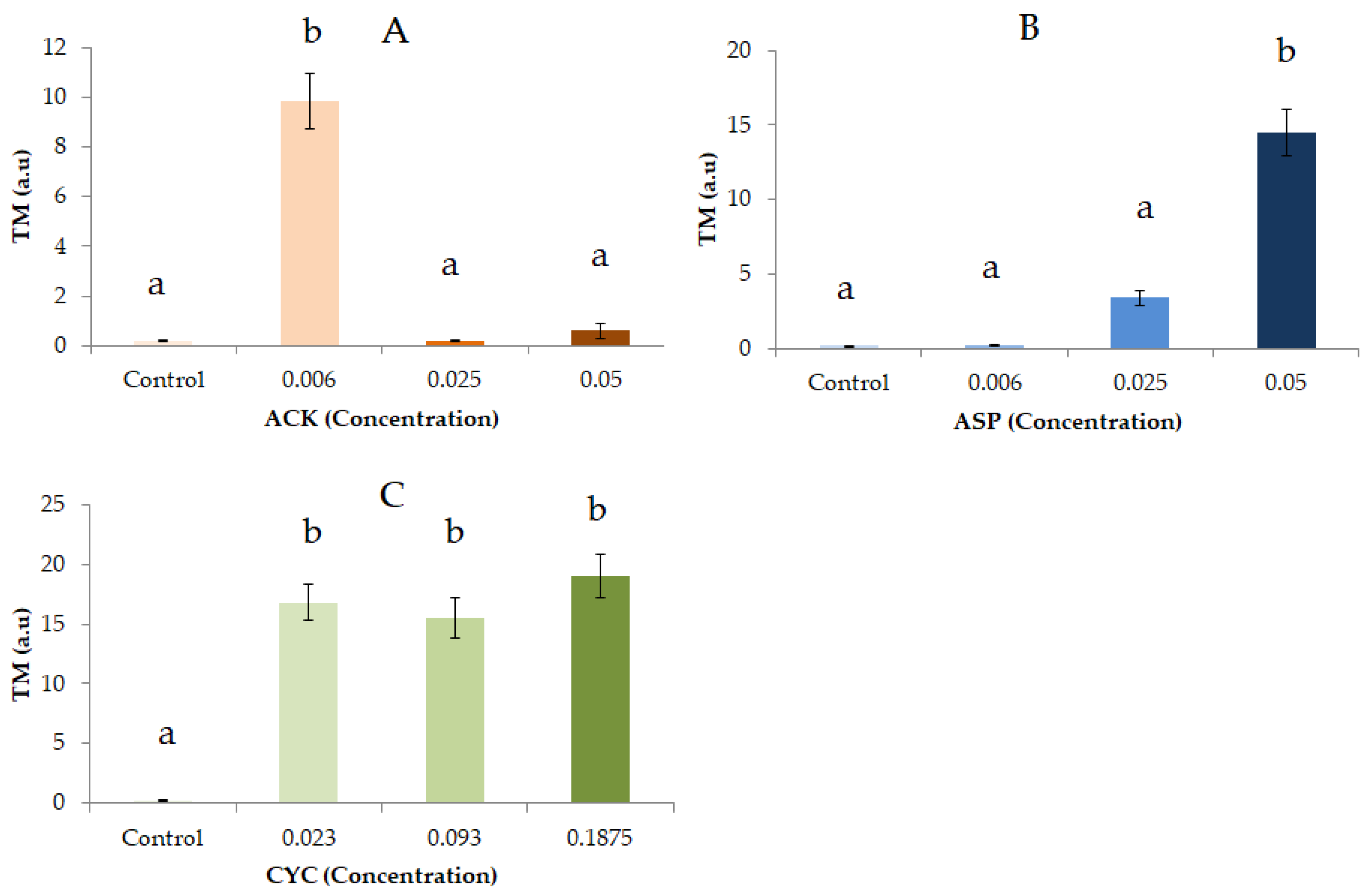
3.6. Comet Assay
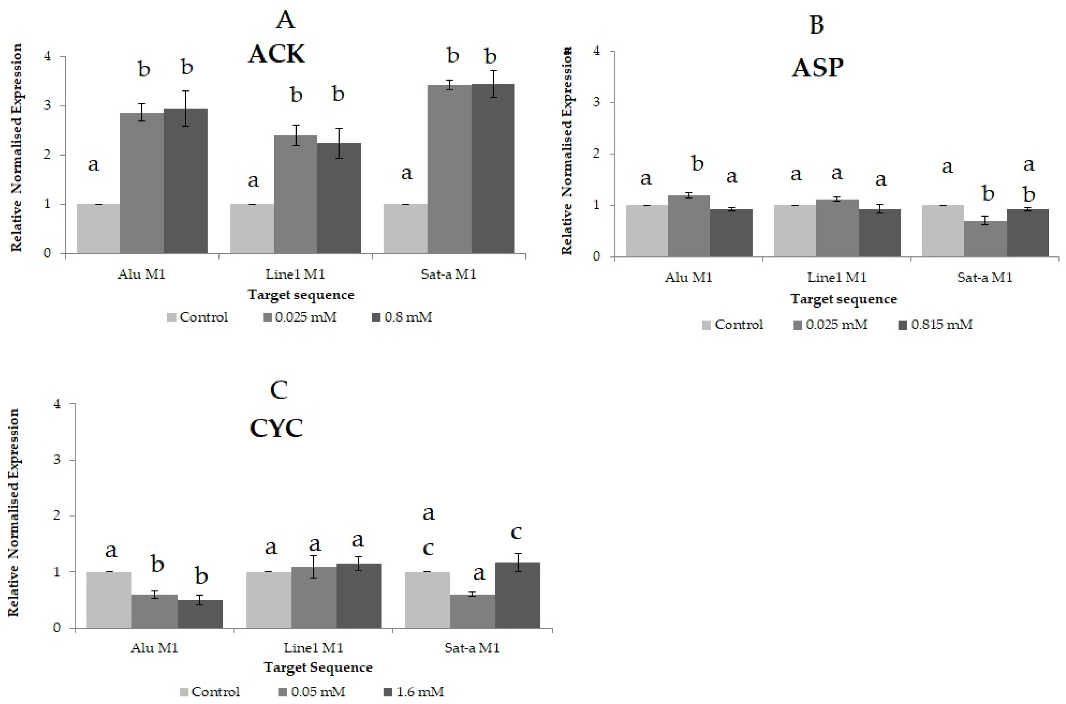
3.7. Methylation Status
4. Discussion
5. Conclusions
Author Contributions
Funding
Data Availability Statement
Conflicts of Interest
References
- Lange, F.T.; Scheurer, M.; Brauch, H.-J. Artificial sweeteners—A recently recognized class of emerging environmental contaminants: A review. Anal. Bioanal. Chem. 2012, 403, 2503–2518. [Google Scholar] [CrossRef] [PubMed]
- Chung, Y.-S.; Lee, M. Genotoxicity assessment of erythritol by using short-term assay. Toxicol. Res. 2013, 29, 249. [Google Scholar] [CrossRef] [PubMed] [Green Version]
- Yang, Q. Gain weight by “going diet?” Artificial sweeteners and the neurobiology of sugar cravings: Neuroscience. Yale J. Biol. Med. 2010, 83, 101–108. [Google Scholar]
- Pang, M.D.; Goossens, G.H.; Blaak, E.E. The Impact of Artificial Sweeteners on Body Weight Control and Glucose Homeostasis. Front. Nutr. 2021, 7, 598340. [Google Scholar] [CrossRef] [PubMed]
- García, A.; Martínez, J.A. Los edulcorantes y su papel sobre el metabolismo humano. Dialnetplus 2016, 4, 13–22. [Google Scholar]
- Marinovich, M.; Galli, C.L.; Bosetti, C.; Gallus, S.; La Vecchia, C. Aspartame, low-calorie sweeteners and disease: Regulatory safety and epidemiological issues. Food Chem. Toxicol. 2013, 60, 109–115. [Google Scholar] [CrossRef]
- Mishra, A.; Ahmed, K.; Froghi, S.; Dasgupta, P. Systematic review of the relationship between artificial sweetener consumption and cancer in humans: Analysis of 599,741 participants. Int. J. Clin. Pract. 2015, 69, 1418–1426. [Google Scholar] [CrossRef] [Green Version]
- Cillo, F. 0% Calorías, % azúcares. Un análisis sobre las bebidas light del mercado. La tendencia a endulzar naturalmente: Stevia. ISDe Sports Mag. 2011, 3, 8. [Google Scholar]
- Durán, S.; Cordón, K.; Rodríguez, M.D.P. Edulcorantes no nutritivos, riesgos, apetito y ganancia de peso. Rev. Chil. Nutr. 2013, 40, 309–314. [Google Scholar] [CrossRef] [Green Version]
- Wu, G.D.; Chen, J.; Hoffmann, C.; Bittinger, K.; Chen, Y.-Y.; Keilbaugh, S.A.; Bewtra, M.; Knights, D.; Walters, W.A.; Knight, R.; et al. Linking Long-Term Dietary Patterns with Gut Microbial Enterotypes. Science 2011, 334, 105. [Google Scholar] [CrossRef] [Green Version]
- Bian, X.; Chi, L.; Gao, B.; Tu, P.; Ru, H.; Lu, K. The artificial sweetener acesulfame potassium affects the gut microbiome and body weight gain in CD-1 mice. PLoS ONE 2017, 12, e0178426. [Google Scholar] [CrossRef]
- Shanmugasundaram, K.; Nayak, B.K.; Friedrichs, W.E.; Kaushik, D.; Rodriguez, R.; Block, K. NOX4 functions as a mitochondrial energetic sensor coupling cancer metabolic reprogramming to drug resistance. Nat. Commun. 2017, 8, 997. [Google Scholar] [CrossRef] [PubMed] [Green Version]
- Ibi, D.; Suzuki, F.; Hiramatsu, M. Effect of AceK (acesulfame potassium) on brain function under dietary restriction in mice. Physiol. Behav. 2018, 188, 291–297. [Google Scholar] [CrossRef]
- Merinas-Amo, T.; Lozano-Baena, M.-D.; Obregón-Cano, S.; Alonso-Moraga, Á.; de Haro-Bailón, A. Role of Glucosinolates in the Nutraceutical Potential of Selected Cultivars of Brassica rapa. Foods 2021, 10, 2720. [Google Scholar] [CrossRef]
- Arnold, D.; Boyes, B. The toxicological effects of saccharin in short-term genotoxicity assays. Mutat. Res. Rev. Genet. 1989, 221, 69–132. [Google Scholar] [CrossRef]
- Graf, U.; Wurgler, F.E.; Katz, A.J.; Frei, H.; Juon, H.; Hall, C.B.; Kale, P.G. Somatic mutation and recombination test in Drosophila melanogaster. Environ. Mutagen. 1984, 6, 153–188. [Google Scholar] [CrossRef]
- Rojas Molina, M.; Campos Sánchez, J.; Analla, M.; Muñoz Serrano, A.; Alonso Moraga, A. Genotoxicity of vegetable cooking oils in the Drosophila wing spot test. Environ. Mol. Mutag. 2005, 45, 90–95. [Google Scholar] [CrossRef] [PubMed]
- Graf, U.; Abraham, S.K.; Guzman-Rincon, J.; Wurgler, F.E. Antigenotoxicity studies in Drosophila melanogaster. Mutat. Res. 1998, 402, 203–209. [Google Scholar] [CrossRef]
- Fernandez-Bedmar, Z.; Anter, J.; de La Cruz-Ares, S.; Munoz-Serrano, A.; Alonso-Moraga, A.; Perez-Guisado, J. Role of citrus juices and distinctive components in the modulation of degenerative processes: Genotoxicity, antigenotoxicity, cytotoxicity, and longevity in Drosophila. J. Toxicol. Environ. Health A 2011, 74, 1052–1066. [Google Scholar] [CrossRef] [PubMed]
- Bell, R.; Hubbard, A.; Chettier, R.; Chen, D.; Miller, J.P.; Kapahi, P.; Tarnopolsky, M.; Sahasrabuhde, S.; Melov, S.; Hughes, R.E. A human protein interaction network shows conservation of aging processes between human and invertebrate species. PLoS Genet. 2009, 5, e1000414. [Google Scholar] [CrossRef] [Green Version]
- Li, S.; Chen, K.; Li, X.; Zhang, X.; Liu, S.V. A new cultivation system for studying chemical effects on the lifespan of the fruit fly. Exp. Gerontol. 2010, 45, 158–162. [Google Scholar] [CrossRef] [PubMed]
- Olive, P.L.; Frazer, G.; Banath, J.P. Radiation-induced apoptosis measured in tk6-human b-lymphoblast cells using the comet assay. Radiat. Res. 1993, 136, 130–136. [Google Scholar] [CrossRef] [PubMed]
- Collins, A.R. The comet assay for DNA damage and repair: Principles, applications, and limitations. Mol. Biotechnol. 2004, 26, 249–261. [Google Scholar] [CrossRef]
- Anter, J.; Demyda-Peyras, S.; Delgado-Torre, M.P.; Campos-Sanchez, J.; Luque De Castro, M.D.; Muñoz-Serrano, A.; Alonso-Moraga, Á. Biological and health promoting activity of vinification byproducts produced in Spanish vineyards. S. Afr. J. Enol. Vitic. 2015, 36, 126–133. [Google Scholar] [CrossRef] [Green Version]
- Yan, J.; Huen, D.; Morely, T.; Johnson, G.; Gubb, D.; Roote, J.; Adler, P.N. The multiple-wing-hairs gene encodes a novel GBD-FH3 domain-containing protein that functions both prior to and after wing hair initiation. Genetics 2008, 180, 219–228. [Google Scholar] [CrossRef] [PubMed] [Green Version]
- Ren, N.; Charlton, J.; Adler, P.N. The flare gene, which encodes the AIP1 protein of Drosophila, functions to regulate F-actin disassembly in pupal epidermal cells. Genetics 2007, 176, 2223–2234. [Google Scholar] [CrossRef] [Green Version]
- Lehkozivova, J.; Karovicova, J.; Kohajdova, Z.; Suhaj, M.; Simonova, I. Isotachophoretic analysis of the artificial sweeteners and time-intensity sweetness evaluation of soft drinks. Żywność Nauka Technol. Jakość 2006, 3, 76–85. [Google Scholar]
- Romero-Jiménez, M.; Campos-Sánchez, J.; Analla, M.; Muñoz-Serrano, A.; Alonso-Moraga, A. Genotoxicity and anti-genotoxicity of some traditional medicinal herbs. Mutat. Res. Genet. Toxicol. Environ. Mutagen. 2005, 585, 47–155. [Google Scholar] [CrossRef]
- Tasset-Cuevas, I.; Fernandez-Bedmar, Z.; Lozano-Baena, M.D.; Campos-Sanchez, J.; de Haro-Bailon, A.; Munoz-Serrano, A.; Alonso-Moraga, A. Protective effect of borage seed oil and gamma linolenic acid on DNA: In vivo and in vitro studies. PLoS ONE 2013, 8, e56986. [Google Scholar] [CrossRef] [PubMed] [Green Version]
- Frei, H.; Wurgler, F.E. Optimal experimental design and sample size for the statistical evaluation of data from somatic mutation and recombination tests (SMART) in Drosophila. Mutat. Res. 1995, 334, 247–258. [Google Scholar] [CrossRef]
- Anter, J.; Fernandez-Bedmar, Z.; Villatoro-Pulido, M.; Demyda-Peyras, S.; Moreno-Millan, M.; Alonso-Moraga, A.; Munoz-Serrano, A.; Luque de Castro, M.D. A pilot study on the DNA-protective, cytotoxic, and apoptosis-inducing properties of olive-leaf extracts. Mutat. Res. 2011, 723, 165–170. [Google Scholar] [CrossRef] [PubMed]
- Abraham, S.K. Antigenotoxicity of coffee in the Drosophila assay for somatic mutation and recombination. Mutagenesis 1994, 9, 383–386. [Google Scholar] [CrossRef] [PubMed]
- Anter, J.; Tasset, I.; Demyda-Peyras, S.; Ranchal, I.; Moreno-Millan, M.; Romero-Jimenez, M.; Muntane, J.; Luque de Castro, M.D.; Munoz-Serrano, A.; Alonso-Moraga, A. Evaluation of potential antigenotoxic, cytotoxic and proapoptotic effects of the olive oil by-product "alperujo", hydroxytyrosol, tyrosol and verbascoside. Mutat. Res. Genet. Toxicol. Environ. Mutagen. 2014, 772, 25–33. [Google Scholar] [CrossRef] [PubMed]
- Mateo-Fernández, M.; Merinas-Amo, T.; Moreno-Millán, M.; Alonso-Moraga, Á.; Demyda-Peyrás, S. In vivo and in vitro genotoxic and epigenetic effects of two types of cola beverages and caffeine: A multiassay approach. BioMed Res. Int. 2016, 2016. [Google Scholar] [CrossRef] [Green Version]
- Gyori, B.M.; Venkatachalam, G.; Thiagarajan, P.S.; Hsu, D.; Clement, M.V. OpenComet: An automated tool for comet assay image analysis. Redox Biol. 2014, 2, 457–465. [Google Scholar] [CrossRef] [Green Version]
- Merinas-Amo, T.; Tasset-Cuevas, I.; Díaz-Carretero, A.M.; Alonso-Moraga, A.; Calahorro, F. Role of Choline in the Modulation of Degenerative Processes: In Vivo and In Vitro Studies. J. Med. Food 2017, 20, 223–234. [Google Scholar] [CrossRef]
- Weisenberger, D.J.; Campan, M.; Long, T.I.; Kim, M.; Woods, C.; Fiala, E.; Ehrlich, M.; Laird, P.W. Analysis of repetitive element DNA methylation by MethyLight. Nucleic Acids Res. 2005, 33, 6823–6836. [Google Scholar] [CrossRef] [Green Version]
- Nikolaidis, G.; Raji, O.Y.; Markopoulou, S.; Gosney, J.R.; Bryan, J.; Warburton, C.; Walshaw, M.; Sheard, J.; Field, J.K.; Liloglou, T. DNA methylation biomarkers offer improved diagnostic efficiency in lung cancer. Cancer Res. 2012, 72, 5692–5701. [Google Scholar] [CrossRef] [Green Version]
- Liloglou, T.; Bediaga, N.G.; Brown, B.R.; Field, J.K.; Davies, M.P. Epigenetic biomarkers in lung cancer. Cancer Lett. 2014, 342, 200–212. [Google Scholar] [CrossRef]
- Martínez Becerra, R.; Robles González, J. Methodology for the desing of bioassays in aquatic toxicity. Agron. Colombiana 1999, 16, 40–45. [Google Scholar]
- Frei, H.; Wurgler, F.E. Statistical methods to decide whether mutagenicity test data from Drosophila assays indicate a positive, negative, or inconclusive result. Mutat. Res. 1988, 203, 297–308. [Google Scholar] [CrossRef]
- Karstadt, M. Inadequate toxicity tests of food additive acesulfame. Int. J. Occup. Environ. Health. 2010, 16, 89–96. [Google Scholar] [CrossRef] [PubMed]
- Jung, R.; Kreiling, R.; Acesulfame, K. Edited by Mayer and Kemper. Marcel Dekker 1991, 35, 87–104. [Google Scholar]
- Mukherjee, A.; Chakrabarti, J. In vivo cytogenetic studies on mice exposed to acesulfame-K—A non-nutritive sweetener. Food Chem. Toxicol. 1997, 35, 1177–1179. [Google Scholar] [CrossRef]
- Mayer, D. Acesulfame-K; CRC Press: Boca Ratón, FL, USA, 1991; p. 47. [Google Scholar]
- Reuzel, P.; van der Heijden, C. Long-Term Oral Toxicity Study with Acesulfame-K in Beagles. Marcel Dekker 1991, 6, 71. [Google Scholar]
- Butchko, H.H.; Stargel, W.W.; Comer, C.P.; Mayhew, D.A.; Benninger, C.; Blackburn, G.L.; de Sonneville, L.M.; Geha, R.S.; Hertelendy, Z.; Koestner, A. Aspartame: Review of safety. Regul. Toxicol. Pharmacol. 2002, 35, S1–S93. [Google Scholar] [CrossRef] [Green Version]
- von Rymon Lipinski, G.-W.; Hanger, L.Y.; Acesulfame, K. Alternative Seeteners. In Food science and technology, 3rd ed.; Marcel Dekker: New York, NY, USA, 2001; pp. 13–30. [Google Scholar]
- Bandyopadhyay, A.; Ghoshal, S.; Mukherjee, A. Genotoxicity testing of low-calorie sweeteners: Aspartame, acesulfame-K, and saccharin. Drug Chem. Toxicol. 2008, 31, 447–457. [Google Scholar] [CrossRef]
- Martins, M.R.I.; Azoubel, R. Effects of aspartame on fetal kidney: A morphometric and stereological study. Int. J. Morphol. 2007, 25, 689–694. [Google Scholar] [CrossRef]
- Simintzi, I.; Schulpis, K.H.; Angelogianni, P.; Liapi, C.; Tsakiris, S. The effect of aspartame on acetylcholinesterase activity in hippocampal homogenates of suckling rats. Pharmacol. Res. 2007, 56, 155–159. [Google Scholar] [CrossRef]
- Lindseth, G.N.; Coolahan, S.E.; Petros, T.V.; Lindseth, P.D. Neurobehavioral effects of aspartame consumption. Res. Nurs. Health 2014, 37, 185–193. [Google Scholar] [CrossRef] [Green Version]
- Siddique, Y.H.; Anjum, S.; Jyoti, S. Evaluation of the toxic potential of aspartame in third instar larvae of transgenic Drosophila melanogaster (hsp70-lacZ) Bg9. All Results J. Biol. 2017, 8, 16–24. [Google Scholar]
- Shankar, P.; Ahuja, S.; Sriram, K. Non-nutritive sweeteners: Review and update. Nutrition 2013, 29, 1293–1299. [Google Scholar] [CrossRef] [PubMed]
- Weerasooriyagedara, M. Toxicity effects of aspartame on embryonic development of Zebrafish (Danio rerio). Int. J. Eng. Manag. Res. 2018, 8, 183–188. [Google Scholar]
- Taylor, J.D.; Richards, R.K.; Wiegand, R.G.; Weinberg, M.S. Toxicological studies with sodium cyclamate and saccharin. Food Cosmet. Toxicol. 1968, 6, 313–327. [Google Scholar] [CrossRef]
- Brantom, P.G.; Gaunt, I.F.; Grasso, P. Long-term toxicity of sodium cyclamate in mice. Food Cosmet. Toxicol. 1973, 11, 735–746. [Google Scholar] [CrossRef]
- Serra-Majem, L.; Bassas, L.; García-Glosas, R.; Ribas, L.; Inglés, C.; Casals, I.; Saavedra, P.; Renwick, A.G. Cyclamate intake and cyclohexylamine excretion are not related to male fertility in humans. Food Addit. Contam. 2003, 20, 1097–1104. [Google Scholar] [CrossRef]
- Brusick, D.; Cifone, M.; Young, R.; Benson, S. Assessment of the genotoxicity of calcium cyclamate and cyclohexylamine. Environ. Mol. Mutag. 1989, 14, 188–199. [Google Scholar] [CrossRef]
- Bopp, B.A.; Sonders, R.C.; Kesterson, J.W.; Renwick, A. Toxicological aspects of cyclamate and cyclohexylamine. Crit. Rev. Toxicol. 1986, 16, 213–306. [Google Scholar] [CrossRef]
- Martins, A.T.; Santos, S.F.; Scannavino, F.L.F.; Pires, J.R.; Zuza, E.P.; Padovani Junior, J.A.; Azoubel, R.; Mateo, M.A.S.D.; Lopes, R.A. Effect of sodium cyclamate on the rat fetal exocrine pancreas: A karyometric and stereological study. Int. J. Morphol. 2010, 28, 899–904. [Google Scholar] [CrossRef]
- Chen, Z.; Chen, G.; Zhou, K.; Zhang, P.; Ren, X.; Mei, X. Toxicity of food sweetener-sodium cyclamate on osteoblasts cells. Biochem. Biophys. Res. Commun. 2019, 508, 507–511. [Google Scholar] [CrossRef]
- Hu, Y.; Xie, M.; Wu, X. Interaction studies of sodium cyclamate with DNA revealed by spectroscopy methods. Spectrochim. Acta A Mol. Biomol. Spectrosc. 2019, 220, 117085. [Google Scholar] [CrossRef] [PubMed]
- Marquadt, H.; Marquadt, H. Induction of malignant transformation and mutagenesis in cell cultures by cancer chemothcrapeutic agents. Cancer 1977, 40, 1930–1934. [Google Scholar] [CrossRef]
- Jung, R.; Hollander, H. Sunett. Study of the mutagenic potential in strains of Salmonella typhimurium (Ames test) and Escherichia coli. Pharm. Res. Toxicol. Unpublished Report No.86.0811. 1986. [Google Scholar]
- Rencüzoğulları, E.; Tüylü, B.A.; Topaktaş, M.; Ila, H.B.; Kayraldız, A.; Arslan, M.; Diler, S.B. Genotoxicity of aspartame. Drug Chem. Toxicol. 2004, 27, 257–268. [Google Scholar] [CrossRef]
- Soffritti, M.; Belpoggi, F.; Tibaldi, E.; Degli Esposti, D.; Lauriola, M. Life-span exposure to low doses of aspartame beginning during prenatal life increases cancer effects in rats. Environ. Health Perspect. 2007, 115, 1293. [Google Scholar] [CrossRef]
- AlSuhaibani, E.S. In vivo cytogenetic studies on aspartame. Comp. Funct. Genom. 2010, 2010, 1–4. [Google Scholar] [CrossRef] [Green Version]
- Elfatah, A.A.A.; Ghaly, I.S.; Hanafy, S.M. Cytotoxic effect of aspartame (diet sweet) on the histological and genetic structures of female albino rats and their offspring. Pak. J. Biol. Sci. 2012, 15, 904. [Google Scholar]
- Kashanian, S.; Khodaei, M.M.; Kheirdoosh, F. In vitro DNA binding studies of aspartame, an artificial sweetener. J. Photochem. Photobiol. B Biol. 2013, 120, 104–110. [Google Scholar] [CrossRef] [PubMed]
- Soffritti, M.; Belpoggi, F.; Esposti, D.D.; Lambertini, L.; Tibaldi, E.; Rigano, A. First experimental demonstration of the multipotential carcinogenic effects of aspartame administered in the feed to Sprague-Dawley rats. Environ. Health Perspect. 2006, 114, 379–385. [Google Scholar] [CrossRef] [Green Version]
- Schernhammer, E.S.; Bertrand, K.A.; Birmann, B.M.; Sampson, L.; Willett, W.C.; Feskanich, D. Consumption of artificial sweetener–and sugar-containing soda and risk of lymphoma and leukemia in men and women. Am. J. Clin. Nutr. 2012, 96, 1419–1428. [Google Scholar] [CrossRef]
- Kamath, S.; Vijaynarayana, K.; Shetty, D.P.; Shetty, P. Evaluation of genotoxic potential of aspartame. Pharmacologyonline 2010, 1, 753–769. [Google Scholar]
- Demir, E.; Turna, F.; Aksakal, S.; Kaya, B.; Marcos, R. Genotoxicity of different sweeteners in Drosophila. Fresenius Environ. Bull. 2014, 23, 3427–3433. [Google Scholar]
- Mohammed, M. Assessment of mutagenic potentiality of stevioside in comparison with some other sweeteners using Drosophila. AGRIS 2011. [Google Scholar]
- Cruzan, G. Assessment of the cancer potential of methanol. Crit. Rev. Toxicol. 2009, 39, 347–363. [Google Scholar] [CrossRef] [PubMed]
- Ahmed, F.E.; Thomas, D.B. Assessment of the carcinogenicity of the nonnutritive sweetener cyclamate. Crit. Rev. Toxicol. 1992, 22, 81–118. [Google Scholar] [CrossRef]
- Jeffrey, A.M.; Williams, G.M. Lack of DNA-damaging activity of five non-nutritive sweeteners in the rat hepatocyte/DNA repair assay. Food Chem. Toxicol. 2000, 38, 335–338. [Google Scholar] [CrossRef]
- Yılmaz, S.; Uçar, A. A review of the genotoxic and carcinogenic effects of aspartame: Does it safe or not? Cytotechnology 2014, 66, 875–881. [Google Scholar] [CrossRef] [PubMed] [Green Version]
- Grim, J.M.; Simonik, E.A.; Semones, M.C.; Kuhn, D.E.; Crockett, E.L. The glutathione-dependent system of antioxidant defense is not modulated by temperature acclimation in muscle tissues from striped bass, Morone saxatilis. Comp. Biochem. Physiol. Part A Mol. Integr. Physiol. 2013, 164, 383–390. [Google Scholar] [CrossRef]
- Choudhary, A.K.; Devi, R.S. Imbalance of the oxidant-antioxidant status by aspartame in the organs of immune system of Wistar albino rats. Afr. J. Biotechnol. 2014, 8, 220–230. [Google Scholar]
- Creppy, E.E.; Baudrimont, I.; Marie, A. How aspartame prevents the toxicity of ochratoxin A. J. Toxicol. Sci. 1998, 23, 165–172. [Google Scholar] [CrossRef] [Green Version]
- Abhilash, M.; Paul, M.S.; Varghese, M.V.; Nair, R.H. Effect of long term intake of aspartame on antioxidant defense status in liver. Food Chem. Toxicol. 2011, 49, 1203–1207. [Google Scholar] [CrossRef] [PubMed]
- Ashok, I.; Wankhar, D.; Sheeladevi, R.; Wankhar, W. Long-term effect of aspartame on the liver antioxidant status and histopathology in Wistar albino rats. Biomed. Prev. Nutr. 2014, 4, 299–305. [Google Scholar] [CrossRef]
- Choudhary, A.K.; Devi, R.S. Longer period of oral administration of aspartame on cytokine response in Wistar albino rats. Endocrinol. Nutr. 2015, 62, 114–122. [Google Scholar] [CrossRef] [PubMed]
- Cruz-Rojas, C.; SanJuan-Reyes, N.; Fuentes-Benites, M.P.A.G.; Dublan-García, O.; Galar-Martínez, M.; Islas-Flores, H.; Gómez-Oliván, L.M. Acesulfame potassium: Its ecotoxicity measured through oxidative stress biomarkers in common carp (Cyprinus carpio). Sci. Total Environ. 2019, 647, 772–784. [Google Scholar] [CrossRef]
- Mchunu, N.; Chukwuma, C.I.; Ibrahim, M.A.; Oyebode, O.A.; Dlamini, S.N.; Islam, M.S. Commercially available non-nutritive sweeteners modulate the antioxidant status of type 2 diabetic rats. J. Food Biochem. 2019, 43, e12775. [Google Scholar] [CrossRef]
- Fleming, J.E.; Reveillaud, I.; Niedzwiecki, A. Role of oxidative stress in Drosophila aging. Mutat. Res. 1992, 275, 267–279. [Google Scholar] [CrossRef]
- Gomez, I. The Effect of Aspartame & Sodium Nitrite on Drosophila melanogaster. In Proceedings of the AAAS 2015 Annual Meeting, San Jose, CA, USA, 12–16 February 2015. [Google Scholar]
- Soffritti, M.; Belpoggi, F.; Manservigi, M.; Tibaldi, E.; Lauriola, M.; Falcioni, L.; Bua, L. Aspartame administered in feed, beginning prenatally through life span, induces cancers of the liver and lung in male Swiss mice. Am. J. Ind. Med. 2010, 53, 1197–1206. [Google Scholar] [CrossRef]
- Huang, D. The Effect of Acesulfame Potassium on Phosphoinositide 3-Kinase-Induced Viability and Lifespan in Drosophila melanogaster. Acesulfame Potassium Drosoph. Melanogaster 2011, 1–26. [Google Scholar]
- Lee, K.P.; Simpson, S.J.; Clissold, F.J.; Brooks, R.; Ballard, J.W.O.; Taylor, P.W.; Soran, N.; Raubenheimer, D. Lifespan and reproduction in Drosophila: New insights from nutritional geometry. Proc. Natl. Acad. Sci. USA 2008, 105, 2498–2503. [Google Scholar] [CrossRef] [Green Version]
- Anbara, H.; Sheibani, M.T.; Razi, M. Long-term effect of aspartame on male reproductive system: Evidences for testicular histomorphometrical, Hsp70-2 protein expression and biochemical status. Int. J. Fertil. Steril. 2019, 14, 91–101. [Google Scholar]
- Dalderup, L.; Visser, W. Influence of extra sucrose, fats, protein and of cyclamate in the daily food on the life-span of rats. Experientia 1971, 27, 519–521. [Google Scholar] [CrossRef]
- Findikli, Z.; Turkoglu, S. Determination of the effects of some artificial sweeteners on human peripheral lymphocytes using the comet assay. J. Toxicol. Environ. Health Part A 2014, 6, 147–153. [Google Scholar]
- Pandurangan, M.; Enkhtaivan, G.; Mistry, B.; Chandrasekaran, M.; Noorzai, R.; Kim, D.H. Investigation of role of aspartame on apoptosis process in HeLa cells. Saudi J. Biol. Sci. 2016, 23, 503–506. [Google Scholar] [CrossRef] [PubMed] [Green Version]
- Oyama, Y.; Sakai, H.; Arata, T.; Okano, Y.; Akaike, N.; Sakai, K.; Noda, K. Cytotoxic effects of methanol, formaldehyde, and formate on dissociated rat thymocytes: A possibility of aspartame toxicity. Cell Biol. Toxicol. 2002, 18, 43–50. [Google Scholar] [CrossRef] [PubMed]
- Whitcutt, J.; Bey, E. Toxicity evaluation of synthetic food sweeteners by means of the Weaver Human Cell Test. S. Afr. J. Sci. 2008, 104, 36. [Google Scholar]
- Forchhammer, L.; Ersson, C.; Loft, S.; Möller, L.; Godschalk, R.W.; van Schooten, F.J.; Jones, G.D.; Higgins, J.A.; Cooke, M.; Mistry, V. Inter-laboratory variation in DNA damage using a standard comet assay protocol. Mutagenesis 2012, 27, 665–672. [Google Scholar] [CrossRef] [Green Version]
- Yedjou, C.; Tchounwou, P. In-vitro cytotoxic and genotoxic effects of arsenic trioxide on human leukemia (HL-60) cells using the MTT and alkaline single cell gel electrophoresis (Comet) assays. Mol. Cell Biochem. 2007, 301, 123–130. [Google Scholar] [CrossRef] [Green Version]
- Olive, P.L.; Banáth, J.P. The comet assay: A method to measure DNA damage in individual cells. Nat. Protoc. 2006, 1, 23–29. [Google Scholar] [CrossRef]
- Fabiani, R.; Rosignoli, P.; De Bartolomeo, A.; Fuccelli, R.; Morozzi, G. Genotoxicity of alkene epoxides in human peripheral blood mononuclear cells and HL60 leukaemia cells evaluated with the comet assay. Mutat. Res. 2012, 747, 1–6. [Google Scholar] [CrossRef]
- Fairbairn, D.W.; O’Neill, K.L. Necrotic DNA degradation mimics apoptotic nucleosomal fragmentation comet tail length. In Vitro Cell. Dev. Biol. Anim. 1995, 31, 171–173. [Google Scholar] [CrossRef]
- Nakao, H.; Umebayashi, C.; Nakata, M.; Nishizaki, Y.; Noda, K.; Okano, Y.; Oyama, Y. Formaldehyde-induced shrinkage of rat thymocytes. J. Pharmacol. Sci. 2003, 91, 83–86. [Google Scholar] [CrossRef] [Green Version]
- Horio, Y.; Sun, Y.; Liu, C.; Saito, T.; Kurasaki, M. Aspartame-induced apoptosis in PC12 cells. Environ. Toxicol. Pharmacol. 2014, 37, 158–165. [Google Scholar] [CrossRef] [PubMed]
- Durnev, A.D.; Oreshchenko, A.V.; Kulakova, A.V.; Beresten, N.F.; Seredenin, S.B. Clastogenic activity of dietary sugar substitutes. Vopr. Med. Khim. 1995, 41, 31–33. [Google Scholar] [PubMed]
- Knaap, A.G.A.C.; Kramers, P.G.N.; Sobels, F.H. Lack of mutagenicity of the cyclamate metabolites in Drosophila. Mutat. Res. Sect. Environ. Mutagen. Relat. Subj. 1973, 21, 341–344. [Google Scholar] [CrossRef]
- Sasaki, Y.F.; Kawaguchi, S.; Kamaya, A.; Ohshita, M.; Kabasawa, K.; Iwama, K.; Taniguchi, K.; Tsuda, S. The comet assay with 8 mouse organs: Results with 39 currently used food additives. Mutat. Res. Genet. Toxicol. Environ. Mutagen. 2002, 519, 103–119. [Google Scholar] [CrossRef]
- van Eyk, A.D. The effect of five artificial sweeteners on Caco-2, HT-29 and HEK-293 cells. Drug Chem. Toxicol. 2015, 38, 318–327. [Google Scholar] [CrossRef] [PubMed]
- Abdulfattah, S.Y. Studying some Cytotoxic Parameters of Aspartame (Diet sweet) on Mature Albino Male Mice. J. Biotech. Res. Center 2017, 11, 41–44. [Google Scholar] [CrossRef]
- Murray, A.B.; Lomelino, C.L.; Supuran, C.T.; McKenna, R. “Seriously Sweet”: Acesulfame K Exhibits Selective Inhibition Using Alternative Binding Modes in Carbonic Anhydrase Isoforms. J. Med. Chem. 2018, 61, 1176–1181. [Google Scholar] [CrossRef]
- Lopez-Serra, L.; Esteller, M. Proteins that bind methylated DNA and human cancer: Reading the wrong words. Br. J. Cancer 2008, 98, 1881–1885. [Google Scholar] [CrossRef]
- Qin, T.; Jelinek, J.; Si, J.; Shu, J.; Issa, J.-P.J. Mechanisms of resistance to 5-aza-2′-deoxycytidine in human cancer cell lines. Blood 2009, 113, 659–667. [Google Scholar] [CrossRef] [PubMed] [Green Version]
- Wild, L.; Flanagan, J.M. Genome-wide hypomethylation in cancer may be a passive consequence of transformation. Biochim. Biophys. Acta Rev. Cancer 2010, 1806, 50–57. [Google Scholar] [CrossRef] [PubMed]
- Waye, J.; Willard, H. Structure, organization, and sequence of alpha satellite DNA from human chromosome 17: Evidence for evolution by unequal crossing-over and an ancestral pentamer repeat shared with the human X chromosome. Mol. Cell. Biol. 1986, 6, 3156–3165. [Google Scholar] [PubMed] [Green Version]
- Lander, E.S.; Linton, L.M.; Birren, B.; Nusbaum, C.; Zody, M.C.; Baldwin, J.; Devon, K.; Dewar, K.; Doyle, M.; FitzHugh, W. Initial sequencing and analysis of the human genome. Nature 2001, 409, 860–921. [Google Scholar]
- Martínez, J.G.; Pérez-Escuredo, J.; Castro-Santos, P.; Marcos, C.Á.; Pendás, J.L.L.; Fraga, M.F.; Hermsen, M.A. Hypomethylation of LINE-1, and not centromeric SAT-α, is associated with centromeric instability in head and neck squamous cell carcinoma. Cell. Oncol. 2012, 35, 259–267. [Google Scholar] [CrossRef]
- Wilson, A.S.; Power, B.E.; Molloy, P.L. DNA hypomethylation and human diseases. Biochim. Biophys. Acta Rev. Cancer 2007, 1775, 138–162. [Google Scholar] [CrossRef]
- Ting, D.T.; Lipson, D.; Paul, S.; Brannigan, B.W.; Akhavanfard, S.; Coffman, E.J.; Contino, G.; Deshpande, V.; Iafrate, A.J.; Letovsky, S. Aberrant overexpression of satellite repeats in pancreatic and other epithelial cancers. Science 2011, 331, 593–596. [Google Scholar] [CrossRef] [Green Version]
- Schiffman, S.S. Rationale for further medical and health research on high-potency sweeteners. Chem. Senses 2012, 37, 671–679. [Google Scholar] [CrossRef] [Green Version]


| Primer | Forward Primer Sequence 5′ to 3′ (N) | Reverse Primer Sequence 5′ to 3′ (N) |
|---|---|---|
| ALU-C4 | GGTTAGGTATAGTGGTTTATATTTGTAATTTTAGTA (-36) | ATTAACTAAACTAATCTTAAACTCCTAACCTCA (-33) |
| ALU-M1 | ATTATGTTAGTTAGGATGGTTTCGATTTT (-29) | CAATCGACCGAACGCGA (-17) |
| LINE-1-M1 | GGACGTATTTGGAAAATCGGG (-21) | AATCTCGCGATACGCCGTT (-19) |
| SAT-α-M1 | TGATGGAGTATTTTTAAAATATACGTTTTGTAGT (-34) | AATTCTAAAAATATTCCTCTTCAATTACGTAAA (-33) |
| ACK | Survival | ASP | Survival | CYC | Survival | |||
|---|---|---|---|---|---|---|---|---|
| (mM) | (%) | (mM) | (%) | (mM) | (%) | |||
| Simple | Combined | Simple | Combined | Simple | Combined | |||
| Treatment 1 | Treatment 2 | Treatment | Treatment | Treatment | Treatment | |||
| 0 | 100 | 100 | 0 | 100 | 100 | 0 | 100 | 100 |
| H2O2 | - | 53.00 | H2O2 | - | 40.32 | H2O2 | - | 52.30 |
| 0.006 | 89.32 | 50.68 | 0.0063 | 100.00 | 91.98 *,4 | 0.0125 | 99.00 | 46.65 |
| 0.025 | 91.00 | 53.00 | 0.025 | 100.00 | 88.65 * | 0.050 | 95.65 | 94.00 * |
| 0.05 | 87.00 *,3 | 53.00 | 0.050 | 100.00 | 82.32 * | 0.100 | 98.00 | 84.67 * |
| 0.2 | 86.36 * | 54.00 | 0.200 | 93.68 | 75.68 * | 0.400 | 99.00 | 51.64 |
| 0.8 | 85.68 * | 57.00 | 0.815 | 95.97 | 64.35 * | 1.600 | 98.30 | 59.35 |
| Clones per Wings (Number of Spots) 1 | |||||||
|---|---|---|---|---|---|---|---|
| Compound | Wings | Small Single Spots | Large Simple Spots | Twin Spots | Total Spots | Mann-Whitney Test 2 | IP (%) 3 |
| Number | (1–2 Cells) | (>2 Cells) | m = 5 | m = 2 | |||
| m = 2 | m = 5 | ||||||
| H2O | 41 | 0.147 (6) | 0.048 (2) | 0 | 0.195 (8) | ||
| H2O2 (0.15 M) | 40 | 0.375 (15) | 0.050 (2) | 0 | 0.425 (17) + | ||
| Simple Treatment | |||||||
| ACK (mM) | |||||||
| [0.025] | 39 | 0.231 (9) | 0.000 | 0 | 0.231 (9) i | λ | |
| [0.8] | 40 | 0.200 (8) | 0.025 (1) | 0 | 0.225 (9) i | λ | |
| ASP (mM) | |||||||
| [0.025] | 40 | 0.250 (10) | 0.100 (4) | 0 | 0.350 (14) i | λ | |
| [0.82] | 40 | 0.200 (8) | 0.075 (3) | 0 | 0.275 (11) i | λ | |
| CYC (mM) | |||||||
| [0.05] | 40 | 0.250 (10) | 0 | 0.350 (14) i | λ | ||
| [0.16] | 40 | 0.175 (7) | 0.000 | 0 | 0.175 (7) i | λ | |
| Combined Treatment (mwh/flr 3) | |||||||
| ACK (mM) | |||||||
| [0.025] | 40 | 0.200 (8) i | 0.050 (2) i | 0 − | 0.250 (10) Δ | 41 | |
| [0.8] | 40 | 0.275 (11) i | 0.000 − | 0 − | 0.275 (11) Δ | 35 | |
| ASP (mM) | |||||||
| [0.025] | 38 | 0.130 (5) | 0.050 (2) | 0 | 0.184 (7) Δ | 57 | |
| [0.82] | 40 | 0.250 (10) | 0.125 (5) | 0 | 0.375 (15) Δ | 12 | |
| CYC (mM) | |||||||
| [0.05] | 40 | 0.125 (5) | 0.025 (1) | 0 | 0.150 (6) Δ | 65 | |
| [0.16] | 39 | 0.175 (7) | 0.026 (1) | 0 | 0.205 (8) Δ | 52 | |
| Compound (Concentration) | Mean Lifespan (Days) | Mean Lifespan Difference (%) a | Healthspan (80th Percentile) (Days) | Healthspan Difference (%) a |
|---|---|---|---|---|
| ACK (mM) | ||||
| Control | 57.630 ± 2.108 | 0 | 38.630 ± 1.600 | 0.00 |
| 0.006 | 51.360 ± 2.720 | −10.87 | 40.600 ± 3.560 | 5.10 |
| 0.025 | 53.182 ± 2.707 | −7.70 | 27.50 ± 2.100 ** | −28.80 |
| 0.05 | 58.022 ± 2.635 | 0.70 | 43.830 ± 5.000 | 13.46 |
| 0.2 | 54.783 ± 2.560 | −4.94 | 28.100 ± 2.210 ** | −27.25 |
| 0.8 | 59.702 ± 2.300 | 3.60 | 23.200 ± 0.990 *** | −40.00 |
| ASP (mM) | ||||
| Control | 71.830 ± 1.980 | 0.00 | 52.524 ± 2.000 | 0.00 |
| 0.0063 | 68.520 ± 2.950 | −4.60 | 38.150 ± 3.640 ** | −27.36 |
| 0.025 | 71.636 ± 2.300 | −0.27 | 48.540 ± 1.540 | −7.57 |
| 0.05 | 77.193 ± 200 * | 7.46 | 56.080 ± 2.040 | 6.77 |
| 0.2 | 77.720 ± 2.400 ** | 8.20 | 49.300 ± 3.000 | −6.13 |
| 0.815 | 67.135 ± 2.680 | −6.54 | 36.450 ± 2.970 * | −30.60 |
| CYC (mM) | ||||
| Control | 69.200 ± 2.350 | 0.00 | 40.889 ± 2.210 | 0.00 |
| 0.0125 | 76.460 ± 2.260 | 10.50 | 44.300 ± 1.300 | 8.34 |
| 0.05 | 64.720 ± 2.640 | −6.47 | 29.850 ± 1.720 ** | −27.00 |
| 0.1 | 63.970 ± 2.900 | −7.56 | 34.170 ± 2.265 | −16.43 |
| 0.4 | 75.387 ± 2.212 | 8.94 | 51.000 ± 4.000 | 24.72 |
| 1.6 | 66.550 ± 2.340 | −3.83 | 41.700 ± 2.900 | 1.98 |
Publisher’s Note: MDPI stays neutral with regard to jurisdictional claims in published maps and institutional affiliations. |
© 2022 by the authors. Licensee MDPI, Basel, Switzerland. This article is an open access article distributed under the terms and conditions of the Creative Commons Attribution (CC BY) license (https://creativecommons.org/licenses/by/4.0/).
Share and Cite
Mateo-Fernández, M.; González-Jiménez, M.J.; Celestino, M.D.R.; Font, R.; Alonso-Moraga, Á.; Merinas-Amo, T. Toxicological and Nutraceutical Screening Assays of Some Artificial Sweeteners. Processes 2022, 10, 410. https://0-doi-org.brum.beds.ac.uk/10.3390/pr10020410
Mateo-Fernández M, González-Jiménez MJ, Celestino MDR, Font R, Alonso-Moraga Á, Merinas-Amo T. Toxicological and Nutraceutical Screening Assays of Some Artificial Sweeteners. Processes. 2022; 10(2):410. https://0-doi-org.brum.beds.ac.uk/10.3390/pr10020410
Chicago/Turabian StyleMateo-Fernández, Marcos, Miguel Josué González-Jiménez, Mercedes Del Río Celestino, Rafel Font, Ángeles Alonso-Moraga, and Tania Merinas-Amo. 2022. "Toxicological and Nutraceutical Screening Assays of Some Artificial Sweeteners" Processes 10, no. 2: 410. https://0-doi-org.brum.beds.ac.uk/10.3390/pr10020410