Anti-Melanogenesis, Antioxidant and Anti-Tyrosinase Activities of Scabiosa columbaria L.


School of Biology and Environmental Sciences, University of Mpumalanga, Private Bag X11283, Mbombela 1200, South Africa
*
Author to whom correspondence should be addressed.
Processes 2020, 8(2), 236; https://0-doi-org.brum.beds.ac.uk/10.3390/pr8020236
Submission received: 17 January 2020
/
Revised: 1 February 2020
/
Accepted: 10 February 2020
/
Published: 19 February 2020
(This article belongs to the Special Issue Extraction Optimization Processes of Antioxidants)
Abstract
:Scabiosa columbaria is a plant traditionally used to treat skin ailments, such as scabies, wound bruises, sores and hyperpigmentation. To find a novel skin depigmenting agent, the present study was investigated to determine the possible anti-melanogenesis, antioxidant and anti-tyrosinase effects of methanol extract of S. columbaria leaves. Cytotoxicity towards human dermal fibroblast (MRHF) cells was assessed using the live-cell fluorescence imaging microscopy. The inhibitory effects of the extract on tyrosinase, collagenase and melanin synthesis were also investigated using standard in vitro method, while ferric reducing power (FRAP) was used to determine the antioxidant potential of the plant extract. The effect of the extract on collagen content in MRHF cells was also investigated. The plant extract displayed no meaningful cytotoxicity towards MRHF cells and no significant cell death was recorded at all the tested concentrations. The extract (25–100 µg/mL) effectively decreased melanin content in B16F10 (mouse melanoma) cells with moderate inhibition of tyrosinase enzyme in a dose-dependent manner. However, the extract also demonstrated no significant effect on collagenase and collagen content in MRHF cells, but showed strong antioxidant activity at the concentrations tested. The results suggest that S. columbaria could be a promising candidate in the treatment of skin hyperpigmentation disorders
1. Introduction
Skin hyperpigmentation is one of the common skin complaints that affect people of all skin types. It is described as increased production and dispersion of melanin, the pigment that gives human skin its color [1,2]. Some types of hyperpigmentation, including post-inflammatory hyperpigmentation, age spots, solar lentigo and melisma occur in many human populations and are considered to be skin diseases [3]. Many depigmenting agents, such as hydroquinone, arbutin, kojic acid and corticosteroids have been used to treat hyperpigmentation disorders [4]. In spite of their efficacy, many of these agents are frequently reported to have numerous side effects (for example, erythema, sensitization, contact dermatitis and mutagenic effect with prolonged exposures) [4,5]. In the pursuit for novel depigmenting agents with fewer side effects, the investigation of natural plant products has prompted the identification of many potentially active compounds, including flavonoids, polyphenols, licorice, hesperidin and yeast derivatives. Studies have also revealed that plants are more effective inhibitors of melanogenesis without melanocytoxicity than hydroquinone, kojic acid, linoleic acid and arbutin [6]. Hence, it is imperative to explore novel natural sources to combat the problem of skin hyperpigmentation.
Scabiosa columbaria (Caprifoliaceae family) is commonly called wild scabious (English); bitterwortel (Afrikaans); makgha (Xhosa); ibheka (Zulu). It is an evergreen perennial herb with branches developing from a woody rootstock [7]. S. columbaria is widely distributed throughout South Africa and particularly common in the Western Cape where it prefers growing in a sunny location, in a well-drained, sandy soil, with plenty of compost. Traditionally, the leaves and roots of the plant are used medicinally to treat various ailments, such as skin diseases, sterility and heartburn [8]. In addition, the powdered roots of the plant are also used to make baby powder [9]. Observed pharmacological activities of S. columbaria include antifungal, antibacterial and antiprotozoan [10,11]. Some of the compounds previously isolated from the root extract of S. columbaria include loganin and sweroside glycosides [12]. Vinnitska [13] also isolated palmitic acid, phthalic acid, diisooctyl phthalate, bis-(ethylhexyl) phthalate, and dibutyl phthalate from the aerial parts of S. columbaria. Previous study also showed that S. columbaria leaves contain polyphenols, flavonoids, terpenoids and coumarins (unpublished).
In this study, S. columbaria was selected based on its extensive traditional usage for the treatment of skin-related diseases and the presence of some chemical constituents associated with skin toning and depigmentation, such as polyphenols and flavonoids. For this reason, we investigated the possible anti-melanogenesis, antioxidant and anti-tyrosinase effects of methanol extract of S. columbaria leaves.
2. Results
2.1. Tyrosinase
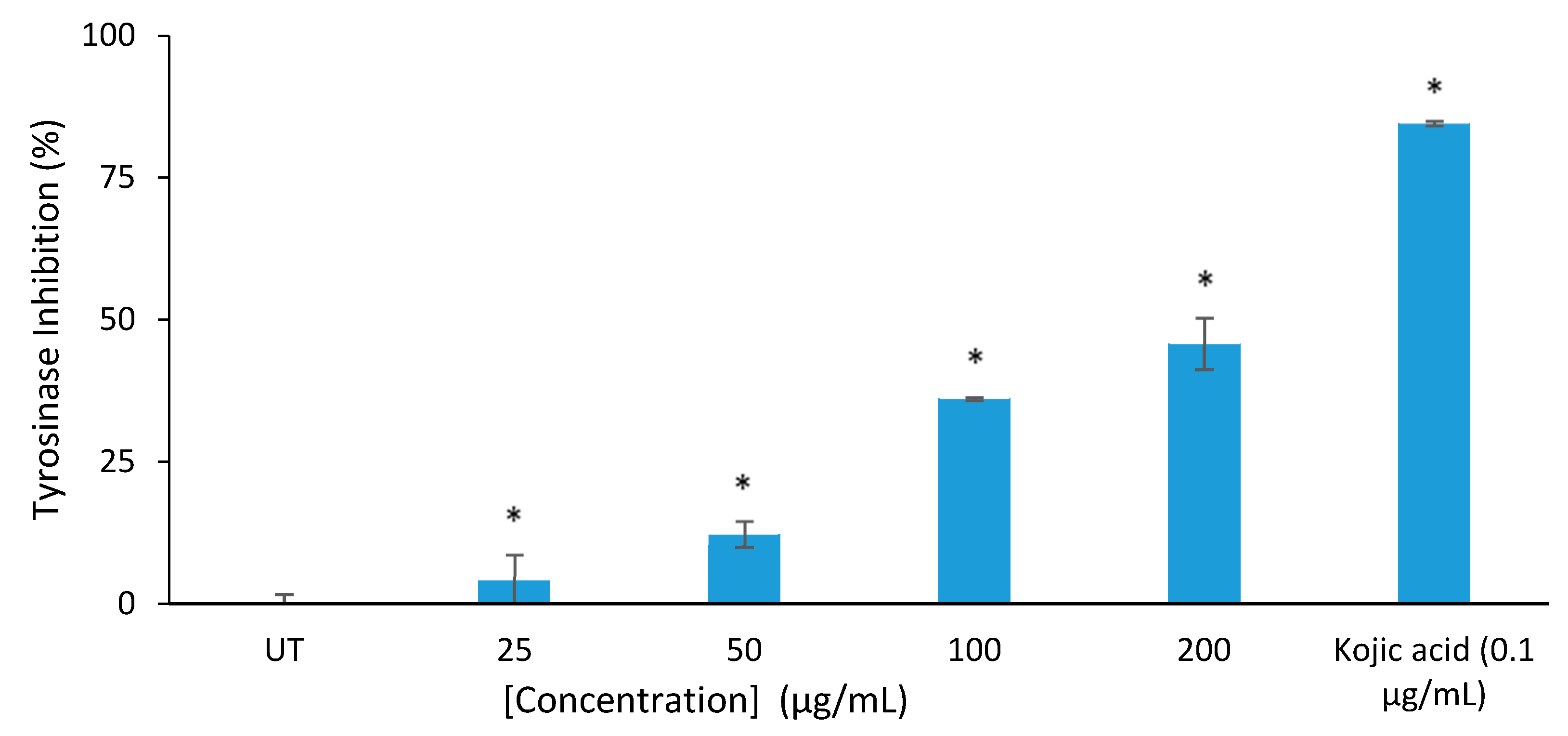
As shown in Figure 1, the tyrosinase inhibitory activity of the plant extract (25–200 µg/mL) exhibited a dose-dependent manner. The inhibitory effect of the extract was higher than that of the control at all the tested concentrations, but lower than kojic acid (84.6%).
2.2. Collagenase
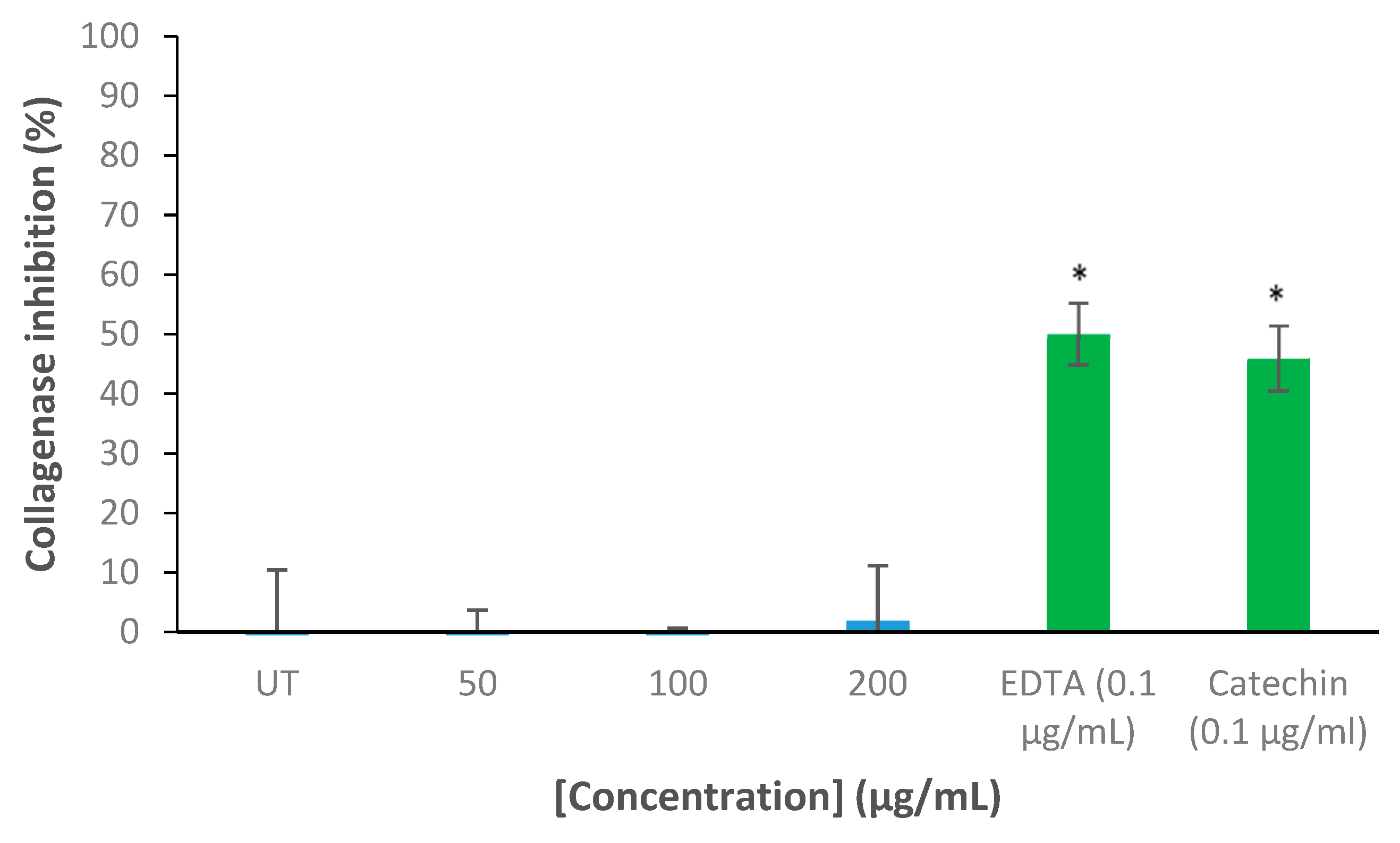
The inhibition of collagenase by the extract is presented in Figure 2. The extract exhibited no inhibition on collagenase at all the tested concentrations (25–200 µg/mL). The positive controls, Ethylenediaminetetraacetic acid (EDTA) and catechin demonstrated significant inhibition on collagenase by 50% and 46%, respectively. EDTA and catechin are well-known collagenase inhibitors.
2.3. Ferric Reducing Power (FRAP)
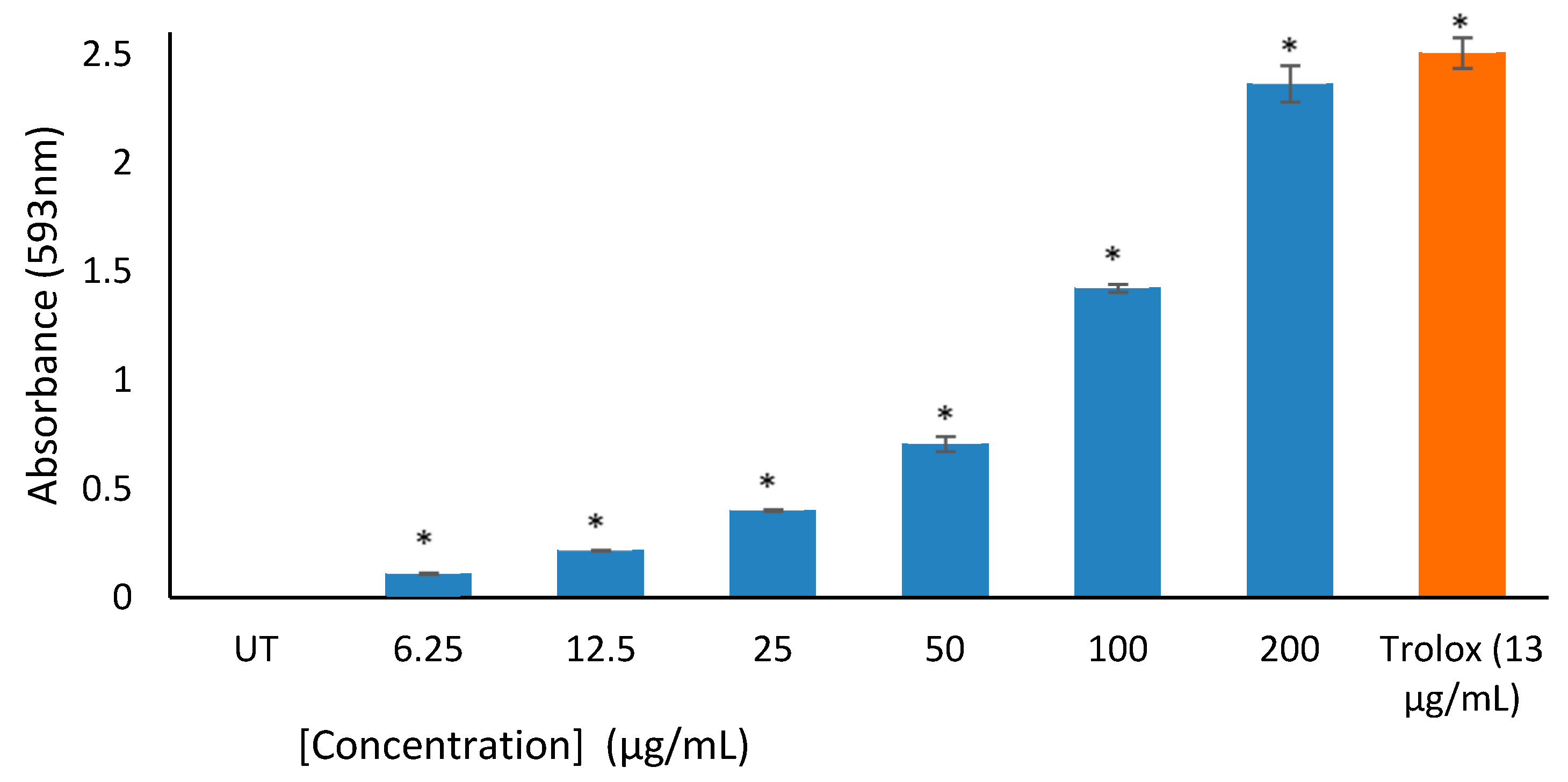
The result of the FRAP assay shows that the extract demonstrated significant FRAP values in a concentration-dependent relationship (Figure 3). At the highest concentration (200 μg/mL) tested, S. columbaria extract demonstrated a remarkable FRAP activity than the control. The positive control Trolox displayed strong ferric reducing potential than both the extract and control.
2.4. Cytotoxicity/Proliferation/Apoptosis Assay
The cytotoxic/proliferation/apoptosis effect of S. columbaria was determined by using the ImageXpress Micro XLS Widefield High-Content Analysis System. The result showed no significant cytotoxicity against the MRHF cells at the tested concentrations, as confirmed by the percentage of live cells. As indicated in Figure 4, there was also a slight increase (less than 7% cell death) in the percentage of apoptotic and necrotic cells at all the tested concentrations (25–200 µg/mL), but physiologically not relevant. This was supported by the trend seen in the percentage of relative cell count (%RCC). The positive control, Melphalan, a potent cytotoxicity drug, decreased MRHF cell viability to 75% as compared to both the extract and untreated control.
2.5. Collagen Production Assay
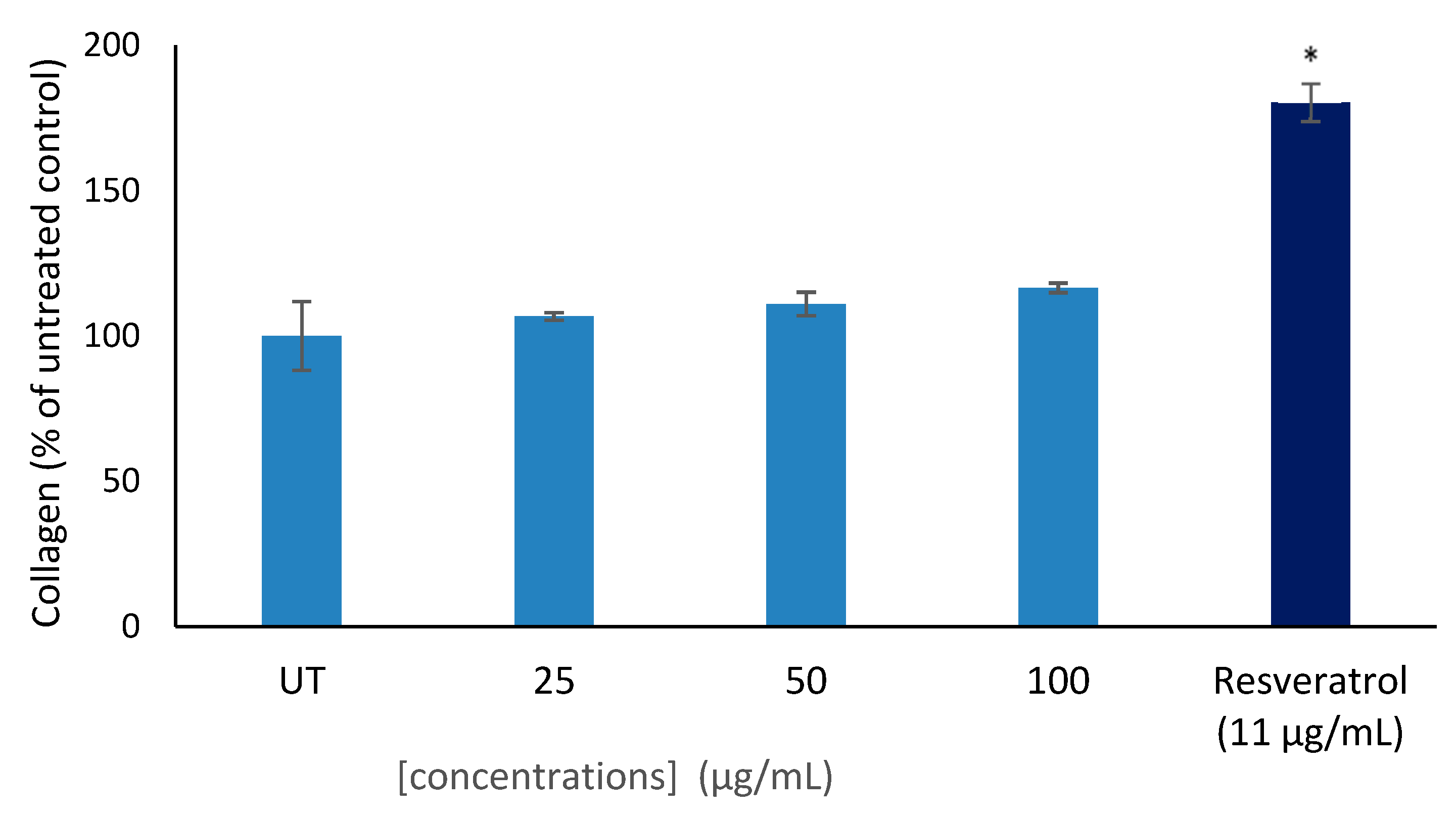
The extract was tested for its effects on collagen production in MRHF cells. The result recorded revealed a weak increase of collagen production in fibroblast cells when treated with different concentrations (25–100 μg/mL) of the plant extract (Figure 5). However, the positive control resveratrol, a well-known collagen booster significantly increased the collagen production in fibroblast cells by 180%.
2.6. Melanin Synthesis Inhibition
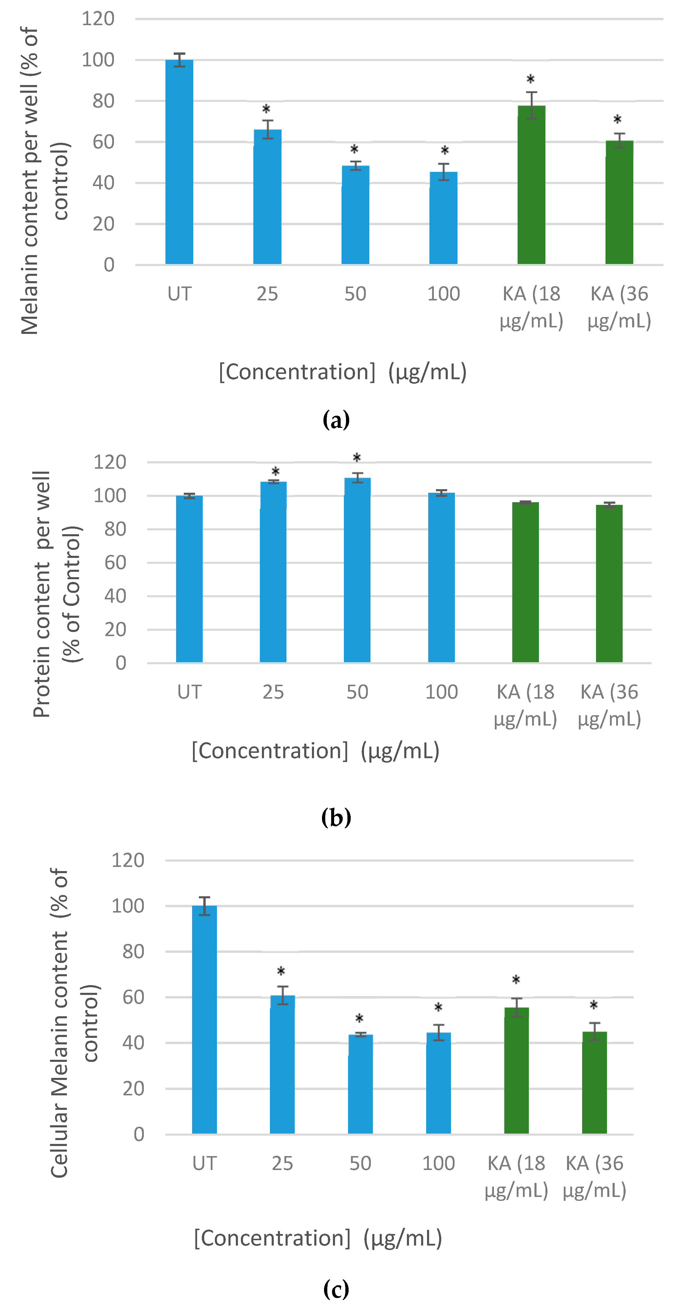
In this experiment, the increasing concentrations of the extract caused inhibition of melanogenesis induced by alpha-melanocyte-stimulating hormone (α-MSH) in B16F10 cells. In addition, at 100 µg/mL, as much as 60% of cellular melanin content was reduced compared to the trend seen in the untreated cells (100%). Kojic acid a well-known inhibitor of melanin exhibited a reduction of cellular melanin content at the tested concentration (Figure 6).
3. Discussion
Tyrosinase, a rate-limiting enzyme, plays a vital role in melanin synthesis within melanocytes. It catalyzes the conversion of tyrosine to 3,4-dihydroxyphenylalanine (DOPA) followed by the conversion of DOPA to DOPA chrome, thereby polymerises to form melanin. Inhibition of tyrosinase represents an important strategy in preventing melanin accumulation in the skin. Thus, tyrosinase inhibitors are attractive targets in cosmetics and treatments for skin pigmentation disorders. In this study, the extract of S. columbaria leaves inhibited tyrosinase activity in a moderate dose-dependent manner with the greatest inhibition observed at the highest concentration tested (200 μg/mL). This inhibition of tyrosinase could be due to the extract competing against the melanin substrate, such as L-DOPA, for the same active site of the enzyme or affects the chelating activity of copper at the active site of the enzyme, which in turn preventing the binding of copper ions to oxygen, thereby leading to the irreversible deactivation of tyrosinase enzyme [14,15]. Therefore, the plant extract may be a copper chelator which functions as a competitive inhibitor. This is the first study of the effect of S. columbaria on tyrosinase enzyme, suggesting its possible use for the treatment of skin pigmentation.
Collagen functions to provide firmness to the skin and consequently damage and loss of skin collagen results in the appearance of thin and wrinkled skin. Collagenase, a key enzyme in collagen degradation is therefore a potential therapeutic target against collagen decline. As such, the activity of the enzyme has to be inhibited to prevent loss of skin collagen. Unfortunately, our result revealed no inhibition of the collagenase enzyme at all the tested concentrations. The extract also demonstrated a slight increase of collagen production in MRHF cells but physiologically not relevant. The findings suggest that S. columbaria extract may not be useful as a collagen booster, benefiting the skin via reduced collagen breakdown.
Skin hyperpigmentation has been linked to free radicals [16] and antioxidants or free radical scavengers have been reported to play a significant role in suppressing hyperpigmentation [16]. FRAP assay has been employed to measure the reducing potential of an antioxidant reacting with a Fe3+-tripyridyltriazine (colorless complex) and producing a Fe2+-tripyridyltriazine (blue-colored complex) [17]. In this study, the FRAP assay result revealed higher absorbance of the reaction mixture with increasing concentration of the extract indicating a remarkable reducing power. This reducing properties of the S. columbaria may be linked to the presence of polyphenols compounds in the extract, which exert their action by breaking the free radical chain thereby donating a hydrogen atom [18]. The results are in accordance with those reported by Zheng et al. [19], who found a strong correlation between polyphenols compounds and FRAP assay. It could be deduced from this study that the methanol extract of S. columbaria may be beneficial in eradicating oxidative damage caused by free radicals which plays a major contributory role in hyperpigmentation. It is imperative to note that the strong antioxidant activity as measured by FRAP assay may not be relevant in vivo, since it is purely chemical not biological. Therefore we recommend antioxidant in in vivo studies.
The result of the cytotoxic assay revealed that S. columbaria extract is not toxic to the MRHF cells. This absence of cytotoxicity supports the ethno-pharmacological usage of this plant. To the best of our knowledge, none of the previous studies had comprehensively analyzed the cytotoxicity of Scabiosa species. However, it is imperative to note that cytotoxicity in vitro does not always equate to in vivo. This is based on the fact that within a biological system, there are possible interactions in the gut and biotransformation issues [20,21].
Melanin is a key determinant of color and its accumulation plays a significant role in abnormal skin pigmentation disorders [22]. In this study, we evaluated whether the extract of S. columbaria leaves may suppress melanin production in B16F10 melanoma cells. The extract demonstrated a dose-dependent decrease of melanin synthesis in α-MSH-stimulated B16F10 melanoma cells without inducing cytotoxicity. Interestingly, this result corresponds with tyrosinase inhibition in the cell-free assay, suggesting that the inhibition may be due to a decreased tyrosinase activity. Tyrosinase and tyrosinase-related protein-1 (TRP-1) play a major role in melanogenesis pathways [23]. Activation of tyrosinase and TRP-1 enhances melanocyte inducing transcription factor (MITF) protein expression, which in turn causes the increase of melanin synthesis [24]. It could be deduced from this study that S. columbaria has the potential to attenuate overproduction of melanin and hyperpigmentation in skin cells.
4. Experimental Procedure
4.1. Reagents
Human foreskin fibroblast (MRHF) cells were obtained from Celonex, Johannesburg, South Africa. While mouse melanoma (B16F10) cells were obtained from Highveld Biologicals, (Johannesburg, South Africa). FBS (etal bovine serum) was purchased from Biowest, Logan, UT, USA. Trypsin-EDTA, RPMI I640 (Roswell Park Memorial Institute), DMEM (Dulbecco’s modified Eagle’s medium) and DPBS (Dulbecco’s phosphate buffered saline) with/without Ca2+ and Mg2+ were purchased from HyClone (Longa, UT, USA). The Annexin V-FITC/PI kit was obtained from MACS Miltenyi Biotec, (Cologne, Germany). CellRox® Orange reagent was obtained from Molecular Probes®-Life Technologies-Thermo Fisher Scientific, Logan, UT, USA. All other reagents used in this study were obtained from Sigma-Aldrich, St. Louis, MI, USA.
4.2. Plant Materials and Extraction Procedure
The leaves of S. columbaria were harvested from Alice, Eastern Cape province of South Africa and the identity was proven by comparing with the Giffen Herbarium specimen from the University of Fort Hare and voucher specimens were kept in the same institution (University of Fort Hare).
The methanol extract of S. columbaria was prepared by pulverizing about 60 g of leaves and then extracting the resulting powder with 1000 mL of methanol. After filtering through Whatman No. 1 filter paper, the resulting filtrate was then concentrated to dryness with rotary evaporator (Heidolph Laborata 4000, Heidolph Instruments, GmbH & Co, Schwabach, Germany). The final extract was weighed to determine the yield (4.54%).
4.3. Mushroom Tyrosinase Inhibition Assay
Briefly, 20 μL of test sample (diluted in assay buffer; phosphate buffered saline pH 6.8), blank (assay buffer) or positive control, Kojic acid (0.1 μg/mL) were pipetted into the wells of a 96-well plate. Then, 20 μL of enzyme solution and 10 μL of buffer was added, whereas for the control wells only 30 µL of buffer was added. Thereafter, 50 μL of L-DOPA (4 mM) solution was added to the resulting mixture to start the reaction and the absorbance was measured at 475 nm for 6 min at 1 min intervals.
4.4. Collagenase Inhibition Assay
The collagenase assay was performed as described previously [25]. Briefly, 10 µL of the test sample or positive controls, EDTA (0.1 μg/mL) or catechin (0.1 μg/mL) were pipetted into the 96-wells plate. Then, 10 µL of gelatin (2 mg/mL) and 10 µL of collagenase enzyme solution were then added to the respective wells. The resulting mixture was incubated for 1 h at 37 °C. After the incubation period, 20 µL of Coomassie Brilliant Blue (CBB) was added, then the plate was shaken for 5 min and then centrifuged for 5 min at 500 rcf. Thereafter, the supernatant was removed and 50 µL of washing solution (containing 40% methanol and 10% acetic acid) was added to wash the pellet. The remaining pellet was then dissolved by adding DMSO (50 µL) and the absorbance was read at 600 nm.
4.5. Ferric Reducing Antioxidant Power (FRAP) Assay
The FRAP assay was performed as described previously [26]. The working FRAP reagent containing 20 mL of sodium acetate buffer (30 mM; pH 3.6), 2 mL of ferric chloride solution (20 mM), 2.5 mL of TPTZ (tripyridyltriazine) solution (prepared at 10 mM TPTZ in 40 mM HCl) and distilled water (2 mL) was freshly prepared. The mixture was incubated at 37 °C for 15 min before use. Fifty microliters of the test sample or Trolox (13 μg/mL) and FRAP reagent (200 μL) were pipetted into the well of the 96-well plate. Then, the mixture was incubated for 30 min at 37 °C, and then the absorbance was read at 593 nm.
4.6. Cell Culture Conditions
B16F10 and MRHF cells were maintained separately in culture dishes (10 cm) in DMEN (low glucose) supplemented with 10% FBS and incubated in a humidified incubator with 5% CO2. The cell number and viability were performed using a LunaTM cell Counter (Logos Biosystems, Inc., Anyang, South Korea) after the staining of cells with trypan blue.
4.7. Imaging and Analysis for Cell-Based Assays
For the cell-based assays experiments, the ImageXpress Micro XLS Widefield High-Content Analysis System (Molecular Devices®, San Jose, CA, USA) was used and then analyzed using the MetaXpress® High-Content Image Acquisition & Analysis Software supplied by Molecular Devices®, San Jose, CA, USA.
4.8. Cytotoxicity/Proliferation/Apoptosis Assay
MRHF cells were seeded in 96-well plate at a density of 8000 cells/well, using 100 µL aliquots, and allowed to attach overnight. Cells were treated by adding 100 µL of the plant extract (prepared at varying concentrations of 25, 50 and 100 µg/mL). The treated cells were then incubated for 24 h at 37 °C in a humidified incubator with 5% CO2, then the treatment medium was removed. Thereafter, the treatment medium was replaced with 50 µL of staining solution containing 5 mL binding buffer (PBS with Ca2+ and Mg2+), 50 µL Annexin V-FITC reagent and 2 µL of Hoechst dye solution (10 mg/mL in DMSO). After 15 min of incubation at 37 °C, 50 µL of propidium iodide (PI) (prepared at 2 µg/mL in binding buffer) was added to the cells and further incubated for additional 5 min, then the images were acquired and analyzed.
4.9. Collagen Production Assay
MRHF cells were seeded into 96-well plates at a density of 8000 cells per well, maintained in DMEM (low glucose) supplemented with 10% FCS and then incubated at 37 °C in a humidified atmosphere with 5% CO2. The cells were then allowed to adhere and grow to confluence. Thereafter, the medium was removed by aspiration and then replaced with fresh DMEN containing ascorbic acid and plant extract (prepared at 25, 50, 100 µg/mL) or resveratrol (11 μg/mL). After 72 h of incubation, the spent medium was then removed by aspiration and fixed overnight with 4% formaldehyde in PBS. The fix solution was aspirated and replaced by adding 50 µL of Sirius Red solution to each well and further incubated at room temperature for 60 min. After the incubation period, the dye solution was aspirated and washed with 100 μL acidified water then the attached dye was eluted in 100 μL of 0.1 N sodium hydroxide (15 min at room temperature), and the absorbance was measured at 510 nm. Cell viability in the representative wells was measured after removing the solubilized dye and replacing it with 50 µL of crystal violet stain and then incubating at room temperature for 30 min. The dye was removed by washing the plates with tap water and allowed to dry in an oven at 37 °C. Thereafter, the dye taken up by the cells was extracted with 100 µL of 10% acetic acid. The absorbance was then read at 595 nm.
4.10. Melanin Synthesis Inhibition
B16F10 cells were seeded in 96-well plate at a density of 5000 cells per well and allowed to attain approximately 90% confluence. After the spent medium was removed, the cells were treated by adding 100 µL of the plant extract at varying concentrations (0–200 µg/mL), then 100 µL 2X melanogenesis medium (DMEM containing 1 mM Theophylline, 0.1 mM l-tyrosine and 100 nM α-MSH) was added and then cultured for a further 5 days. The medium was removed by aspiration and the cells were washed with PBS, then 100 µL 1 N NaOH containing 10% DMSO was added. The plate was then incubated at 60 °C for 1–2 h to solubilise melanin and the absorbance was read at 475 nm. Then, 20 µL aliquot was transferred to a new micro-plate and 200 µL of Bradford reagent was added. The plate was further incubated at room temperature for 20 min and the absorbance was read at 595 nm.
4.11. Statistical Analysis
Statistical analysis was performed using Student’s t-test (two-tailed). Triplicate values for each test sample were compared with triplicate values of the controls. Error bars represent the standard deviation (SD) of the mean.
5. Conclusions
In summary, the leaf extract of S. columbaria possessed strong antioxidant, tyrosinase and antimelanogenic effects, with no significant effect on collagenase and collagen production in MRHF cells. However, the extract also displayed no cytotoxic effect, which further supports the safe use of this plant, but should be confirmed by further in-depth investigations. The results of this present study have demonstrated that S. columbaria could be a useful therapeutic agent for the treatment of skin hyperpigmentation. Future studies will focus on the isolation and characterization of the active component(s) of the plant extract.
Author Contributions
Investigation, W.O.-M., I.J.S.; supervision, W.O.-M., I.J.S.; writing—original draft, W.O.-M.; writing—review and editing, W.O.-M., I.J.S. All authors have read and agreed to the published version of the manuscript.
Funding
Research was funded by the National Research Foundation (NRF), South Africa (Grant no: 105161).
Acknowledgments
National Research Foundation (NRF) and University of Mpumalanga, South Africa.
Conflicts of Interest
The authors declare no conflict of interest.
References
- Rigopoulos, D.K.; Gregoriou, S. Hyperpigmentation and melasma. J. Cosmet. Derm. 2007, 6, 195–202. [Google Scholar] [CrossRef] [PubMed]
- Ghafari, S.S.; Fahimi, S.H. Plants used to treat hyperpigmentation in Iranian traditional medicine: A review. Res. J. Pharm. 2017, 4, 71–85. [Google Scholar]
- Adhikari, A.; Devkota, H.P.; Takano, A.; Masuda, K.; Nakane, T.; Basnet, P.; Skalko-Basnet, N. Screening of Nepalese crude drugs traditionally used to treat hyperpigmentation: In vitro tyrosinase inhibition. Int. J. Cosmet. Sci. 2008, 30, 353–360. [Google Scholar] [CrossRef] [PubMed]
- Zhu, W.; Gao, J. The use of botanical extracts as topical skin-lightening agents for the improvement of skin pigmentation disorders. J. Investig. Dermatol. Symp. Proc. 2008, 13, 20–24. [Google Scholar] [CrossRef] [PubMed] [Green Version]
- Chen, C.Y.; Lin, L.C.; Yang, W.F.; Bordon, J.; Wang, H.M.D. An updated organic classification of tyrosinase inhibitors on melanin biosynthesis. Curr. Org. Chem. 2015, 19, 4–18. [Google Scholar] [CrossRef]
- Wu, S.; Ng, C.C.; Tzeng, W.S.; Ho, K.C.; Shyu, Y.T. Functional antioxidant and tyrosinase inhibitory properties of extracts of Taiwanese pummelo (Citrus grandis Osbeck). Afr. J. Biotechnol. 2011, 10, 7668–7674. [Google Scholar]
- Van Wyk, B.E.; van Oudtshoorn, B.; Gericke, N. Medicinal Plants of South Africa; Sun Press: Bloemfontein, South Africa, 2009. [Google Scholar]
- Maroyi, A. Scabiosa columbaria: A review of its medicinal uses, phytochemistry, and biological activities. Asian J. Pharm. Clin. Res. 2019, 12. [Google Scholar] [CrossRef]
- Von Koenen, E. Medicinal, Poisonous and Edible Plants in Namibia; Klaus Hess Pubkishers: Windhoek, Africa, 2001. [Google Scholar]
- van Vuuren, S.F.; Naidoo, D. An antimicrobial investigation of plants used traditionally in Southern Africa to treat sexually transmitted infections. J. Ethnopharmacol. 2010, 130, 552–558. [Google Scholar] [CrossRef]
- Seleteng-Kose, L. Evaluation of Commonly used Medicinal Plants of Maseru District in Lesotho for their Ethnobotanical Uses, Antimicrobial Properties and Phytochemical Compositions. Ph.D. Thesis, University of Johannesburg: Johannesburg, South Africa, 2017. [Google Scholar]
- Horn, M.M.; Drewes, S.E.; Brown, N.J.; Munro, O.Q.; Meyer, J.J.; Mathekga, A.D. Transformation of naturally-occurring 1,9-trans-9,5-cis sweroside to all trans sweroside during acetylation of sweroside aglycone. Phytochemistry 2001, 57, 51–56. [Google Scholar] [CrossRef]
- Vinnitska, R.B. Studies of phthalates pigeon scabious (Scabiosa columbaria L.). Farmatsevtychnyi Zhurnal 2018, 1, 59–63. [Google Scholar]
- Sarikurkcu, C.; Zengin, G.; Oskay, M.; Uysal, S.; Ceylan, R.; Aktumsek, A. Composition, antioxidant, antimicrobialand enzyme inhibition activities of twoOriganum vulgaresubspecies (subsp.vulgareand subsp.hirtum)essentialoils. Ind. Crops Prod. 2015, 70, 178–184. [Google Scholar] [CrossRef]
- Souza, P.M.; Elias, S.T.; Simeoni, L.A.; de Paula, J.E.; Gomes, S.M.; Guerra, E.S.N.; Fonseca, Y.M.; Silva, E.C.; Silveira, D.; Magalhaes, P. Plants from brazilian cerrado with potent tyrosinase inhibitory activity plants from brazilian cerrado with potent tyrosinase inhibitory activity. PLoS ONE 2012, 7. [Google Scholar] [CrossRef] [PubMed] [Green Version]
- Yasui, H.; Sakurai, H. Age-dependent generation of reactive oxygen species in the skin of live hairless rats exposed to UVA light. Exp. Dermatol. 2003, 12, 655–661. [Google Scholar] [CrossRef] [PubMed]
- Benzie, I.F.; Strain, J.J. Ferric reducing/antioxidant power assay: Direct measure of total antioxidant activity of biological fluids and modified version for simultaneous measurement of total antioxidant power and ascorbic acid concentration. Methods Enzymol. 1999, 299, 15–27. [Google Scholar] [PubMed]
- Pavithra, K.; Vadivukkarasi, S. Evaluation of free radical scavenging activity of various extracts of leaves from Kedrostis foetidissima (Jacq.) Cogn. Food Sci. Hum. Williness 2015, 4, 42–46. [Google Scholar] [CrossRef] [Green Version]
- Zheng, W.; Wang, S.Y. Effect of the plant growth temperature on antioxidant capacity in strawberry. J. Agric. Food Chem. 2001, 49, 4977–4982. [Google Scholar]
- Nchu, F.; Githiori, J.B.; McGaw, L.J.; Elo, J.N. Anthelmintic and cytotoxic activities of extracts of Markhamia obtusifolia Sprague (Bignoniaceae). Vet. Parasitol. 2007, 2011, 184–188. [Google Scholar] [CrossRef] [Green Version]
- Kudumela, R.G.; McGaw, L.J.; Masoko, P. Antibacterial interactions, anti-inflammatory and cytotoxic e ects of four medicinal plant species. BMC Complement. Altern. Med. 2018, 18, 199. [Google Scholar] [CrossRef] [Green Version]
- Parvez, S.; Kang, M.; Chung, H.S.; Bae, H. Naturally occurring tyrosinase inhibitors: Mechanism and applications in skin health, cosmetics and agriculture industries. Phytother. Res. 2007, 21, 805–816. [Google Scholar] [CrossRef]
- Slominski, A.; Tobin, D.J.; Shibahara, S.; Wortsman, J. Melanin pigmentation in mammalian skin and its hormonal regulation. Physiol. Rev. 2004, 84, 1155–1228. [Google Scholar] [CrossRef]
- Liu, Z.; Wang, Y.; Li, Q.; Yang, L. Improved antimelanogenesis and antioxidant effects of polysaccharide from Cuscuta chinensis Lam seeds after enzymatic hydrolysis. Braz. J. Med. Biol. Res. 2018, 51, 1–8. [Google Scholar] [CrossRef] [PubMed]
- Sagbo, I.J.; Van De Venter, M.; Koekemoer, T.; Bradley, G. In vitro antidiabetic activity and mechanism of action of Brachylaena elliptica (Thunb.) DC. Evid. Based Complementary Altern. Med. 2018. [Google Scholar] [CrossRef] [PubMed]
- Odeyemi, S. A Comparative Study of the in vitro Antidiabetic Properties, Mechanism of Action and Cytotoxicity of Albuca setosa and Albuca bracteata Bulb Extracts. Ph.D. Thesis, University of Fort Hare, Alice, South Africa, 2016. [Google Scholar]
Figure 1.
Effect of methanol extract of S. columbaria on tyrosinase activity using L-DOPA as substrate. Values represent the mean ± SD, n = 3. * p < 0.05 compared to untreated (UT) control.
Figure 1.
Effect of methanol extract of S. columbaria on tyrosinase activity using L-DOPA as substrate. Values represent the mean ± SD, n = 3. * p < 0.05 compared to untreated (UT) control.

Figure 2.
Effect of methanol extract of S. columbaria on collagenase activity. Values represent the mean ± SD, n = 3. * p < 0.05 compared to untreated (UT) control.
Figure 2.
Effect of methanol extract of S. columbaria on collagenase activity. Values represent the mean ± SD, n = 3. * p < 0.05 compared to untreated (UT) control.

Figure 3.
Antioxidant activity of leaf extract of S. columbaria using ferric reducing power (FRAP) Method. Values represent the mean ± SD, n = 3. * p < 0.05 compared to untreated (UT) control.
Figure 3.
Antioxidant activity of leaf extract of S. columbaria using ferric reducing power (FRAP) Method. Values represent the mean ± SD, n = 3. * p < 0.05 compared to untreated (UT) control.

Figure 4.
Cytotoxic effect in human dermal fibroblast (MRHF) cells when treated with extract of S. columbaria using the staining method. % relative cell count (%RCC) represents the mean number of cells per site expressed as a percentage of the untreated control. Values represent the mean ± SD, n = 3. * p < 0.05 compared to untreated (UT) control.

Figure 5.
Collagen content of MRHF cells when treated with methanol extract of S. columbaria. Collagen content was measured using Sirius Red and normalised for cell density based on crystal violet staining after removal of the bound Sirius Red. Values represent the mean ± SD, n = 3. * p < 0.05 compared to untreated (UT) control.

Figure 6.
Effect of methanol extract of S. columbaria on melanin production in B6F10 cells. Melanin content, protein content and melanin normalised relative to the total cellular protein. Values represent the mean ± SD, n = 3. * p < 0.05 compared to untreated (UT) control. KA: Kojic acid. (a): Melanin content. (b): Protein content. (c): Melanin normalised relative to the total cellular protein

© 2020 by the authors. Licensee MDPI, Basel, Switzerland. This article is an open access article distributed under the terms and conditions of the Creative Commons Attribution (CC BY) license (http://creativecommons.org/licenses/by/4.0/).
Share and Cite
MDPI and ACS Style
Otang-Mbeng, W.; Sagbo, I.J. Anti-Melanogenesis, Antioxidant and Anti-Tyrosinase Activities of Scabiosa columbaria L. Processes 2020, 8, 236. https://0-doi-org.brum.beds.ac.uk/10.3390/pr8020236
AMA Style
Otang-Mbeng W, Sagbo IJ. Anti-Melanogenesis, Antioxidant and Anti-Tyrosinase Activities of Scabiosa columbaria L. Processes. 2020; 8(2):236. https://0-doi-org.brum.beds.ac.uk/10.3390/pr8020236
Chicago/Turabian StyleOtang-Mbeng, Wilfred, and Idowu Jonas Sagbo. 2020. "Anti-Melanogenesis, Antioxidant and Anti-Tyrosinase Activities of Scabiosa columbaria L." Processes 8, no. 2: 236. https://0-doi-org.brum.beds.ac.uk/10.3390/pr8020236
Note that from the first issue of 2016, this journal uses article numbers instead of page numbers. See further details here.