Article

Ginsenosides Conversion and Anti-Oxidant Activities in Puffed Cultured Roots of Mountain Ginseng

1
Graduate School of Biotechnology, College of Life Sciences, Kyung Hee University, Yongin-si 17104, Gyeonggi-do, Korea
2
Department of Oriental Medicinal Biotechnology, College of Life Science, Kyung Hee University, Yongin-si 17104, Gyeonggi-do, Korea
3
Medical School, Institute of Reproductive Medicine, Nantong University, Nantong 226019, China
*
Authors to whom correspondence should be addressed.
These authors have contributed equally.
Submission received: 19 November 2021 / Revised: 10 December 2021 / Accepted: 14 December 2021 / Published: 17 December 2021
(This article belongs to the Topic Innovative Food Processing Technologies)

Abstract

:
CRMG (Cultured Roots of Mountain Ginseng) have the advantages in scale-up production, safety, and pharmacological efficacies. Though several methods are available for the conversion of major to minor ginsenosides, which has more pharmacological activities, a single step process with high temperature and pressure as a puffing method took place in this study to gain and produce more pharmacologically active compounds. Puffed CRMG exhibited an acceleration of major ginsenosides to minor ginsenosides conversions, and released more phenolic and flavonoid compounds. HPLC analysis was used to detect a steep decrease in the contents of major ginsenosides (Re, Rf, Rg1, Rg2, Rb1, Rb2, Rb3, Rc and Rd) with increasing pressure; on the contrary, the minor ginsenosides (20 (S, R)-Rg3, Rg5, Rk1, Rh1, Rh2, Rg6, F4 and Rk3) contents increased. Minor ginsenosides, such as Rg6, F4 and Rk3, were firstly reported to be produced from puffed CRMG. After the puffing process, phenolics, flavonoids, and minor ginsenoside contents were increased, and also, the antioxidant properties, such as DPPH inhibition and reducing the power of puffed CRMG, were significantly enhanced. Puffed CRMG at 490.3 kPa and 588.4 kPa had a low toxicity on HaCaT (immortalized human epidermal keratinocyte) cells at 200 μg/mL, and could significantly reduce ROS by an average of 60%, compared to the group treated with H2O2. Therefore, single step puffing of CRMG has the potential to be utilized for functional food and cosmeceuticals.

Graphical Abstract

1. Introduction

The root of Panax ginseng Meyer has been used as a medicinal herb over thousands of years [1]. The wild mountain ginseng (WMG) is expensive and scarce in the market due to the difficulties involved in supplying materials in the required quantity, and high costs due to its quality [2]. WMG has also suffered from environmental pressure [3], having more bio-activities than field cultivated ginseng [2]. However, the problem can be solved with cell and tissue cultures of ginseng. The technology of cultured roots of mountain ginseng (CRMG) has some advantages in growth acceleration, safety, and pharmacological activities [4,5,6]. The ginsenosides are the triterpenoid saponins which present in the ginseng, classified into five different types, such as protopanaxadiol (PPD), protopanaxatriol (PPT), ocotillol type (OT), oleanolic acid (OA), C17 side-chain varied (C17SCV), and miscellaneous [5,7]. Recently, more than 170 ginsenosides have been identified from P. ginseng, and 289 saponins have been identified from various Panax species [8]. These different types also play a major role in the identification of different panax species, such as the ginsenosides Rf (PPT type) and Rs1 (PPD type) in P. ginseng (Korean ginseng). F11 (OT type) in P. quinquefolius and Noto-R1 (PPT type) in P. notoginseng are unique ginsenosides [7,9]. In addition, PPD exhibited excellent anticancer potentials [10] and PPT were reported to be helpful for skin treatments [11].
These ginsenosides are further grouped into major and minor ginsenosides, based on the number of sugar moieties; those with a smaller or no number of sugars are called minor. Moreover, the major ginsenosides, after oral administration, are mainly converted into minor ginsenosides after the deglycosylation of intestinal microbial glycosidase enzymes [12]. In addition, those minor ginsenosides also exhibited better pharmacological activities than major ginsenosides, such as anticancer [13], antitumor [14], antiwrinkle [15], antioxidant [16], and reach systemic circulation [17]. To obtain the best efficacy of minor ginsenosides, various processing techniques are utilized to convert major ginsenosides into minor ginsenosides [7]. Amongst these, red ginseng (RG), and black ginseng (BG) are used widely as medicine, supplements, and cosmetics.
The RG and BG processing takes a longer time as well as laborious processes. Also, RG is manufactured by the steaming at about 100 °C for 2–3 h of white ginseng (WG), which is obtained from fresh ginseng by drying under sunlight. BG needs to be prepared from WG for 3–9 times of the steaming process [7,18,19]. Thus, a single step and fast processing technique is required to obtain minor ginsenosides. For that reason, we tried to utilize the puffing method. The puffing process is cost effective and rapid (less than 20 min) [20,21]. This method contains two processes: (1) the prior phase of increasing the temperature and pressure; (2) the following phase of the rapid release to the atmosphere [22]. It is confirmed that the amounts of minor ginsenosides [22,23] and the antioxidant capability [23,24] could be improved by puffing. Shin et al. [25] reported puffed raw white, red, and black ginseng to effectively yield some minor PPD-ginseng saponins, such as F2, Rg3, Rk1 and Rg5. Kim et al. [26] also demonstrated minor PPD-ginsenosides, such as Rg3, F2 and Rh2, and antioxidant properties of American and Canadian ginseng were improved by puffing.
However, the study of puffed CRMG and their antioxidant activities have still been limited until now. In addition, it was reported that ROS generation could accelerate skin aging [27]. Based on these facts, this research aimed to investigate the relationship between the pressure in the puffing process and antioxidant properties, as well as their cytotoxicity effect and intracellular ROS generation on HaCaT cells.

2. Material and Method

2.1. Material

Fresh CRMG were provided from Hanbang Biotechnology Company of Kyung Hee University (Yongin, Korea). The ginsenoside standards, Rb1, Rb2, Rb3, Rc, Rd, 20 (S)-Rg3, 20 (R)-Rg3, Rk1, Rg5, Rh2, Re, Rf, Rg1, Rg2, Rh1, Rg6, F4, Rk3 and PPT were supplied by Lab of Hanbangbio, Kyung Hee University, South Korea. The immortalized human epidermal keratinocyte (HaCaT) cell line was obtained from Korean Cell Line Bank. The HaCaT cells were cultured in Dulbecco’s Modified Eagle’s medium (DMEM) with 10% heat-inactivated Fetal Bovine Serum (FBS) and 1% penicillin-streptomycin at 37 °C in a humidified incubator with 5% CO2 atmosphere.

2.2. Chemicals

HPLC-grade methanol and water were obtained from Honeywell (New Jersey, USA). HPLC-grade acetonitrile was obtained from J. T. Baker (Phillipsburg, NJ, USA). Analytical grade ethanol and hydrogen peroxide solutions were purchased from Samchun (Hwaseong, South Korea). 1,1-Diphenyl-2-picrylhydrazyl (DPPH), gallic acid, Folin-Ciocalteu reagent, rutin, phosphate buffer solution, potassium ferricyanide, and trichloroacetic acid were purchased from Sigma-Aldrich (St. Louis, MO, USA).

2.3. Puffing of CRMG

To minimize the carbonization of CRMG at high temperature and pressure, dried CRMG were added with rice (CRMG: Rice = 1:50, w/w) [22]. The mixture of dried CRMG and rice (about 1 kg) was placed in the traditional puffing equipment (PPsori Co., Namyangju, Korea). The samples were divided into five groups and placed in the cylinder under different pressure of 196.1 kPa, 294.2 kPa, 392.3 kPa, 490.3 kPa, and 588.4 kPa, respectively. After the inner pressure reached the target level, the lid was instantly opened, and the gas was released and then recovered. The puffed CRMG were separated by sieving and cooled at room temperature. The dried CRMG that were not puffed were used as a control, and the moisture contents of all samples were less than 5%.

2.4. Extraction Process

The Soxhlet was chosen for the extraction method, according to Suleman et al. [28], with minor modifications. 1 g of dried puffed CRMG was extracted with 50 mL of 70% ethanol for one hour at 70 °C and repeated three times. Then, the mixture was filtered, collected and evaporated with the rotary evaporator at 45 °C. The extraction yield (%) was calculated by dividing the weight of crude extract by the weight of dried CRMG.

2.5. Crude Ginsenosides Content

The crude ginsenosides were obtained by liquid-liquid extraction. The crude extract was dissolved in 20 mL of distilled water by sonication and mixed three times with 20 mL of water-saturated n-butanol. The mixture was vortexed and centrifuged (8000 rpm) for 15 min at room temperature. The supernatant was collected and mixed with 60 mL of water. After evaporation, the residue was collected, dissolved in 1 mL of methanol, filtered via the filter (0.45 μm, ADVANTEC, CA, USA), and then detected by HPLC (Agilent 1260, Santa Clara, CA, USA) [21]. The crude ginsenosides content was measured by weight changes (mg crude saponin/dried CRMG).

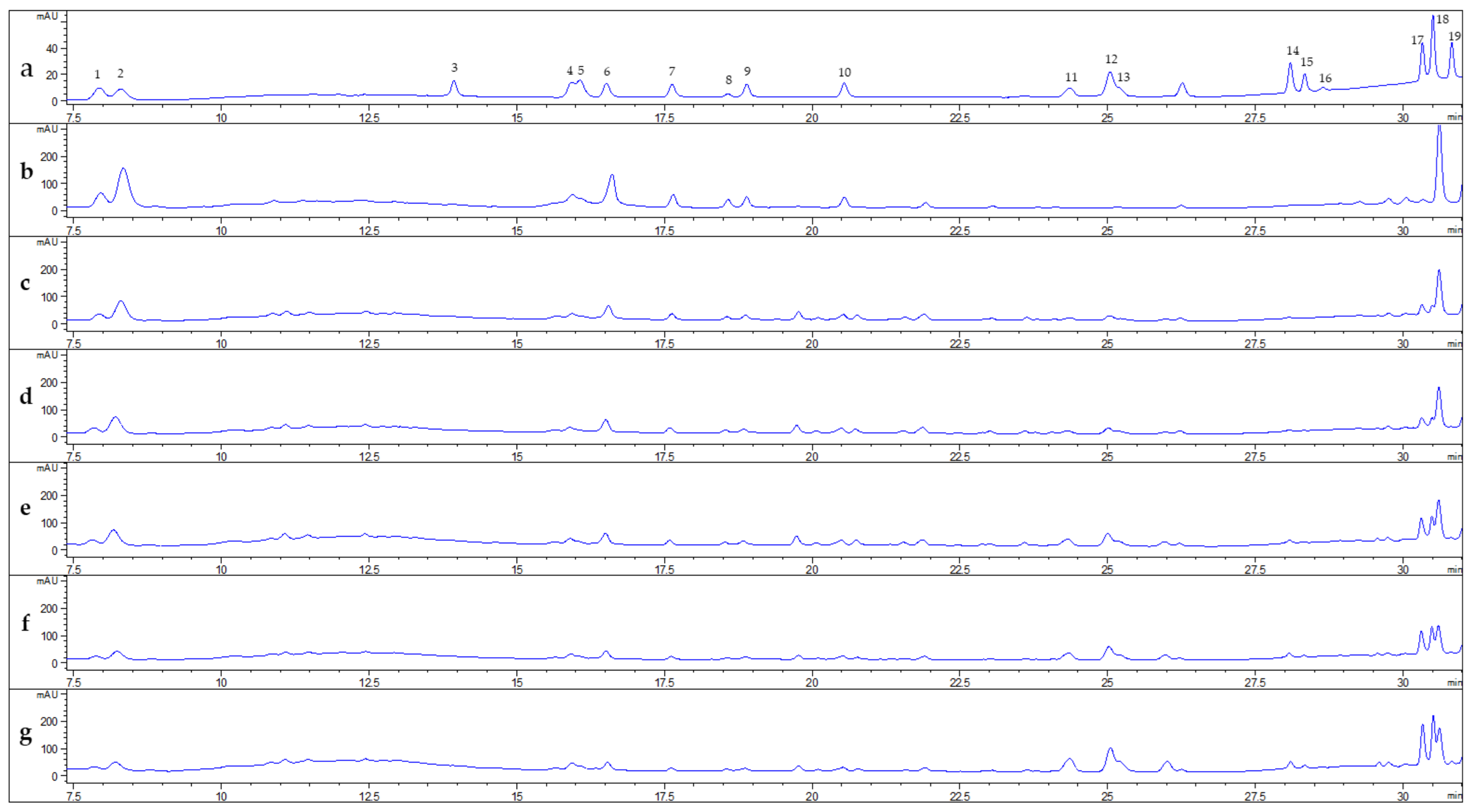
2.6. Ginsenoside Analysis

HPLC was equipped with UV detector and a C18 column (Kinetex ID 2.6 μm, 50 mm × 4.6 mm) with H2O (solvent A) and acetonitrile (solvent B) under the following gradient program: 0–7 min, 19% B; 7–11 min, 29% B; 11–14 min, 29% B; 14–25 min, 40% B; 25–28 min, 56% B; 28–30 min, 70% B; 30–31.5 min, 90% B; 31.5–34 min, 90% B; 34–34.5 min, 90% B; 34.5–40 min, 90% B. The flow rate was set at 0.6 mL/min and UV detection wavelength was set at 203 nm [29].

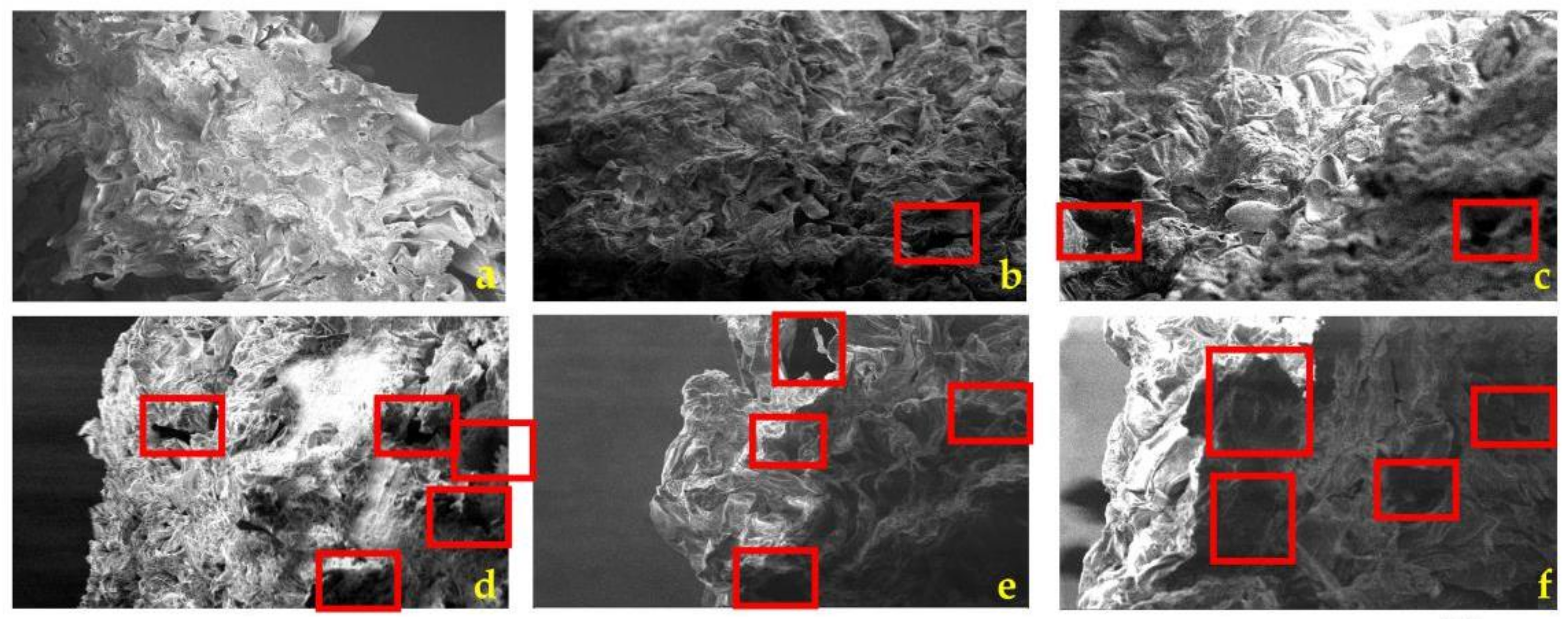
2.7. Scanning Electron Microscope

A scanning electron microscope (SEM) (LEO SUPRA 55; Carl Zeiss AG, Jena, Germany) was applied to view the cross-section microstructure of puffed CRMG. Ten pieces of each sample, after being cut, were pasted on double-sided tapes, which also were fixed on an aluminum specimen holder. Then, the test was examined at 2 kV and magnified at 1 k X.

2.8. Analysis of Total Phenolics

According to Folin–Ciocalteu colorimetric method, total phenolic content (TPC) was detected with slight modification [30]. 30 μL of the extract solution was added to 150 μL of 10% 2N Folin–Ciocalteu reagent. After shaking thoroughly and kept for 5 min, 160 μL of 7.5% (w/v) Na2CO3 solution was added. Then the mixture was placed in the dark for 30 minutes. The absorbance was determined at 715 nm. The gallic acid was used as a standard to calculate TPC from calibration curve.

2.9. Analysis of Total Flavonoids

According to the aluminum chloride colorimetric method, total flavonoids content (TFC) was detected with slight modifications [31]. 30 μL of the extract was added into 110 μL of distilled water, and 8 μL of 5% NaNO2 was added subsequently. After 5 min, 8 μL of 10% AlCl3 was added and left for 6 min, before 50 μL of 1 M NaOH and 70 μL of distilled water was added. The absorbance was measured at 510 nm. The rutin was used as a standard, and TPC was calculated from calibration curve.

2.10. DPPH (2,2-Diphenyl-1-Picrylhydrazyl) Radical Scavenging Activity

According to the method of Su et al. [32], DPPH was detected with slight modification. A 100 μL of the sample was added to 900 μL of 0.2 mM ethanolic solution of DPPH. The DPPH solution was mixed well and kept in the dark for 30 min. The absorbance was adjusted at 517 nm. Gallic acid was used as standard to calculate DPPH from a calibration curve. The DPPH inhibition rate (%) was calculated as follow:
Inhibition (%) = [(Ablank − Asample)/Ablank] × 100
where: Ablank was the absorbance of the control, and Asample was the absorbance of the sample solution.

2.11. Reducing Power Test

Reducing power assay (RPA) was carried out according to Bhalodia et al. with a modification [33]. 1 mL of the extract solution was added to 2.5 mL of 0.2 mM phosphate buffer (pH 6.6), followed by an addition of 2.5 mL of 1% potassium ferricyanide. The solution was incubated in a water bath at 50 °C for 20 min, the mixture was added to 2.5 mL of 10% trichloroacetic acid, followed by centrifugation at 3000 rpm for 10 min. 0.5 mL of 0.1% FeCl3 was added to 2.5 mL of supernatant and 2.5 mL of distilled water. The absorbance was adjusted at 700 nm. The RPA was calculated from calibration curve with the gallic acid as a standard.

2.12. Cytotoxic Effect of Puffed CRMG Extracts on HaCaT Cells

3-(4,5-dimethylthiazol-2-yl-2,5-diphenyl tetrazolium bromide) (MTT) was used to determine cytotoxic effect of puffed CRMG extracts [34]. The HaCaT cells were seeded in 96-well plates (Nest Inc., Corning, NY, USA) at 5 × 10⁴ cells/well, and incubated at 37 °C in the humidified atmosphere of 5% CO2 for 24 h. Subsequently, the cells were washed with PBS, then treated with all samples with various concentrations (50, 100, and 200 μg/mL) in serum-free media and incubated for 24 h. The serum-free medium without sample was used as a control. Then, cells were added with 20 μL of MTT (5 mg/mL, PBS, Life Technologies, Eugene, OR, USA) for four hours at 37 °C. The insoluble formazan was dissolved by 100 μL of DMSO addition to each well. The absorbance was measured at 570 nm with an Enzyme-Linked Immunosorbent Assay (ELISA) microplate reader (Bio-Tek, Instruments, Inc., Winooski, VT, USA).

2.13. Effect of H2O2 on Cell Viability of HaCaT Cells

The required concentration of H2O2 to reduce the cell viability of HaCaT cells by 50% was selected using the MTT assay, the cells were treated with different concentrations of H2O2 (100, 200, 300, 400 and 500 μmol/L) in serum-free media at 37 °C for 24 h. The serum-free medium without H2O2 was used as a control. After the incubation, the cells were washed twice with PBS, and treated with 20 μL of MTT for 4 h at 37 °C, then the supernatant was replaced by 100 μL of DMSO in each well. The absorbance was measured at 570 nm.

2.14. Effect of Puffed CRMG Extracts on ROS Production in HaCaT Cells under Oxidative Stress

The intracellular ROS was determined by dichloro-dihydro-fluorescein diacetate (DCFH-DA) reagent, as described by Zhang et al. [35], with slight modification. The HaCaT cells (5 × 10⁴ cells/well) were seeded in a 96-well plate and incubated for 24 h at 37 °C and 5% CO2. After incubation, the cells were washed with excess PBS and treated with various sample concentrations in serum-free media (at 50, 100, and 200 μg/mL) for 24 h. Vitamin C was used as a positive control. To assess antioxidant activity, the cells were then washed with an excess of PBS and pre-treated with 20 μM of DCFH-DA in PBS at 37 °C for 30 min. After washing with PBS, the cells were added to H2O2 (500 μmol/L) in serum-free media prior to incubation at 37 °C for two hours. Finally, the cells were washed twice with PBS. The ROS levels were measured with a multi-model plate reader at 485 nm for excitation and 528 nm for emission.

2.15. Statistical Analysis

Data analysis was performed by the statistical package SPSS 20.0. The data were shown as the mean ± standard deviation (SD). The mean values were compared by one-way analysis of variance (ANOVA) with Dunnett’s test. All experiments were repeated at least three times independently, and differences were considered significant at * p < 0.05 or ** p < 0.01. Heatmap which containing TFC, TPC, ginsenosides content, DPPH, and RPA, was performed using GraphPad Prism 8.0 (GraphPad Software, San Diego, CA, USA).

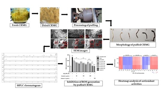
3. Results and Discussion

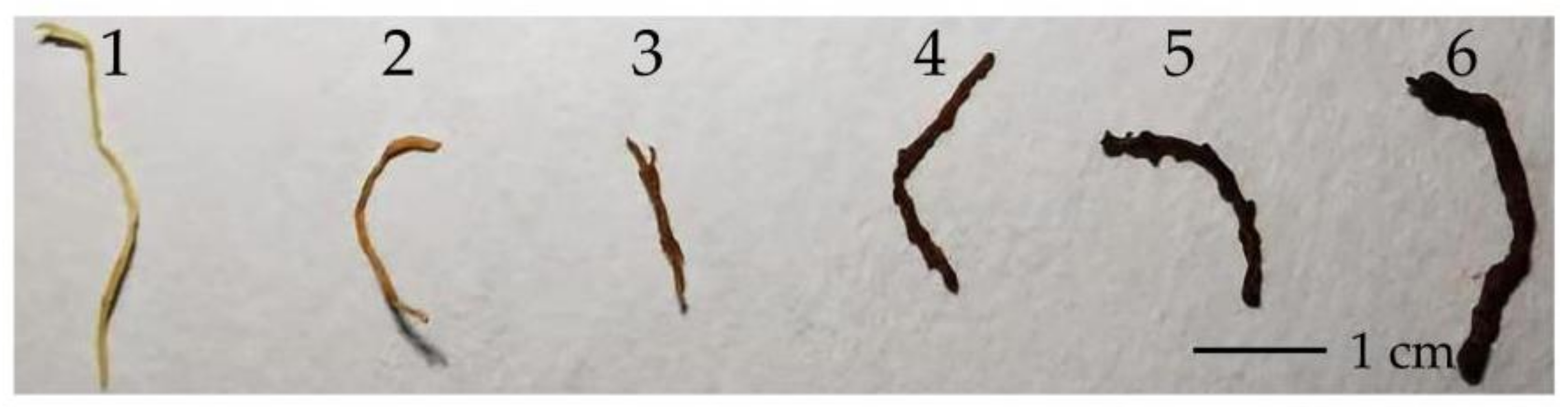
3.1. Morphological Characteristics of Puffed CRMG

The color changes of CRMG displayed in Figure 1 show a gradual darkening in color with the increase in temperature and time. The reason for color changes have been already determined to be caused by the Maillard reaction and caramelization [22]. The puffed CRMG tinct became dark as the surface became frayed and coarse, but the roots became enlarged in volume with increased pressure. Moreover, CRMG would burn when the pressure exceeded 588.4 kPa due to their low moisture contents and environmental temperature. As Figure 2 shows, there were some air pockets produced in cross-section since 490.3 kPa, particularly. The textural property of porosity was modified thoroughly by high pressure. The significant changes in the micro-structures were observed after the puffing processing was observed.

3.2. Extraction Yield and Crude Ginsenoside Contents

The extraction yield (360.77–409.03 mg/g DW) and crude ginseng saponin contents (63.33–88.57 mg/g DW) of puffed CRMG are shown in Table 1. Both the yield extraction and crude ginsenoside contents decreased at first, and then arose with increased pressure. The puffed CRMG showed the most extraction yield and crude ginsenosides at 588.4 kPa. Additionally, the trends of them were similar.

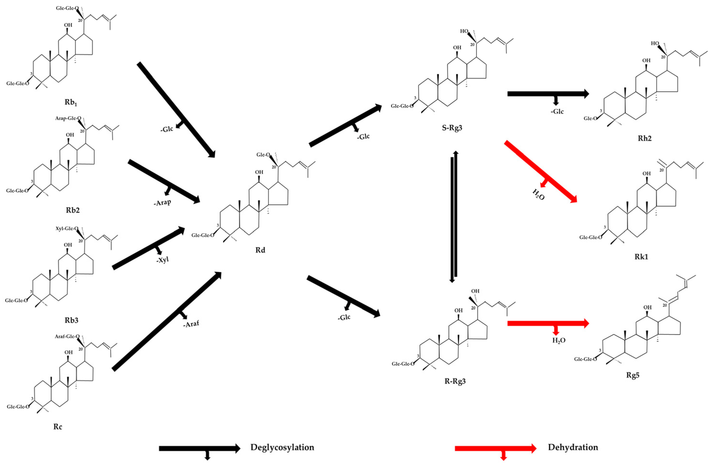
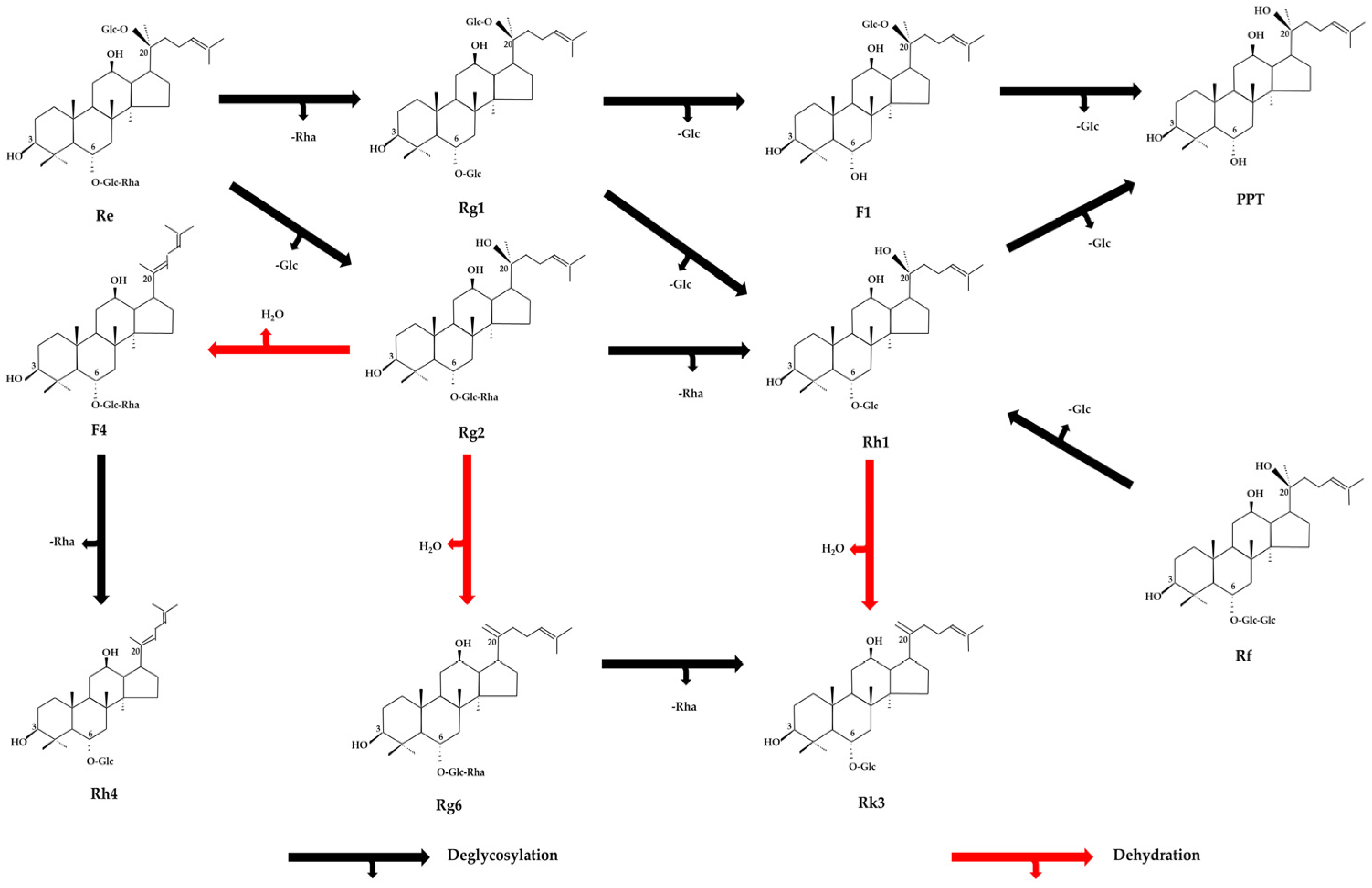
3.3. Ginsenosides Analysis

The HPLC chromatograms of puffed CRMG are shown in Figure 3. The ginsenoside contents of puffed CRMG, divided into PPD-type and PPT-type, were shown in Table 2 and Table 3, respectively. Figure 3 presents 11 major ginsenosides detected in dried CRMG. The amount of major ginsenosides (Rb1, Rb2, Rb3, Rc, and Rd) showed a sharp decrease in the peak at first, and then was delayed with time. Ginsenosides 20 (S)-Rg3, 20 (R)-Rg3, Rk1, and Rg5 were yielded after the puffing process, and Rh2 was produced from 490.3 kPa. The ginsenoside Rd was generated from the ginseng saponins Rb1, Rb2, Rb3 and Rc by removing sugar at C-20. Meanwhile, it transformed into ginsenoside 20 (S, R)-Rg3 by losing one GLC-residue at C-20 under a high-temperature and pressure. Moreover, the conversions were also accelerated by dehydration, from 20 (S)-Rg3 and 20 (R)-Rg3 to ginsenosides Rk1 and Rg5, respectively. In addition, ginsenoside 20 (S)-Rg3 can be transformed to ginsenoside Rh2 by losing one glucose at C-3.
As for the conversion of PPT-type ginsenosides (Figure 4), it showed a similar trend with PPD-type ginsenosides (Figure 5). The amounts of the major ginsenosides Rg1 and Re decreased, while the quantities of minor ginsenosides Rg6, F4, Rk3 were produced with increasing pressure. The amounts of the ginsenosides Rf, Rg2, and Rh1 did not show significant changes. Some transformations from major ginsenosides to minor ginsenosides occurred via the deglycosylation at C-6 or C-20. For example, ginsenosides Rh1, PPT, Rk3 were produced by Rg2, Rh1, and Rg6, respectively. Furthermore, there were other conversions through dehydration at C-20, including ginsenoside Rg2 to Rg6 and F4, and ginsenoside Rh1 to Rk3. Though Shin et al. [25] reported that puffed raw white, red, and black ginseng effectively yielded some minor PPD-ginseng saponins, such as F2, Rg3, Rk1, and Rg5, and Kim et al. [26] demonstrated that the minor PPD-ginsenosides, such as Rg3, F2, and Rh2, in American and Canadian ginseng were improved by puffing, the minor PPT-ginsenosides, such as F4, Rg6 and Rk3, were produced by puffing of CRMG in our study.
According to the present trends in the transformation of ginsenosides, the total ginsenosides are divided into total major ginsenosides, and total minor ginsenosides, including total PPD-minor ginsenosides and total PPT-minor ginsenosides (Table 1). The results showed PPD-type minor ginsenosides ranged from 0 to 1.18 mg/g and PPT-type minor ginsenosides ranged from 0 to 2.01 mg/g. Furthermore, total minor ginsenosides ranged from 0 to 3.18 mg/g, total major ginsenosides ranged from 3.11 to 10.36 mg/g, and total ginsenosides ranged from 5.46 to 10.36 mg/g. The content of minor ginsenosides arose by the increased puffing temperature and pressure, while the major ginsenosides decreased significantly by puffing. These results were consistent with previous studies [36]. In addition, the amounts of total ginsenosides descended from 196.1 kPa, and then rose at 588.4 kPa, according to Table 1. However, dried CRMG could not be used in a higher pressure process due to the possible production of burned samples of a porous structure, since the samples had low moisture and a slender shape.

3.4. TPC, TFC and Antioxidant Activities

The activities of TPC, TFC, and antioxidant activities, including DPPH and RPA of puffed CRMG, are displayed in Table 4. TPC ranged from 3.85 to 11.81 mg GAE/g DW. TFC ranged from 3.40 to 6.43 mg RE/g DW. The scavenging of DPPH ranged from 0.18 to 1.69 mg GAE/g DW. RPA ranged from 1.37 to 5.22 mg GAE/g DW. The puffed CRMG at 588.4 kPa had the most phenolics and TFC contents. However, puffed CRMG at 490.3 kPa showed the best antioxidant in DPPH scavenging and RPA, although some data of 490.3 kPa and 588.4 kPa were closed. We infer that the TPC and TFC were affected by the breakage of molecular bonds and the loosened texture caused by the high temperature and pressure [26]. Based on SEM, we found the puffing method is advantageous on more surfaces in favor of extraction. Antioxidant activities were enhanced by higher amounts of phenolics, including flavonoids. This consequence was in accordance with previous findings [26,37].

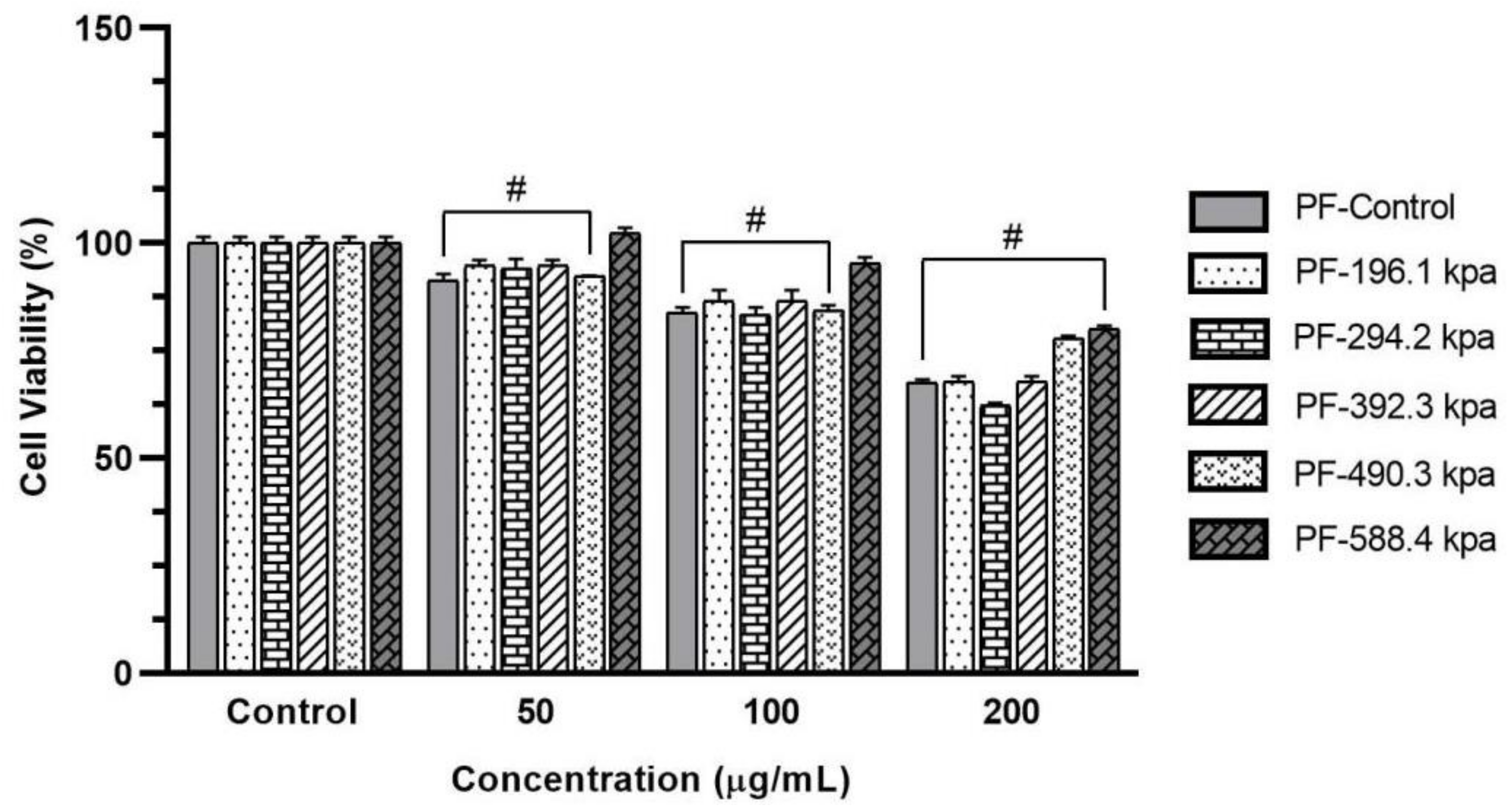
3.5. Effect of Puffed CRMG Extracts on Cell Viability of HaCaT Cells

We also studied the puffed CRMG for the cytotoxicity effect on HaCaT cells using the MTT assay. Each sample was evaluated with various concentrations of 50, 100 and 200 μg/mL. As shown in Figure 6, puffed CRMG of 490.3 kPa and 588.4 kPa displayed low toxicity on HaCaT cells at 200 μg/mL compared to other samples. In this study, we have demonstrated, for the first time, that the pressuring (puffing) technique could have an effect to transform some compounds of the sample, which would result in low cytotoxicity in normal cells. This is essential because safety and toxicity effects are significant in drugs, supplements, and cosmetics developments.
In this study, we have demonstrated the pressuring (puffing) technique could have an effect to transform or increase some compounds of the sample, which would result in low cytotoxicity effects in normal cells. Based on HPLC analysis, the minor ginsenosides’ (20 (S, R)-Rg3, Rg5, Rk1, Rh1, Rh2, Rg6, F4 and Rk3) (Table 2 and Table 3) contents, as well as the total phenolic and flavonoid contents, were increased (Table 4). Previous studies proved that 20 (S, R)-Rg3 [38], Rg5, Rk1 [39], Rh1, Rh2, F4 [40] could promote the proliferation of HaCaT cells, MMP-inhibiting, and promote skin-wound healing effects. Furthermore, two studies showed that flavonoid and phenolic compounds increased HaCaT cells viability [41,42]. With those mechanism, it is reasonable to reduce its cytotoxicity effects due to the increasing content of compounds which have biological effects. However, we suppose the toxicity could be related to TPC, TFC, and ginsenoside contents. This study concluded that increasing the content of phenolics, flavonoids and minor ginsenosides may decrease the cytotoxicity level of the puffed CRMG. The mechanism of this phenomenon is still unclear, and further study is required.

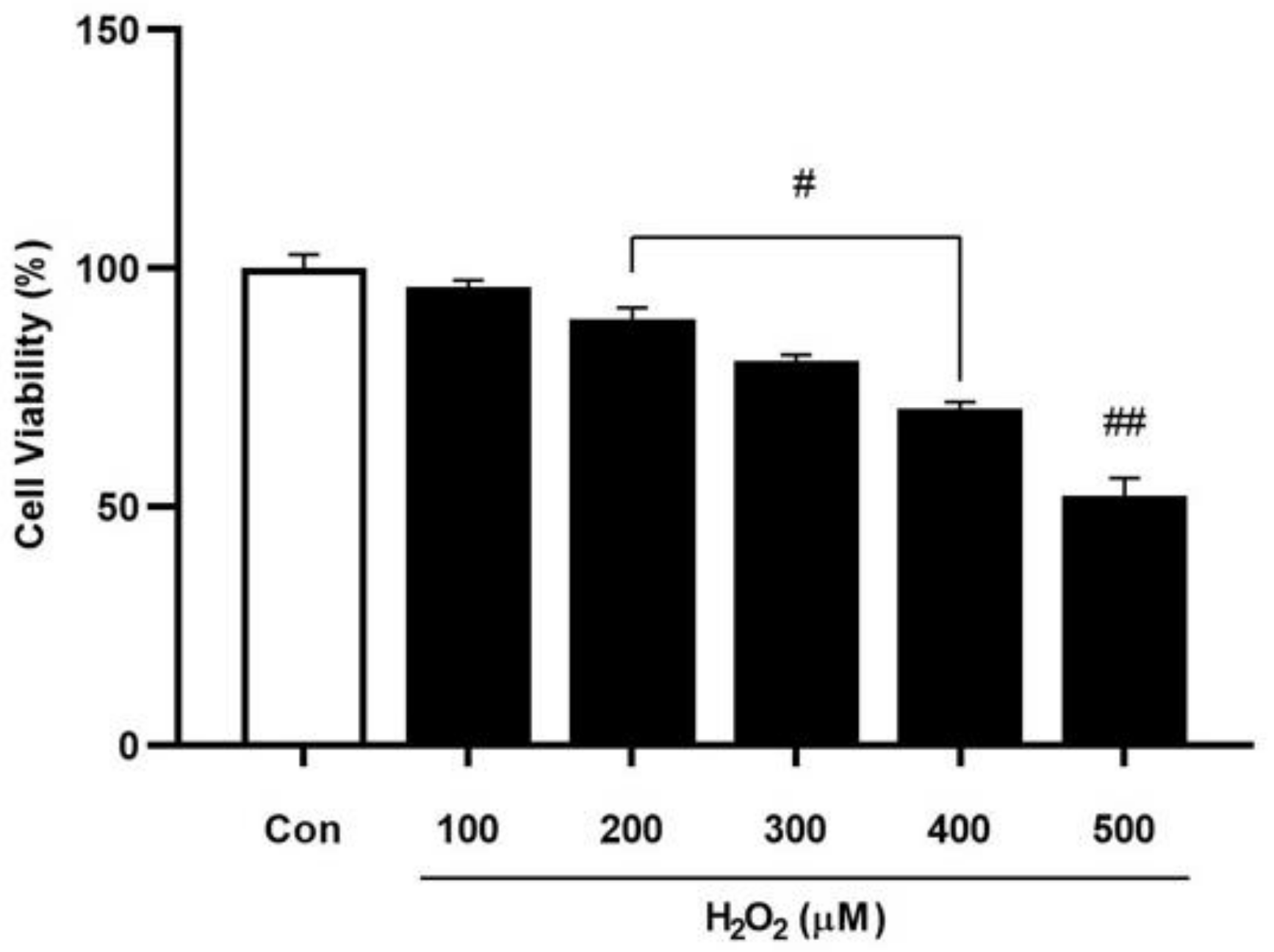
3.6. Effect of H2O2-Induced Oxidative Stress on Cell Viability of HaCaT Cells

The high level of H2O2 in the cell can induce excessive ROS production and correspond to imbalanced levels of oxidants and antioxidants in cells, leading to cell damage, lipid peroxidation, and apoptosis induction that can cause various kinds of diseases [43,44]. To establish the H2O2-induced oxidative stress model, HaCaT cells were treated with different concentrations of H2O2 (100, 200, 300, 400 and 500 μmol), as described previously. The results showed 500 μmol of H2O2 decreased the cell viability by 50%, compared to the untreated group (Figure 7); therefore, we have used this determined concentration for further studies. For inducing oxidative stress in cells, the concentration of H2O2 between 100–500 μmol/L was mainly used and our results, in accordance with previous studies, showed that H2O2 at 500 μmol/L reduced cell viability in the range between 50 and 60%, compared to the control group [45,46].

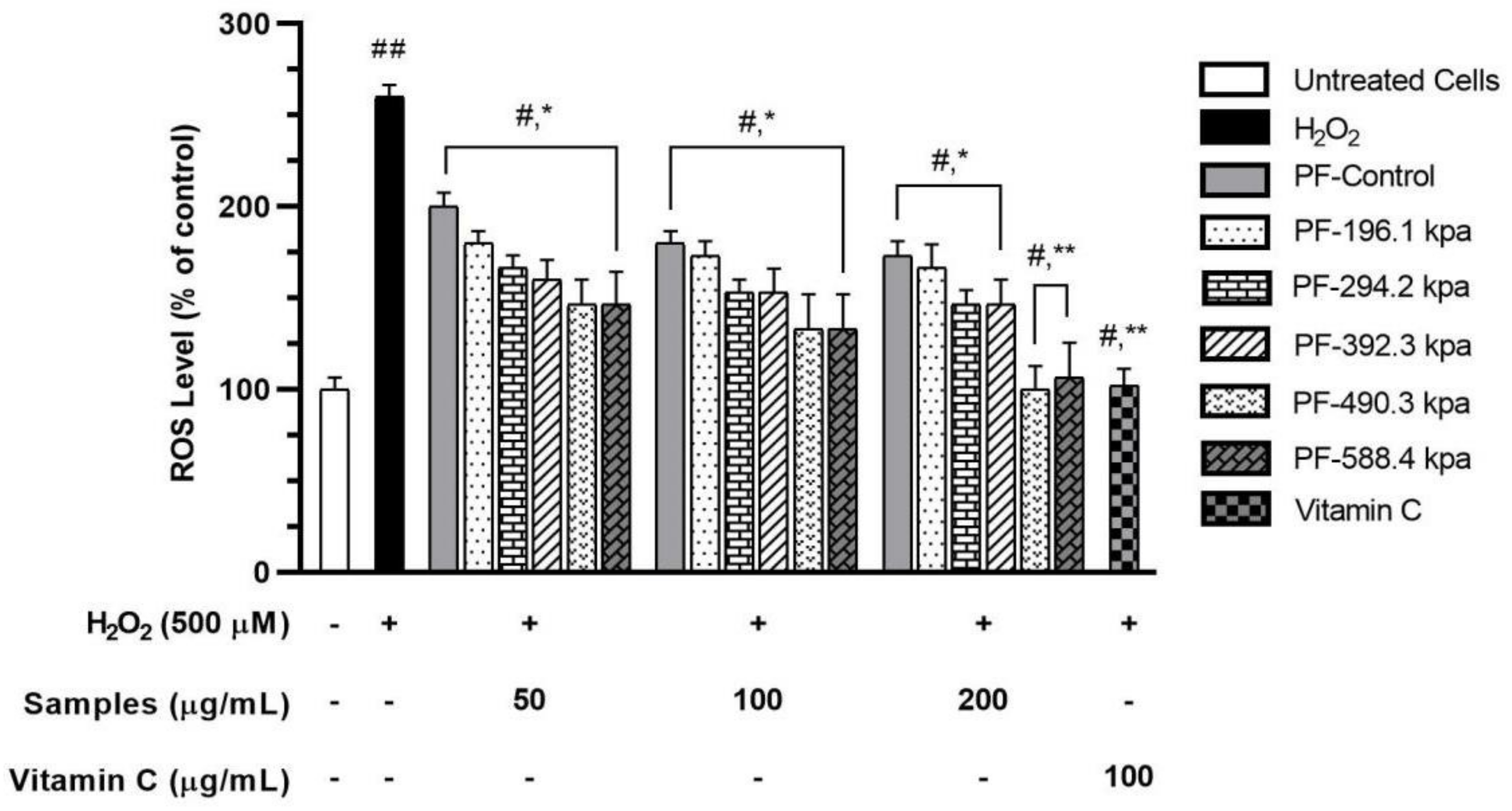
3.7. Effect of Puffed CRMG Samples on ROS Production in H2O2-Induced Oxidative Stress-Treated HaCaT Cells

To ascertain the antioxidant activity of the puffed CRMG in the cellular model, the measurement of ROS levels in H2O2-induced HaCaT cells using the DCFH-DA assay was investigated. The H2O2 was commonly used to induce intracellular ROS levels. The Fenton’s reaction between H2O2 and Fe2+ ions generates the highly reactive OH radicals and is thought to be the main mechanism for oxidative damage [47]. According to the cytotoxicity results, various concentrations were selected for examination. The mean value of the ROS level was measured in 500 μmol of H2O2-treated HaCaT cells, increased to 260% compared to the control group. The trend of decreased cell viability after the H2O2 exposure is shown in Figure 8. Vitamin C was used as a positive control, and, at a concentration of 100 μg/mL, significantly reduced ROS levels.
We found that all the puffed CRMG samples could act against the H2O2-induced oxidative stress of HaCaT cells in a dose-dependent manner. However, 200 μg/mL of the puffed CRMG in 490.3 kPa and 588.4 kPa were able to reduce the ROS level by an average of 60%, which is significant when compared with the H2O2 treatment group (Figure 8). Additionally, the results showed that the puffed CRMG samples have a similar capacity of reducing the ROS level as the positive control (Vitamin C at a concentration 100 μg/mL).
Moreover, these results are in accordance with the in vitro antioxidant capacity (DPPH and RPA) data in the present study. This proves even further that the puffing process induced significant changes in the physical and chemical properties, enhancing the antioxidant properties of puffed CRMG.
These findings agree with previous studies in that the antioxidant effects, TPC and TFC of turmeric, and American and Canadian ginseng were increased by puffing [26,37]. Furthermore, in the case of red and black ginseng, which are products of P. ginseng, by increasing the temperature and repeating the time, it was shown that minor ginsenosides and antioxidant activity could be elevated [48]. Likewise, in this study, minor ginsenosides, TPC, TFC and antioxidant activities of puffed CRMG increased during puffing, with the increasing temperature and holding time.
For some insight, it is well known that hydrophobicity and deglycosylation of the bioactive compounds can enhance the antioxidant properties by counteracting oxidative stress, especially ROS which is located in the inner mitochondrial membrane of cell [49]. Huang et al. [50] reported that the puffing process would expose the non-polar groups buried inside the protein and increase the surface of hydrophobicity. This phenomenon was not elucidated and requires further investigation.

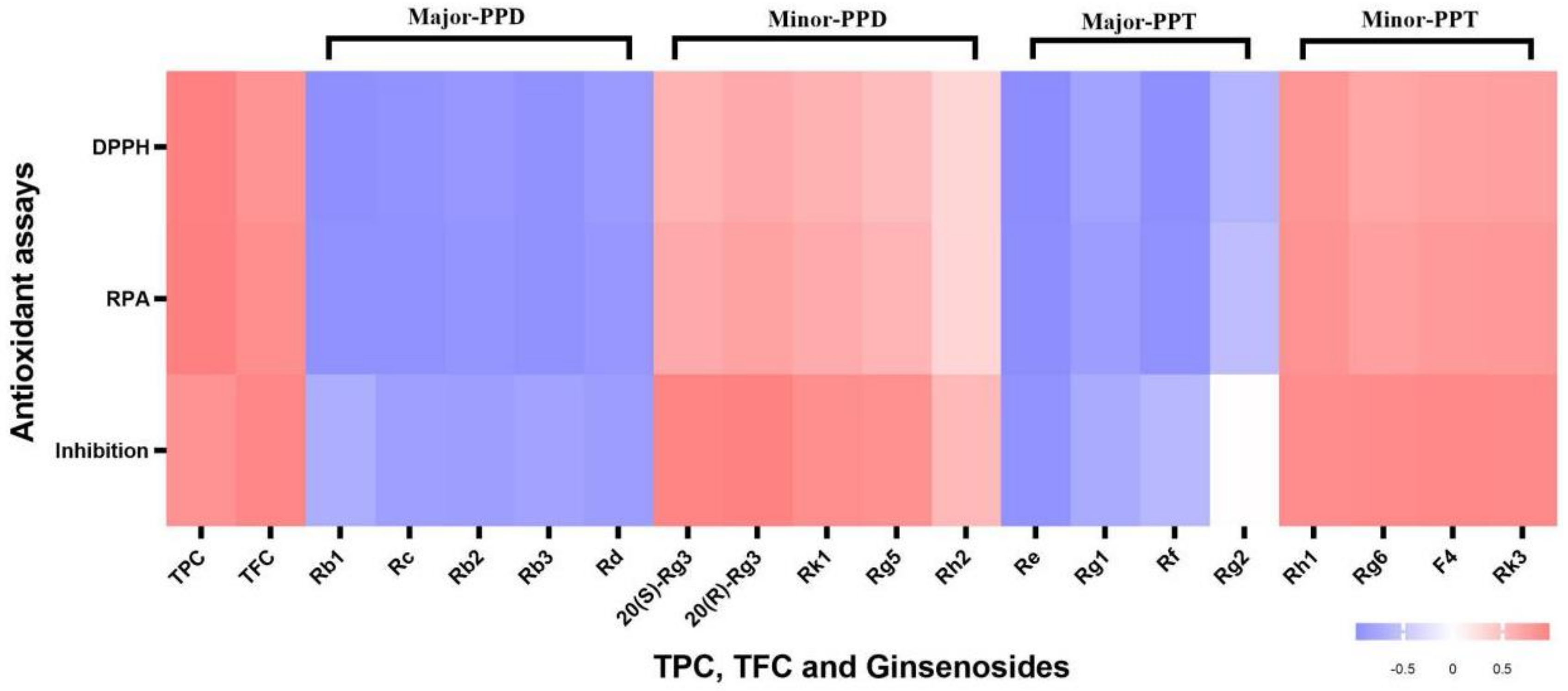
3.8. Correlation Map among Contents of Ginsenosides, TPC, TFC and Antioxidant Assays

There are many kinds of antioxidant compounds, such as polyphenols, flavonoids, and ginsenosides in ginseng roots. Some natural antioxidant substances also have capacities for the inhibition of ROS production [51]. Therefore, it is necessary to use a heatmap to reveal the correlation among the relative abundance of different types of ginsenosides, TPC, and TFC, with antioxidants assays, including DPPH scavenging, RPA, and the inhibition of ROS generation. The Pearson’s correlation coefficient values ranged from −0.998 to 0.985. Obviously, these antioxidant assays showed similar trends of contribution to antioxidant capabilities for those substances. In Figure 9, TPC and TFC had a positive correlation with antioxidant assays. It was demonstrated that more TPC and TFC had a positive correlation with antioxidant capacity, as already reported by Benabderrahim et al. [52]. It was also displayed that Minor-PPD and Minor-PPT had a positive correlation, whereas major-PPD and major-PPT had a negative or no correlation. Minor ginsenosides, such as Rg5 and Rk1 were already proved to have antioxidant capabilities and caused the reduction of ROS generation in our lab [39]. Thus, we inferred that more contents of minor ginsenosides also contributed more to antioxidant capacities. However, much of the literature [53,54] revealed that some major ginseng saponins, such as Rb1, Rb2 and Rc, have also showed antioxidant potentials. In this research, the contents of these major ginseng saponins exhibited a negative correlation with antioxidant assays because the antioxidant capacities of puffed CRMG became enhanced with the increase in pressure, while the contents of major ginsenosides sharply decreased. Furthermore, major ginsenosides transformed into minor ginsenosides during the processing of the ginseng in general, leading to a continuous change in their contents. Choi et al. [55] also investigated that ginsenosides Rb1, Rb2 and Rc showed a negative correlation with antioxidant assays after different pressure treatments in ginseng berries.

4. Conclusions

The various reports showed the minor ginsenosides exhibit more pharmacological activities than major ginsenosides. In that case, though various processing (physical, chemical, and biological) methods are available to convert major ginsenosides into minor ginsenosides, this puffing process is a single step method and, therefore, a time-saving procedure, compared to chemical and biological methods. Overall, for the first time, the present study shows that puffing is a promising method for the conversion of the ginsenosides of CRMG. Through the puffing process, phenolics, flavonoids, and minor ginsenosides contents were increased, and the antioxidant properties, such as DPPH inhibition and reducing the power of puffed CRMG, were significantly enhanced. Minor ginsenosides, such as Rg6, F4, and Rk3, were firstly reported to be produced from puffing CRMG. Moreover, the puffed CRMG of 490.3 kPa and 588.4 kPa displayed less toxicity compared to other samples in HaCaT cells. In addition, the inhibition of ROS production was increased in 490.3 kPa and 588.4 kPa puffed CRMG in H2O2-induced oxidative stress of HaCaT cells. In a word, puffed CRMG enhance their safety and efficacy as bioactive materials for health care products and cosmeceuticals.

Author Contributions

Conceptualization, D.C.Y., S.C.K. and J.Y.P.; methodology, J.Y.P., Z.M.R., Y.H. (Yue Huo) and D.W.L.; software, J.Y.P., Z.M.R., J.F.L. and Y.H. (Yaxi Han); validation, D.C.Y., S.C.K., X.H.Z. and D.W.L.; formal analysis, J.Y.P. and Z.M.R.; investigation, X.H.Z., J.C.A. and Y.H. (Yaxi Han); resources, S.C.K. and D.C.Y.; data curation, J.Y.P., Z.M.R. and Y.H. (Yue Huo); writing—original draft preparation, J.Y.P. and Z.M.R.; writing—review and editing, F.J.X., J.Y.P., G.-Y.K., R.M. and Z.M.R.; visualization, J.Y.P., Y.H. (Yaxi Han) and Z.M.R.; supervision, D.C.Y. and S.C.K.; project administration, D.C.Y., S.C.K. and J.Y.P.; funding acquisition, D.C.Y., S.C.K., R.M. and J.C.A. All authors have read and agreed to the published version of the manuscript.

Funding

This work was supported by the Korea Institute of Planning and Evaluation for Technology in Food, Agriculture and Forestry (IPET) through the Agri-Food Export Business Model Development Program, funded by Ministry of Agriculture, Food and Rural Affairs (MAFRA) (Project No: 320104-03), and the study was also supported by a grant from The National Research Foundation of Korea (NRF) (Project No: NRF-2020R1I1A1A01070867).

Institutional Review Board Statement

Not applicable.

Informed Consent Statement

Not applicable.

Data Availability Statement

Not applicable.

Conflicts of Interest

The authors declare that there are no financial interests or personal relationships regarding the publication of this paper.

References

  1. Kim, D.H. Chemical Diversity of Panax ginseng, Panax quinquifolium, and Panax notoginseng. J. Ginseng Res. 2012, 36, 1–15. [Google Scholar] [CrossRef] [PubMed] [Green Version]
  2. Ma, G.-D.; Chiu, C.-H.; Hsu, Y.-J.; Hou, C.-W.; Chen, Y.-M.; Huang, C.-C. Changbai Mountain ginseng (Panax ginseng CA Mey) extract supplementation improves exercise performance and energy utilization and decreases fatigue-associated parameters in mice. Molecules 2017, 22, 237. [Google Scholar] [CrossRef] [PubMed]
  3. Guo, N.; Yang, Y.; Yang, X.; Guan, Y.; Yang, J.; Quan, J.; Yan, H.; Hou, W.; Zhang, G. Growth age of mountain cultivated ginseng affects its chemical composition. Ind. Crops Prod. 2021, 167, 113531. [Google Scholar] [CrossRef]
  4. Murthy, H.N.; Dandin, V.S.; Park, S.-Y.; Paek, K.-Y. Quality, safety and efficacy profiling of ginseng adventitious roots produced in vitro. Appl. Microbiol. Biotechnol. 2018, 102, 7309–7317. [Google Scholar] [CrossRef] [PubMed]
  5. Paek, K.-Y.; Murthy, H.N.; Hahn, E.-J.; Zhong, J.-J. Large scale culture of ginseng adventitious roots for production of ginsenosides. Biotechnol. China I 2009, 113, 151–176. [Google Scholar]
  6. Jiménez-Pérez, Z.E.; Kim, Y.-J.; Castro-Aceituno, V.; Mathiyalagan, R.; Markus, J.; Ahn, S.; Simu, S.Y.; Yang, D.-C. Novel application of cultured roots of mountain ginseng (Panax ginseng meyer) and ginsenoside re as safe antimelanogenic cosmeceutical components. Afr. J. Tradit. Complementary Altern. Med. 2017, 14, 209–218. [Google Scholar] [CrossRef] [Green Version]
  7. Piao, X.M.; Huo, Y.; Kang, J.P.; Mathiyalagan, R.; Zhang, H.; Yang, D.U.; Kim, M.; Yang, D.C.; Kang, S.C.; Wang, Y.P. Diversity of ginsenoside profiles produced by various processing technologies. Molecules 2020, 25, 4390. [Google Scholar] [CrossRef]
  8. Yang, W.-Z.; Hu, Y.; Wu, W.-Y.; Ye, M.; Guo, D.-A. Saponins in the genus Panax L. (Araliaceae): A systematic review of their chemical diversity. Phytochemistry 2014, 106, 7–24. [Google Scholar] [CrossRef]
  9. Yang, W.; Qiao, X.; Li, K.; Fan, J.; Bo, T.; Guo, D.-a.; Ye, M. Identification and differentiation of Panax ginseng, Panax quinquefolium, and Panax notoginseng by monitoring multiple diagnostic chemical markers. Acta Pharm. Sin. B 2016, 6, 568–575. [Google Scholar] [CrossRef] [Green Version]
  10. Du, G.-J.; Dai, Q.; Williams, S.; Wang, C.-Z.; Yuan, C.-S. Synthesis of protopanaxadiol derivatives and evaluation of their anticancer activities. Anti-Cancer Drugs 2011, 22, 35. [Google Scholar] [CrossRef] [Green Version]
  11. Lee, J.-O.; Hwang, S.-H.; Shen, T.; Kim, J.H.; You, L.; Hu, W.; Cho, J.Y. Enhancement of skin barrier and hydration-related molecules by protopanaxatriol in human keratinocytes. J. Ginseng Res. 2021, 45, 354–360. [Google Scholar] [CrossRef] [PubMed]
  12. Peng, D.; Wang, H.; Qu, C.; Xie, L.; Wicks, S.M.; Xie, J. Ginsenoside Re: Its chemistry, metabolism and pharmacokinetics. Chin. Med. 2012, 7, 1–6. [Google Scholar] [CrossRef] [Green Version]
  13. Nakhjavani, M.; Palethorpe, H.M.; Tomita, J.; Smith, E.; Price, T.J.; Yool, A.J.; Pei, J.V.; Townsend, A.R.; Hardingham, J.E. Stereoselective Anti-Cancer Activities of Ginsenoside Rg3 on Triple Negative Breast Cancer Cell Models. Pharmaceuticals 2019, 12, 117. [Google Scholar] [CrossRef] [Green Version]
  14. Liu, Z.; Liu, T.; Li, W.; Li, J.; Wang, C.; Zhang, K. Insights into the antitumor mechanism of ginsenosides Rg3. Mol. Biol. Rep. 2021, 48, 2639–2652. [Google Scholar] [CrossRef]
  15. Jin, J.P.; An, J.; Lee, J.D.; Kim, H.Y.; Kim, K.B. Effects of anti-wrinkle and skin-whitening fermented black ginseng on human subjects and underlying mechanism of action. J. Toxicol. Environ. Health Part A 2020, 83, 1–15. [Google Scholar]
  16. Song, Y.N.; Hong, H.G.; Son, J.S.; Kwon, Y.O.; Yoon, M.H. Investigation of Ginsenosides and Antioxidant Activities in the Roots, Leaves, and Stems of Hydroponic-Cultured Ginseng (Panax ginseng Meyer). Prev. Nutr. Food Sci. 2019, 24, 283–292. [Google Scholar] [CrossRef]
  17. Liu, Y.; Zhang, J.-W.; Li, W.; Ma, H.; Sun, J.; Deng, M.-C.; Yang, L. Ginsenoside metabolites, rather than naturally occurring ginsenosides, lead to inhibition of human cytochrome P450 enzymes. Toxicol. Sci. 2006, 91, 356–364. [Google Scholar] [CrossRef] [PubMed] [Green Version]
  18. Sun, B.S.; Gu, L.J.; Fang, Z.M.; Wang, C.Y.; Wang, Z.; Lee, M.R.; Zheng, L.; Li, J.J.; Sung, C.K. Simultaneous quantification of 19 ginsenosides in black ginseng developed from Panax ginseng by HPLC-ELSD. J. Pharm. Biomed. Anal. 2009, 50, 15–22. [Google Scholar] [CrossRef]
  19. Jin, Y.; Kim, Y.-J.; Jeon, J.-N.; Wang, C.; Min, J.-W.; Noh, H.-Y.; Yang, D.-C. Effect of white, red and black ginseng on physicochemical properties and ginsenosides. Plant Foods Hum. Nutr. 2015, 70, 141–145. [Google Scholar] [CrossRef] [PubMed]
  20. Subramani, D.; Tamilselvan, S.; Murugesan, M.; Shivaswamy, M. Optimization of Sand Puffing Characteristics of Quinoa using Response Surface Methodology. Curr. Res. Nutr. Food Sci. J. 2020, 8, 496–503. [Google Scholar] [CrossRef]
  21. Farh, M.E.-A.; Kim, Y.-J.; Abbai, R.; Singh, P.; Jung, K.-H.; Kim, Y.-J.; Yang, D.-C. Pathogenesis strategies and regulation of ginsenosides by two species of Ilyonectria in Panax ginseng: Power of speciation. J. Ginseng Res. 2020, 44, 332–340. [Google Scholar] [CrossRef]
  22. An, Y.E.; Ahn, S.C.; Yang, D.C.; Park, S.J.; Baik, M.Y. Chemical conversion of ginsenosides in puffed red ginseng. LWT—Food Sci. Technol. 2011, 44, 370–374. [Google Scholar] [CrossRef]
  23. Hwang, I.G.; Kim, H.Y.; Joung, E.M.; Woo, K.S.; Jeong, J.H.; Yu, K.W.; Lee, J.; Jeong, H.S. Changes in ginsenosides and antioxidant activity of Korean ginseng (Panax ginseng C.A. Meyer) with Heating Temperature and Pressure. Food Sci. Biotechnol. 2010, 19, 941–949. [Google Scholar] [CrossRef]
  24. Guo, H.; Saravanakumar, K.; Wang, M.-H. Total phenolic, flavonoid contents and free radical scavenging capacity of extracts from tubers of Stachys affinis. Biocatal. Agric. Biotechnol. 2018, 15, 235–239. [Google Scholar] [CrossRef]
  25. Shin, J.-H.; Park, Y.J.; Kim, W.; Kim, D.-O.; Kim, Y.; Hyungjae, L.; Baik, M.-Y. Change of Ginsenoside Profiles in Processed Ginseng by Drying, Steaming, and Puffing. J. Microbiol. Biotechnol. 2019, 29, 222–229. [Google Scholar] [CrossRef] [Green Version]
  26. Kim, M.-S.; Jeon, S.-J.; Youn, S.J.; Lee, H.; Park, Y.-J.; Kim, D.-O.; Kim, B.-Y.; Kim, W.; Baik, M.-Y. Enhancement of minor ginsenosides contents and antioxidant capacity of american and canadian ginsengs (Panax quinquefolius) by puffing. Antioxidants 2019, 8, 527. [Google Scholar] [CrossRef] [PubMed] [Green Version]
  27. Noh, E.-M.; Park, J.; Song, H.-R.; Kim, J.-M.; Lee, M.; Song, H.-K.; Hong, O.-Y.; Whang, P.H.; Han, M.-K.; Kwon, K.-B. Skin aging-dependent activation of the PI3K signaling pathway via downregulation of PTEN increases intracellular ROS in human dermal fibroblasts. Oxidative Med. Cell. Longev. 2016, 2016. [Google Scholar] [CrossRef]
  28. Abid, S.; Kaliraj, L.; Rahimi, S.; Kim, Y.J.; Yang, D.C.; Kang, S.C.; Balusamy, S.R. Synthesis and characterization of glycol chitosan coated selenium nanoparticles acts synergistically to alleviate oxidative stress and increase ginsenoside content in Panax ginseng. Carbohydr. Polym. 2021, 267, 118195. [Google Scholar] [CrossRef] [PubMed]
  29. Park, J.K.; Yang, D.U.; Arunkumar, L.; Han, Y.; Lee, S.J.; Arif, M.H.; Li, J.F.; Huo, Y.; Kang, J.P.; Hoang, V.A. Cumulative Production of Bioactive Rg3, Rg5, Rk1, and CK from Fermented Black Ginseng Using Novel Aspergillus niger KHNT-1 Strain Isolated from Korean Traditional Food. Processes 2021, 9, 227. [Google Scholar] [CrossRef]
  30. Ali, M.B.; Hahn, E.J.; Paek, K.Y. CO2-induced total phenolics in suspension cultures of Panax ginseng C. A. Mayer roots: Role of antioxidants and enzymes. Plant Physiol. Biochem. 2005, 43, 449–457. [Google Scholar] [CrossRef]
  31. Ffda, A.; Ddpf, A.; Nn, A.; Da, B.; Jd, B.; Fgds, A.; Rrc, B.; Ckds, C.; Gmp, A. Influence of high-intensity ultrasound on color, chemical composition and antioxidant properties of araá-boi pulp. Food Chem. 2021, 338, 127747. [Google Scholar]
  32. Su, Y.; Li, L. Structural Characterization and Antioxidant Activity of polysaccharide from four Auriculariales. Carbohydr. Polym. 2019, 229, 115407. [Google Scholar] [CrossRef]
  33. Bhalodia, N.R.; Nariya, P.B.; Acharya, R.N.; Shukla, V.J. In vitro antioxidant activity of hydro alcoholic extract from the fruit pulp of Cassia fistula Linn. AYU 2013, 34, 209–214. [Google Scholar] [CrossRef] [PubMed] [Green Version]
  34. Warinhomhoun, S.; Muangnoi, C.; Buranasudja, V.; Mekboonsonglarp, W.; Rojsitthisak, P.; Likhitwitayawuid, K.; Sritularak, B. Antioxidant Activities and Protective Effects of Dendropachol, a New Bisbibenzyl Compound from Dendrobium pachyglossum, on Hydrogen Peroxide-Induced Oxidative Stress in HaCaT Keratinocytes. Antioxidants 2021, 10, 252. [Google Scholar] [CrossRef]
  35. Tsuda, T.; Horio, F.; Uchida, K.; Aoki, H.; Osawa, T. Dietary cyanidin 3-O-β-D-glucoside-rich purple corn color prevents obesity and ameliorates hyperglycemia in mice. J. Nutr. 2003, 133, 2125–2130. [Google Scholar] [CrossRef] [PubMed]
  36. Yoon, S.R.; Lee, G.D.; Park, J.H.; Lee, I.S.; Kwon, J.H. Ginsenoside composition and antiproliferative activities of explosively puffed ginseng (Panax ginseng CA Meyer). J. Food Sci. 2010, 75, C378–C382. [Google Scholar] [CrossRef]
  37. Choi, Y.; Ban, I.; Lee, H.; Baik, M.-Y.; Kim, W. Puffing as a novel process to enhance the antioxidant and anti-inflammatory properties of Curcuma longa L. (turmeric). Antioxidants 2019, 8, 506. [Google Scholar] [CrossRef] [Green Version]
  38. Kim, S.-W.; Noh, Y.-C.; Park, S.-Y.; Lee, M.; Choi, H.-J.; Song, G.-Y.; Kim, H. Effects of 20 (R)-Ginsenoside Rg3 and 20 (S)-Ginsenoside Rg3 on the migration and invasion of HaCaT cells. Yakhak Hoeji 2021, 65, 65–71. [Google Scholar] [CrossRef]
  39. Ahn, S.; Siddiqi, M.H.; Aceituno, V.C.; Simu, S.Y.; Zhang, J.; Perez, Z.E.J.; Kim, Y.-J.; Yang, D.-C. Ginsenoside Rg5: Rk1 attenuates TNF-α/IFN-γ-induced production of thymus-and activation-regulated chemokine (TARC/CCL17) and LPS-induced NO production via downregulation of NF-κB/p38 MAPK/STAT1 signaling in human keratinocytes and macrophages. Vitr. Cell. Dev. Biol.—Anim. 2016, 52, 287–295. [Google Scholar] [CrossRef]
  40. Lee, S.Y. Anti-metastatic and anti-inflammatory effects of matrix metalloproteinase inhibition by ginsenosides. Biomedicines 2021, 9, 198. [Google Scholar] [CrossRef] [PubMed]
  41. Lee, E.-R.; Kim, J.-H.; Kang, Y.-J.; Cho, S.-G. The anti-apoptotic and anti-oxidant effect of eriodictyol on UV-induced apoptosis in keratinocytes. Biol. Pharm. Bull. 2007, 30, 32–37. [Google Scholar] [CrossRef] [PubMed] [Green Version]
  42. Kim, S.; You, D.H.; Han, T.; Choi, E.-M. Modulation of viability and apoptosis of UVB-exposed human keratinocyte HaCaT cells by aqueous methanol extract of laver (Porphyra yezoensis). J. Photochem. Photobiol. B Biol. 2014, 141, 301–307. [Google Scholar] [CrossRef] [PubMed]
  43. Liu, Y.-H.; Lin, Y.-S.; Huang, Y.-W.; Fang, S.-U.; Lin, S.-Y.; Hou, W.-C. Protective effects of minor components of curcuminoids on hydrogen peroxide-treated human HaCaT keratinocytes. J. Agric. Food Chem. 2016, 64, 3598–3608. [Google Scholar] [CrossRef]
  44. Sritularak, B.; Likhitwitayawuid, K. New bisbibenzyls from Dendrobium falconeri. Helv. Chim. Acta 2009, 92, 740–744. [Google Scholar] [CrossRef]
  45. Ransy, C.; Vaz, C.; Lombès, A.; Bouillaud, F. Use of H2O2 to cause oxidative stress, the catalase issue. Int. J. Mol. Sci. 2020, 21, 9149. [Google Scholar] [CrossRef]
  46. Yoon, Y.; Lee, Y.M.; Song, S.; Lee, Y.Y.; Yeum, K.J. Black soybeans protect human keratinocytes from oxidative stress-induced cell death. Food Sci. Nutr. 2018, 6, 2423–2430. [Google Scholar] [CrossRef]
  47. Zhang, J.; Wang, W.; Mao, X. Chitopentaose protects HaCaT cells against H2O2-induced oxidative damage through modulating MAPKs and Nrf2/ARE signaling pathways. J. Funct. Foods 2020, 72, 104086. [Google Scholar] [CrossRef]
  48. Choudhry, Q.N.; Kim, J.H.; Cho, H.T.; Heo, W.; Lee, J.-J.; Lee, J.H.; Kim, Y.J. Ameliorative effect of black ginseng extract against oxidative stress-induced cellular damages in mouse hepatocytes. J. Ginseng Res. 2019, 43, 179–185. [Google Scholar] [CrossRef]
  49. Laguerre, M.; Wrutniak-Cabello, C.; Chabi, B.; López Giraldo, L.J.; Lecomte, J.; Villeneuve, P.; Cabello, G. Does hydrophobicity always enhance antioxidant drugs? A cut-off effect of the chain length of functionalized chlorogenate esters on ROS-overexpressing fibroblasts. J. Pharm. Pharmacol. 2011, 63, 531–540. [Google Scholar] [CrossRef] [PubMed]
  50. Huang, R.; Pan, X.; Lv, J.; Zhong, W.; Yan, F.; Duan, F.; Jia, L. Effects of explosion puffing on the nutritional composition and digestibility of grains. Int. J. Food Prop. 2018, 21, 2193–2204. [Google Scholar] [CrossRef]
  51. He, L.; He, T.; Farrar, S.; Ji, L.; Liu, T.; Ma, X. Antioxidants maintain cellular redox homeostasis by elimination of reactive oxygen species. Cell. Physiol. Biochem. 2017, 44, 532–553. [Google Scholar] [CrossRef] [PubMed]
  52. Benabderrahim, M.A.; Yahia, Y.; Bettaieb, I.; Elfalleh, W.; Nagaz, K. Antioxidant activity and phenolic profile of a collection of medicinal plants from Tunisian arid and Saharan regions. Ind. Crops Prod. 2019, 138, 111427. [Google Scholar] [CrossRef]
  53. Park, S.K.; Hyun, S.H.; In, G.; Park, C.-K.; Kwak, Y.-S.; Jang, Y.-J.; Kim, B.; Kim, J.-H.; Han, C.-K. The antioxidant activities of Korean Red Ginseng (Panax ginseng) and ginsenosides: A systemic review through in vivo and clinical trials. J. Ginseng Res. 2021, 45, 41–47. [Google Scholar] [CrossRef] [PubMed]
  54. Chae, S.; Kang, K.A.; Youn, U.; Park, J.S.; Hyun, J.W. A comparative study of the potential antioxidant activities of ginsenosides. J. Food Biochem. 2010, 34, 31–43. [Google Scholar] [CrossRef]
  55. Choi, S.R.; Lee, M.Y.; Reddy, C.K.; Lee, S.J.; Lee, C.H. Evaluation of Metabolite Profiles of Ginseng Berry Pomace Obtained after Different Pressure Treatments and Their Correlation with the Antioxidant Activity. Molecules 2021, 26, 284. [Google Scholar] [CrossRef] [PubMed]
Figure 1. Morphology of puffed CRMG: (1) Control; (2) 196.1 kPa; (3) 294.2 kPa; (4)392.3 kPa; (5) 490.3 kPa; (6) 588.4 kPa.
Processes 09 02271 g001
Figure 2. SEM photo of puffed CRMG ×1000 magnification: (a) Control; (b) 196.1 kPa; (c) 294.2 kPa; (d)392.3 kPa; (e) 490.3 kPa; (f) 588.4 kPa.
Processes 09 02271 g002
Figure 3. HPLC chromatograms of puffed CRMG: (a) Standard; (b) Control; (c) 196.1 kPa; (d) 294.2 kPa; (e)392.3 kPa; (f) 490.3 kPa; (g) 588.4 kPa. Peaks: 1, Rg1; 2, Re; 3, Rf; 4, Rg2; 5, Rh1; 6, Rb1; 7, Rc; 8, Rb2; 9, Rb3; 10, Rd; 11, Rg6; 12, F4; 13, Rk3; 14 and 15, 20 (S)- and 20 (R)-Rg3; 16, PPT; 17, Rk1; 18, Rg5; 19, Rh2.
Processes 09 02271 g003
Figure 4. Transformation trends of PPD-type ginsenosides.
Processes 09 02271 g004
Figure 5. Transformation trends of PPT-type ginsenosides.
Processes 09 02271 g005
Figure 6. Effect of puffed CRMG on cell viability of HaCaT cells. The graph showed mean ± SD values of four replications. # p < 0.05 indicates significant differences from control group.
Processes 09 02271 g006
Figure 7. HaCaT cells were treated with H2O2 (100–500 μmol) for 24 h. Graphs exhibits mean ± SD values of four replications. The graph represents the mean ± SD values of four replications. # p < 0.05, ## p < 0.01 indicates significant differences from control group.
Processes 09 02271 g007
Figure 8. Intracellular ROS level (% of control) in HaCaT cells induced oxidative stress with H2O2. Cells were exposed to 500 μmol/L of H2O2 for 2 h and treated with various concentrations of samples (50, 100 and 200 μg/mL) for 24 h. The graph represents the mean ± SD values of four replications. # p < 0.05 ## p < 0.01 indicates significant differences from control group, * p < 0.05 ** p < 0.01 indicates significant differences from the H2O2 stimulation group.
Processes 09 02271 g008
Figure 9. Heatmap of ginsenosides, TPC, TFC and antioxidants activities, including DPPH scavenging, RPA and inhibition. Inhibition meant inhibition of ROS generation at 200 μg/mL, compared to H2O2. The square of blue or pink, depending on Pearson’s correlation coefficient values (r), displayed negative (−1 < r < 0) or positive (0 < r < 1), respectively. Major-PPD and Major-PPT meant major PPD-ginsenosides and major PPT-ginsenosides, respectively.
Processes 09 02271 g009
Table 1. Contents of ginsenosides and extraction yield of puffed CRMG (mg/g) by increasing pressure.
Puffed CRMGMinor GinsenosidesTotal Major GinsenosidesTotal GinsenosidesExtraction YieldCrude Ginsenosides
Minor-PPDMinor-PPTTotal Minor
mg/g DWmg/g DWmg/g DWmg/g DWmg/g DWmg/g DWmg/g DW
ControlNDNDND10.36 ± 0.42 a10.36 ± 0.42 a403.43 ± 26.61 a85.23 ± 5.25 a
196.1 kpaND0.34 ± 0.02 d0.35 ± 0.02 e5.74 ± 0.13 b6.09 ± 0.14 bc385.07 ± 12.23 bc66.87 ± 4.07 d
294.2 kpa0.06 ± 0 d0.38 ± 0.02 d0.45 ± 0.02 d5.02 ± 0.16 c5.46 ± 0.14 d372.63 ± 14.81 c63.33 ± 2.7 d
392.3 kpa0.31 ± 0.02 c0.93 ± 0.05 c1.24 ± 0.04 c4.48 ± 0.1 d5.72 ± 0.13 cd360.77 ± 22.34 c71.17 ± 4.34 cd
490.3 kpa0.66 ± 0.02 b1.33 ± 0.03 b2 ± 0.05 b3.74 ± 0.08 e5.74 ± 0.07 cd385.57 ± 15.6 bc78.67 ± 2.8 c
588.4 kpa1.18 ± 0.05 a2.01 ± 0.06 a3.18 ± 0.06 a3.11 ± 0.05 f6.3 ± 0.05 b409.03 ± 15.72 b88.57 ± 5.72 b
DW: Dry weight. Minor-PPD, Minor-PPT and Total minor meant the total minor PPD-ginsenosides, total minor PPT-ginsenosides and total minor ginsenosides, respectively. Values in the same column followed by a different letter (a–d) are significantly different at p < 0.05.
Table 2. PPD-type ginsenoside contents of puffed CRMG ginsenoside contents (mg/g DW).
Puffed CRMGContents of PPD-Type Ginsenosides (mg/g DW)
Rb1RcRb2Rb3Rd20(S)-Rg320(R)-Rg3Rk1Rg5Rh2
Control1.12 ± 0.07 a0.75 ± 0.03 a0.99 ± 0.06 a0.45 ± 0.04 a0.51 ± 0.03 aNDNDNDNDND
196.1 kpa0.64 ± 0.03 b0.51 ± 0.04 b0.8 ± 0.03 b0.33 ± 0.01 b0.4 ± 0.01 bNDNDNDNDND
294.2 kpa0.63 ± 0.03 bc0.41 ± 0.03 c0.72 ± 0.04 c0.3 ± 0 bc0.38 ± 0.02 b0.01 ± 0 d0.01 ± 0 d0.05 ± 0 dNDND
392.3 kpa0.61 ± 0.04 bc0.36 ± 0.02 d0.69 ± 0.02 cd0.28 ± 0.01 c0.29 ± 0.01 c0.04 ± 0 c0.02 ± 0 c0.22 ± 0.02 c0.04 ± 0 cND
490.3 kpa0.56 ± 0.03 c0.31 ± 0.01 e0.64 ± 0.03 d0.26 ± 0.02 c0.28 ± 0.03 c0.13 ± 0.01 b0.04 ± 0 b0.35 ± 0.02 b0.14 ± 0.01 bND
588.4 kpa0.47 ± 0.02 d0.22 ± 0.01 f0.52 ± 0.02 e0.21 ± 0.01 d0.22 ± 0.01 d0.18 ± 0.02 a0.05 ± 0 a0.64 ± 0.04 a0.28 ± 0.02 a0.02 ± 0 a
DW: Dry weight; ND: Not detected. Values in the same column followed by a different letter (a–d) are significantly different at p < 0.05.
Table 3. PPT-type ginsenoside contents of puffed CRMG ginsenoside contents (mg/g DW).
Puffed CRMGContents of PPT-Type Ginsenosides (mg/g DW)
Rg1ReRfRg2Rh1Rg6F4Rk3PPT
Control0.63 ± 0.07 a5.31 ± 0.35 a0.16 ± 0.02 a0.33 ± 0.01 a0.09 ± 0 cNDNDNDND
196.1 kpa0.56 ± 0.01 b2.08 ± 0.11 b0.11 ± 0 b0.21 ± 0.01 e0.1 ± 0 bc0.13 ± 0.01 d0.13 ± 0.01 d0.09 ± 0.01 dND
294.2 kpa0.47 ± 0.05 c1.67 ± 0.04 c0.11 ± 0 b0.22 ± 0.01 de0.1 ± 0 bc0.14 ± 0.02 d0.15 ± 0.01 d0.09 ± 0.01 dND
392.3 kpa0.42 ± 0.01 c1.39 ± 0.07 c0.11 ± 0 b0.23 ± 0.01 d0.1 ± 0 b0.38 ± 0.01 c0.34 ± 0.02 c0.21 ± 0.01 cND
490.3 kpa0.32 ± 0.01 d0.9 ± 0.08 d0.1 ± 0 b0.26 ± 0.01 c0.11 ± 0 ab0.55 ± 0.03 b0.49 ± 0.01 b0.29 ± 0.01 bND
588.4 kpa0.26 ± 0.01 d0.71 ± 0.03 d0.1 ± 0 b0.29 ± 0.01 b0.11 ± 0 a0.87 ± 0.03 a0.71 ± 0.03 a0.43 ± 0.02 aND
DW: Dry weight; ND: Not detected. Values in the same column followed by a different letter (a–d) are significantly different at p < 0.05.
Table 4. The contents of TPC, TFC and antioxidant activities (including DPPH and RPA) of puffed CRMG.
Puffed CRMGTPCTFCIn Vitro Antioxidant
DPPHRPA
mg GAE/g DWmg RE/g DWmg GAE/g DWmg GAE/g DW
Control3.85 ± 0.12 f3.4 ± 0.03 e0.18 ± 0.05 d1.37 ± 0.01 h
196.1 kpa8.41 ± 0.21 e4.3 ± 0.15 d1.31 ± 0.04 c3.82 ± 0.09 e
294.2 kpa8.99 ± 0.27 d4.44 ± 0.14 d1.45 ± 0.03 b4.23 ± 0.13 c
392.3 kpa9.94 ± 0.38 c5.07 ± 0.03 c1.51 ± 0.02 b4.56 ± 0.19 b
490.3 kpa11.14 ± 0.33 b5.76 ± 0.05 b1.69 ± 0.02 a5.22 ± 0.19 a
588.4 kpa11.81 ± 0.26 a6.43 ± 0.11 a1.66 ± 0.01 a4.95 ± 0.31 d
PC: Total phenolic content; TFC: Total flavonoids content; DPPH: DPPH (2,2-diphenyl-1-picrylhydrazyl) radical scavenging assay; RPA: Reducing powder assay; mg GAE/g DW: mg gallic acid equivalents (mg GAE)/g dry weight (DW); mg RE/g DW: mg rutin equivalents (mg RE)/g dry weight (DW). Values in the same column followed by a different letter (a–e) are significantly different at p < 0.05.
Publisher’s Note: MDPI stays neutral with regard to jurisdictional claims in published maps and institutional affiliations.

