Article

Secretome Analysis of Clavibacter nebraskensis Strains Treated with Natural Xylem Sap In Vitro Predicts Involvement of Glycosyl Hydrolases and Proteases in Bacterial Aggressiveness

1
Department of Plant Science, University of Manitoba, Winnipeg, MB R3T 2N2, Canada
2
Department of Genetics, Faculty of Agriculture, University of Tanta, Tanta, Gharbiya 31111, Egypt
3
Lethbridge Research and Development Centre, Agriculture and Agri-Food Canada, Lethbridge, AB T1J 4B1, Canada
4
Morden Research and Development Centre, Agriculture and Agri-Food Canada, Morden, MB R6M 1Y5, Canada
5
Agriculture and Agri-Food Canada, Ottawa, ON K1A 0C6, Canada
*
Author to whom correspondence should be addressed.
Submission received: 30 November 2020 / Revised: 27 December 2020 / Accepted: 30 December 2020 / Published: 9 January 2021

Abstract

:
The Gram-positive bacterium Clavibacter nebraskensis (Cn) causes Goss’s wilt and leaf blight on corn in the North American Central Plains with yield losses as high as 30%. Cn strains vary in aggressiveness on corn, with highly aggressive strains causing much more serious symptoms and damage to crops. Since Cn inhabits the host xylem, we investigated differences in the secreted proteomes of Cn strains to determine whether these could account for phenotypic differences in aggressiveness. Highly and a weakly aggressive Cn strains (Cn14-15-1 and DOAB232, respectively) were cultured, in vitro, in the xylem sap of corn (CXS; host) and tomato (TXS; non-host). The secretome of the Cn strains were extracted and processed, and a comparative bottom-up proteomics approach with liquid chromatography–tandem mass spectrometry (LC–MS/MS) was used to determine their identities and concentration. Relative quantitation of peptides was based on precursor ion intensities to measure protein abundances. In total, 745 proteins were identified in xylem sap media. In CXS, a total of 658 and 396 proteins were identified in strains Cn14-5-1 and DOAB232, respectively. The unique and the differentially abundant proteins in the secretome of strain Cn14-5-1 were higher in either sap medium compared to DOAB232. These proteins were sorted using BLAST2GO and assigned to 12 cellular functional processes. Virulence factors, e.g., cellulase, β-glucosidase, β-galactosidase, chitinase, β-1,4-xylanase, and proteases were generally higher in abundance in the aggressive Cn isolate. This was corroborated by enzymatic activity assays of cellulase and protease in CXS. These proteins were either not detected or detected at significantly lower abundance levels in Cn strains grown in non-host xylem sap (tomato), suggesting potential factors involved in Cn–host (corn) interactions.

1. Introduction

The phytopathogenic bacterial genus Clavibacter belongs to the Microbacteriaceae family (Class Actinobacteria). Members of this genus are pathogens of economically important agricultural crops [1,2], causing significant yield losses [1,3]. One of the species, Clavibacter nebraskensis, is the causal agent of the Goss’s wilt and leaf blight disease of corn [4,5,6]. This bacterial pathogen causes foliar infections by entering wounds in leaf tissues produced by mechanical damage, e.g., hail, blowing sandy soil, farm machinery, and/or herbivore grazing [7,8,9]. After a heavy rain or hail, bacteria in infected debris splash onto wounded leaves allowing Cn to enter plant tissues. Disease symptoms first appear in the form of gray lesions with streaks of small water-soaked spots (freckles), before turning necrotic. Bacterial exudates appear shiny after drying on the infected tissues [10]. In systemic infection phase of the disease, the pathogen colonizes the xylem vessels and spreads throughout the plant [11], which might lead to colonization of the seeds, providing another source of inoculum [5]. Infection in the absence of wounding can also occur via natural openings under high humidity conditions [12]. Cn overwinters in corn stubble and in alternate host plants such as barnyardgrass (Echinochloa crus-galli), foxtail (Setaria spp.), shattercane (Sorghum bicolor), annual ryegrass, Johnson grass (Sorghum halepense), and large crabgrass [5,7,13,14].
A few studies have investigated Clavibacter–host interactions at the proteome level [15,16]. The secretome of Clavibacter michiganensis subsp. michiganensis (Cmm), the causal agent of tomato stem canker, was investigated in minimal and xylem-mimicking media, revealing that Cmm responds to xylem sap similarly as to sugar-depleted media by secreting virulence factors such as Pat-1 and CelA [16]. In another study, Savidor and colleagues [15] investigated the secretome and showed that Cmm secretes multiple hydrolytic proteins, such as serine proteases and glycosyl hydrolases, which promote infection and disable plant defense components. Infected tomato responds by activating defense genes such as pathogenesis-related (PR) and other defense-related proteins [15]. These studies demonstrated the reliability of using xylem sap to better understand host–pathogen interactions, especially for Cmm. This xylem-sap approach could be a suitable medium for studying Cn–corn interactions in vitro. However, to date, no published reports exit on the use of xylem sap to study Cn.
Here, we report differences in the secreted proteomes between a highly aggressive (Cn14-5-1) and a weakly aggressive (DOAB232 = NCPPB2581 = LMG 5627T) strains of Cn grown in corn xylem sap (CXS). These differences were less marked or, even, not observed when the strains were grown in a non-host tomato xylem sap (TXS), indicating that the host sap might contain relevant factors involved in triggering or inducing Cn pathogenicity traits. Our results indicated that the Cn-secreted proteins might be involved in scavenging and mobilizing host plant nutrients for bacterial growth and proliferation. In addition, Cn deployed known virulence factors that affect the integrity of the host plant’s cell walls and membranes, e.g., cell wall-degrading enzymes (CWDEs) and proteases, especially in the aggressive isolate. The proteomics-based observations were supported by in vitro enzymatic assays of total cellulases and proteases, which showed significantly higher activities in the aggressive isolate, Cn14-5-1. Finally, secretion of chaperone proteins, part of a repair mechanism for stress-damaged proteins is potential evidence of the ability of Cn to counteract and evade the host’s defense strategies.

2. Materials and Methods

2.1. Bacterial Growth Conditions

Two Cn strains, the highly aggressive Cn14-5-1, and the weakly aggressive DOAB232T (=NCPPB2581T), were streaked on nutrient broth yeast extract (NBY), containing 0.8% (w/v) nutrient broth, 0.2% (w/v) yeast extract, 0.2% (w/v) K2HPO4, 0.05% (w/v) KH2PO4, 0.5% (w/v) glucose, and 1.5% (w/v) agar. After autoclaving, 1 mL of 1 M MgSO4·7H2O was added [17]. The bacterial cultures were incubated at 23–24 °C for 4–5 days. The term aggressiveness of Cn strains refers to the level of leaf damage induced by tested strains.

2.2. Aggressiveness Assay

Corn plants were inoculated with the two Cn strains (Cn14-5-1 and DOAB232). Corn hybrid A4631G2 (RIB Pride Seeds), rated as Goss’s wilt-tolerant (2015 Crop Production Services Seed guide, Loveland, CO, USA), was used to rate the ability of the two strains to induce leaf damage. In brief, Cn strains were grown on NBY plates for 4–5 days at 23–24 °C, and then loops of bacterial colonies were suspended and diluted in phosphate buffer (10 mM potassium phosphate dibasic, and 10 mM potassium phosphate monobasic, pH 6.7) and the concentration was adjusted to 7 × 10⁸ cfu·mL−1. Three leaves (third, fourth, and fifth) of corn plants at the V4-V5 stage were wounded on both sides of the middle vein using a 1 mL disposable syringe plunger mounted with a 5 mm sand paper disk. Twenty microliters of inoculum were applied onto each wound. The wounded control plants were inoculated with phosphate buffer. Plants were placed in a humidity tent with 100% relative humidity overnight. The area under the disease progress curve (AUDPC) was calculated for both lesion length and disease severity as previously described [18]. The experiment was performed in 3 biological replicates and the entire experiment was repeated once.

2.3. Hydrogen Peroxide and Superoxide Localization

Hydrogen peroxide (H2O2) was visualized in the infected corn leaves using 3,3′-diaminobenzidine tetrahydrochloride (DAB; Sigma-Aldrich, St. Louis, MO, USA). Infected leaves were submerged in DAB solution (1 mg·mL−1 DAB in 50 mM Tris–acetate buffer (pH 5.0) and vacuumed for 5 min, and thereafter kept in the dark for 24 h. Infiltrated leaves were subsequently bleached using ethanol/glycerol/glacial acidic acid (3:1:1) for 15 min in a water bath at 95 °C. The latter step was repeated 3–4 times to clear out the remaining chlorophyll. Superoxide (O2) was visualized using 0.5 mg·mL−1 nitroblue tetrazolium (NBT) (Sigma-Aldrich, St. Louis, MO, USA) dissolved in 10 mM potassium phosphate buffer (pH 7.6) for 6 h in the dark and then chlorophyll was bleached out as described above and photographed [19].

2.4. Xylem Sap Collection

Corn xylem sap was collected from 6–8-week-old plants grown in a greenhouse (22 °C, 18 h light and 16 °C, 8 h dark). Stems were cut 10–15 cm above the soil and cleaned twice using autoclaved wipes to remove leaking sap. The sap was collected using a micropipette for 1 h, filter-sterilized using 0.22 µm filters (Millipore, Burlington, MA, USA), and stored at −20 °C. Tomato xylem sap was collected from 4-week-old plants grown under the same conditions and using the same procedure.

2.5. Bacterial Induction with Xylem Sap

The two Cn strains were grown in NBY liquid media for 12 h at 23–24 °C. The concentration of the bacterial cultures was adjusted to OD600 = 1.0, and then equal volumes of the cultures were centrifuged for 20 min at 6000× g. The bacterial pellets were washed twice with sterilized distilled water. Bacterial pellets were suspended and incubated on an orbital shaker at 200 rpm for 12 h at room temperature in 20 mL in either corn xylem sap (CXS) or tomato xylem sap (TXS). The experiment was carried out with 3 biological replicates and the entire experiment was repeated once.

2.6. Protein Extraction, Reduction, Alkylation and Digestion

Bacterial cells were removed from the xylem sap by centrifugation for 20 min at 12,000× g, and the supernatant was filtered through 0.22 μm filters to remove any remaining cells. The filtrates were concentrated to approximately 300 µL using centrifugal devices (10 kDa cut-off, Millipore Sigma, Burlington, MA, USA). Proteins were harvested by precipitation in 10% (w/v) trichloroacetic acid (TCA)-acetone and 0.07% (w/v) dithiothreitol (DTT) overnight at −20 °C. Samples were then centrifuged at 12,000× g for 20 min and protein pellets washed 3 times with acetone–DTT. The final pellet was air-dried and suspended in 100 mM ammonium bicarbonate.
Proteins were reduced in 5 mM DTT and incubated for 25 min at 56 °C. After cooling to room temperature, alkylation was performed in 14 mM iodoacetamide in the dark for 30 min. Protein samples were then dialyzed using Slide-A-Lyzer MINI Dialysis Device (ThermoFisher Scientific, Waltham, MA, USA) in 100 mM ammonium bicarbonate buffer for 2 h at room temperature, and then overnight in fresh dialysis buffer at 4 °C. Proteins were quantified using a Bradford assay [20] with bovine serum albumin (BioRad Laboratories, Hercules, CA, USA) as the standard. Approximately 2 µg of soluble protein was digested using Gold MS-grade trypsin (Promega, San Luis Obispo, CA, USA) at 37 °C for 18 h. The digest was terminated by adding 0.1% (v/v) formic acid (FA), and the samples were dried under vacuum. Tryptic peptides were dissolved in 0.1% (v/v) trifluoroacetic acid (TFA) in 2% (v/v) acetonitrile (ACN) desalted using C18 spin cartridges (ThermoFisher Scientific) and eluted using 0.1% (v/v) FA in ACN, vacuum dried, and stored at −20 °C.

2.7. LC–MS Analysis

Mass spectrometry analysis of 2 µg tryptic peptides was performed using a Q Exactive hybrid quadrupole-Orbitrap mass spectrometer (ThermoFisher Scientific) coupled with a nanoflow NLeasy1000 HPLC unit (Thermofisher Scientfic). All 3 biological replicates from the TXS- and CXS-treated bacterial samples were analyzed, creating raw MS files for further analysis. Peptides were separated on an in-house packed C18 column (5 μm particles, 300 Å pores, 10 cm) eluting at 300 nL/min. A 2% (v/v) ACN to 80% ACN (v/v) gradient in 0.1% (v/v) FA was used for HPLC, with a total run time of 2 h. After acquisition, spectra were converted to Mascot Generic Format (MGF) files using Mascot Distiller v2.0: (Matrix Science, London, UK) and queried against the C. nebraskensis genomic sequence using the Mascot search engine v2.4 (Matrix Science). The following parameters were applied: trypsin with 1 missed cleavage permitted, fixed modification of carbamidomethyl on Cys, variable modifications of oxidation on Met, and deamidation on Asn or Gln. The precursor ion tolerance was ±5 ppm, and MS/MS tolerance was ±0.1 Da. Decoy searches of reversed sequences was enabled. The spectra acquired during all of the LC–MS runs were deposited to the ProteomeXchange Consortium via the PRIDE partner repository [21], with the dataset identifier PXD014510.

2.8. LC–MS Data Analysis

Data from LC–MS were loaded into Scaffold (v4.8.6: Proteome Software Inc., Portland, OR, USA). Scaffold normalizes the runs by multiplying each spectrum count in each sample by the average count over total spectrum count of the replicate. Scaffold v4.8.6 (Proteome Software Inc.) was used to validate MS/MS-based peptide and protein identifications. Peptide identifications were accepted if they could be established at greater than 5% probability to achieve a false discovery rate (FDR) less than 0.5% by the Peptide Prophet algorithm [22], with Scaffold delta-mass correction. Protein identifications were accepted if they could be established at greater than 95% probability and contained at least 2 identified peptides. Protein probabilities were assigned by the Protein Prophet algorithm [23]. Proteins that contained similar peptides and could not be differentiated on the basis of MS/MS analysis alone were grouped to satisfy the principles of parsimony. Proteins sharing significant peptide evidence were grouped into clusters. The differential abundant proteins were statistically analyzed using Fisher’s exact test with probability ˂0.05 based on total spectra counts.

2.9. Cellulase Activity Assay

Total cellulase activity assay was carried out as previously described [24] on the concentrated Cn filtrates with minor modifications. Where necessary, different Cn filtrate volumes (250, 500, and 1000 µL) were adjusted to 1 mL with filtered xylem sap and added to 0.5 mL of 1% carboxymethyl cellulose (CMC) as a substrate. The mixture was incubated at 40 °C in a water bath for 30 min. Thereafter, reactions were stopped by adding 3 mL of 3,5-dinitrosalicylic acid reagent (DNS) and then the color was developed by placing the samples in boiling water for 5 min. Reaction mixtures were placed in ice-cooled water to quench the reaction, and then 20 mL distilled water was added to each mixture. Absorbance was measured with the Harvard Biochrom Ultrospec 2100 pro UV/Visible Spectrophotometers (ThermoFisher Scientific) at 540 nm against the blank. The experiment was repeated once with 3 replications each. A standard curve was prepared using 0.2% (w/v) glucose.

2.10. Protease Activity Assay

Total protease activity was measured in the concentrated filtrates using casein as substrate [25]. In brief, different Cn filtrate volumes (250, 500, and 1000 µL) were adjusted to 1 mL with the enzyme solution (10 mM sodium acetate buffer with 5mM calcium, pH 7.5) to make a final volume of 1 mL. The assay was carried out by using 5 mL casein solution equilibrated at 37 °C in a water bath, and then 1 mL filtrate concentrates was added to casein solution and incubated at 37 °C for 10 min. After incubation, 5 mL TCA solution was added to stop the reaction. The final volume in the blank tubes was adjusted by adding 1 mL of the enzyme solution. Samples were incubated at 37 °C for 30 min in a water bath, then filtered using a 0.45 µm filter. Color was developed by adding 2 mL sample mixture with 5 mL sodium carbonate solution and 1 mL Folin’s reagent. Samples were incubated at 37 °C for 30 min in a water bath, then filtered using 0.45 µm filters. The absorbance of the developed blue color was measured with the Harvard Biochrom Ultrospec 2100 pro UV/Visible Spectrophotometers (ThermoFisher Scientific) at a wavelength of 660 nm. The activity was calculated on the basis of a standard curve generated using l-tyrosine (Sigma-Aldrich, St. Louis, MO, USA). The experiment was carried out with 3 biological replications and the entire experiment was repeated once.

2.11. Statistical Analysis

The analysis was performed on data collected from 3 biological replicates (n = 3) for each strain. Standard curves were used to convert spectra values into concentrations for total cellulases and proteases activities, while the disease severity scale values were converted into a percentage prior to statistical analysis. Statistical analysis was performed by the Statistical Analysis Software (SAS) (SAS Institute, Cary, NC, USA; release 9.1) using PROC MIXED module. Data were tested for normality using PROC UNIVARIATE and outliers were removed using Lund’s test [26] (Lund, 1975). The mean values were separated by least squared means and letters assigned by the macro PDMIX800.sas [27] (Saxton, Tadcaster, UK, 1998) with α = 0.05.

3. Results

3.1. Aggressiveness Assay

The total area under the disease progress curve (total AUDPC) of both disease severity and lesion length (Figure S1) indicated that the Cn strain Cn14-5-1 was significantly more aggressive than strain DOAB232. Strain Cn14-5-1 induced significant damage to corn leaves beginning with developing water-soaked spots, which later turned necrotic. Strain DOAB232 induced significantly low damage, limited to a few millimeters in size, from the inoculation site, leaving the rest of the inoculated leaves undamaged (Figure S1).

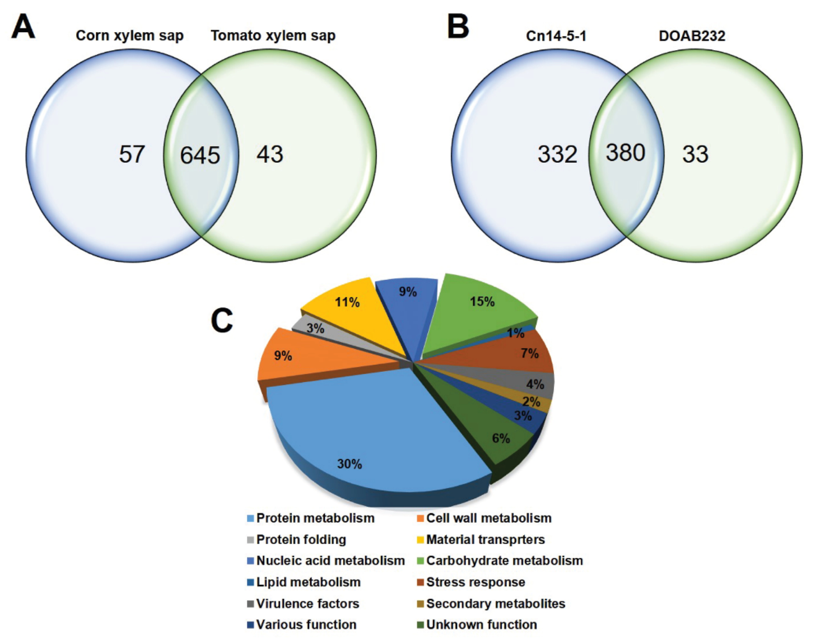
3.2. Characterisation of Cn Secretome by LC–MS Analysis

The total secretome profile of Cn strains Cn14-5-1 and DOAB232, grown in CXS and TXS, is shown in Table S1. A total of 745 proteins were identified in the bacterial secretomes for both strains in both xylem sap media (Table S1). A total of 645 proteins were observed in both sap media with only 57 and 43 proteins identified in CXS and TXS, respectively, for the two Cn strains (Figure 1A). On the other hand, in sap media, a total of 380 proteins were detected between the two Cn strains, with 332 and 33 proteins uniquely recorded by Cn14-5-1 and DOAB232, respectively (Figure 1B). Using the BLAST2GO algorithm, we categorized these secreted proteins into 12 cellular functional processes involved in protein metabolism (30.6%), cell wall metabolism, protein folding, material transporters, nucleic acid metabolism, carbohydrate metabolism, lipid metabolism, stress response, secondary metabolites, various functions, and a group of proteins with unknown function (Figure 1C).
Table 1 shows the number of proteins on the basis of their status in the different secretome pools—presence/absence and uniquely or differentially abundance. The total identified proteins of Cn14-5-1 were approximately double those of DOAB232 in both sap media. Strains Cn14-5-1 and DOAB232 responded differently to CXS and TXS. The secretome patterns of strain Cn14-5-1 in both CXS and TXS were similar while strain DOAB232 exhibited more secreted proteins in CXS. Proteins that were identified only in CXS could more likely contribute to the bacterial colonization and/or pathogenicity on corn rather than those identified in both sap media.

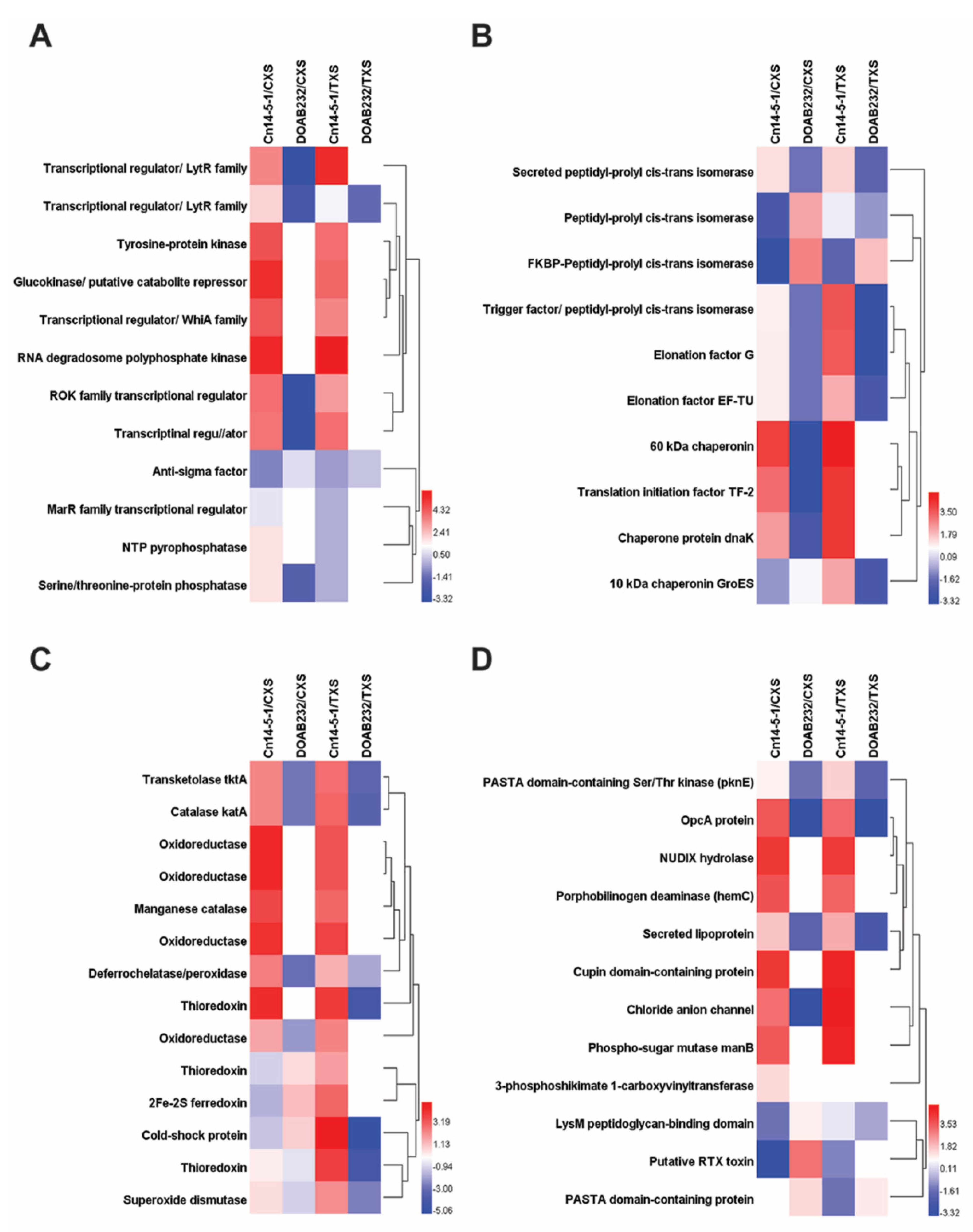
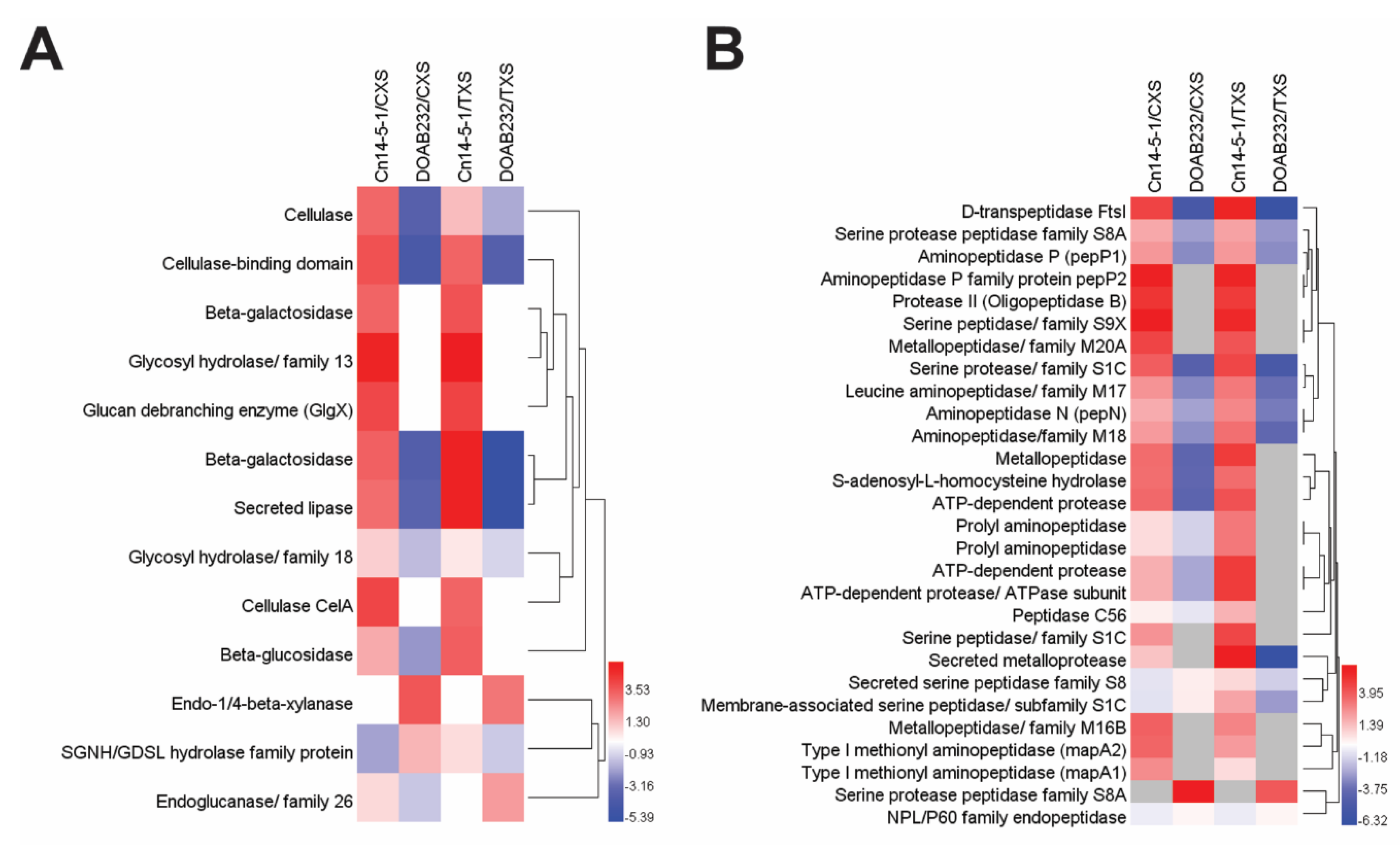
3.3. Abundance of Secreted Proteins in Xylem Sap Media

Proteins with various abundance levels were identified in the secretome of the Cn strains in the xylem sap media. Differentially or uniquely abundant proteins were identified in each strain under each sap medium. A total of 269 and 459 proteins were identified only in Cn14-5-1, the highly aggressive strain, under CXS (Table S2) and TXS (Table S3), respectively. These include well-characterized protein families involved in carbon metabolism (e.g., glucose-6-phosphate dehydrogenase), protein degradation (e.g., peptidases), and antibiotic resistance (e.g., putative multidrug export ABC transporters as well as a CelA-like protein, a known pathogenicity trait) (Tables S2 and S3). Further, a total of 270 and 200 proteins were differentially abundant in CXS (Table S4) and TXS (Table S5), respectively. Examples of uniquely and differentially abundant proteins secreted in both media are presented in Table 2 and Table 3, respectively, and were shown to exhibit the presence of a wide range of functional groups such as stress response, pathogenicity, protein degradation/folding, virulence, and carbohydrate metabolism. The majority of the unique proteins in Cn14-5-1 strain were identified under CXS conditions (Table 2). Some proteins with cellulase activity exhibited increased abundance in Cn14-5-1 under CXS, e.g., secreted cellulase, and cellulase-binding and expansin domain-containing proteins (Table 3). Unlike Cn14-5-1, only 40 and 10 secreted proteins were uniquely observed for DOAB232, the low aggressive strain, in CXS (Table S6) and TXS (Table S7), respectively. The number of differentially abundant proteins secreted in both sap media by DOAB232 also showed low counts of 40 and 18 for CXS (Table S8) and TXS (Table S9), respectively. The majority of uniquely abundant proteins of DOAB232 strain was identified in CXS, which mainly belonged to stress response proteins. A few proteins, such as endo-1,4-beta-xylanase and secreted serine protease, which are suggested to be potential pathogenicity factors, were identified in both sap media (Table 4). Several proteins that were differentially abundant in either sap media belonged to stress response and pathogenicity groups, e.g., 10 Kb chaperonin, serine protease, and endo-1,4-beta-xylanase (Table 5).
Figure 2 shows the fold change patterns of key stress-related protein families of the two Cn strains treated with CXS and TXS media. Figure 2A shows the changes in 12 secreted proteins that are related to signal perception and transduction. All the secreted proteins exhibited an increment with the exception of the anti-sigma factor, which showed a differentially reduced abundance in strain Cn14-5-1. RNA degradosome polyphosphate kinase and glucokinase (putative catabolite repressor) exhibited the highest fold changes in both xylem sap media for the highly aggressive strain (Figure 2B). The weakly pathogenic strain (DOAB232) either showed no fold change or a reduced abundance for all the 12 secreted proteins. With respect to the 10 protein folding enzymes analyzed, significant increases were observed only in 3 (60 kDa chaperonin, translation initiation factor TF-2, and chaperone protein dnaK) for strain Cn-14-5-1, while DOAB232 had significant increases only for FKBP-peptidyl-polyl-cis-transisomerase in both xylem sap media (Figure 2B).
In addition, proteins with predicted antioxidant activities were identified in both strains when treated with both xylem sap media (Figure 2C). Of the 14 secreted proteins, 9 differentially increased in both xylem sap media for strain Cn14-5-1, while none exhibited significant increase for DOAB232 (Figure 2C). These include catalase (KatA), transketolase tktA, oxidorectuxtase, manganese catalase, organic hydroperoxide resistance protein (Ohr), and thioredoxins differentially increased abundance levels in both xylem sap media in Cn14-5-1 (Figure 2C).
Figure 2D shows the changes in patterns of 12 secreted proteins reported to have potential virulence activities. Strain Cn14-5-1 exhibited significant fold increases in 7 (OpcA protein, NUDIX hydrolase, porphobilinogen deaminase, secreted lipoprotein, Cupin domain-containing protein, chloride anion channel, and phospo-sugar mutase manB) of the 12 virulence-related proteins in CXS and TXS (Figure 2D). On the other hand, fibronectin type III domain-containing protein (putative RTX toxin) was the only virulence-related protein out of 12 that exhibited a differentially increased abundance in DOAB232 in CXS (Figure 2D). PASTA domain-containing protein was the only secreted protein that showed moderate (about 1.5-fold change) in both xylem sap media by strain DOAB232 (Figure 2D).
Different cell wall-degrading enzymes (CWDEs), belonging to four glycosyl hydrolase families (GH3, GH5, GH18, and GH26), varied in abundance in the Cn secetome (Figure 3). However, the abundance levels of the proteins with hydrolytic activity were higher in the secretome of the Cn14-5-1 than DOAB232 (Figure 3A). Significant fold changes were recorded in 10 of the 13 secreted proteins detected in the secretome of strain Cn14-5-1 in both sap media (Figure 3A). The 10 proteins included cellulase, beta-galactosidase, glycosyl hydrolase, glucan debrabching enzyme (GigX), CelA, and beta-glucosidase (Figure 3A). On the other hand, the weakly aggressive strain (DOAB232) exhibited significant positive changes for only endo-1,4-β-xylanases anddetected in both sap media (Figure 3A).
Two classes of peptidases, serine peptidase and metallopeptidase families, with seven different peptidase families from each class, were identified in the Cn secretomes (Figure 3B). In general, the majority of the proteins with proteolytic activity were detected in the secretome of the Cn14-5-1 strain (Figure 3B). Different protein peptidases were observed in the secretome of strain Cn14-5-1 only. However, some aminopeptidase classes belonging to various subfamilies, and metallopeptidase were differentially abundant in Cn14-5-1 (Figure 3B). Some serine protease peptidase family S8A such as secreted subtilase B (sbtB) were differentially abundant only in DOAB232 in both xylem sap media (Figure 3B).

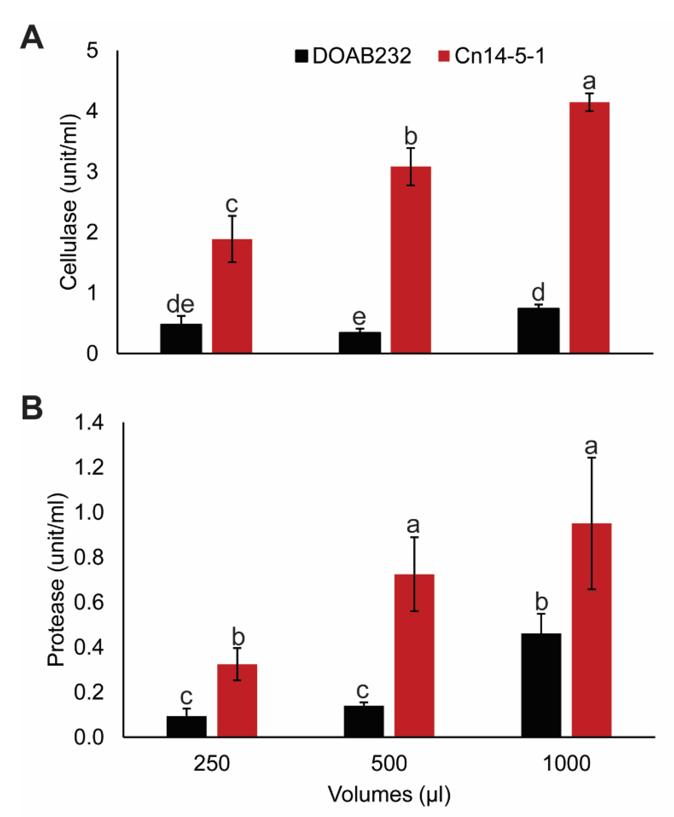
3.4. Hydrolytic Enzyme Activity Assays

Cellulase activity in the Cn14-5-1 strain increased significantly with increasing volume of CXS filtrates, which ranged from 2–4 units·mL−1, while the cellulose activity in the filtrate of the DOAB232 strain showed significantly lower increases in activity (Figure 4A). A similar trend was observed for protease activity with strain Cn14-5-1, showing significantly higher activity of 0.3 to 0.9 units·mL−1 compared to 0.09 to 0.4 units·mL−1 for DOAB232 (Figure 4B). Both strains showed increases in protease activity with increasing filtrate volumes (Figure 4B).

3.5. Hydrogen Peroxide and Superoxide Assays

Reactive oxygen species, hydrogen peroxide (H2O2), and superoxide (O2) were detected using DAB and NBT, respectively, and localized in corn leaves infected with Cn strains Cn14-5-1 and DOAB232. They were compared to non-infected controls, which exhibited very low staining. Leaves infected with either Cn strains showed deep brown and blue staining for H2O2 and O2, respectively, around the inoculation sites (Figure S2), suggesting the host cell response by the production of reactive oxygen species.

4. Discussion

This study profiled the secretomes of two Cn strains that differ in the level of aggressiveness or pathogenicity to corn. This is the first report of the use of LC–MS analysis to investigate the secreted proteins of the corn pathogen, C. nebraskensis, and the data presented here will enable a better understanding of the pathogenicity traits in Cn. The generated proteomic data would be a step forward to refine the annotation of the sequenced genome of C. nebraskensis. A recent study conducted by Peritore-Galve and colleagues, investigated the implement of proteome peptide sequences to refine the genome annotations of the Clavibacter michiganensis susp. michiganensis, the causal agent of bacterial canker of tomato, to create a system called proteogenomics [28].
The discrepancies in aggressiveness between Cn14-5-1 and DOAB232, which presented different symptoms clearly on corn, and resulted in different levels of plant damage, provided a platform to study, in vitro, Cn aggressiveness at the protein level using natural xylem sap media. Strain Cn14-5-1 was isolated in 2014, while DOAB 232 was isolated over 40 years ago. Agarkova and colleagues [9] indicated that strain DOAB 232 was pathogenic by direct injection of the bacterial suspension into corn stem. In our study, we used minimal foliar abrasion method, but only Cn14-5-1 inflicted typical symptoms of the Goss’s disease on corn. The differences in aggressiveness could be due to a combination of factors including the effect of long-term storage on DOAB 232 as well as changes at the genome level. Comparative genomics analysis of DOAB 232 and two strains, DOAB395 and DOAB397, isolated at the same year with Cn14-5-1 in 2014, revealed a proteome homology of only 92%, an indication of potential differences in genome arrangements [29]. The LC–MS data presented here contribute to the understanding of this genome-level differences and may explain the variation in aggressiveness of the two Cn strains.
This study identified secreted proteins reported to be involved in potential virulence of Cn strains in response to the corn xylem sap (CXS, host plant) and the tomato xylem sap (TXS, non-host plant) as an alternative medium. Tomato xylem sap (TXS) was used as an alternative induction medium because tomato is a host for C. michiganensis subsp. michiganensis that does not infect corn plants. The use of synthetic media such as minimal media or rich media, e.g., NBY, would either stress Cn or produce high background noise that might impair reliable mass spectrometric analyses, respectively. We hypothesized that Cn proteins secreted in equal abundance in both CXS and TXS were less likely to be directly involved in the pathogenicity of corn. The identified Cn secreted proteins were assigned into five different categories as follows: adaptation to new ecological niches, signal perception and transduction, stress-responsive proteins, virulence-related proteins, and hydrolytic enzymes for cell wall or protein degradation.
Degradation of plant cell walls is usually a major part of plant pathogen infections [30,31], since it can be a critical step in establishing a successful infection. Plant pathogens, including bacteria, secrete cell wall-degrading enzymes (CWDEs) to break down the cell walls of host plants, which releases nutrients and promotes colonization [30,32,33].
We identified different CWDEs belonging to four glycosyl hydrolase families (GH3, GH5, GH18, and GH26) showing variation in abundance. These enzymes are involved in pectin, hemicellulose, and cellulose hydrolysis [15], and could be required for Cn to colonize corn tissues resulting in the establishment of C. nebraskensis secreted cellulase (CelA, GH5), β-glucosidase (GH3), β-galactosidase (GH5), and chitinase (GH18), which hydrolyze cellulose, pectin, and chitin, and were found in Cn14-5-1 grown in CXS. In contrast, only β-1,4-xylanase was identified in DOAB232, which could contribute to its lower aggressiveness and milder symptoms of this strain. In the potato–Clavibacter sepedonicus system, cellulases (celA and celB) and xylanases were reported to be upregulated [34]. The putative plasmid-encoded celA gene was also implicated in the pathogenicity of C. sepedonicus strains to eggplant [35,36], and concluded that cellulase is a major plasmid-borne virulence determinant [36]. It is unclear whether the lack of plasmid in strain DOAB232 may contribute to its low aggressiveness. The identification of secreted CelA in extracts of Cn14-5-1 in CXS suggests the presence of a plasmid or an unknown production pathway. Some Cn strains contain single large plasmids with a proximate size of 70 kb, which have shown no correlation with Cn strain pathogenicity [37]. Further research is required to determine the involvement of any such plasmid in the virulence of Cn14-5-1. However, our results clearly showed that Cn14-5-1 is a very aggressive strain. The watery-soaked spots seen in symptomatic infected corn leaves infected by Cn14-5-1 suggested the efficiency of the CWDEs and can be considered as direct evidence of the role of CWDEs as virulence factors in this system. Furthermore, the high activity of total cellulase in Cn14-5-1 compared to DOAB232, measured in the filtrates of Cn grown in CXS, provided additional support to the assumption that CWDEs are involved in Cn aggressiveness.
Establishing a successful infection relies not only on degrading host plant cell walls but also on neutralizing host protein-based defense factors. We identified proteins belonging to two peptidase families in Cn14-5-1 and DOAB232. Secreted peptidases are frequently implicated in overcoming host protein defense components, and as such a large arsenal of such enzymes could lead to higher aggressiveness [38]. Secreted proteases are used by bacterial pathogens to evade plant immune systems and maintain homeostasis [39]. CXS medium boosted the secretion of proteases, e.g., subtilase C (sbtC, S8A) and subtilase B (subtB, S8A), especially in Cn14-5-1, which secreted more proteases and peptidases than DOAB232. Similar proteolytic enzymes, specifically from the serine proteases families Ppa, Sbt. and Chp, were identified in tomato infected by C. michiganensis subsp. michiganensis [15]. The authors concluded that these enzymes were likely targeting host immune components as well as structural components to facilitate disease progression. The protease assay indicated significantly higher levels of proteases in Cn14-5-1, which may contribute to degradation of plant defense proteins and promote colonization. A similar conclusion was reported on the involvement of proteases in colonization of Clavibacter michiganensis and Clavibacter sepedonicus [3]. An important next step will be to find targets for these enzymes, as these would be host factors potentially involved in resistance to Goss’s wilt disease. Besides secreted hydrolytic proteins, other secreted proteins with various cellular functions might also be important in Cn–host interaction.
The ability of bacteria to utilize carbohydrates from the surrounding environment is a positive sign of adaptation. Unlike DOAB232, secretion of carbohydrate metabolism-related proteins was obvious in strain Cn14-5-1 treated with CXS. Furthermore, Cn14-5-1 secreted some carbohydrate metabolism-related proteins in TXS medium, an indication of the ability of Cn14-5-1 to utilize carbohydrates from non-host plants. Some of these identified proteins, e.g., glucokinase (GLK) and glycerol kinase, function as transcriptional regulators or catalytic enzymes for carbohydrate metabolism, respectively [40,41,42]. Identifying these proteins in the highly aggressive Cn14-5-1 strain but not in DOAB232 provided further evidence that this isolate utilizes host carbon resources.
Activity of transcriptional and translational machineries in bacterial cells are essential for cellular perception and signal transduction. The ability to perceive and transmit signals is crucial to prepare and respond to plant defense factors. The majority of transcriptional regulators and DNA-binding proteins were identified in Cn14-5-1 treated with CXS, e.g., antibiotic resistance regulator (MarR) protein family, a one-component signal transduction [43], transcriptional regulator (TetR) [44,45], and ROK family transcriptional regulator [46,47], may provide evidence of the efficiency of strain Cn14-5-1 to recognize and respond to plant stimuli.
Furthermore, phosphorylation is a crucial process that modulates protein activities. Secreted proteins with high abundance in Cn14-5-1 that are involved in protein phosphorylation, e.g., serine/threonine protein phosphatase, HAD family phosphatase, and NTP phosphatase, when treated with CXS may indicate the preference to the host stimuli.
The Cn–corn interaction occurs in the apoplast and extracellular spaces, where bacterial cells secrete virulence factors to evade plant defense factors and promote infection. These secreted virulence factors are translocated via transporter proteins [15,48]. We identified a larger number of transporters in Cn14-5-1 than DOAB232, which could be another potential piece of evidence to explain variations in Cn strain aggressiveness. Plant pathogens negatively affect the integrity of host plant cell walls and cell membranes to cause cell death and release nutrients [49]. Thereafter, pathogens employ different strategies to take up these nutrients, including passive diffusion and active uptake. Various types of membrane transporters facilitate the latter, e.g., ABC transporters [50,51] and ATP-binding cassette ABC transporters [52].
Under stress conditions, e.g., environmental changes, bacterial cellular proteins are subjected to alterations. Chaperones are responsible for folding either newly synthesized polypeptides or refolding stress-denatured proteins, or both [53,54]. The secretome of Cn14-5-1 had various types of chaperones with different levels of abundance, e.g., peptidyl-poly cis/trans isomerase (PPIase), which reacts with the chaperone DnaK system to fold newly synthesized polypeptides [55]. In addition, 60 kDa chaperonin and 10 kDa chaperonin (GroES) are essential in newly synthesized polypeptides, as well as stress-denatured proteins [56,57]. Abundance of these types of chaperones in Cn14-5-1 treated with CXS may demonstrate efficient re-use of resources, which may enhance its ecological fitness leading to high aggressiveness.
Phytopathogens cope with host plant reactive oxygen species (ROS)-based defenses [58] by secreting antioxidant enzymes. Corn plants accumulate ROS in the infection site to restrict bacterial growth and to hinder disease progress. High abundance of secreted ROS scavenging proteins in Cn14-5-1 secretome, e.g., catalase (KatA), and manganese catalase were deployed to mitigate the deleterious effect of ROS as the first host plant chemical defense barrier [59]. Finally, lipoproteins [60,61], cupin domain-containing proteins [62], and Stk1 family PASTA domain-containing Ser/Thr kinase [63] were also observed in Cn14-5-1, and these may also contribute to the higher aggressiveness of this strain.

5. Conclusions and Future Perspectives

Host and non-host xylem sap represent a convenient and biologically relevant model for culturing Cn and performing comparative proteomics of the Cn–corn interaction in vitro. We found that the pathogen secretome was altered in response to host xylem sap compared to non-host xylem sap. We identified significant changes in the abundance of key proteins implicated in pathogenicity. Since these changes are in response to host xylem, it is likely that these proteins could play important roles in the corn-pathogen interaction through responding to chemical stimuli present in the host xylem sap. The xylem sap system has the advantage that the host proteome is very minute from the protein extracts. This minimizes background noise from LC–MS data and greatly simplifies downstream proteomic analyses. A comparison of the secretomes of weakly and highly aggressive strains of Cn indicated that cell wall-degrading enzymes, proteases, and other types of proteins might have important roles in Goss’s wilt disease establishment. Further experiments on Cn gene knockout for CWDEs and/or proteases encoding genes will be useful in deciphering the potential role of these two major secreted proteins in Cn aggressiveness.

Supplementary Materials

The following are available online at https://0-www-mdpi-com.brum.beds.ac.uk/2227-7382/9/1/1/s1: Figure S1: Aggressiveness assay of the two strains of Clavibacter nebraskensis. Two disease assessment methods were used to evaluate the aggressiveness of Cn14-5-1 and DOAB232 on the basis of the total area under the disease progress curve (AUDPC) using (A) lesion length and (B) disease severity. Figure S2: Reactive oxygen species (ROS) localization in corn leaves inoculated with the two strains of Clavibacter nebraskensis. Localization of hydrogen peroxide (A) and superoxide (B) in corn leaves. 3,3′-Diaminobenzidine tetrahydrochloride (DAB) (brown) staining of the infected corn leaves with Cn strains Cn14-5-1 and DOAB232 vs. non-infected leaf (control). Nitroblue tetrazolium (NBT) (blue) staining of the infected corn leaves with Cn strains Cn14-5-1 and DOAB232 vs. non-infected leaf (control). Table S1: Seven hundred and forty-five total secreted proteins identified in the corn xylem sap (host) and tomato xylem sap (non-host). Table S2: Secreted proteins identified only for strain Cn14-5-1 in corn xylem sap medium. Table S3: Secreted proteins identified only for strain Cn14-5-1 in tomato xylem sap medium. Table S4: Increased differentially abundant secreted proteins of Cn14-5-1 in corn xylem sap medium. Table S5: Differentially abundant proteins of Cn14-5-1 with an increased fold change in tomato xylem sap medium. Table S6: Secreted proteins identified only for strain DOAB232 in corn xylem sap medium. Table S7: Secreted proteins identified only for strain DOAB232 in tomato xylem sap medium. Table S8: Differentially abundant proteins of DOAB232 with an increased fold change in corn xylem sap medium. Table S9: Differentially abundant proteins of DOAB232 with an increased fold change in tomato xylem sap medium.

Author Contributions

A.S. contributed to the idea, carried out experiments, and contributed to data analysis and writing the manuscript; C.R. processed samples for mass spectrometry, helped in data analysis, and revised the manuscript; J.T.T. contributed to experimental design and revised the manuscript; F.D. contributed the idea, supervised the work, and contributed to the writing of the manuscript. All authors have read and agreed to the published version of the manuscript.

Funding

This research was funded by the Manitoba Corn Growers Association and the Manitoba Growing Innovation Capacity and Knowledge Development program, Grain Innovation Hub—Corn Development Project # 1000135719.

Institutional Review Board Statement

Not applicable.

Informed Consent Statement

Not applicable.

Data Availability Statement

Data is contained within the article and the supplementary materials.

Acknowledgments

We would like to thank Lorne Adam for his assistance in reviewing the manuscript, and Bryan Waller, Pride Seeds, for providing hybrid A4631G2 RIB.

Conflicts of Interest

The authors declare no conflict of interest.

References

  1. Tambong, J.T. Comparative genomics of Clavibacter michiganensis subspecies, pathogens of important agricultural crops. PLoS ONE 2017, 12, e0172295. [Google Scholar] [CrossRef] [PubMed] [Green Version]
  2. Jacques, M.A.; Durand, K.; Orgeur, G.; Balidas, S.; Fricot, C.; Bonneau, S.; Quillévéré, A.; Audusseau, C.; Olivier, V.; Grimault, V.; et al. Phylogenetic analysis and polyphasic characterization of Clavibacter michiganensis strains isolated from tomato seeds reveal that nonpathogenic strains are distinct from C. michiganensis subsp. michiganensis. Appl. Environ. Microbiol. 2012, 78, 8388–8402. [Google Scholar] [CrossRef] [PubMed] [Green Version]
  3. Eichenlaub, R.; Gartemann, K.H. The Clavibacter michiganensis subspecies: Molecular investigation of Gram-positive bacterial plant pathogen. Annu. Rev. Phytopathol. 2011, 49, 445–464. [Google Scholar] [CrossRef] [PubMed]
  4. Vidaver, A.K.; Mandel, M. Corynebacterium nebraskense, a new orange-pigmented phytopathogenic species. Int. J. Syst. Bacteriol. 1974, 24, 482–485. [Google Scholar] [CrossRef] [Green Version]
  5. Schuster, M.L. Leaf Freckles and Wilt of Corn Incited by Corynebacterium nebraskense Schuster, Hoff, Mandel, Lazar, 1972; Agricultural Experiment Station, University of Nebraska: Lincoln, NE, USA, 1975. [Google Scholar]
  6. Li, X.; Tambong, J.; Yuan, K.X.; Chen, W.; Xu, H.; Lévesque, C.A.; De Boer, S.H. Reclassification of Clavibacter michiganensis subspecies on the basis of whole-genome and multi-locus sequence analyses. Int. J. Syst. Evol. Microbiol. 2018, 68, 234–240. [Google Scholar] [CrossRef]
  7. Wysong, D.S.; Doupnik, J.B.; Lane, L. Goss’s wilt and corn lethal necrosis—Can they become a major problem? In Proceedings of the Annual Corn Sorghum Research Conference; 36th American Seed Trade Association: Washington, DC, USA, 1981; pp. 104–130. [Google Scholar]
  8. Rocheford, T.R.; Vidaver, A.K.; Gardner, C.O.; Arbust, D.L. Effect of wind-generated sand abrasion on infection of corn (Zea mays L.) by Corynebacterium michiganense ssp. nebraskense. Phytopathology 1985, 75, 1378. [Google Scholar]
  9. Agarkova, I.V.; Lambrecht, P.A.; Vidaver, A.K. Genetic diversity and population structure of Clavibacter michiganensis subsp. nebraskensis. Can. J. Microbiol. 2011, 57, 366–374. [Google Scholar] [CrossRef]
  10. Langemeier, C.B.; Robertson, A.E.; Wang, D.; Jackson-Ziems, T.A. Factors affecting the development and severity of Goss’s bacterial wilt and leaf blight of corn, caused by Clavibacter michiganensis subsp. nebraskensis. Plant Dis. 2017, 101, 54–61. [Google Scholar] [CrossRef] [Green Version]
  11. Claflin, L.E. Goss’s bacterial wilt and blight. In Compendium of Corn Diseases; White, D.H., Ed.; American Phytopathological Society: St. Paul, MN, USA, 1999. [Google Scholar]
  12. Mallowa, S.; Mbofung, G.; Eggenberger, S.; Den Adel, R.; Sheiding, S.R.; Robertson, A.E. Infection of maize by Clavibacter michiganensis subsp. nebraskensis does not require severe wounding. Plant Dis. 2016, 100, 724–731. [Google Scholar] [CrossRef] [Green Version]
  13. Langemeier, C.B.; Jackson-Ziems, T.A.; Kruger, G.R. Four common Setaria Species are alternative hosts for Clavibacter michiganensis subsp. nebraskensis, causal agent of Goss’s bacterial wilt and blight of corn. Plant Health Prog. 2014, 15, 57–60. [Google Scholar] [CrossRef] [Green Version]
  14. Ikley, J.T.; Wise, K.A.; Johnson, W.G. Annual ryegrass (Lolium multiflorum), Johnsongrass (Sorghum halepense), and large crabgrass (Digitaria sanguinalis) are alternative hosts for Clavibacter michiganensis subsp. nebraskensis, causal agent of Goss’s wilt of corn. Weed Sci. 2015, 63, 901–909. [Google Scholar]
  15. Savidor, A.; Teper, D.; Gartemann, K.-H.; Eichenlaub, R.; Chalupowicz, L.; Manulis-Sasson, S.; Barash, I.; Tews, H.; Mayer, K.; Giannone, R.; et al. The Clavibacter michiganensis subsp. michiganensis–tomato interactome reveals the perception of pathogen by the host and suggests mechanisms of infection. J. Proteome Res. 2012, 11, 736–750. [Google Scholar] [CrossRef] [PubMed]
  16. Hiery, E.; Poetsch, A.; Moosbauer, T.; Amin, B.; Hofmann, J.; Burkovski, A. A proteomic study of Clavibacter michiganensis subsp. michiganensis culture supernatants. Proteomes 2015, 3, 411–423. [Google Scholar] [CrossRef] [PubMed] [Green Version]
  17. Gross, D.C.; Vidaver, A.K. A selective medium for isolation of Corynebacterium nebraskense from soil and plant parts. Phytopathology 1979, 69, 82–87. [Google Scholar] [CrossRef]
  18. Soliman, A.; Gulden, R.H.; Tambong, J.T.; Bajracharya, P.; Adam, L.R.; Xu, R.; Cott, M.; Daayf, F. Developed and validated inoculation and disease assessment methods for Goss’s bacterial wilt and leaf blight disease of corn. Crop Prot. 2018, 112, 59–167. [Google Scholar] [CrossRef]
  19. Campbell, M.T.; Proctor, C.A.; Dou, Y.; Schmitz, A.J.; Phansak, P.; Kruger, G.R.; Walia, H. Genetic and molecular characterization of submergence response identifies Subtol6 as a major submergence tolerance locus in maize. PLoS ONE 2015, 10, e0120385. [Google Scholar] [CrossRef]
  20. Bradford, M. A rapid and sensitive method for the quantification of microgram quantities of protein utilizing the principle of protein-dye binding. Anal. Biochem. 1979, 72, 248–254. [Google Scholar] [CrossRef]
  21. Perez-Riverol, Y.; Csordas, A.; Bai, J.; Bernal-Llinares, M.; Hewapathirana, S.; Kundu, D.J.; Inuganti, A.; Griss, J.; Mayer, G.; Eisenacher, M.; et al. The PRIDE database and related tools and resources in 2019: Improving support for quantification data. Nucleic Acids Res. 2019, 47, D442–D450. [Google Scholar] [CrossRef]
  22. Keller, A.; Nesvizhskii, A.I.; Kolker, E.; Aebersold, R. Empirical statistical model to estimate the accuracy of peptide identifications made by MS/MS and database search. Anal. Chem. 2002, 74, 5383–5392. [Google Scholar] [CrossRef]
  23. Nesvizhskii, A.I.; Keller, A.; Kolker, E.; Aebersold, R.A. A statistical model for identifying proteins by tandem mass spectrometry. Anal. Chem. 2003, 75, 4646–4658. [Google Scholar] [CrossRef]
  24. Zhang, Y.H.P.; Hong, J.; Ye, X. Cellulase Assays. In Biofuels: Methods in Molecular Biology; Mielenz, J., Ed.; Humana Press: Totowa, NJ, USA, 2009; Volume 581. [Google Scholar]
  25. Cupp-Enyard, C. Sigma’s non-specific protease activity assay—Casein as a substrate. JoVE 2008, 19, 899. [Google Scholar] [CrossRef] [PubMed]
  26. Lund, R.E. Tables for an approximate test for outliers in linear models. Technometrics 1975, 17, 473–476. [Google Scholar] [CrossRef]
  27. Saxton, A.M. A macro for converting mean separation output to letter groupings in PROC MIXED. In Proceedings of the 23rd SAS Users Group International; SAS Institute: Cary, NC, USA, 1998; pp. 1243–1246. [Google Scholar]
  28. Peritore-Galve, F.C.; Schneider, D.J.; Yang, Y.; Thannhauser, T.W.; Smart, C.D.; Stodghill, P. Proteome profile and genome refinement of the tomato-pathogenic bacterium Clavibacter michiganensis subsp. michiganensis. Proteomics 2019, 19, e1800224. [Google Scholar] [CrossRef] [PubMed]
  29. Tambong, J.T.; Xu, R.; Daayf, F.; Brière, S.; Bilodeau, G.J.; Tropiano, R.; Hartke, A.; Reid, L.M.; Cott, M.; Cote, T.; et al. Genome analysis and development of a multiplex TaqMan real-time PCR for specific identification and detection of Clavibacter michiganensis subsp. nebraskensis. APS Phytopathol. 2016, 106, 1473–1485. [Google Scholar] [CrossRef] [PubMed] [Green Version]
  30. Bellincampi, D.; Cervone, F.; Lionetti, V. Plant cell wall dynamics and wall-related susceptibility in plant–pathogen interactions. Front. Plant Sci. 2014, 28, 228. [Google Scholar] [CrossRef] [Green Version]
  31. Lalaoui, F.; Halama, P.; Dumortier, V.; Paul, B. Cell wall-degrading enzymes produced in vitro by isolates of Phaeosphaeria nodorum differing in aggressiveness. Plant Pathol. 2000, 49, 727–733. [Google Scholar] [CrossRef]
  32. Barras, F.; Gijsegem, F.V.; Chatterjee, A.K. Extracellular enzymes and pathogenesis of soft-rot Erwinia. Annu. Rev. Phytopathol. 1994, 32, 201–234. [Google Scholar] [CrossRef]
  33. Melotto, M.; Kunkel, B.N. Virulence strategies of plant pathogenic bacteria. In The Prokaryotes; Rosenberg, E., DeLong, E.F., Lory, S., Stackebrandt, E., Thompson, F., Eds.; Springer: Berlin/Heidelberg, Germany, 2013; pp. 61–82. [Google Scholar]
  34. Holtsmark, I.; Takle, G.; Brurberg, M. Expression of putative virulence factors in the potato pathogen Clavibacter michiganensis subsp. sepedonicus during infection. Arch. Microbiol. 2008, 189, 131–139. [Google Scholar]
  35. Laine, M.J.; Haapalainen, M.; Wahlroos, T.; Kankare, K.; Nissinen, R.; Kassuwi, S.; Metzler, M.C. The cellulase encoded by the native plasmid of Clavibacter michiganensis ssp. sepedonicus plays a role in virulence and contains an expansin-like domain. Phys. Mol. Plant Pathol. 2000, 57, 221–233. [Google Scholar]
  36. Nissinen, R.; Kassuwi, S.; Peltola, R.; Metzler, M.C. In planta—Complementation of Clavibacter michiganensis subsp. sepedonicus strains deficient in cellulase production or HR induction restores virulence. Eur. J. Plant Pathol. 2001, 107, 175–182. [Google Scholar] [CrossRef]
  37. Ahmad, A.; Mbofung, G.Y.; Acharya, J.; Schmidt, C.L.; Robertson, A.E. Characterization and comparison of Clavibacter michiganensis subsp. nebraskensis strains recovered from epiphytic and symptomatic infections of maize in Iowa. PLoS ONE 2015, 10, e0143553. [Google Scholar] [CrossRef] [PubMed]
  38. Hoston, A.; Mudgett, M.B. Cysteine proteases in phytopathogenic bacteria: Identification of plant targets and activation of innate immunity. Curr. Opin. Plant Biol. 2004, 7, 384–390. [Google Scholar]
  39. Figaj, D.; Ambroziak, P.; Przepiora, T.; Skorko-Glonek, J. The role of proteases in the virulence of plant pathogenic bacteria. Int. J. Mol. Sci. 2019, 20, 672. [Google Scholar] [CrossRef] [PubMed] [Green Version]
  40. Boos, W.; Shuman, H. Maltose/maltodextrin system of Escherichia coli: Transport, metabolism, and regulation. Microbiol. Mol. Biol. Rev. 1998, 62, 204–229. [Google Scholar] [CrossRef] [PubMed] [Green Version]
  41. Lengsfeld, C.; Schönert, S.; Dippel, R.; Boos, W. Glucose-and Glucokinase-controlled mal gene expression in Escherichia coli. J. Bacteriol. 2009, 191, 701–712. [Google Scholar] [CrossRef] [PubMed] [Green Version]
  42. Yeh, J.I.; Kettering, R.; Saxl, R.; Bourand, A.; Darbon, E.; Joly, N.; Briozzo, P.; Deutscher, J. Structural characterizations of glycerol kinase: Unraveling phosphorylation-induced long-range activation. Biochemistry 2009, 48, 346–356. [Google Scholar] [CrossRef] [Green Version]
  43. Grove, A. Regulation of metabolic pathways by MarR family transcription factors. Comput. Struct. Biotechnol. J. 2017, 15, 366–371. [Google Scholar] [CrossRef]
  44. Ramos, J.L.; Martínez-Bueno, M.; Molina-Henares, A.J.; Terán, W.; Watanabe, K.; Zhang, X.; Gallegos, M.T.; Brennan, R.; Tobes, R. The TetR family of transcriptional repressors. Mol. Biol. Rev. 2005, 69, 326–356. [Google Scholar] [CrossRef] [Green Version]
  45. Cuthbertson, L.; Nodwella, J.R. The TetR Family of regulators. Microbiol. Mol. Biol. Rev. 2013, 77, 440–475. [Google Scholar] [CrossRef] [Green Version]
  46. Titgemeyer, F.; Reizer, J.; Reizer, A.; Saier, M.H.J. Evolutionary relationships between sugar kinases and transcriptional repressors in bacteria. Microbiology 1994, 140, 2349–2354. [Google Scholar] [CrossRef] [Green Version]
  47. Kazanov, M.D.; Li, X.; Gelfand, M.S.; Osterman, A.L.; Rodionov, D.A. Functional diversification of ROK-family transcriptional regulators of sugar catabolism in the Thermotogae phylum. Nucleic Acids Res. 2012, 41, 790–803. [Google Scholar] [CrossRef] [PubMed] [Green Version]
  48. Thapa, S.P.; Pattathil, S.; Hahn, M.G.; Jacques, M.-A.; Gilbertson, R.L.; Coaker, G. Genomic analysis of Clavibacter michiganensis reveals insight into virulence strategies and genetic diversity of a Gram-positive bacterial pathogen. Mol. Plant Microbe Interact 2017, 30, 786–802. [Google Scholar] [CrossRef] [PubMed] [Green Version]
  49. Laluk, K.; Mengiste, T. Necrotroph attacks on plants: Wanton Destruction or Covert Extortion? Arabidopsis Book 2010, 8. [Google Scholar] [CrossRef] [PubMed] [Green Version]
  50. Fatima, U.; Senthil-Kumar, M. Plant and pathogen nutrient acquisition strategies. Front. Plant Sci. 2015, 6, 750. [Google Scholar] [CrossRef]
  51. Delmotte, N.; Knief, C.; Chaffron, S.; Innerebner, G.; Roschitzki, B.; Schlapbach, R.; von Mering, C.; Vorholt, J.A. Community proteogenomics reveals insights into the physiology of phyllosphere bacteria. Proc. Natl. Acad. Sci. USA 2009, 106, 16428–16433. [Google Scholar] [CrossRef] [Green Version]
  52. Yu, J.; Ge, J.; Heuveling, J.; Schneider, E.; Yang, M. Structural basis for substrate specificity of an amino acid ABC transporter. Proc. Natl. Acad. Sci. USA 2015, 112, 5243–5248. [Google Scholar] [CrossRef] [Green Version]
  53. Braakman, I.; Hebert, D.N. Protein folding in the endoplasmic reticulum. Cold Spring Harb. Perspect. Biol. 2013, 5, a013201. [Google Scholar] [CrossRef] [Green Version]
  54. Park, C.-J.; Seo, Y.-S. Heat Shock Proteins: A Review of the Molecular Chaperones for Plant Immunity. Plant Pathol. J. 2015, 31, 323–333. [Google Scholar] [CrossRef] [Green Version]
  55. Stoller, G.; Rucknagel, K.P.; Nierhaus, K.H.; Schmid, F.X.; Fischer, G.; Rahfeld, J.-U. A ribosome-associated peptidyl-prolyl cis/trans isomerase identified as the trigger factor. EMBO J. 1995, 20, 4939–4948. [Google Scholar] [CrossRef]
  56. Rospert, S.; Glick, B.S.; Jenö, P.; Schatz, G.; Todd, M.J.; Lorimer, G.H.; Viitanen, P.V. Identification and functional analysis of chaperonin 10, the groES homolog from yeast mitochondria. Proc. Natl. Acad. Sci. USA 1993, 90, 10967–10971. [Google Scholar] [CrossRef] [Green Version]
  57. Levy-Rimler, G.; Viitanen, P.; Weiss, C.; Sharkia, R.; Greenberg, A.; Niv, A.; Lustig, A.; Delarea, Y.; Azem, A. The effect of nucleotides and mitochondrial chaperonin 10 on the structure and chaperone activity of mitochondrial chaperonin 60. Eur. J. Biochem. 2001, 268, 3465–3472. [Google Scholar] [CrossRef] [PubMed]
  58. Fones, H.; Preston, G.M. Reactive oxygen and oxidative stress tolerance in plant pathogenic Pseudomonas. FEMS Microbiol. Lett. 2012, 327, 1–8. [Google Scholar] [CrossRef] [PubMed] [Green Version]
  59. Hasset, D.J.; Cohen, M.S. Bacterial adaptation to oxidative stress: Implications for pathogenesis and interaction with phagocytic cells. FASEB J. 1989, 3, 2574–2582. [Google Scholar] [CrossRef] [PubMed] [Green Version]
  60. Kovacs-Simon, A.; Titball, R.W.; Michell, S.L. Lipoproteins of bacterial pathogens. Infect. Immun. 2011, 79, 548–561. [Google Scholar] [CrossRef] [PubMed] [Green Version]
  61. Zückert, W.R. Secretion of bacterial lipoproteins: Through the cytoplasmic membrane, the periplasm and beyond. Biochim. Biophys. Acta 2014, 1843, 1509–1516. [Google Scholar] [CrossRef] [Green Version]
  62. El Hadrami, A.; Islam, M.R.; Adam, L.R.; Fouad, D. A cupin domain-containing protein with a quercetinase activity (VdQase) regulates Verticillium dahliae’s pathogenicity and contributes to counteracting host defenses. Front. Plant Sci. 2015, 6, 440. [Google Scholar] [CrossRef] [Green Version]
  63. Débarbouillé, M.; Dramsi, S.; Dussurget, O.; Nahori, M.-A.; Vaganay, E.; Jouvion, G.; Cozzone, A.; Msadek, T.; Duclos, B. Characterization of a serine/threonine kinase involved in virulence of Staphylococcus aureus. J. Bacteriol. 2009, 191, 4070–4081. [Google Scholar] [CrossRef] [Green Version]
Figure 1. Categories of the identified secretome proteins of Cn strains and their abundance. (A) Venn diagram shows unique and differentially abundant proteins under corn xylem sap (CXS) and tomato xylem sap (TXS) media. (B) Venn diagram shows unique and differentially abundant proteins of Cn14-5-1 and DOAB232 strains. (C) Different classes and the abundance of the identified protein shown on the basis of the predicted biological function.
Proteomes 09 00001 g001
Figure 2. Stress-related proteins patterns of the two strains of Clavibacter nebraskensis treated with different xylem sap media. Secretome profile of Cn14-5-1 was normalized by that of the DOAB232 strain, and vice versa in each sap medium to identify the differential stress-related protein profile under corn xylem sap (CXS) and tomato xylem sap (TXS) induction. (A) Signal perception and transduction; (B) protein folding; (C) reactive oxygen species (ROS) scavenging proteins; and (D) virulence-related proteins. Hierarchical clustering was performed using the group average (unweighted pair-group method (UWPGM)) method.
Proteomes 09 00001 g002
Figure 3. Hydrolytic proteins patterns of the two strains of Clavibacter nebraskensis treated with different xylem sap media. Secretome profile of Cn14-5-1 was normalized by that of the DOAB232 strain, and vice versa in each sap medium to identify the differential stress-related protein profile under corn xylem sap (CXS) and tomato xylem sap (TXS) induction. (A) Cell wall-degrading enzymes, (B) proteases. Hierarchical clustering was performed using the group average (unweighted pair-group method (UWPGM)) method.
Proteomes 09 00001 g003
Figure 4. Enzymatic activity of secreted cellulases and proteases in filtrates of Clavibacter nebraskensis strains. The weakly aggressive DOAB232 strain in black and the highly aggressive Cn14-5-1 strain in red. (A) Enzymatic activity of total cellulase, and (B) protease were assayed in Cn filtrates. The mean values (n = 3) were separated by least squared means, and values with the same letter are not significantly different at (p < 0.05).
Proteomes 09 00001 g004
Table 1. Total secreted proteins in Cn isolates under different sap media.
Identified ProteinsCorn Xylem Sap (CXS)Tomato Xylem Sap (TXS)
Cn14-5-1DOAB232Cn14-5-1DOAB232
Total proteins658396678229
Unique proteins2694045910
Differentially abundant2704020018
(CSX) corn xylem sap, (TXS) tomato xylem sap.
Table 2. Proteins identified only in Cn14-5-1 in the different sap media.
Accession No.Identified ProteinsMW (KDa)Gene/LocusType of SapFunctional Group
CXSTXS
CCE76040.1Serine peptidase, family S9X76CMN_02099+Protein degradation
CCE75111.1pepP2 protein57pepP2+Protein degradation
CCE76391.1Polyphosphate kinase82ppK+Protein phosphorylation
CCE75799.1Glucokinase33glkA+Catabolite repressor
CCE74648.1Radical SAM domain-containing protein46CMN_00689+Stress response
CCE76022.1Protease II (oligopeptidase B)80ptrB+Protein degradation
CCE75620.1Metallopeptidase, peptidase family M20A47CMN_01675+Protein degradation
CCE74827.1Putative tyrosine-protein kinase49CMN_00871+Stress response
CCE74652.1Oxidoreductase35CMN_00693++Stress response
CCE75671.1Transcriptional regulator, WhiA family35whiA+Signal transduction
CCE74496.1Thioredoxin21ccbD+Stress response
CCE75993.1Oxidoreductase36CMN_02051++Stress response
CCE75476.1NUDIX hydrolase19CMN_01528++Virulence
CCE76025.1Cupin_2 domain-containing protein32CMN_02083++Virulence
AHN16207.1Cellulase78CelA++Pathogenicity
CCE75945.1Metallopeptidase, family M16B48pepR+Protein degradation
CCE76486.1Methionine aminopeptidase29mapA2+Protein degradation
CAN00621.1Porphobilinogen deaminase34hemC++Pathogenicity
AJW80248.1Manganese catalase31DZF93_04220++Stress response
CCE74906.1ManB protein61manB++Pathogenicity
CCE74104.1Putative glycosyl hydrolase, family 269CMN_00123+Pathogenicity
AJW78817.1ATP-dependent Clp protease proteolytic subunit21clpP++Stress response
CCE76317.1Serine peptidase, family S1C51CMN_02381+Protein degradation
CCE75886.1NTP pyrophosphatase22CMN_01943+Signal transduction
CCE74756.1Putative secreted metalloprotease46CMN_00799+Protein degradation
CCE76463.1Alanine racemase/kinase fusion protein59alr2+Signal transduction
AJW79024.1HAD family phosphatase25VO01_07690+Signal transduction
AJW80208.1MarR family transcriptional regulator17DZF93_09435+Signal transduction
CCE74692.1Chloride anion channel25CMN_00734+Pathogenicity
CCE74774.1ATP-dependent protease, ATPase subunit92clpC+Protein degradation
CCE74648.1Radical SAM domain-containing protein46CMN_00689+Stress response
CCE76686.1Alpha-glucosidase, glycosyl hydrolase family 1364aglC+Pathogenicity
CCE75391.1ATP-dependent protease, proteolytic subunit25clpP2+Stress response
CCE76068.11-Deoxy-D-xylulose 5-phosphate reductoisomerase38dxr+Pathogenicity
CCE75358.1Glucan debranching enzyme82treX+Pathogenicity
CCE74827.1Putative tyrosine-protein kinase49CMN_00871+Stress response
CCE76839.12-Keto acid dehydrogenase41CMN_02915+Energy production
CCE74969.1Lipoprotein59lpqB+Pathogenicity
CCE74801.1Beta-glycosidase54bglJ+Pathogenicity
CCE76286.1Alpha glycosidase81CMN_02350+Pathogenicity
AJW79410.1Sugar kinase32VO01_09940+Signal transduction
CCE74250.1Alpha-L-arabinofuranosidase55abfA1+Pathogenicity
AJW78269.1Organic hydroperoxide resistance protein14Ohr+Stress response
(CSX) corn xylem sap, (TXS) tomato xylem sap, (+) presence, (−) absence.
Table 3. Differentially abundant proteins in Cn14-5-1 in different sap media.
Accession No.Identified ProteinsMW (KDa)Gene/LocusFisher’s Exact TestType of SapFold ChangeFunctional Group
p-Value (p < 0.05)CXSTXS
CCE76069.1Secreted peptidyl-prolyl cis-trans isomerase34CMN_021280.00013+3.2Protein folding
ALD12817.1Cold-shock protein7AES38_077500.00018+38Stress response
CCE74756.1Putative secreted metalloprotease46CMN_007990.00029+80Protein degradation
CCE75860.1Secreted lipase30CMN_019170.00047+10Pathogenicity
CCE76016.1Beta-galactosidase, lactase113lacZ0.001+42Pathogenicity
CCE75767.1Metallopeptidase49CMN_018220.0016+14Protein degradation
CCE74047.1Chaperone protein dnaK67dnaK0.0017+6.1Protein folding
CCE74654.1Catalase57katA0.0018+5.8Stress response
CCE75796.1Non-specific serine/threonine protein kinase69pknE0.0051+3.3Virulence
CCE75569.1Leucine aminopeptidase, family M1752pepA0.006+6.9Protein degradation
CCE74438.1Transcriptional regulator, LytR family43CMN_004720.009+13Virulence
CCE74438.1Transcriptional regulator, LytR family43CMN_004720.013+48Virulence
CCE75659.1Transketolase75tktA0.015+5.8Stress response
CCE74692.1Chloride anion channel25CMN_007340.038+11Virulence
CCE75389.1Peptidyl-prolyl cis-trans isomerase52tig0.043+14Pathogenicity
CCE74969.1Lipoprotein59lpqB0.044+22Virulence
CCE74654.1Catalase57katA0.071+9.2Stress response
CCE75600.1Endo-1,4-beta-xylanase71xysB0.084+2.5Pathogenicity
AJW79410.1Sugar kinase32VO01_099400.089+18Signal transduction
CCE76365.160 KDa chaperonin57groEL<0.00010+19Protein folding
CCE74122.1Cellulose-bindingand an expansin domain37CMN_00144<0.00010+16Pathogenicity
CCE76326.1Secreted cellulase58cel<0.00010+11Pathogenicity
CCE75788.1FtsI protein62ftsI<0.00010+30Pathogenicity
CCE75200.1Serine protease, family S1C50CMN_01248<0.00010+18Protein degradation
CCE76016.1Beta-galactosidase, lactase113lacZ<0.00010+13Pathogenicity
CCE75860.1Secreted lipase30CMN_01917<0.00010+41Pathogenicity
CCE76365.160 KDa chaperonin57groEL<0.00010+37Protein folding
CCE75200.1Serine protease, family S1C50CMN_01248<0.00010+29Protein degradation
CCE74047.1Chaperone protein dnaK67dnaK<0.00010+22Protein folding
CCE74122.1Cellulose-binding and an expansin domain37CMN_00144<0.00010+12Pathogenicity
CCE76326.1Endoglucanase58cel<0.00010+3Pathogenicity
CCE76173.1Secreted serine peptidase family S843CMN_02235<0.00010+2.1Protein degradation
CAQ02078.1Glucose-6-phosphate 1-dehydrogenase58Zwf0.025+4.9Carb. Metabolism
CCE75669.1Glyceraldehyde 3-phosphate dehydrogenase36gapA<0.00010+7.9Carb. Metabolism
CCE74538.1Putative levansucrase65sacB0.00051+4.3Carb. Metabolism
AJW79067.1OpcA protein35DZF93_012300.013+14Virulence
CCE75796.1PASTA domain containing Ser/Thr kinase69pknE0.025+2.3Virulence
CCE74059.1Glycosyl hydrolase, (chitinase) family 1839CMN_000770.0026+1.6Pathogenicity
(CSX) corn xylem sap, (TXS) tomato xylem sap, (+) presence, (−) absence.
Table 4. Proteins identified only in DOAB232 in different sap media.
Accession No.Identified ProteinsMW (KDa)Gene/LocusType of SapFunctional Group
CXSTXS
CCE75047.1Putative secreted 5’-nucleotidase73CMN_01095++Nucleotide catabolism
CAQ00484.1Putative solute-binding lipoprotein46CMS0363++Virulence
AJW80270.1Integration host factor10VO01_15120+Stress response
CCE74134.1Rhodanese domain-containing protein10CMN_00157+Stress response
CCE74199.1Sugar ABC transporter43CMN_00224+Cell surface
CCE75732.1Rhodanese domain-containing protein12CMN_01787+Stress response
AJW79484.1Exodeoxyribonuclease VII small subunit9xseB+DNA catabolic
AJW78093.1General stress protein CsbD6DZF93_05670+Stress response
CCE74980.1Alkyl hydroperoxide reductase17bcp+Stress response
AJW79401.1Antibiotic biosynthesis monooxygenase11DZF93_04600+Stress response
CCE74048.1Heat shock chaperone GrpE24grpE+Protein folding
CCE76425.1Secreted serine protease, peptidase family S8A121sbtB++Protein degradation
CCE75599.1Endo-1,4-beta-xylanase45xysA++Pathogenicity
CCE76583.1Endoglucanase, glycosyl hydrolase family 2647CMN_02651+Pathogenicity
CCE74634.1Esterase25CMN_00675+Virulence
(CSX) corn xylem sap, (TXS) tomato xylem sap, (+) presence, (−) absence.
Table 5. Differentially abundant proteins in strain DOAB232 grown in the different sap media.
Accession No.Identified ProteinsMW (KDa)Gene/LocusFisher’s Exact TestType of SapFold ChangeFunctional Group
p-Value (p < 0.05)CXSTXS
CCE74709.1Putative extracellular nuclease74CMN_00751<0.00010+6.0Nucleotide degradation
A5CU64.110 kDa chaperonin11groES<0.00010+1.8Protein folding
ALD12817.1Cold-shock protein7AES38_07750<0.00010+2.1Stress response
CCE76393.1Phosphate-binding protein PstS37pstS<0.00010+1.2Transport protein
CCE76173.1Secreted serine peptidase family S843CMN_02235<0.00010+1.4Protein degradation
CCE74340.1Levan fructotransferase57CMN_00371<0.00010+4.1Carb. metabolism
CCE76401.1Putative hydrolase22CMN_02466<0.00010+3.4Protein degradation
CCE76077.1Putative RTX toxin204CMN_02136<0.00010+10.0Virulence
CCE76590.1Sugar ABC transporter48CMN_02658<0.00010+7.5Transport protein
CCE75600.1Endo-1,4-beta-xylanase71xysB<0.00010+6.0Pathogenicity
CCE75697.1FKBP-type peptidyl-prolyl cis-trans isomerase13CMN_01752<0.00010+8.0Protein folding
CCE76397.1Anti-sigma factor29CMN_024620.0017+1.8Stress response
CCE75288.1Membrane-associated serine peptidase40CMN_013370.0057+1.5Protein degradation
CCE74709.1Putative extracellular nuclease74CMN_00751<0.00010+8.4Pathogenicity
CCE75697.1FKBP-type peptidyl-prolyl cis-trans isomerase13CMN_017520.001+4.0Protein folding
CCE76165.1Ferritin-like domain-containing protein32CMN_02227<0.00010+3.5Element acquisition
CCE74340.1Levan fructotransferase57CMN_00371<0.00010+2.7Carb. metabolism
CCE75152.1Agglutinin receptor precursor48CMN_01200<0.00010+2.5Signal transduction
CCE76077.1Putative RTX toxin204CMN_02136<0.00010+2.0Pathogenicity
CCE76397.1Anti-sigma factor29CMN_02462<0.00010+1.3Stress response
CCE74757.1NPL/P60 family endopeptidase45CMN_00800<0.00010+1.3Protein degradation
AJW79074.1Superoxide dismutase23SOD0.027+5.2Stress response
CCE76077.1Putative RTX toxin204CMN_02136<0.00010+10Virulence
CCE75795.1Secreted LysM peptidoglycan-binding protein43CMN_01850<0.00010+2.4Virulence
(CSX) corn xylem sap, (TXS) tomato xylem sap, (+) presence, (−) absence.
Publisher’s Note: MDPI stays neutral with regard to jurisdictional claims in published maps and institutional affiliations.

