Case Report

A Case Report of a Solitary Fibrous Tumor of the Maxillary Sinus

1
Unit of Dentistry and Maxillo-Facial Surgery, University of Verona, 37134 Verona, Italy
2
Cranio-Maxillo-Facial Surgery Unit, University Hospital, 41124 Modena, Italy
3
Department of Pathology, University of Modena and Reggio Emilia, 41124 Modena, Italy
4
Interventional Neuroradiology Unit and Stroke Unit, Ospedale Civile S. Agostino-Estense-University Hospital, 41124 Modena, Italy
5
Unit of Otorhinolaryngology, Department of Experimental, Diagnostic and Specialty Medicine—DIMES, University of Bologna, 40126 Bologna, Italy
6
Cranio-Maxillo-Facial Unit and Biomaterials Laboratory, Department of Medical and Surgical Sciences for Children & Adults, University of Modena and Reggio Emilia, 41124 Modena, Italy
*
Author to whom correspondence should be addressed.
Submission received: 2 September 2021 / Revised: 24 September 2021 / Accepted: 27 September 2021 / Published: 8 October 2021

Abstract

:
A solitary fibrous tumor (SFT) is a benign neoplasm, firstly described as a mesenchymal tumor of the pleura. Its incidence range in the head and neck region is about 5–27%, but only rarely does it affect paranasal sinuses. The differential diagnosis is challenging, owing to its erosive growth pattern and immuno-histochemical features. SFTs have an aggressive behavior and an important recurrence potential. Therefore, a radical surgical excision is the gold standard therapeutic procedure. A rare SFT originating from the right maxillary sinus is reported here. The 37-year-old patient presented to the outpatient clinic with a painful expansive lesion in the whole right maxillary region. The overlying skin was inflamed and the patient had no epistaxis episodes. The 1.5 dentary element tested negative for vitality; however, a puncture of the lesion led to a hematic spill and no purulent discharge. An endoscopic-guided biopsy was suggestive either of SFT or hemangioperictoma, excluding a malignant neoplasm. A multi-equipe surgical team was activated. The lesion was embolized in order to achieve a good hemostatic control and, after 48 h, the neoplasm was radically excised with a combined open and endoscopic approach. The patient was disease-free at 12-month radiological and clinical follow-up. Given the rarity of this lesion and the delicacy required in addressing head and neck neoplasms, we believe that the present case report might be of help in further understanding how to approach cranio-facial SFTs.

1. Introduction

A solitary fibrous tumor (SFT) is a rare mesenchymal tumor, firstly described by Klemperer and Rabin in 1931 as a pleural neoplasm [1]. Nevertheless, it can derive from other serous membranes and in any anatomic site, with approximately 5–27% of SFTs arising in the head and neck region [2,3]. Within this anatomic region, it more frequently affects the oral cavity and the orbit, while its incidence in paranasal sinuses and nasal cavities is very rare [4,5].
Given its rarity and the paucity of pathognomonic clinico-radiological features, the differential diagnosis of SFT can be very tricky. Moreover, only recently have SFTs’ main immunohistochemical characteristics been delineated [2,3].
We describe here an interesting case of SFT arising from the right maxillary sinus and involving the ipsilateral infratemporal fossa, nasal fossa, vestibular space, and even orbital floor.

2. Case Presentation

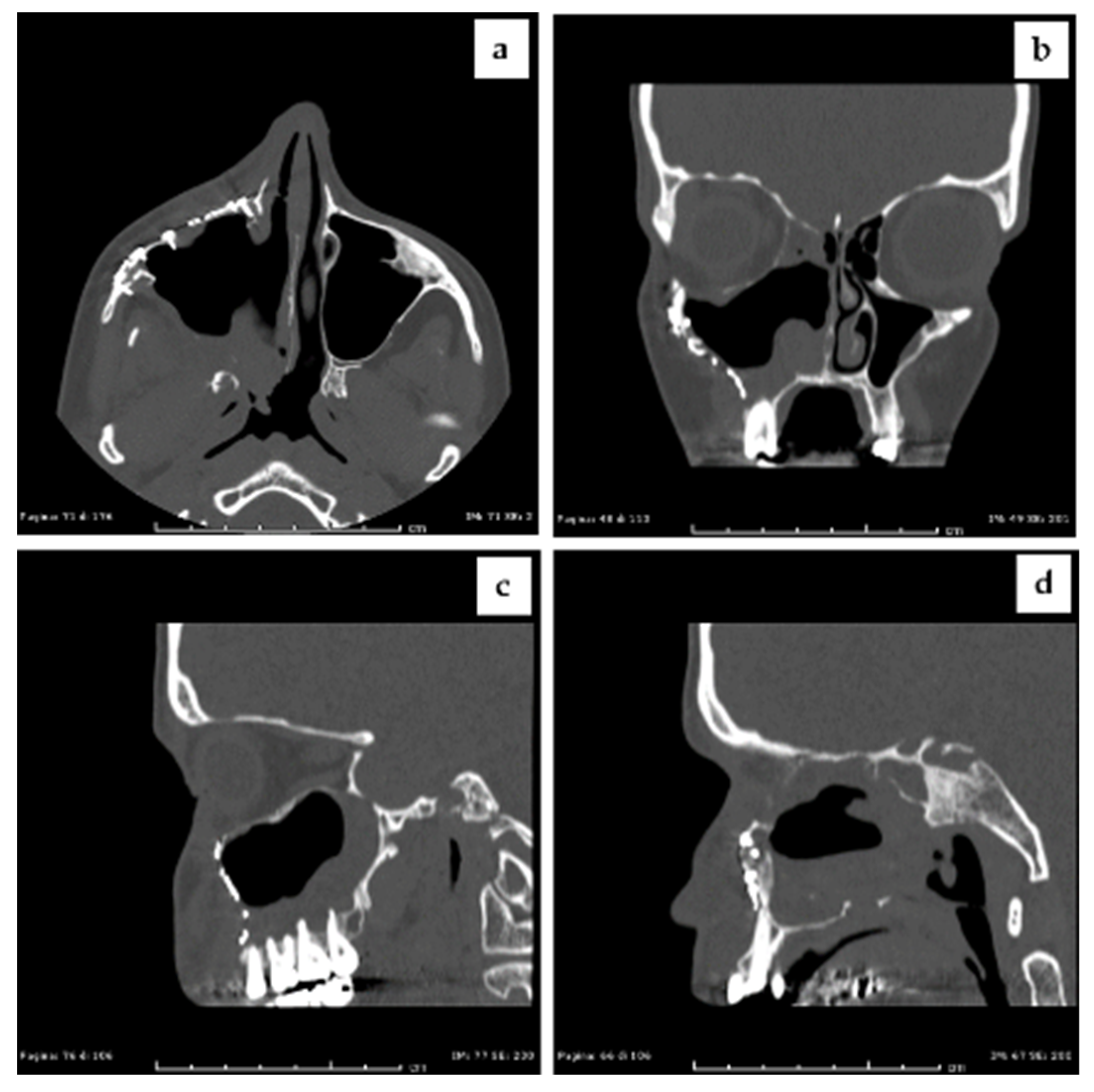
2.1. Clinical Presentation and Preoperative Imaging

A thirty-seven-year-old woman presented at the outpatient clinic with an ingravescent right nasal obstruction beginning 12 months previously and a right canine fossa swelling that appeared 4 months earlier. The patient was previously evaluated by the ear, nose, and throat (ENT) service and underwent a computed tomography scan (CT) of the paranasal sinuses, without contrast enhancement (Figure 1a,b).
The radiological exam showed an expansive hypodense lesion occupying the whole right maxillary sinus, with bone erosion of the surrounding structures (right orbital floor, antero-lateral, posterior, and medial walls of the right maxillary sinus) and the upper right vestibular fornix, the right nasal fossa, and the right infratemporal fossa invasion. The mass was located periapically to the last right upper premolar and the first two upper molars. At the endoscopic evaluation, the mass was covered by intact respiratory mucosa and a complete obstruction of the right middle meatus and of the right osteo-meatal complex was determined. Therefore, magnetic resonance imaging (MRI) with medium contrast and a maxillo-facial evaluation were prescribed.
The MRI showed a neoformation of 50 × 49 × 44 mm in size, with regular margins. The mass was iso-intense compared with the muscle tissue in T1-weighted sequences, dishomogeneously hyper-intense in T2-weighted sequences, and showed a massive and irregular contrast enhancement (Figure 1c,d).
At the physical examination, the overlying skin appeared reddened and inflamed. The patient denied previous epistaxis episodes and denied dysesthesia of the right infraorbital nerve, while she referred to hypoesthesia of the ipsilateral hemipalate. The patient denied tobacco and alcohol use. Cold testing tooth vitality of the last right upper premolar proved no response. The puncture of the swelling on the vestibular side determined a hematic spill and no purulent discharge. An intraoral biopsy was performed, showing only respiratory mucosa with chronic phlogistic infiltration and without neoplastic elements; the histopathological examination result had no diagnostic value. Shortly after the bioptic procedure, the patient referred to the onset of a right infraorbital nerve paresthesia. Therefore, an endoscopic biopsy under general anesthesia was scheduled. During the surgical intervention, a profuse hemorrhage from the mass bulging in the right nasal fossa occurred, which was controlled with bipolar cautery and right nasal cavity packing (Figure 2).

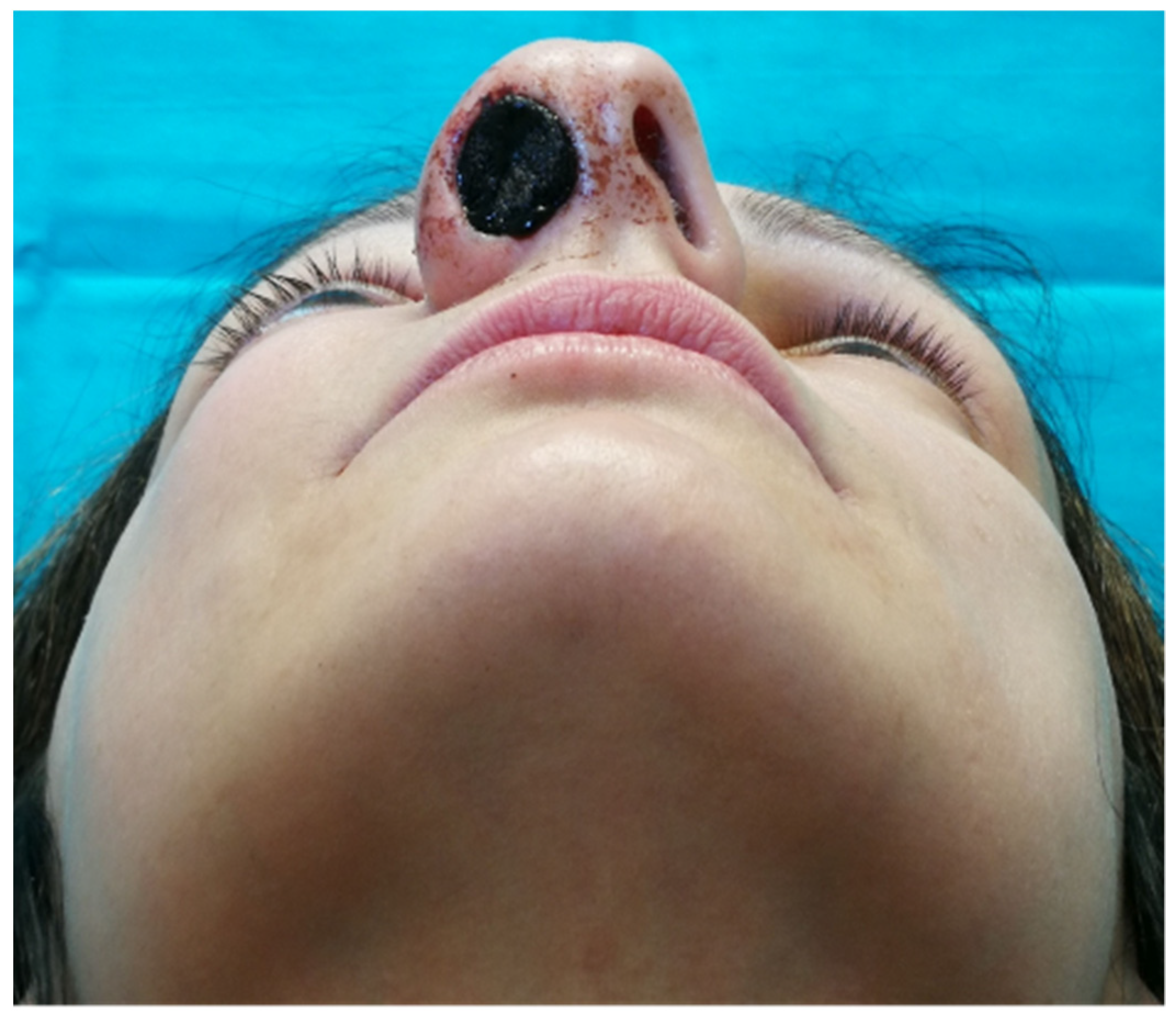
2.2. Incisional Biopsy Findings

The new histopathological result showed monomorphic spindle cells combined with vascular elements. At the immunohistochemical evaluation, the cells intensively expressed bcl2 and had a weak, but wide CD34 expression. There was no smooth muscle actin (SMA), S-100 protein, and MNF-116 cytokeratin expression (Figure 3).
The cytoproliferative activity (evaluated with Mib-1 expression) was lower than 10%. The described characteristics suggested a mesenchymal tumor diagnosis and did not show any signs of malignancy.

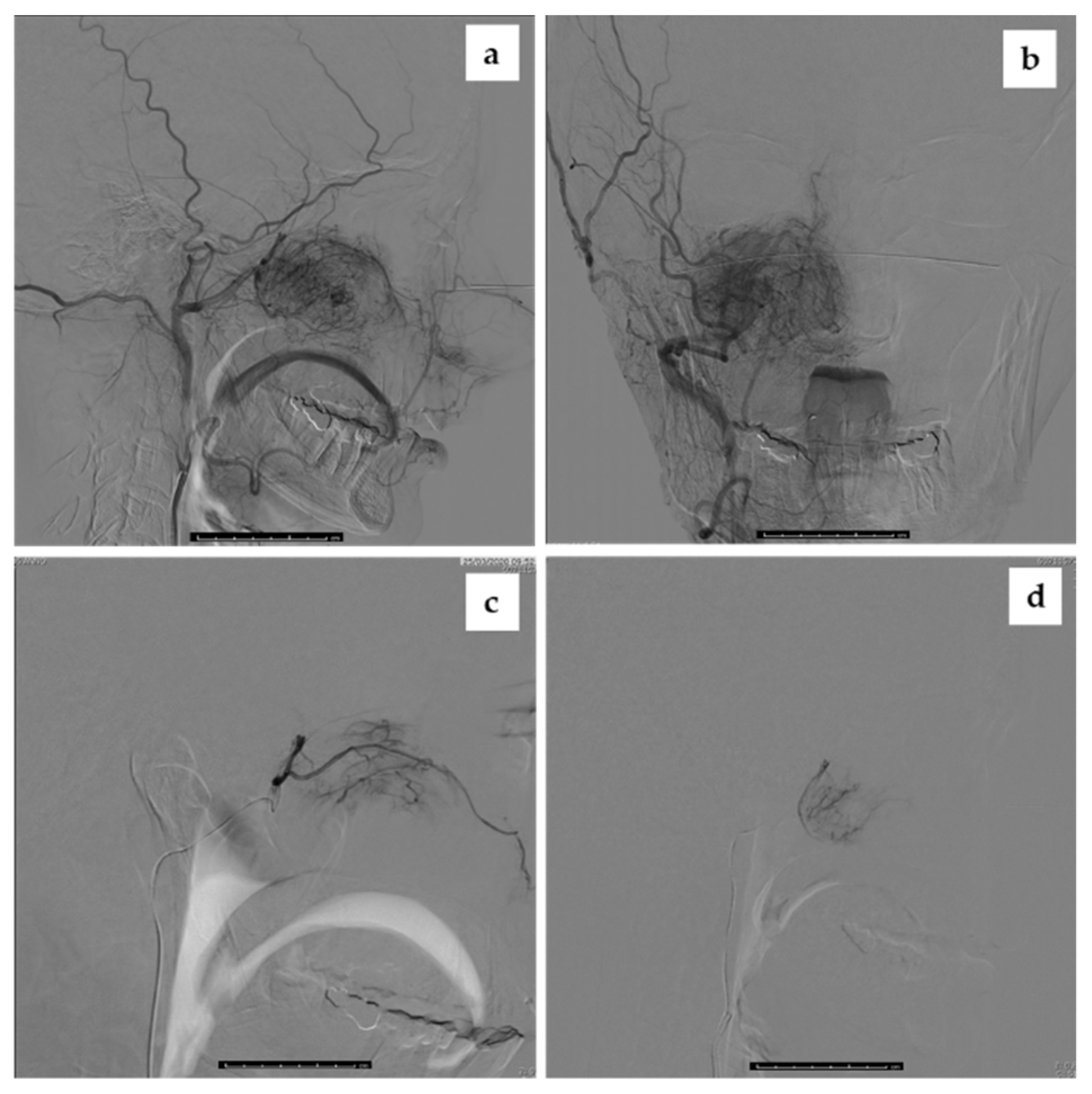
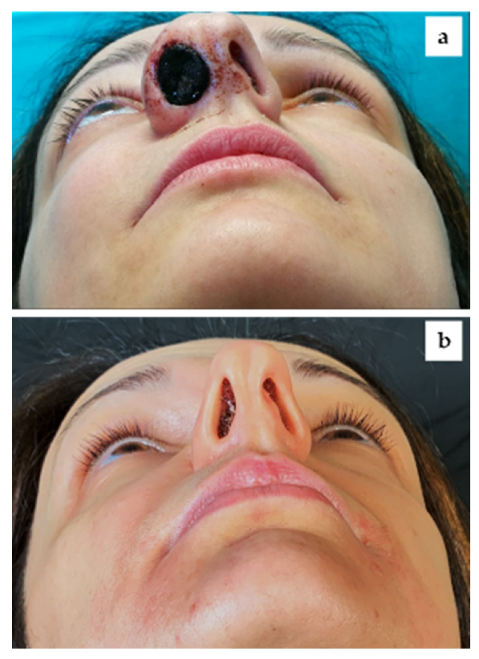
2.3. Intraoperative Imaging and Embolization Procedure

Given the important vascular component of the lesion, a preoperative head and neck angiography was performed. The exam showed a mild contrast enhancement in the late arterial phase and progressively higher enhancement in capillary and venous phases. The mass was vascularized by the terminal branches of the ophthalmic artery, of the internal maxillary artery (especially the sphenopalatine artery), and of the facial artery (Figure 4a,b).
Therefore, the vessels derived from the internal maxillary and facial arteries were embolized during the procedure. The subsequent angiography only showed the enhancement of the antero-superior compartment (Figure 3d and Figure 4c), supplied by the ophthalmic artery. No periprocedural complications were determined.

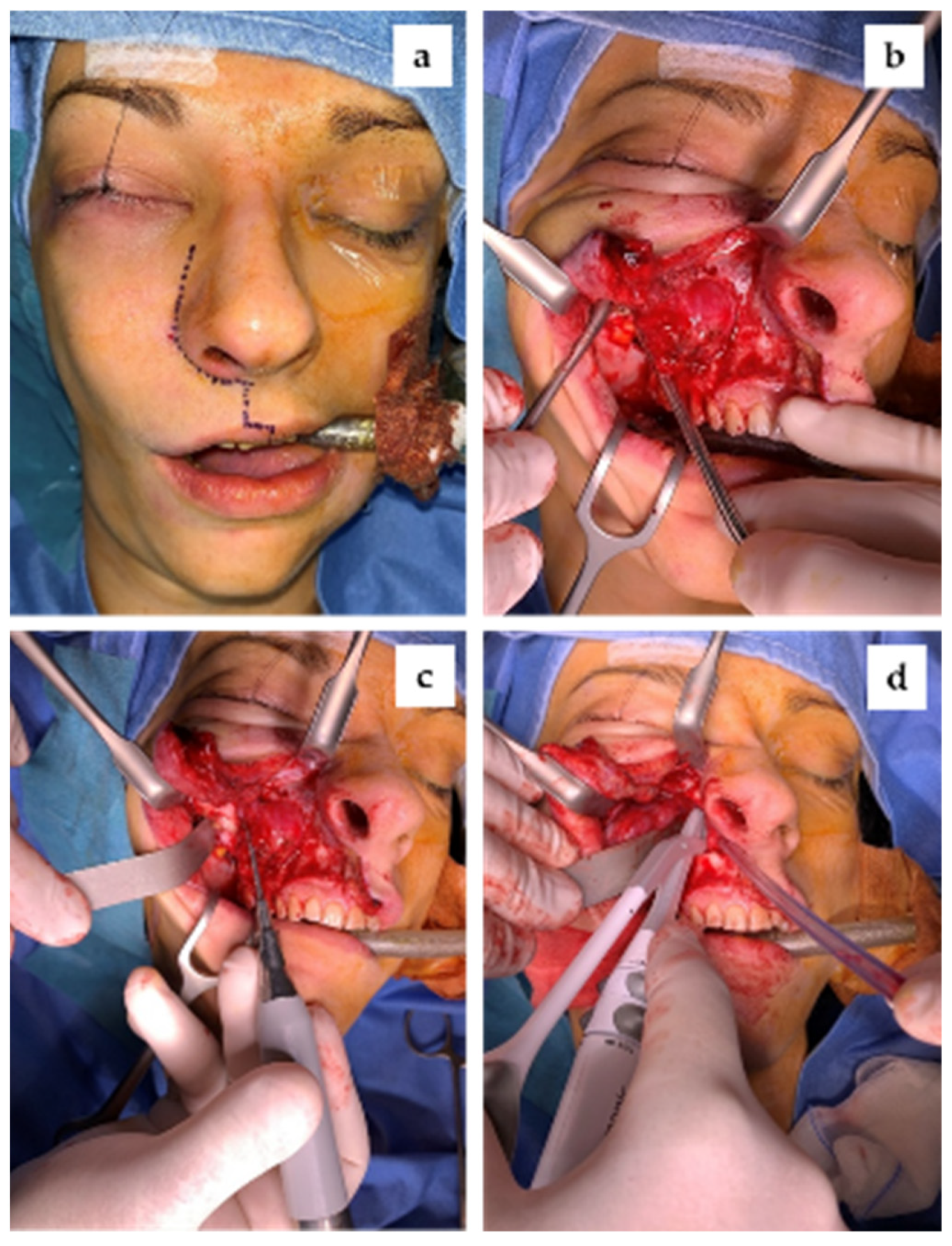
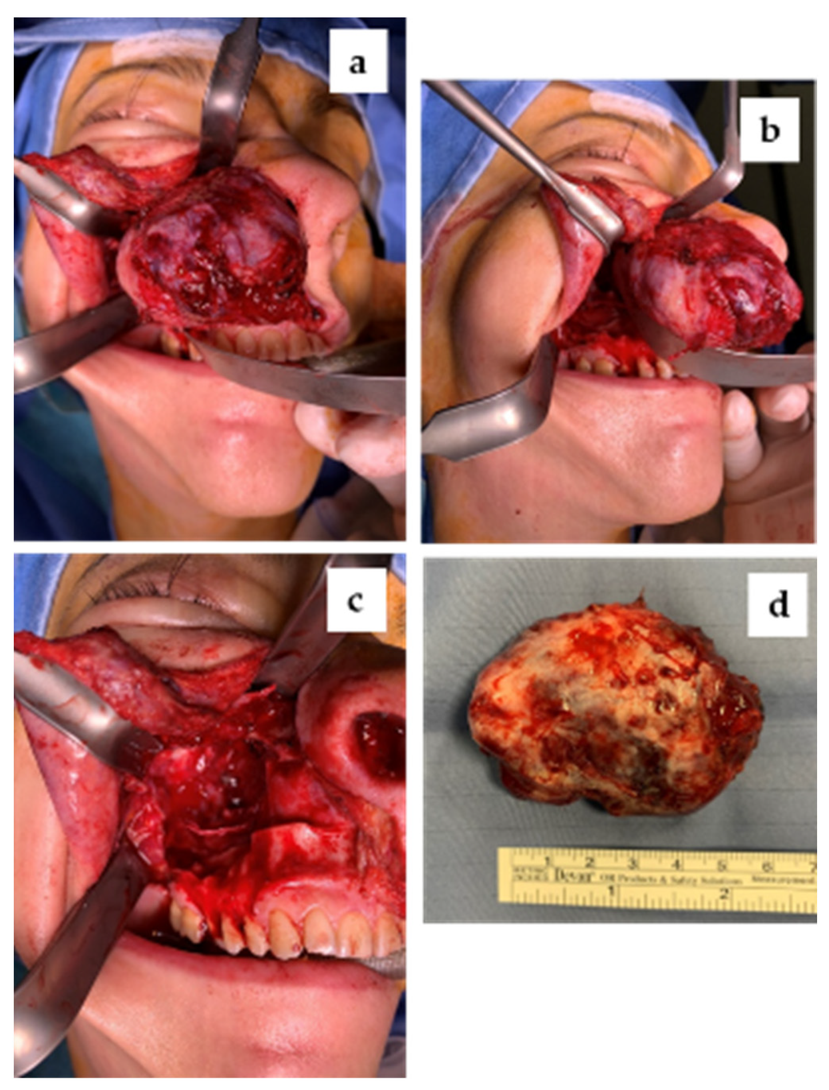
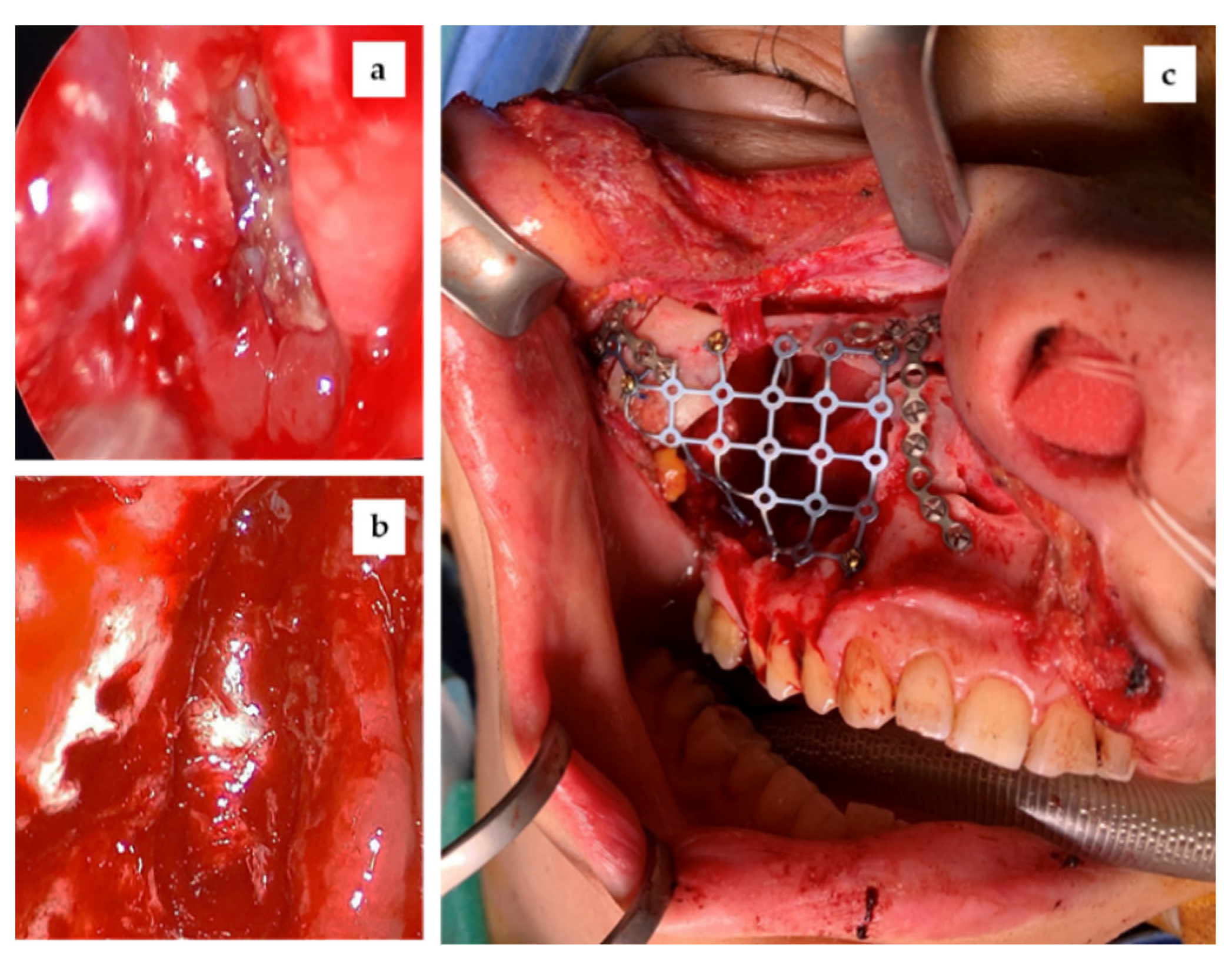
2.4. Surgical Management

Forty-eight hours after the angiographic procedure, the patient underwent the surgical excision of the neoformation throughout a combined endoscopic and transfacial approach. A Weber–Ferguson approach was carried out, with an intrasulcular incision of the right maxillary elements and a right tuber maxillae extension (Figure 5a,b).
The antero-lateral wall of the right maxillary sinus appeared expanded and thinned, with infero-distal erosion. The right infraorbitary nerve was identified at the exit from the homonymous foramen and preserved. Osteotomies of the right naso-frontal pillar and of the right infero-lateral orbital frame were performed with a piezosurgical scalpel (Figure 5c) (Piezosurgery®, Mectron Medical Technology, Carasco, Italy), without periosteal detachment, consenting their upward flipping. This surgical maneuver allowed a wider view of the lesion. The intraoperative evaluation of the mass confirmed the right infratemporal fossa and right nasal fossa invasion, but showed no orbital floor erosion, which was only thinned. We used the harmonic scalpel (Harmonic Focus®+, Ethicon Endo-Surgery, Inc, Cincinnati, OH, USA) (Figure 5d) characterized by high-frequency mechanical energy to offer the surgeon controlled and precise incision and haemostasis. The mass appeared well capsulated and easily cleavable from circumstantial tissues. The tumor was then directly removed (Figure 6).
An endoscopic revision was performed, with excision of the residual neoplastic tissues from right anterior and posterior ethmoidal structures and from the tail of the right inferior turbinate. The endoscopic evaluation excluded the invasion of the lamina cribrosa and of the right ethmoidal roof (Figure 7a,b).
An accurate hemostasis control was performed. The right naso-frontal pillar and the right infero-lateral orbital frame were osteosynthetized back in their position with titanium plate, while the anterior maxillary wall was reconstructed with a titanium mesh (Figure 7c). The transfacial approach was sutured and a right nasal packing was performed.

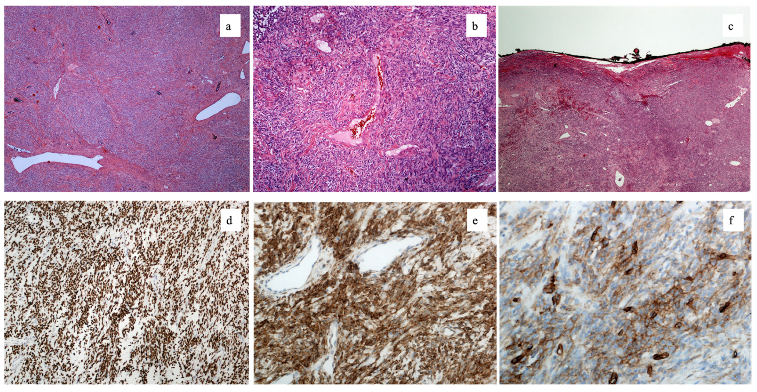
2.5. Solitary Fibrous Tumor Diagnosis

Pathological examination of the surgical specimens revealed a 5.5 × 4 × 3 cm solitary, well-circumscribed, gray-brownish nodule (Figure 6d), confirming the pre-operative immunohistochemical diagnosis.
Microscopically, the tumour was composed of monomorphic spindle cells arranged in fascicles, with a vascular component showing a peculiar “hemangiopericytomatous” pattern (Figure 3a,b). The only positive surgical margin is the one inside the maxillary sinus, which is not in contact with any other tissue (Figure 3c).
Spindle cells featured strong immunostaining for STAT6 (Figure 3d) and Bcl-2 (Figure 1e), and faint staining for CD34 (Figure 3f), whereas smooth muscle actin, S-100 protein, and cytokeratins were negative.
A diagnosis of solitary fibrous tumour was rendered. The surgical margins were clear from disease.

2.6. Follow Up

There were no postoperative complications. The patient did not refer to diplopia. The nasal packing was removed 48 h after the surgery, without subsequent epistaxis episodes. Immediate post-operative CT showed no macroscopic evidence of residual tumor and an appropriate bone reconstruction. The patient referred to transient infraorbital nerve hypoesthesia, which improved in three months. The 6-month post-operative CT excluded a relapse and sinusitis and confirmed the good reconstruction results obtained (Figure 8).
Clinically, the patient showed good facial symmetry, without anesthetic and visible scars (Figure 9).
The patient referred to a complete disappearance of the right hemipalate hypoesthesia, while a mild paresthesia of the right infraorbitary nerve was still present one year after the surgery.

2.7. Patient Consent

A written consent statement was provided by the patient to allow the publication of her case, including non-anonymized photos of her face during and after the surgical procedure.

3. Discussion

Without sex predilection, SFT generally occurs in the third to fourth decade of life [6]. This is consistent with our patient, a young 37-year-old woman.
In the scientific literature, SFT is generally described as a slow-growing, painless, asymptomatic mass [4,5]. However, it also can determine progressive nasal obstruction, rhinorrhea, intermittent epistaxis, headache and anosmia, and even exophthalmos. On nasal endoscopy, SFT usually appears as a mass covered by an intact surface respiratory epithelium and/or metaplastic squamous mucosa.
In the case described here, the patient referred only to a unilateral persistent nasal obstruction, and the nasoendoscopic exam showed an intact respiratory mucosa. While these features are consistent with those previously reported in literature, it has to be said that none of them are pathognomonic of SFT [3]. Therefore, the differential diagnosis must include glomangiopericytoma, inverted papilloma, hemangioma, leiomyomas, schwannoma, squamous cell carcinoma, juvenile angiofibroma, angiomatous polyps, nerve sheets tumors, mesenchymal chondrosarcoma, and byphenotipic synovial sarcoma [7,8,9]. The former definition of hemangiopericytoma has been integrated under the SFT nomenclature in the 2013 WHO classification [6]. Given the complexity of the clinical case, the treatment was given top priority even during the COVID-19 pandemic [10].
The differential diagnosis between SFTs of the paranasal sinuses and the previously described tumors is very difficult on a clinical basis, given the non-specific clinical presentation of them all. In fact, they can all manifest with nasal obstruction, recurrent epistaxis, sinusitis, and rhinorrhea [11,12,13,14].
In non-contrast to CT, SFT appears as a homogeneous mass and isodense with surrounding tissue. Thinning and local remodeling of adjacent bone was observed, owing to a pressure effect (Figure 1).
MRI findings included the following: well-circumscribed mass homogeneously isointense on T1-weighted sequences, associated with heterogeneously marked enhancement after gadolinium administration, and a possible hyperintensity on T2 image sequences [15,16], as described in the present case (Figure 1). Therefore, it is clear that SFT radiological findings are also not completely diriment [17].
Moreover, the radiological exams usually cannot be completely diriment regarding the final diagnosis, because of the similar aspects of SFTs and other mesenchymal tumors [18,19,20,21,22].
Given the great diversity of possible clinical pictures and the multiplicity of applicable surgical strategies, the need to perform an incisional biopsy of the lesion is evident, in order to establish the diagnosis and the correct therapeutic procedure.
Histopathologically, SFT consists of diffuse spindle cells within an attenuated collagenous stroma and a characteristic prominent vascular network. SFT cells also usually show low mitotic activity and the absence of nuclear pleomorphism [23].
At the immunochemical evaluation, SFT cells express CD34 and vimentin and moderately express Bcl-2, while being negative for cytokeratin, S-100 protein, smooth muscle actin, and desmin [24]. CD34 positivity is not pathognomonic; nevertheless, the presence of CD34 and the contextual negativity of other reported markers can help to exclude a variety of soft tissue tumors, such as epithelial tumors, neurogenic tumors, and sarcomas [24]. Moreover, it has to be said that a typical rearrangement in chromosome 12q13, with a NAB2–STAT6 gene fusion, is present [25,26]. In our case, a STAT6 strong and diffuse nuclear expression pointed toward a diagnosis of SFT, with this marker being highly specific for this tumor. In fact, nuclear expression of STAT6 reflects the NAB2–STAT6 gene fusion that characterizes SFT [27]. Unfortunately, this molecular investigation could not be performed in our case, but it has to be said that only occasionally can other mesenchymal tumors express STAT-6, both in cytoplasm and nucleus, rather than exclusively in the nucleus [28].
Glomangiopericytoma shows the same cellular component of SFT and the same presence of collagen areas. Moreover, it is positive for muscular markers (such as smooth muscle actin) as well as leiomyomas. Nasopharingeal angiofibromas, on the other side, are usually positive for androgen receptor expression. None of the benign tumors mentioned here show CD34 and STAT6 expression [8]. Tumors with neural differentiation, such as neurofibroma or schwannoma, can histologically mimic SFTs, but they differ by expressing S-100 protein and SOX10 while lacking STAT6 expression [8]. Histologically, synovial sarcomas appear to be made of spindle cells, with the possible presence of collagen bands and branching vessels, similarly to SFTs. On the other side, they usually are more densely cellular and they lack of CD34 and STAT6 expressions, while being positive to keratin and EMA [28].
Even though there are not definite treatment guidelines for head and neck localization, resectability is the most important prognostic factor; the en bloc radical surgical excision is thus the treatment of choice [29]. However, surgical margins’ control is frequently difficult in the paranasal region, and an endoscopic approach can be an efficient tool to grant magnification for an accurate resection with limited morbidity. Of course, in the present case, the only positive surgical margin is the one inside the maxillary sinus, which is not in contact with any other tissue (Figure 3c). For this reason, the surgical margins can be considered clear from disease.
Nevertheless, in a highly vascularized lesion, neither a transfacial approach nor an exclusively endoscopic approach grant an excellent control of the surgical margins in the paranasal sinuses. SFTs have an aggressive behavior and an important recurrence potential.
Owing to the highly vascular nature of SFT, a surgical intervention can often determine a consistent blood loss. In the present case report, severe bleeding was also encountered during the endoscopic bioptic procedure. Therefore, we decided to preventively perform a selective endovascular embolization to obtain improved control of the bleeding during the following resection procedure. This foresight allowed a dramatic reduction of the blood supply to the neoformation, only leaving the vascularization from some branches of the ophthalmic artery. Indeed, the definitive resection procedure did not determine a major blood loss.
In order to obtain radicality and increase surgical safety, we planned a multidisciplinary approach, combining the transfacial Weber–Ferguson approach with the magnification power of endoscopy for better control and radicalization.
Given the delicacy of the osteotomies performed, the use of the piezosurgical scalpel (Piezosurgery®, Mectron Medical Technology, Carasco, Italy) was preferred owing to its better impact on bone healing dynamics [30,31].
Moreover, to obtain an effective hemostasis in the nasal fossa and less extensive tissue damage to reminder nasal mucosa, the harmonic scalpel (Harmonic Focus®+, Ethicon Endo-Surgery, Inc, Cincinnati, OH, USA) was used [32].
SFTs are generally indolent, with a low recurrence rate. Many risk assessment methods have been developed, while a universal system has not been approved yet [33,34,35]. Regarding intrathoracic SFTs, some inflammatory markers have also been proven to be of prognostic importance, such as fibrinogen levels and the neutrophil-to-lymphocyte-ratio (NLR) [36].
Overall, the recurrence rate accounts to 10–25%, with the possibility of a late relapse occurring even 10 years after the surgical resection [37,38]. Despite the limited number of reported cases, head and neck SFTs seem to have good prognostic results after radical surgical excision alone. Generally, no adjuvant therapies are required and surgical resection is considered curative.

4. Conclusions

Owing to its rarity and its aspecific morphologic appearance, sinonasal SFT may be difficult to distinguish from other mesenchymal lesions that more commonly arise in this area.
An accurate differential diagnosis is necessary in order to avoid confusion with more aggressive lesions, which are more frequent in the head and neck region and which require an extensive resection, possibly functionally and aesthetically disabling.
Radical surgical resection is the treatment of choice for SFT. Hopefully preceded by embolization, a combined open and endoscopic approach, in the context of a multi-equipe surgical team, can be helpful for an accurate resection with better visibility and improved hemostasis.

Author Contributions

Conceptualization, M.D.B. and S.N.; methodology, A.A.; validation, A.A., S.N. and M.D.B.; formal analysis, A.P.; investigation, L.C., S.V., A.M.C. and L.P.; data curation, A.P.; writing—original draft preparation, M.D.B. and A.A.; writing—review and editing, A.A.; visualization, A.P.; supervision, A.A. All authors have read and agreed to the published version of the manuscript.

Funding

This research received no external funding.

Institutional Review Board Statement

Due to the retrospective nature of this study, it was granted an exemption by the Institutional Review Board of the University Hospital of Modena, Italy. All procedures performed involving the human participant were in accordance with the 1964 Helsinki Declaration and its later amendments or comparable ethical standards.

Informed Consent Statement

A written consent statement was provided by the patient to allow the publication of her case, including non-anonymized photos of her face during and after the surgical procedure.

Conflicts of Interest

The authors declare no conflict of interest.

References

  1. Klemperer, P.; Rabin, C.B. Primary Neoplasms of the pleura. A report of five cases. Am. J. Ind. Med. 1992, 22, 4–31. [Google Scholar] [CrossRef]
  2. Demicco, E.G.; Park, M.S.; Araujo, D.M.; Fox, P.S.; Bassett, R.L.; Pollock, R.E.; Lazar, A.J.; Wang, W.-L. Solitary fibrous tumor: A clinicopathological study of 110 cases and proposed risk assessment model. Mod. Pathol. 2012, 25, 1298–1306. [Google Scholar] [CrossRef] [Green Version]
  3. Gold, J.S.; Antonescu, C.R.; Hajdu, C.; Ferrone, C.R.; Hussain, M.; Lewis, J.J.; Brennan, M.F.; Coit, D.G. Clinicopathologic correlates of solitary fibrous tumors. Cancer 2002, 94, 1057–1068. [Google Scholar] [CrossRef] [PubMed]
  4. Smith, S.C.; Gooding, W.E.; Elkins, M.; Patel, R.M.; Harms, P.W.; McDaniel, A.S.; Palanisamy, N.; Uram-Tuculescu, C.; Balzer, B.B.; Lucas, D.R.; et al. Solitary Fibrous Tumors of the Head and Neck: A Multi-Institutional Clinicopathologic Study. Am. J. Surg. Pathol. 2017, 41, 1642–1656. [Google Scholar] [CrossRef] [PubMed]
  5. Bernardini, F.P.; de Conciliis, C.; Schneider, S.; Kersten, R.C.; Kulwin, D.R. Solitary fibrous tumor of the orbit: Is it rare? Report of a case series and review of the literature. Ophthalmology 2003, 110, 1442–1448. [Google Scholar] [CrossRef]
  6. Fletcher, C.D.M.; World Health Organization. International Agency for Research on Cancer. In WHO Classification of Tumours of Soft Tissue and Bone; International Agency for Research on Cancer: Lyon, France, 2013; ISBN 9789283224341. [Google Scholar]
  7. Martin-Broto, J.; Mondaza-Hernandez, J.L.; Moura, D.S.; Hindi, N. A Comprehensive Review on Solitary Fibrous Tumor: New Insights for New Horizons. Cancers 2021, 13, 2913. [Google Scholar] [CrossRef] [PubMed]
  8. Tariq, M.U.; Din, N.U.; Abdul-Ghafar, J.; Park, Y.-K. The many faces of solitary fibrous tumor; diversity of histological features, differential diagnosis and role of molecular studies and surrogate markers in avoiding misdiagnosis and predicting the behavior. Diagn. Pathol. 2021, 16, 32. [Google Scholar] [CrossRef] [PubMed]
  9. Thompson, L.D.R.; Lau, S.K. Sinonasal Tract Solitary Fibrous Tumor: A Clinicopathologic Study of Six Cases with a Comprehensive Review of the Literature. Head Neck Pathol. 2018, 12, 471–480. [Google Scholar] [CrossRef]
  10. Di Bartolomeo, M.; Pellacani, A.; Negrello, S.; Chiarini, L.; Anesi, A. Emerging challenges and possible strategies in maxillo-facial and oral surgery during the COVID-19 pandemic. J. Oral Sci. 2020, 62, 452–454. [Google Scholar] [CrossRef]
  11. Thobejane, O.; Maharaj, S. Nasal Septal Angiofibroma. Ear Nose Throat J. 2021, 1455613211026517. [Google Scholar] [CrossRef]
  12. Ghaloo, S.K.; Dhanani, R.; Pasha, H.A.; Wasif, M.; Fatima, S.; Ikram, M. Glomangiopericytoma: A rare tumour of sinonasal cavity. J. Pak. Med. Assoc. 2020, 70, 2469–2471. [Google Scholar] [CrossRef] [PubMed]
  13. Assiri, K.S.; Al-Ahmari, M.S.; Alshahrani, M.S.; Mastor, A.; Elhawary, R. Clinical and Pathological Features of Angiomatous Nasal Polyps: A Report of Four Cases and Review of Literature. Cureus 2020, 12, e7642. [Google Scholar] [CrossRef] [PubMed] [Green Version]
  14. Katre, M.I. Neurofibroma of Nasal Cavity and Nasopharynx. Adv. Case Stud. 2017, 1. [Google Scholar] [CrossRef]
  15. Bowe, S.N.; Wakely, P.E.; Ozer, E. Head and neck solitary fibrous tumors: Diagnostic and therapeutic challenges. Laryngoscope 2012, 122, 1748–1755. [Google Scholar] [CrossRef]
  16. Ganly, I.; Patel, S.G.; Stambuk, H.E.; Coleman, M.; Ghossein, R.; Carlson, D.; Edgar, M.; Shah, J.P. Solitary fibrous tumors of the head and neck: A clinicopathologic and radiologic review. Arch. Otolaryngol. Head Neck Surg. 2006, 132, 517–525. [Google Scholar] [CrossRef] [Green Version]
  17. Yang, B.T.; Song, Z.L.; Wang, Y.Z.; Dong, J.Y.; Wang, Z.C. Solitary fibrous tumor of the sinonasal cavity: CT and MR imaging findings. AJNR Am. J. Neuroradiol. 2013, 34, 1248–1251. [Google Scholar] [CrossRef] [Green Version]
  18. Suroyo, I.; Budianto, T. The role of diagnostic and interventional radiology in juvenile nasopharyngeal angiofibroma: A case report and literature review. Radiol. Case Rep. 2020, 15, 812–815. [Google Scholar] [CrossRef]
  19. Wang, X.; Liu, Y.; Chen, Q.; Xian, J. Evaluation of multiparametric MRI differentiating sinonasal angiomatous polyp from malignant tumors. Neuroradiology 2019, 61, 891–896. [Google Scholar] [CrossRef]
  20. Lin, N.; Liu, X.; Zhang, F.; Pan, Y.; Qi, M.; Sha, Y. Sinonasal synovial sarcoma: Evaluation of the role of radiological and clinicopathological features in diagnosis. Clin. Radiol. 2021, 76, 78. [Google Scholar] [CrossRef]
  21. Yang, B.T.; Wang, Z.C.; Xian, J.F.; Hao, D.P.; Chen, Q.H. Leiomyoma of the sinonasal cavity: CT and MRI findings. Clin. Radiol. 2009, 64, 1203–1209. [Google Scholar] [CrossRef]
  22. Suh, C.H.; Lee, J.H.; Lee, M.K.; Cho, S.J.; Chung, S.R.; Choi, Y.J.; Baek, J.H. CT and MRI Findings of Glomangiopericytoma in the Head and Neck: Case Series Study and Systematic Review. AJNR Am. J. Neuroradiol. 2020, 41, 155–159. [Google Scholar] [CrossRef] [PubMed]
  23. Kao, Y.-C.; Lin, P.-C.; Yen, S.-L.; Huang, S.-C.; Tsai, J.-W.; Li, C.-F.; Tai, H.-C.; Lan, J.; Chuang, I.-C.; Yu, S.-C.; et al. Clinicopathological and genetic heterogeneity of the head and neck solitary fibrous tumours: A comparative histological, immunohistochemical and molecular study of 36 cases. Histopathology 2016, 68, 492–501. [Google Scholar] [CrossRef] [PubMed]
  24. Han, Y.; Zhang, Q.; Yu, X.; Han, X.; Wang, H.; Xu, Y.; Qiu, X.; Jin, F. Immunohistochemical detection of STAT6, CD34, CD99 and BCL-2 for diagnosing solitary fibrous tumors/hemangiopericytomas. Int. J. Clin. Exp. Pathol. 2015, 8, 13166–13175. [Google Scholar] [PubMed]
  25. Chmielecki, J.; Crago, A.M.; Rosenberg, M.; O’Connor, R.; Walker, S.R.; Ambrogio, L.; Auclair, D.; McKenna, A.; Heinrich, M.C.; Frank, D.A.; et al. Whole-exome sequencing identifies a recurrent NAB2-STAT6 fusion in solitary fibrous tumors. Nat. Genet. 2013, 45, 131–132. [Google Scholar] [CrossRef] [PubMed] [Green Version]
  26. Robinson, D.R.; Wu, Y.-M.; Kalyana-Sundaram, S.; Cao, X.; Lonigro, R.J.; Sung, Y.-S.; Chen, C.-L.; Zhang, L.; Wang, R.; Su, F.; et al. Identification of recurrent NAB2-STAT6 gene fusions in solitary fibrous tumor by integrative sequencing. Nat. Genet. 2013, 45, 180–185. [Google Scholar] [CrossRef] [PubMed] [Green Version]
  27. Koelsche, C.; Schweizer, L.; Renner, M.; Warth, A.; Jones, D.T.W.; Sahm, F.; Reuss, D.E.; Capper, D.; Knösel, T.; Schulz, B.; et al. Nuclear relocation of STAT6 reliably predicts NAB2-STAT6 fusion for the diagnosis of solitary fibrous tumour. Histopathology 2014, 65, 613–622. [Google Scholar] [CrossRef]
  28. Doyle, L.A.; Vivero, M.; Fletcher, C.D.; Mertens, F.; Hornick, J.L. Nuclear expression of STAT6 distinguishes solitary fibrous tumor from histologic mimics. Mod. Pathol. 2014, 27, 390–395. [Google Scholar] [CrossRef] [Green Version]
  29. van Houdt, W.J.; Westerveld, C.M.A.; Vrijenhoek, J.E.P.; van Gorp, J.; van Coevorden, F.; Verhoef, C.; van Dalen, T. Prognosis of Solitary Fibrous Tumors: A Multicenter Study. Ann. Surg. Oncol. 2013, 20, 4090–4095. [Google Scholar] [CrossRef]
  30. Anesi, A.; Ferretti, M.; Cavani, F.; Salvatori, R.; Bianchi, M.; Russo, A.; Chiarini, L.; Palumbo, C. Structural and ultrastructural analyses of bone regeneration in rabbit cranial osteotomy: Piezosurgery versus traditional osteotomes. J. Cranio-Maxillofac. Surg. 2018, 46, 107–118. [Google Scholar] [CrossRef]
  31. Anesi, A.; Di Bartolomeo, M.; Pellacani, A.; Ferretti, M.; Cavani, F.; Salvatori, R.; Nocini, R.; Palumbo, C.; Chiarini, L. Bone Healing Evaluation Following Different Osteotomic Techniques in Animal Models: A Suitable Method for Clinical Insights. Appl. Sci. 2020, 10, 7165. [Google Scholar] [CrossRef]
  32. Sherman, J.A.; Davies, H.T. Ultracision: The harmonic scalpel and its possible uses in maxillofacial surgery. Br. J. Oral Maxillofac. Surg. 2000, 38, 530–532. [Google Scholar] [CrossRef] [PubMed]
  33. Lu, C.; Ji, Y.; Shan, F.; Guo, W.; Ding, J.; Ge, D. Solitary fibrous tumor of the pleura: An analysis of 13 cases. World J. Surg. 2008, 32, 1663–1668. [Google Scholar] [CrossRef] [PubMed]
  34. Gholami, S.; Cassidy, M.R.; Kirane, A.; Kuk, D.; Zanchelli, B.; Antonescu, C.R.; Singer, S.; Brennan, M. Size and Location are the Most Important Risk Factors for Malignant Behavior in Resected Solitary Fibrous Tumors. Ann. Surg. Oncol. 2017, 24, 3865–3871. [Google Scholar] [CrossRef]
  35. Demicco, E.G.; Wagner, M.J.; Maki, R.G.; Gupta, V.; Iofin, I.; Lazar, A.J.; Wang, W.-L. Risk assessment in solitary fibrous tumors: Validation and refinement of a risk stratification model. Mod. Pathol. 2017, 30, 1433–1442. [Google Scholar] [CrossRef]
  36. Ghanim, B.; Hess, S.; Bertoglio, P.; Celik, A.; Bas, A.; Oberndorfer, F.; Melfi, F.; Mussi, A.; Klepetko, W.; Pirker, C.; et al. Intrathoracic solitary fibrous tumor—An international multicenter study on clinical outcome and novel circulating biomarkers. Sci. Rep. 2017, 7, 12557. [Google Scholar] [CrossRef] [Green Version]
  37. Baldi, G.G.; Stacchiotti, S.; Mauro, V.; Dei Tos, A.P.; Gronchi, A.; Pastorino, U.; Duranti, L.; Provenzano, S.; Marrari, A.; Libertini, M.; et al. Solitary fibrous tumor of all sites: Outcome of late recurrences in 14 patients. Clin. Sarcoma Res. 2013, 3, 4. [Google Scholar] [CrossRef] [PubMed] [Green Version]
  38. Park, C.K.; Lee, D.H.; Park, J.Y.; Park, S.H.; Kwon, K.Y. Multiple recurrent malignant solitary fibrous tumors: Long-term follow-up of 24 years. Ann. Thorac. Surg. 2011, 91, 1285–1288. [Google Scholar] [CrossRef] [Green Version]
Figure 1. Preoperative imaging. (a) CT, axial view; (b) CT, coronal view; (c) MRI, sagittal view; (d) MRI, coronal view.
Reports 04 00033 g001
Figure 2. Picture following the endoscopic bioptic procedure. A profuse bleeding occurred, controlled with bipolar cautery and right nasal cavity packing. The bulging of the right cheek because of the mass is clearly evident.
Reports 04 00033 g002
Figure 3. Microscopically, the tumor was composed of monomorphic spindle cells arranged in fascicles, with a vascular component showing a peculiar “hemangiopericytomatous” pattern. Spindle cells featured strong immunostaining for Bcl-2, and faint staining for CD34, whereas smooth muscle actin, S-100 protein, and cytokeratins were negative. (a,b) The nodule was composed of spindle cells arranged in fascicles; vessels with hemangiopericytomatous pattern were visible (H&E staining: 40× magnification (a); 100× magnification (b); 40× magnification (c) the only positive surgical margin is the one inside the maxillary sinus, which is not in contact with any other tissue. Spindle cells were positive for (d) STAT6: 100× magnification, (e) Bcl-2: 100× magnification, and (f) CD34: 100× magnification.
Reports 04 00033 g003
Figure 4. Angiographic imaging. (a,b) Pre-embolization images, showing the vascularization by terminal branches of the ophthalmic artery, of the internal maxillary artery (especially the sphenopalatine artery), and of the facial artery; (c,d) images after the embolization of the internal maxillary and facial arteries, showing only the enhancement of the antero-superior compartment, supplied by the ophthalmic artery.
Reports 04 00033 g004
Figure 5. Intraoperative pictures. (a) Preoperative drawing; (b) isolation of the neoformation; (c) fine maxillary osteotomies performed with the piezosurgical scalpel; and (d) resection of the nasal part of the mass carried out with the harmonic scalpel.
Reports 04 00033 g005
Figure 6. Intraoperative pictures. (a,b) Resection of the neoformation; (c) post-resective site. (d) Macroscopically, a 5.5 × 4 × 3 cm well-circumscribed nodule was visible.
Reports 04 00033 g006
Figure 7. Intraoperative pictures. (a) Endoscopic image of the nasal fossa after the macroscopic resection (b) and at the end of the endoscopic one. (c) Titanium mesh reconstruction of the anterior maxillary sinus wall.
Reports 04 00033 g007
Figure 8. Post-operative CT. (a) Axial view; (b) coronal view; and (c,d) sagittal views.
Reports 04 00033 g008
Figure 9. Clinical pictures: (a) picture following the bioptic procedure with right nasal packing; (b) 12 months post-operative picture. The bulging under the right cheek is not evident anymore.
Reports 04 00033 g009
Publisher’s Note: MDPI stays neutral with regard to jurisdictional claims in published maps and institutional affiliations.

