Article

Dehydroepiandrosterone (DHEA) Improves the Metabolic and Haemostatic Disturbances in Rats with Male Hypogonadism

1
Medical Physiology Department, Faculty of Medicine, Mansoura University, Mansoura 35516, Egypt
2
Department of endocrinology and Internal Medicine, Delta University for Science and Technology, Gamasa 35712, Egypt
3
Medical Biochemistry and Molecular Biology Department, Faculty of Medicine, Mansoura University, Mansoura 35516, Egypt
4
Department of Clinical Laboratory Sciences, College of Applied Medical Sciences, Taif University, P.O. Box 11099, Taif 21944, Saudi Arabia
5
Department of Physiology, College of Medicine and Health Sciences, Sultan Qaboos University, Muscat P.O. Box 123, Oman
*
Author to whom correspondence should be addressed.
Submission received: 21 October 2021 / Revised: 19 December 2021 / Accepted: 21 December 2021 / Published: 20 January 2022

Abstract

:
Objectives: The current work was designed to study the effect of dehydroepiandrosterone (DHEA) on glucose homeostasis, liver functions and hemostatic disturbances in a rat model of bilateral orchidectomy (ORCH). Methods: 32 male rats (n = 8) were randomly assigned into 4 groups; (i) control (sham operated) group; were normal rats in which all surgical procedures were done without ORCH, (ii) Control + DHEA group: as control group but rats were treated with DHEA for 12 weeks, (iii) orchiectomized (ORCH) group: rats had bilateral orchidectomy and (iv) ORCH + DHEA group: orchiectomized rats treated with DHEA for 12 weeks. Four weeks after ORCH, DHEA treatment began and lasted for twelve weeks. By the end of the experiment, the parameters of glucose homeostasis, lipid profile, liver enzymes, bleeding and clotting times (B.T. and C.T.), prothrombin time (P.T.), activated partial thromboplastin time (aPTT), platelet count and aggregation, von-Willebrand factor (vWF), fibrinogen, plasminogen activator inhibitor (PAI-1), fibrin degradation products (FDP), intercellular adhesion molecule (ICAM-1), vascular cell adhesion molecule (VCAM-1), endothelin-1 were measured. Results: ORCH caused significant deteriorations in the parameters of glucose homeostasis, lipid profile, and liver functions (p < 0.05). In addition, lower androgenicity-induced by ORCH caused a significant rise in PAI-1, fibrinogen, FDPs, ET-1 (p < 0.01) with significant shortening of bleeding and clotting times. DHEA replacement therapy significantly decreased glucose, insulin, PAI-1, fibrinogen, ICAM-1, and VCAM-1 when compared to ORCH rats. Conclusion: DHEA ameliorated the metabolic, hepatic, hypercoagulable, and hypofibrinolysis disturbances induced by ORCH.

1. Introduction

Male hypogonadism that is associated with a low serum testosterone level is commonly seen in male diabetic patients and metabolic syndrome [1]. In addition, low serum androgens are commonly associated with insulin resistance, central obesity, elevated LDL, hypertension, hypercoagulability, hypofibrinolysis, poor cardiac function, and ischemic heart diseases [2]. Recent studies have documented that about 50% of men with type 2 diabetes mellitus (T2DM) have impaired testicular functions [1]. Furthermore, previous studies reported a strong relation between insulin resistance and testosterone deficiency [3,4,5]. Furthermore, the mechanisms underlying obesity-induced androgen deficiency are not fully understood. Federica et al. [6] reported that insulin resistance and metabolic syndrome are key players in the pathogenesis of androgen deficiency and the link between them appears to be is bidirectional. Nonalcoholic fatty liver disease (NAFLD) which encompasses a wide range of hepatic damage, from simple macrovesicular steatosis to steatohepatitis, advanced fibrosis, and cirrhosis [7] is referred to as a hepatic component of metabolic syndrome (MS).
Previous studies reported that androgen deficiency in obese men is associated with higher plasma levels of some prothrombotic factors such as plasminogen activator inhibitor (PAI-1) [8], which provides an explanation for the higher cardiovascular risk in patients with central obesity [9]. It has been demonstrated that androgens have clear impacts on endothelial levels prothrombotic proteins like von Willibrand factor, and PAI-1 [10]. Furthermore, Webb et al. [11] documented that intracoronary testosterone infusions improve coronary blood flow by causing dilatation of coronary vessels and long-term therapy with testosterone in males with low plasma testosterone and coronary heart disease significantly improves the signs of heart ischemia. In comparison, a study by Kojima et al. [12] showed that orchidectomy caused an obvious elevation in plasminogen activator activity in male rats and did not influence the plasminogen activator inhibitor activity. Recently, Demirci et al. [13] found high levels of platelet count and surrogate markers of cardiovascular risk, e.g., mean platelet volume (MPV), platelet-to-lymphocyte ratio (PLR), and neutrophil-to-lymphocyte ratio (NLR) in congenital hypogonadotropic hypogonadism (CHH) patients. However, they found that hormonal replacement therapy with testosterone did not improve these parameters.
Dehydroepiandrosterone (DHEA) and DHEA-sulphate (S) are considered pre-hormones. In spite of the fact that DHEA-S may be a circulating stock, it is hydrophilic, and as it is lipophilic DHEA can be changed over to more strong androgens and estrogens in fringe tissues [14]. Steroid formation at a tissue level permits for auto-regulation of the neighborhood hormonal environment based on neighborhood prerequisites, with fewer systemic side impacts [15]. DHEA produces 50–70 percent of estrogens and the bulk of androgens in ladies before menopause through intracrine processes. The production of local hormones in males whose testes continue to generate androgens throughout their lives, however, is more difficult to assess [14,16]. To the best of our knowledge, there is no study that has examined the effect of DHEA on prothrombic markers and fibrinolytic system activities, therefore, the current work was designed to investigate the effect of DHEA on the metabolic, hepatic, prothrombotic markers and fibrinolytic system activity disturbances in male hypogonadism in rats.

2. Materials and Methods

2.1. Animals Housing and Diet

32- male Sprague Dawley rats (aged 12–14 weeks, weighing 300 ± 25 g) were purchased and housed in the Nile center for experimental research, Mansoura, Egypt under controlled conditions (temperature of 23 ± 1 °C and a 12 h light:12 h dark cycle). The animals had unrestricted access to food and water. The Guide for the Care and Use of Laboratory Animals published by the US National Institutes of Health (NIH publication No. 85–23, revised 1996) was used as a guide for the animal experiments. All protocols and procedures were approved by our local ethical committee, faculty of medicine, delta university for science and technology (#RP.21.10.26).

2.2. Experimental Design

Rats were allocated into 4 groups (each with eight rats) after a week of acclimatization to the research facility environment; (a) control (sham operated) group: rats in this group underwent all surgical procedures without orchiectomy and treated with saline, (b) Control + DHEA: as control group but rats were treated with subcutaneous DHEA pellets implants (50 mg, Innovative Research America, Cat#, NX-999, USA) that releases DHEA for 12 weeks, (c) orchiectomized (ORCH) group: rats underwent bilateral orchidectomy and treated with saline for 12 weeks, (d) ORCH + DHEA group: as ORCH group, but rats were treated with DHEA for 12 weeks. All medicines began 4 weeks after ORCH and endured for a further 12 weeks.

2.3. The Animal Model

A bilateral orchidectomy was performed according to the technique described by Erben et al. [16]. A minor surgical incision in the middle of the scrotum was done under Na- thiopental (60 mg/Kg i.p.) anesthesia. The surgical hole was used to expose each testicle. The testicle and epididymis were removed after the ductus deferens and principal arteries and veins were identified, ligated, and severed. After that, the wound was sutured and swabbed with povidone-iodine solution.

2.4. Measurement of Systolic Blood Pressure (SBP)

The SBP was measured using the rat-tail sphygmomanometer technique (Harvard Apparatus) at the end of the experiments (15 weeks). Before beginning the measurements, each rat was acclimated to the plastic restrainer in which the SBP was measured by being placed in the restrainer for 1 h daily for 3 days. When the heart rate had steadied, the blood pressure was measured by inflating the tail-cuff once. The average of three SBP and HR recordings taken at three different times was calculated [17].

2.5. Bleeding and Clotting Times

The rodent tail was sanitized and after that, warmed in a water bath (40 °C) for 1 min before being dried. A sterile lancet was used utilized to form a minor cut in the middle of the tail. A stopwatch was immediately begun. Until the bleeding stopped, blood was wiped every 30 s with circular filter paper. The bleeding time was measured by the time it took for the blood to stop flowing. Blood coagulation time was measured as previously reported [18]. Briefly, the animal’s tail was briefly warmed in 40 °C water for 1 min. The tail was dried and a razor blade was used to chop the tip off. In a microhematocrit glass capillary, a 25 µL sample of capillary blood was taken. When the blood initially made contact with the glass capillary tube, the chronometer was started. By tilting the capillary tube to +60° and 60° angles with respect to the horizontal plane until blood stopped flowing, the blood was allowed to flow by gravity between the two markings of the tube, 45 mm apart (reaction end point).

2.6. Animal Euthanasia and Blood Samples Collections

Blood samples were taken by heart puncture at the end of the experiment under sodium thiopental anesthesia (120 mg/kg i.p.). These blood samples were divided into two parts; (i) the first part was collected in tubes without anticoagulant which was left for 10 min, then centrifuged for 10 min at 4000 r/min to obtain serum, which was stored at −20 °C until biochemical analysis and (ii) the second part was collected in sodium citrate tubes for doing of some prothrombotic and fibrinolytic parameters.

2.7. Biochemical Analysis of Serum Glucose, Insulin, Lipids and Liver Enzymes

The serum levels of glucose, triglycerides (TG), total cholesterol (TC), and high-density lipoprotein cholesterol (HDL-C) were measured using commercially available kits (Spinreact, Barcelona, Spain) according to manufacturer instructions. Serum insulin was measured by ELISA insulin kits for rats (Sun-Red biology and technology, Shanghai #cat no 201-11-0708). The liver function markers, including serum levels of ALT, AST, ALP, and GGT were assessed using an enzymatic kit (Randox Laboratories, Crumlin, UK) according to the manufacturer’s recommendations.

2.8. Measurement of Endothelin-1 (ET-1), Intercellular Adhesion Molecule-1 (ICAM-1), and Vascular Cell Adhesion Molecule-1 (VCAM-1)

The serum level of endothelin-1 (ET-1), ICAM-1 and VCAM-1 were measured using ELISA kits which were purchased from Abcam, USA (ET-1, ab285352) and USCN Life Science Inc., Houston, USA (ICAM-1, Cat. No. SEA548Ra and VCAM-1, Cat. No. SEA547Ra).

2.9. Measurement of Clotting Parameters

Blood clotting parameters including fibrinogen, plasminogen activator inhibitor-1 (PAI-1), and fibrin degradation products (FDP) were measured in citrated blood samples. The TEClot FIB kit (Catalog No. 050–500, TECO, GmbH, Niederbayern, Germany) was used to determine the fibrinogen level. A commercial kit was used to measure PAI-1 activity (pg/mL) (Cat #. CSBE07948r, Cusabio, China). Cusabio rat FDP ELISA test kit (Cat #. CSB-E07942r, Cusabio, Wuhan, China) was used to measure fibrin degradation product (FDP) according to the manufacturer’s instructions.

2.10. Estimation of Platelet Count and Aggregation

Estimation of platelet count and aggregation was carried out according to the method of Mustard et al. [19]. A computer and printer were utilized in conjunction with a Chrono-Log Automatic Aggregometer (model 540, Chrono-Log Corporation, Harverton, USA). ADP was utilized as the aggregating agent, with a final concentration of 10 M.

2.11. Assay of Prothrombin Time (PT) and Activated Partial Thromboplastin Time (aPTT)

We measured PT and aPTT using Ca2+ rabbit brain thromboplastin and kaolin platelet substitute procedures (Diagen Diagnostic Reagent Ltd., Oxon, UK). Briefly, PT was assessed by adding 100 µL of plasma to 200 µL of thromboplastin reagents pre-incubated in a water bath for 2 min at 37 °C, then start a stopwatch and the tube is gently tilted at regular intervals till the clot was observed. For assessment of aPTT, 100 µL of plasma was added to 200 µL of kaolin platelet substitute mixture pre-incubated for 2 min at 37 °C and the tube was gently tilted at intervals for 2 min. Then, 100 µL of calcium chloride (pre-incubated at 37 °C) and the tube was tilted at intervals until the clot was observed [20].

2.12. Statistical Analysis

The data were expressed as mean and standard deviation (mean ± SD), and the Statistical Package of Social Science (SPSS, version 10) was used to calculate the statistical significance tests. One-way ANOVA followed by Tukey’s post hoc test was used to find the statistical significance among groups (p < 0.05 is considered significant).

3. Results

3.1. Effect of DHEA on Systolic Blood Pressure (SBP), Body Weight, Weight Gain, Body Mass Index (BMI) and Liver Weight in Orchiectomized Rats

The changes in body weight, weight gain, BMI, liver weight, and liver weight to body weight ratio, as well as systolic blood pressure, are shown in Table 1. In response to DHEA supplementation in control rats, it did not cause any insignificant changes in these parameters (p > 0.05). On the other hand, ORCH caused a significant increase in the body weight, weight gain, BMI, liver weight, and liver weight to body weight ratio in comparison to control rats (p < 0.05). Moreover, DHEA treatment in the orchidectomized rats caused a significant decrease in the body weight, weight gain, BMI, liver weight, and liver weight to body weight ratio versus the orchiectomized rats (p < 0.05).

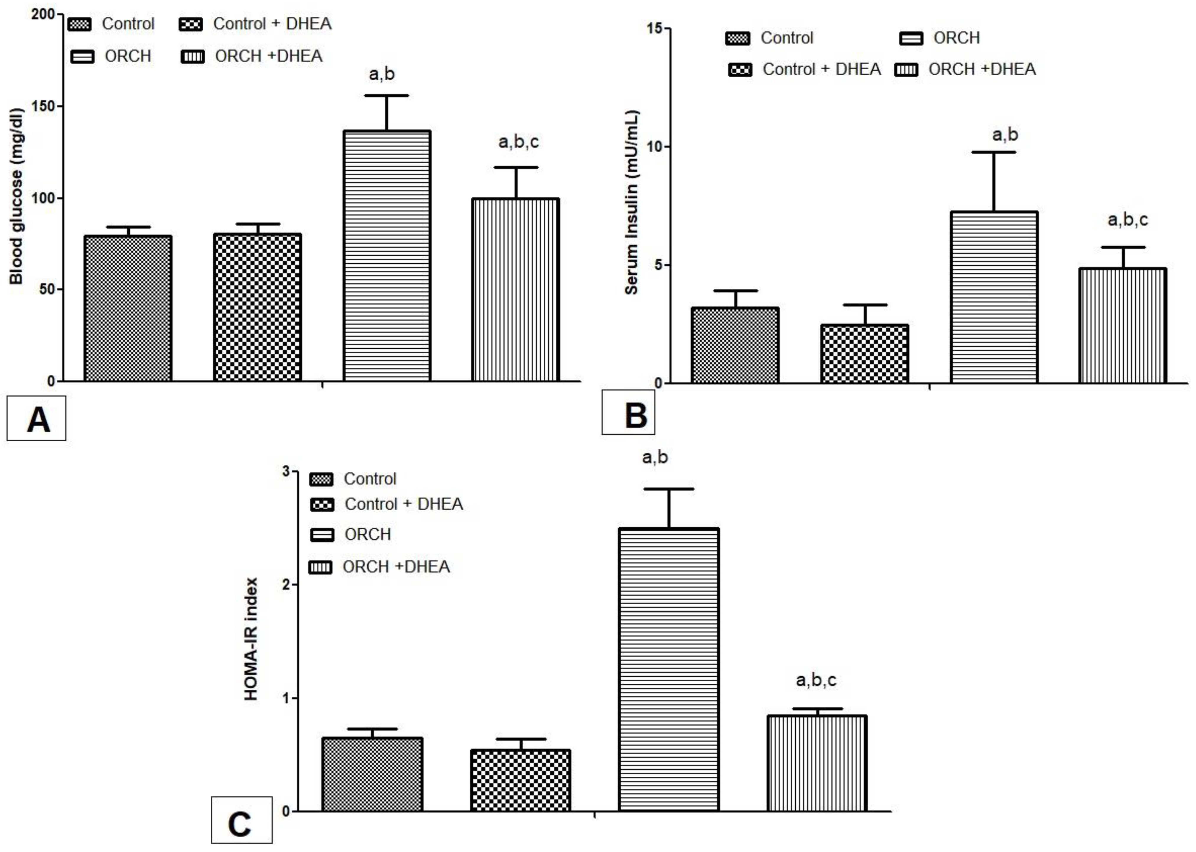
3.2. Effect of DHEA on Fasting Blood Glucose, Insulin, and HOMA-IR Index in Orchiectomized Rats

The levels of fasting blood glucose, fasting insulin, and HOMA-IR were increased significantly (p < 0.05) in ORCH rats when compared to the control group. DHEA treatment significantly attenuated the fasting blood glucose, fasting insulin, and HOMA-IR compared to ORCH-untreated rats (p < 0.05) (Figure 1A–C).

3.3. Effect of DHEA on Lipid Profile and Markers of Liver Functions in Orchiectomized Rats

DHEA treatment in control rats did not significantly change the parameters of the lipid profile and liver enzymes. However, orchidectomy in rats perturbs the lipid profile (TGs, TC, LDL, HDL) by causing a significant elevation in triglycerides, total cholesterol, and LDL-C (p < 0.05) and a significant reduction in HDL-C in comparison with control rats (p < 0.05). Moreover, DHEA treatment after orchiectomized rats significantly improved the ORCH-induced disturbances in lipid profile (p < 0.05) (Table 2). On the other hand, ORCH caused a significant rise in the serum levels of hepatic enzymes (ALT, AST, ALK phosphatase, and GGT), and DHEA treatment significantly attenuated this elevation in the liver enzymes (p < 0.05) (Table 2).

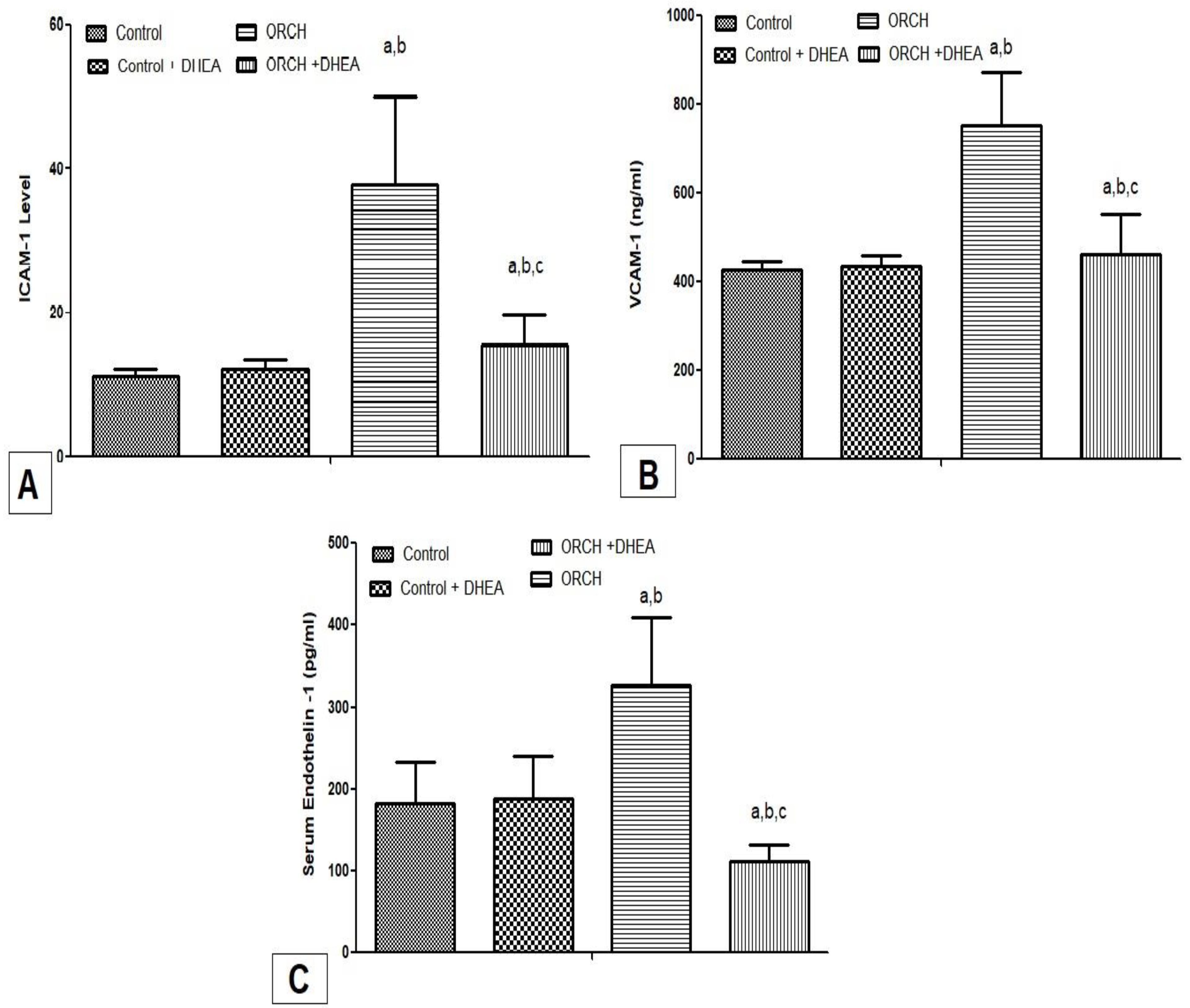
3.4. Effect of DHEA on Inflammatory Cytokines ICAM-1, VCAM-1, and Endothelin-1 in Orchiectomized Rats

ORCH increased significantly the serum levels of ICAM-1, VCAM-1, and endothelin-1 (p < 0.05) versus the control rats. While, DHEA supplementation produced a significant (p < 0.05) decrease in the level of ICAM-1, VCAM-1, endothelin-1 as compared to ORCH rats, but remained significantly (p < 0.05) greater than the control rats (Figure 2A–C).

3.5. Effect of DHEA on Serum Levels of vWF, PAI-1, Fibrinogen, and PDFs in Orchiectomized Rats

ORCH significantly increased the plasma levels of vWF, PAI-1, fibrinogen, and FDPs (p < 0.05) versus control rats. While, DHEA supplementation produced a significant (p < 0.05) decrease in the levels of vWF, PAI-1, fibrinogen, and FDPs as compared to ORCH rats and remained significantly (p < 0.05) greater than the control rats (Figure 3A–D).

3.6. Effect of DHEA on Bleeding Time, Clotting Time, PT, APTT, Platelet Count and ADP-Induced Platelet Aggregations in Orchiectomized Rats

DHEA treatment to control rats produced non-significant change (p > 0.05) in the hemostatic parameters (BT, CT, PT, APTT, platelet count, and ADP-induced platelet aggregations). On the other hand, ORCH in rats caused significant shortening of the BT, CT, PT, and APTT (p < 0.05) as well as a significant increase in the ADP induced platelet aggregation (p < 0.05) without significant change in the platelet count as compared to control rats. DHEA treatment of the orchidectomized rats significantly prolongs the BT, CT, APTT times (p < 0.05) as well as significantly increases the ADP induced platelet aggregation (p < 0.05) without significant change in the platelet count versus ORCH rats (Table 3).

4. Discussion

In the present research, we investigated the possible therapeutic effect of DHEA on platelet dysfunction, hypofibrinolysis, and prothrombotic states in a rat model of male hypogonadism. In the present study, orchidectomy induced a significant increase in body weight, weight gain, BMI, liver weight, and systolic blood pressure. Furthermore, orchidectomized rats showed a significant increase in glucose, insulin, HOMA-IR, serum cholesterol, triglycerides, LDL-C, liver enzymes (ALT, AST, ALK phosphatase and GGT) with a significant decrease in HDL-C. Moreover, bleeding, coagulation, prothrombin, activated partial thromboplastin times were significantly shortened with a significant increase in PAI-1, vW factor, fibrinogen, FDPs, endothelin-1, ICAM, VCAM, and ADP induced platelet aggregation. DHEA treatment modulated the metabolic, hepatic, prothrombotic, and fibrinolytic disturbances in orchidectomized rats.
In the present study, male hypogonadism increased the body weight, BMI, liver weight, and systolic blood pressure as compared to control rats. The above mentioned findings were confirmed by previous research that has linked low circulating testosterone levels in males to white adipose tissue build-up, central obesity, insulin resistance, type 2 diabetes, and metabolic syndrome [8]. DHEA pellets insertion subcutaneously lowered the systolic blood pressure and reduced the increase in body weight, BMI, and liver weight versus the orchidectomized rats. These results affirmed the past hypothesis that lack of testosterone is associated with higher visceral fat deposition [21] and testosterone replacement, in this case, reduced the visceral fats [22]. It has been demonstrated that testosterone administration in euo-and hypogonadal males resulted in a reduction in body weight and fat deposition [23]. DHEA is considered as a weak androgen modulated the changes produced by male hypogonadism. DHEA was also able to reduce both visceral and abdominal fat while enhancing insulin sensitivity, which was in line with our findings [24]. When compared to control rats, orchidectomy and testosterone deprivation raised plasma glucose levels. Ramamani et al. [25] is in agreement with our findings. Orchidectomized rats showed insulin resistance as seen from the increased HOMA-IR as compared to control rats. Visceral fat is considered as an endocrine organ that enhances the release of inflammatory cytokines such as TNF-alpha and IL-6 resulting in chronic inflammation and development of metabolic syndrome [26]. Furthermore, alterations in visceral fat have been shown to alter glucose tolerance and IR [27].
In the present study, we found that ORCD resulted in considerable dyslipidemia, as measured by an increase in the levels of dangerous lipids in the blood (vLDL, LDL, TC, and TGs) with a decrease in beneficial lipids (HDL) that is reversed to nearly its normal levels with testosterone treatment. These results confirmed a previous finding that hypogonadism was associated with dyslipidemia and testosterone improved this dyslipidemia [28]. In addition, the present study showed that DHEA improved those changes, suggesting that it might enhance glucose uptake and insulin sensitivity. These findings are consistent with prior research that showed DHEA supplementation improved glucose tolerance in older, overweight, or obese women and men with impaired glucose tolerance, as well as reduced insulin resistance [29].
Interestingly, male hypogonadism induced by bilateral orchidectomy disturbed platelet functions. The mechanisms of platelet dysfunction are several. The metabolic alterations associated with NAFLD, including IR, dyslipidemia, oxidative stress, adipokines, and inflammation, may modify platelet functioning [30]. Insulin-induced platelet disinhibition has been hypothesized as a major cause of platelet hyperactivity in people with obesity and metabolic syndrome [31]. Weight loss lowers IR and platelet hyperactivity at the same time [32]. In vitro, elevated triglycerides and free fatty acid concentrations have a proaggregating impact [33]. Because HDL inhibits the activation properties of low-density lipoprotein (LDL) on platelets, decreased HDL-C influences platelet aggregation [34]. Platelet hyperactivation in diabetes has been linked to oxidative stress, and advanced products of oxidation such as isoprostanes have been demonstrated to induce platelet aggregation [35]. NAFLD patients’ LDLs have a higher level of oxidative stress and this also stimulates platelet aggregation by activating platelets and priming collagen-induced platelet aggregation [36]. NAFLD may affect platelet characteristics during their production from megakaryocytes, in addition to modifying platelet activities by direct intervention. Resistin and leptin generate IR in megakaryocytes by interfering with insulin receptor substrate 1 [37].
In the current research, DHEA improved the platelet function, prolongs the bleeding time, coagulation time, APTT, and shortens ADP-induced platelet aggregation. These findings were in accordance with previous studies that concluded that DHEA increased the antioxidant enzymes in platelets [38]. Furthermore, DHEA-S prevented platelet activation induced by a modest stimulus without impairing platelet functionality completely, suggesting that DHEA-S may play a role in the physiological mechanisms that keep circulating platelets at rest [39]. Furthermore, ORCH inhibited the fibrinolytic system significantly, as evidenced by the increase of PAI-1. PAI-1 is a significant driver of fibrinolytic activity, influencing both basal and exercise-induced fibrinolytic activity. Subjects with metabolic syndrome and, as a result, hepatic fatty changes had longer clot lysis times (CLT) than those without metabolic syndrome, partly due to higher circulating levels of plasminogen activator inhibitor 1 (PAI-1), the most visible change in the hemostatic system in metabolic syndrome and NAFLD [40]. PAI-1 synthesis by ectopic adipose tissues [41] and fatty liver [42] may be responsible for the rise in plasma PAI-1 levels associated with abdominal obesity.
Many inducers of PAI-1 synthesis during NAFLD have been found, and they may act locally or more distantly in endothelial cells, hepatocytes, Ito cells, fibroblasts, and adipocytes, among other cell types. The mechanisms of increased PAI-1 level in NAFLD [43] include hypetriglyceridemia, which is one of the most important characteristics of NAFLD, raised levels of VLDL stimulate increased PAI-1 synthesis and secretion by cultured human endothelial cells and the ligand-activated transcription factor, peroxisomal proliferator-activated receptor (PPAR-) modulates gene expression in response to a number of stimuli. In human endothelial cells, PPAR- has been demonstrated to influence PAI-1 expression. In addition, endothelial cells produce the Ig-like supergenes intercellular adhesion molecule-1 (ICAM-1) and vascular cell adhesion molecule-1 (VCAM-1). Inflammatory events increase the production of these molecules on the cell surface, and these compounds stimulate leukocyte adherence to the endothelium, which is an early occurrence in the pathophysiology of atherosclerosis [44]. Hyperglycemia increases the expression of cell adhesion molecules on the vascular endothelium. ICAM-1 not only increases atherogenesis [45] but it also exacerbates organ damage [46]. VCAM-1 is also one of the most important endothelial receptors for leukocyte adherence to the endothelium [47]. In vascular stress circumstances such as insulin resistance and persistent hyperglycemia, VCAM-1 expression rises [48]. Endothelial dysfunction is defined as a shift in the endothelium’s functions from vasodilation to vasoconstriction and a proinflammatory state [49].
In the present research, lower androgenicity in orchidectomized rats increased the level of endothelial dysfunction markers as ET-1, ICAM-1, and VCAM-1 in comparison with control rats. Hypogonadism in males triggered the endothelial dysfunction by several mechanisms, either directly or indirectly, through hyperglycemia and IR [50]. On the other hand, DHEA-treated rats showed decreased ET-1, ICAM-1, and VCAM-1 versus the gonadectomized rats. DHEA appeared to modulate the endothelial function through increasing NO generation [51], reducing reactive oxygen species (ROS) generation [52] and producing anti-inflammatory effects [53]. In the present study, orchiectomized rats showed significant shortening of the coagulation, prothrombin, and activated partial thromboplastin times as compared to control rats. Hypercoagulability is one of the complications of male hypogonadism. In the present study, we observed that lower androgenicity was associated with increased prothrombotic factors such as fibrinogen, FDPs, vWF, ET-1, and PAI-1 in orchiectomized rats. These findings are in accordance with previous researches that concluded with a negative correlation between testosterone and PAI-1 as seen in hypogonadal men [8,54]. DHEA pellet treatment to gonadectomized rats improved the fibrinolytic activity as represented by the decrease in PAI-1, fibrinogen, ET-1, and vW as compared to orchiectomized rats. These findings are in agreement with the observation of Beer et al., who found that DHEA administration reduces plasma PAI-1 in men [55]. Furthermore, DHEA therapy at a dose of 150 mg daily for 40 days in men with DHEAS levels less than 2000 mg/l and angiographically confirmed coronary heart disease (CHD) was linked to a significant decrease in fibrinogen concentration and an increase in estradiol levels but had no effect on plasminogen activator inhibitor-1 (PAI-1) or tissue plasminogen activator (tPA) plasma concentrations [56]. The discussed hormone is also functionally connected with coagulation and fibrinolysis, a positive correlation was detected between endogenous testosterone and t-PA and a negative correlation between testosterone and PAI-1, fibrinogen, and alpha 2- antiplasmin [57]. De Pergola et al. [58] demonstrated that there is a negative relationship of both fibrinogen and free testosterone, and another negative relationship between PAI-1 antigen and PAI-1 activity with sex hormone-binding globulin.

5. Conclusions

Collectively, this study demonstrates that bilateral orchidectomy (ORCH) for 12 weeks resulted in a considerable impairment of glucose homeostasis, liver functions with induction of hypercoagulable, and hypofibrinolytic states. On the other hand, administration of DHEA attenuated significantly the metabolic, hepatic, and hemostatic dysfunctions induced by orchidectomy.

Author Contributions

Conceptualization, S.M.S., A.M.H., E.A.E., B.H.E. and H.F.S.; methodology, S.M.S., A.M.H., E.A.E. and M.S.S.; software, E.A.E. and H.F.S.; validation, M.S.S. and H.F.S.; formal analysis, S.M.S., A.M.H. and E.A.E.; investigation, S.M.S., A.M.H. and M.S.S.; data curation, M.S.S., E.A.E., B.H.E. and H.F.S.; writing—original draft preparation, S.M.S. and M.S.S.; writing—review and editing, A.M.H., H.F.S. and E.A.E.; supervision, A.M.H. and H.F.S.; funding acquisition, B.H.E. All authors have read and agreed to the published version of the manuscript.

Funding

This research is supported from Taif University, Taif, Saudi Arabia (TURSP-2020/127).

Institutional Review Board Statement

IRB committee for DUST approved this study with code #RP.21.10.26.

Informed Consent Statement

Not applicable.

Data Availability Statement

All original data will be available on request.

Acknowledgments

We acknowledge Soheir Mohamed (Veterinary research assistant at the Nile center for experimental research, Mansoura, Egypt) for helping us in the induction of the animal model.

Conflicts of Interest

The authors declared no conflict of interest.

References

  1. Corona, G.; Mannucci, E.; Forti, G.; Maggi, M.; Hypogonadism, E.D. Metabolic syndrome and obesity: A pathological link supporting cardiovascular diseases. Int. J. Androl. 2009, 32, 587–598. [Google Scholar] [CrossRef] [PubMed]
  2. Eleawa, S.M.; Sakr, H.F.; Hussein, A.; Assiri, A.S.; Bayoumy, N.M.K.; Alkhateeb, M. Effect of testosterone replacement therapy on cardiac performance and oxidative stress in orchidectomized rats. Acta Physiol. 2013, 209, 136–147. [Google Scholar] [CrossRef] [PubMed]
  3. Brand, J.S.; Van Der Tweel, I.; Grobbee, D.; Emmelot-Vonk, M.H.; Van Der Schouw, Y.T. Testosterone, sex hormone-binding globulin and the metabolic syndrome: A systematic review and meta-analysis of observational studies. Int. J. Epidemiol. 2010, 40, 189–207. [Google Scholar] [CrossRef] [PubMed]
  4. Corona, G.; Monami, M.; Rastrelli, G.; Aversa, A.; Tishova, Y.; Saad, F.; Lenzi, A.; Forti, G.; Mannucci, E.; Maggi, M. Testosterone and Metabolic Syndrome: A Meta-Analysis Study. J. Sex. Med. 2011, 8, 272–283. [Google Scholar] [CrossRef]
  5. Haring, R.; Völzke, H.; Felix, S.B.; Schipf, S.; Dörr, M.; Rosskopf, D.; Nauck, M.; Schöfl, C.; Wallaschofski, H. Prediction of Metabolic Syndrome by Low Serum Testosterone Levels in Men: Results From the Study of Health in Pomerania. Diabetes 2009, 58, 2027–2031. [Google Scholar] [CrossRef] [Green Version]
  6. Gevi, F.; Fanelli, G.; Zolla, L. Metabolic patterns in insulin-resistant male hypogonadism. Cell Death Dis. 2018, 9, 671. [Google Scholar] [CrossRef] [PubMed]
  7. Angulo, P. Nonalcoholic fatty liver disease and liver transplantation. Liver Transplant. 2006, 12, 523–534. [Google Scholar] [CrossRef]
  8. Muraleedharan, V.; Jones, T.H. Testosterone and the metabolic syndrome. Ther. Adv. Endocrinol. Metab. 2010, 1, 207–223. [Google Scholar] [CrossRef]
  9. Cercato, C.; Fonseca, F.A. Cardiovascular risk and obesity. Diabetol. Metab. Syndr. 2019, 11, 1–15. [Google Scholar] [CrossRef]
  10. van Kesteren, P.J.; Kooistra, T.; Lansink, M.; van Kamp, G.J.; Asscheman, H.; Gooren, L.J.; Emeis, J.J.; Vischer, U.M.; Stehouwer, C.D. The effects of sex steroids on plasma levels of marker proteins of endothelial cell functioning. Thromb. Haemost. 1998, 79, 1029–1033. [Google Scholar] [CrossRef]
  11. Webb, C.M.; Elkington, A.G.; Kraidly, M.M.; Keenan, N.; Pennell, D.J.; Collins, P. Effects of Oral Testosterone Treatment on Myocardial Perfusion and Vascular Function in Men With Low Plasma Testosterone and Coronary Heart Disease. Am. J. Cardiol. 2008, 101, 618–624. [Google Scholar] [CrossRef] [PubMed] [Green Version]
  12. Kojima, T.; Doi, K.; Kish, M.; Horiguchi, Y. Sex-related differences in plasminogen activator and plasminogen activator inhibiting activities in young and aged rats. Res. Commun. Mol. Pathol. Pharmacol. 1999, 104, 81–91. [Google Scholar] [PubMed]
  13. Demirci, I.; Haymana, C.; Demir, O.; Akın, O.; Meriç, C.; Aydoğdu, A.; Sönmez, A. Hematological indices in congenital male hypogonadism and the effects of testosterone replacement therapy: A retrospective study. Gulhane Med. J. 2021, 63, 205–211. [Google Scholar] [CrossRef]
  14. Genazzani, A.D.; Lanzoni, C.; Genazzani, A.R. Might DHEA be considered a beneficial replacement therapy in the elderly? Drugs Aging 2007, 24, 173–185. [Google Scholar] [CrossRef]
  15. Davis, S.R.; Panjari, M.; Stanczyk, F.Z. Clinical review: DHEA replacement for postmenopausal women. J. Clin. Endocrinol. Metab. 2011, 96, 1642–1653. [Google Scholar] [CrossRef] [Green Version]
  16. Erben, R.G.; Eberle, J.; Stahr, K.; Goldberg, M. Androgen deficiency induces high turnover osteopenia in aged male rats: A sequential histomorphometric study. J. Bone Miner. Res. 2000, 15, 1085–1098. [Google Scholar] [CrossRef]
  17. Wang, C.; Chao, L.; Chao, J. Direct gene delivery of human tissue kallikrein reduces blood pressure in spontaneously hy-pertensive rats. J. Clin. Investig. 1995, 95, 1710–1716. [Google Scholar] [CrossRef] [Green Version]
  18. Lemini, C.; Rubio-Poo, C.; Silva, G.; Garcia-Mondragon, J.; Zavala, E. Anticoagulant and estrogenic effects of two new 17 beta-aminoestrogens, butolame [17 beta-(4-hydroxy-1-butylamino)-1,3,5(10)-estratrien-3-ol] and pentolame [17 be-ta-(5-hydroxy-1-pentylamino)-1,3,5(10)-estratrien-3-ol]. Steroids 1993, 58, 457–461. [Google Scholar] [CrossRef]
  19. Mustard, J.F.H.T.; RowSell, H.C. Effects of adenosine nucleotides on platelet aggregation and clotting time. J. Lab. Clin. Med. 1964, 64, 548. [Google Scholar]
  20. El-Gendy, A.A.; Abbas, A.M. Effect of omega-3 fatty acids on haemostatic functions in urocortin-treated obese rats. J. Physiol. Biochem. 2014, 70, 809–820. [Google Scholar] [CrossRef]
  21. Rotter, I.; Rył, A.; Grzesiak, K.; Szylińska, A.; Pawlukowska, W.; Lubkowska, A.; Sipak-Szmigiel, O.; Pabisiak, K.; Laszczyńska, M. Cross-Sectional Inverse Associations of Obesity and Fat Accumulation Indicators with Testosterone in Non-Diabetic Aging Men. Int. J. Environ. Res. Public Health 2018, 15, 1207. [Google Scholar] [CrossRef] [Green Version]
  22. Traish, A.M. Testosterone and weight loss: The evidence. Curr. Opin. Endocrinol. Diabetes. Obes. 2014, 21, 313–322. [Google Scholar] [CrossRef] [Green Version]
  23. Behre, H.M.; Tammela, T.L.J.; Arver, S.; Tolrá, J.R.; Bonifacio, V.; Lamche, M.; Kelly, J.; Hiemeyer, F.; Giltay, E.J.; Gooren, L.J.; et al. A randomized, double-blind, placebo-controlled trial of testosterone gel on body composition and health-related quality-of-life in men with hypogonadal to low-normal levels of serum testosterone and symptoms of androgen deficiency over 6 months with 12 months open-label follow-up. Aging Male 2012, 15, 198–207. [Google Scholar] [CrossRef]
  24. Villareal, D.T.; Holloszy, J.O. Effect of DHEA on abdominal fat and insulin action in elderly women and men: A randomized controlled trial. JAMA 2004, 292, 2243–2248. [Google Scholar] [CrossRef]
  25. Ramamani, A.; Aruldhas, M.M.; Govindarajulu, P. Differential response of rat skeletal muscle glycogen metabolism to tes-tosterone and estradiol. Can. J. Physiol. Pharmacol. 1999, 77, 300–304. [Google Scholar] [CrossRef]
  26. Hwang, Y.-C.; Fujimoto, W.Y.; Hayashi, T.; Kahn, S.E.; Leonetti, D.L.; Boyko, E.J. Increased Visceral Adipose Tissue Is an Independent Predictor for Future Development of Atherogenic Dyslipidemia. J. Clin. Endocrinol. Metab. 2016, 101, 678–685. [Google Scholar] [CrossRef] [Green Version]
  27. Verkouter, I.; Noordam, R.; Le Cessie, S.; Van Dam, R.M.; Lamb, H.J.; Rosendaal, F.R.; Van Heemst, D.; De Mutsert, R. The Association between Adult Weight Gain and Insulin Resistance at Middle Age: Mediation by Visceral Fat and Liver Fat. J. Clin. Med. 2019, 8, 1559. [Google Scholar] [CrossRef] [Green Version]
  28. Saad, F.; Haider, A.; Doros, G.; Traish, A. Long-term treatment of hypogonadal men with testosterone produces substantial and sustained weight loss. Obesity 2013, 21, 1975–1981. [Google Scholar] [CrossRef]
  29. Weiss, E.P.; Villareal, D.T.; Fontana, L.; Han, D.-H.; Holloszy, J.O. Dehydroepiandrosterone (DHEA) replacement decreases insulin resistance and lowers inflammatory cytokines in aging humans. Aging 2011, 3, 533–542. [Google Scholar] [CrossRef] [Green Version]
  30. Schafer, K.; Konstantinides, S. Adipokines and thrombosis. Clin. Exp. Pharmacol. Physiol. 2011, 38, 864–871. [Google Scholar] [CrossRef]
  31. Anfossi, G.; Russo, I.; Trovati, M. Platelet dysfunction in central obesity. Nutr. Metab. Cardiovasc. Dis. 2009, 19, 440–449. [Google Scholar] [CrossRef]
  32. Basili, S.; Pacini, G.; Guagnano, M.T.; Manigrasso, M.R.; Santilli, F.; Pettinella, C.; Ciabattoni, G.; Patrono, C.; Davì, G. Insulin Resistance as a Determinant of Platelet Activation in Obese Women. J. Am. Coll. Cardiol. 2006, 48, 2531–2538. [Google Scholar] [CrossRef] [Green Version]
  33. Englyst, N.A.; Taube, J.M.; Aitman, T.J.; Baglin, T.P.; Byrne, C.D. A Novel Role for CD36 in VLDL-Enhanced Platelet Activation. Diabetes 2003, 52, 1248–1255. [Google Scholar] [CrossRef] [Green Version]
  34. Korporaal, S.J.; Akkerman, J.W. Platelet activation by low density lipoprotein and high density lipoprotein. Pathophysiol. Haemost. Thromb. 2006, 35, 270–280. [Google Scholar] [CrossRef]
  35. Akkerman, J.W.N. From low-density lipoprotein to platelet activation. Int. J. Biochem. Cell Biol. 2008, 40, 2374–2378. [Google Scholar] [CrossRef]
  36. Colas, R.; Sassolas, A.; Guichardant, M.; Cugnet-Anceau, C.; Moret, M.; Moulin, P.; Lagarde, M.; Calzada, C. LDL from obese patients with the metabolic syndrome show increased lipid peroxidation and activate platelets. Diabetologia 2011, 54, 2931–2940. [Google Scholar] [CrossRef] [Green Version]
  37. Gerrits, A.J.; Gitz, E.; Koekman, C.A.; Visseren, F.L.; van Haeften, T.W.; Akkerman, J.W.N. Induction of insulin resistance by the adipokines resistin, leptin, plasminogen activator inhibitor-1 and retinol binding protein 4 in human megakaryocytes. Haematologica 2012, 97, 1149–1157. [Google Scholar] [CrossRef] [Green Version]
  38. Bednarek-Tupikowska, G.; Gosk, I.; Szuba, A.; Bohdanowicz-Pawlak, A.; Kosowska, B.; Bidzińska, B.; Milewicz, A. Influence of dehydroepiandrosterone on platelet aggregation, superoxide dismutase activity and serum lipid peroxide concentrations in rabbits with induced hypercholesterolemia. Med. Sci. Monit. 2001, 6, 40–45. [Google Scholar]
  39. Bertoni, A.; Rastoldo, A.; Sarasso, C.; Di Vito, C.; Sampietro, S.; Nalin, M.; Bagarotti, A.; Sinigaglia, F. Dehydroepiandrosterone-sulfate inhibits thrombin-induced platelet aggregation. Steroids 2012, 77, 260–268. [Google Scholar] [CrossRef] [PubMed]
  40. Alessi, M.C.; Juhan-Vague, I. PAI-1 and the metabolic syndrome: Links, causes, and consequences. Arterioscler. Thromb. Vasc. Biol. 2006, 26, 2200–2207. [Google Scholar] [CrossRef] [PubMed]
  41. Fain, J.N.; Madan, A.K.; Hiler, M.L.; Cheema, P.; Bahouth, S.W. Comparison of the Release of Adipokines by Adipose Tissue, Adipose Tissue Matrix, and Adipocytes from Visceral and Subcutaneous Abdominal Adipose Tissues of Obese Humans. Endocrinology 2004, 145, 2273–2282. [Google Scholar] [CrossRef] [Green Version]
  42. Alessi, M.-C.; Bastelica, D.; Mavri, A.; Morange, P.; Berthet, B.; Grino, M.; Juhan-Vague, I. Plasma PAI-1 Levels Are More Strongly Related to Liver Steatosis Than to Adipose Tissue Accumulation. Arter. Thromb. Vasc. Biol. 2003, 23, 1262–1268. [Google Scholar] [CrossRef] [Green Version]
  43. Appel, S.J.; Harrell, J.S.; Davenport, M.L. Central Obesity, the Metabolic Syndrome, and Plasminogen Activator Inhibitor-1 in Young Adults. J. Am. Acad. Nurse Pract. 2005, 17, 535–541. [Google Scholar] [CrossRef]
  44. Abbas, A.M.; Sakr, H.F. Effect of magnesium sulfate and thyroxine on inflammatory markers in a rat model of hypothy-roidism. Can. J. Physiol. Pharmacol. 2016, 94, 426–432. [Google Scholar] [CrossRef]
  45. Cybulsky, M.I.; Iiyama, K.; Li, H.; Zhu, S.; Chen, M.; Iiyama, M.; Davis, V.; Gutierrez-Ramos, J.-C.; Connelly, P.; Milstone, D.S. A major role for VCAM-1, but not ICAM-1, in early atherosclerosis. J. Clin. Investig. 2001, 107, 1255–1262. [Google Scholar] [CrossRef] [Green Version]
  46. Ge, H.; Wen, Y.; Yang, G.; Betz, A.L. Increased expression of intercellular adhesion molecule-1 in mouse focal cerebral ischemia model. Chin. Med. J. 2000, 113, 75–79. [Google Scholar]
  47. van Buul, J.D.; Voermans, C.; van den Berg, V.; Anthony, E.C.; Mul, F.P. Migration of human hematopoietic progenitor cells across bone marrow endothelium is regulated by vascular endothelial cadherin. J. Immunol. 2002, 168, 588–596. [Google Scholar] [CrossRef] [Green Version]
  48. Madonna, R.; Pandolfi, A.; Massaro, M.; Consoli, A.; De Caterina, R. Insulin enhances vascular cell adhesion molecule-1 expression in human cultured endothelial cells through a pro-atherogenic pathway mediated by p38 mitogen-activated pro-tein-kinase. Diabetologia 2004, 47, 532–536. [Google Scholar] [CrossRef] [Green Version]
  49. Rajendran, P.; Rengarajan, T.; Thangavel, J.; Nishigaki, Y.; Sakthisekaran, D.; Sethi, G.; Nishigaki, I. The Vascular Endothelium and Human Diseases. Int. J. Biol. Sci. 2013, 9, 1057–1069. [Google Scholar] [CrossRef] [Green Version]
  50. Antonopoulos, A.S.; Antoniades, C. Mechanisms of testosterone deficiency-related endothelial dysfunction: Invited com-mentary for the Hellenic Journal of Cardiology on: Tsikas et al. “Associations between asymmetric dimethylarginine, ni-trite-dependent renal carbonic anhydrase activity and plasma testosterone levels in hypogonadal men”. Hell. J. Cardiol. 2018, 59, 207–208. [Google Scholar]
  51. Formoso, G.; Chen, H.; Kim, J.-A.; Montagnani, M.; Consoli, A.; Quon, M.J. Dehydroepiandrosterone Mimics Acute Actions of Insulin to Stimulate Production of Both Nitric Oxide and Endothelin 1 via Distinct Phosphatidylinositol 3-Kinase- and Mitogen-Activated Protein Kinase-Dependent Pathways in Vascular Endothelium. Mol. Endocrinol. 2006, 20, 1153–1163. [Google Scholar] [CrossRef] [Green Version]
  52. Maggio, M.; De Vita, F.; Fisichella, A.; Colizzi, E.; Provenzano, S.; Lauretani, F.; Luci, M.; Ceresini, G.; Dall’Aglio, E.; Caffarra, P.; et al. DHEA and cognitive function in the elderly. J. Steroid Biochem. Mol. Biol. 2015, 145, 281–292. [Google Scholar] [CrossRef]
  53. Rutkowski, K.; Sowa, P.; Rutkowska-Talipska, J.; Kuryliszyn-Moskal, A.; Rutkowski, R. Dehydroepiandrosterone (DHEA): Hypes and Hopes. Drugs 2014, 74, 1195–1207. [Google Scholar] [CrossRef]
  54. De Pergola, G.; De Mitrio, V.; Sciaraffia, M.; Pannacciulli, N.; Minenna, A. Lower androgenicity is associated with higher plasma levels of prothrombotic factors irrespective of age, obesity, body fat distribution, and related metabolic parameters in men. Metabolism 1997, 46, 1287–1293. [Google Scholar] [CrossRef]
  55. Beer, N.A.; Jakubowicz, D.J.; Matt, D.W.; Beer, R.M.; Nestler, J.E. Dehydroepiandrosterone reduces plasma plasminogen ac-tivator inhibitor type 1 and tissue plasminogen activator antigen in men. Am. J. Med. Sci. 1996, 311, 205–210. [Google Scholar] [CrossRef]
  56. Rabijewski, M.; Zgliczyński, W. Dehydroepiandrosterone therapy in men with angiographically verified coronary heart disease: The effects on plasminogen activator inhibitor-1 (PAI-1), tissue plasminogen activator (tPA) and fibrinogen plasma concentrations. Endokrynol. Pol. 2007, 58, 213–219. [Google Scholar]
  57. Medraś, M.; Jankowska, E. Testosterone and atherosclerosis in males during andropause. Pol. Merkur. Lekarski. 1999, 6, 205–207. [Google Scholar]
  58. De Pergola, G.; Pannacciulli, N. Coagulation and fibrinolysis abnormalities in obesity. J. Endocrinol. Investig. 2002, 25, 899–904. [Google Scholar] [CrossRef]
Figure 1. Effect of DHEA on (A) blood glucose level (mg/dL), (B) serum insulin (IU/mL), and (C) HOMA-IR index in orchidectomized rats. a: significance relative to control group, b: significance relative to Con + DHEA group, c: significance relative to ORCH. ORCH = orchidectomized rats and DHEA = dehydroepiandrosterone.
Scipharm 90 00006 g001
Figure 2. Effect of DHEA on (A) ICAM-1 (intercellular cell adhesion molecule) level, (B) VCAM-1 (vascular cell adhesion molecule-1) level, and (C) Serum endothelin-1 level in orchidectomized rats. a: significance relative to control group, b: significance relative to Con + DHEA group, c: significance relative to ORCH. ORCH = orchidectomized rats and DHEA = dehydroepiandrosterone.
Scipharm 90 00006 g002
Figure 3. Effect of DHEA on (A) serum VWF (Von Willebrand factor) level, (B) serum PAI-1 (plasminogen activator inhibitor-1) level, (C) serum fibrinogen level and (D) serum FDP (fibrin degradation product) level in orchidectomized rats. a: significance relative to control group, b: significance relative to Con + DHEA group, c: significance relative to ORCH. ORCH = orchidectomized rats and DHEA = dehydroepiandrosterone.
Scipharm 90 00006 g003
Table 1. Effect of DHEA on Systolic blood pressure, body weight, percent of weight gain, body mass index (BMI), liver weight, and liver\body weight % in orchidectomized rats.
ConCon + DHEAORCHORCH + DHEA
Systolic blood pressure (mmHg)122 ± 5125 ± 9144 ± 8132 ± 4
Body weight (g)333.8 ± 4.3345.7 ± 6.7408.5 ± 9.9 ab360 ± 8.4 abc
Percent of weight gain (%)31 ± 4.529.8 ± 3.759.5 ± 7.8 ab35.4 ± 6.08 abc
BMI (g/cm2)0.60 ± 0.040.59 ± 0.070.75 ± 0.95 ab0.63 ± 0.04 abc
Liver weight (g)10.8 ± 0.4811.1 ± 0.7615.00 ± 0.31 ab12.65 ± 0.6 abc
Liver weight/body weight (%)3.04 ± 0.13.09 ± 0.23.55 ± 0.09 ab3.25 ± 0.2 abc
Data were expressed as mean ± SD of 8 rats. Con: control group, Con + DHEA: rats treated with dehydroepiandrosterone, ORCH: orchidectomized rats and ORCH + DHEA: orchidectomized rats treated with dehydroepiandrosterone. BMI: body mass index. a: significance relative to control group, b: significance relative to Con + DHEA group, c: significance relative to ORCH.
Table 2. Effect of DHEA on lipid profile and liver enzymes in orchidectomized rats.
ConCon + DHEAORCHORCH + DHEA
Triglycerides (mg/dL)93.1 ± 5.788.45 ± 6.2166.7 ± 12.8 ab133.7 ± 15.58 abc
Total Cholesterol (mg/dL)88.0 ± 4.5190.45 ±3.41144.7 ± 15.7 ab125.7 ± 14.61 ab
LDL-C (mg/dL)35.6 ± 4.4634.4 ± 5.6177.4 ± 5.2 ab60.8 ± 9.44 abc
HDL-C (mg/dL)48.4 ± 4.8247.7 ± 6.5137.7 ± 4.8 ab44.7 ± 3.6 c
ALT (U/L)50.05 ± 4.3452.1 ± 3.6788.41 ± 9.52 ab67.34 ± 6.05 abc
AST (U/L)58.64 ± 5.0460.45 ± 6.85109.75 ± 11.41 ab84.51 ± 7.93 abc
ALK. Phosphatase (U/L)189.45 ± 12.65200.14 ± 15.7452.68 ± 35.7 ab287.45 ± 23.95 abc
GGT (U/L)0.93 ± 0.130.86 ± 0.242.53 ± 0.34 ab1.69 ± 0.35 abc
Data were expressed as mean ± SD of 8 rats. Con: control group, Con + DHEA: rats treated with dehydroepiandrosterone, ORCH: orchidectomized rats and ORCH + DHEA: orchidectomized rats treated with dehydroepiandrosterone. LDL –C: Low-density lipoprotein cholesterol. HDL-C: High-density lipoprotein cholesterol. ALT: alanine transaminase, ASP: aspartate transaminase, ALK. Phosphatase: alkaline phosphatase, GGT: gamma-glutamyl transferase. a: significance relative to control group, b: significance relative to Con + DHEA group, c: significance relative to ORCH.
Table 3. Bleeding time, clotting time, PT, aPTT, platelets count, and ADP-induced platelet aggregation (%) in control and orchidectomized rats.
ConCon + DHEAORCHORCH + DHEA
PT (s)24.5 ± 0.525.1 ± 0.618.4 ± 0.034 ab23.7 ± 0.58 abc
aPTT (s)19.2 ± 0.620.1 ± 0.713.5 ± 0.6 ab14.8 ± 0.4 abc
Bleeding time (s)84.1 ± 2.985.4 ± 3.566.7 ± 3.2 ab73.2 ± 3.1 abc
Clotting time (s)123.6 ± 3.8124.6 ± 4.189.6 ± 3.1 ab106.5 ± 2.8 abc
Platelet count (×103/µL)915 ± 25933 ± 35942 ± 39919 ± 45
ADP-induced platelet aggregation (%)45.1 ± 2.947.4 ± 3.667.3 ± 3.8 ab59.6 ± 4.1 abc
Data were expressed as mean ± SD of 8 rats. Con: control group, Con + DHEA: rats treated with dehydroepiandrosterone, ORCH: orchidectomized rats and ORCH + DHEA: orchidectomized rats treated with dehydroepiandrosterone. PT: prothrombin time, aPTT: activated partial thromboplastin time, ADP: adenosine diphosphate. a: significance relative to control group, b: significance relative to Con + DHEA group, c: significance relative to ORCH.
Publisher’s Note: MDPI stays neutral with regard to jurisdictional claims in published maps and institutional affiliations.

