
Applying a Hydrophilic Modified Hollow Fiber Membrane to Reduce Fouling in Artificial Lungs


1
Department of Biology, College of Sciences, University of Ha’il, P.O. Box 2440, Ha’il 81411, Saudi Arabia
2
Department of Information and Computer Sciences, College of computer science and Engineering, University of Ha’il, P.O. Box 2440, Ha’il 81411, Saudi Arabia
3
Department of Microbiology, Sree Narayana Guru College, Coimbatore 641105, India
*
Author to whom correspondence should be addressed.
Separations 2021, 8(8), 113; https://0-doi-org.brum.beds.ac.uk/10.3390/separations8080113
Submission received: 20 June 2021
/
Revised: 11 July 2021
/
Accepted: 27 July 2021
/
Published: 30 July 2021
(This article belongs to the Special Issue Modeling, Simulation, and Optimization of Membrane Processes)
Abstract
:Membranes for use in high gas exchange lung applications are riddled with fouling. The goal of this research is to create a membrane that can function in an artificial lung until the actual lung becomes available for the patient. The design of the artificial lung is based on new hollow fiber membranes (HFMs), due to which the current devices have short and limited periods of low fouling. By successfully modifying membranes with attached peptoids, low fouling can be achieved for longer periods of time. Hydrophilic modification of porous polysulfone (PSF) membranes can be achieved gradually by polydopamine (PSU-PDA) and peptoid (PSU-PDA-NMEG5). Polysulfone (PSU-BSA-35Mg), polysulfone polydopamine (PSUPDA-BSA-35Mg) and polysulfone polydopamine peptoid (PSU-PDA-NMEG5-BSA35Mg) were tested by potting into the new design of gas exchange modules. Both surfaces of the modified membranes were found to be highly resistant to protein fouling permanently. The use of different peptoids can facilitate optimization of the low fouling on the membrane surface, thereby allowing membranes to be run for significantly longer time periods than has been currently achieved.
1. Introduction
The basic involuntary activity of gas exchange plays an essential role in sustaining life. Air pollution, because of rapid industrialization and urbanization, has meant that progressing lung diseases have remained a significant healthcare challenge in a large number of adult patients each year in the USA alone [1]. Deforestation is augmenting the number of patients with lung diseases. This has thrived researchers’ interest in the field. However, present respiratory assist devices are not up to the same standard, especially when the patient is waiting for a lung transplant [2,3]. There are several obstructions with available artificial lungs/respiratory assist devices because the main material used are polymeric hollow fibers membranes (HFMs) for the transportations between blood/ biological material and gas pathways. However, these have caused major concern with an accumulation of fouling [4]. The membrane oxygenator is commonly utilized worldwide. There is also a microporous and nonporous oxygenator membrane [5].
Furthermore, polysulfone (PSF) is an important material in membranes used for many purposes such as hemodialysis and apheresis, due to its thermal stability, mechanical strength, and chemical internes. It is also reported that PSF has some defects in hematological application, such as its hydrophobic nature [6,7]. Fouling leads to blockage of the membranes porosity [4,8,9]. Hydrogen bonding, hydrophobic, electrostatic, and van der Waals interactions that occur between the membrane surface and biological foulant, were the culprits of membranes fouling [10]. Many studies have addressed the fouling issue and have tried to solve these problems. Novel coatings and modified fibers are used actively to reduce membrane fouling [10].
Biofouling of membranes can be reduced with a wide variety of surface modification methods; however, there have been relatively few studies with hollow fiber membranes which assess the quality on gas exchange [11]. Modification of the PSF membrane is carried out by immobilizing urea on the membrane surface. It was created so that higher activities were reached when the immobilization procedure was carried out at a lower pH. Meanwhile, Lin et al. (2003) found that the urease immobilized dialyzer provided a faster eliminating rate of urea associated to a regular dialyzer, which can be enhanced by increasing the dialysate velocity [12]. There was a study which recommended the infusion of heparin to enhance hemocompatibility of HFMs [13]. Furthermore, Zhu and others have investigated the primary surface characteristic of PDA-coated hydrophobic substrates. They found that the surface-free energy and hydrophilicity of the polydopamine (PDA)-modified substrates were improved dramatically [14], and that the hydrophilicity of PDA-modified PSF was significantly promoted. Morphologically, the membrane colors turn dark after PDA coating [15,16]. Furthermore, peptoid consisting of PEG-peptoid and the PEG-mimetic side chain was able to resist fouling [17]. In addition, it can be used in vivo because of its low immunogenicity and protease resistance [18]. NMEG has many advantages to play an important role in the modification of membranes. It is polar, uncharged, hydrophilic, has no hydrogen-bond donors, and contains hydrogen bound acceptors. In addition, peptoid is stable and easier to synthesize than peptides, and their chemical diversity is large [19].
The present study was aimed to examine the mode of PDA and PDA-NMEH 5 coating layers affecting the whole act of the PSF membrane, including the effect of top surface PDA and PDA-NMEG 5 coating on the structure of the following selective layer, the hydrophilicity, and the oxygen permeability compared with unmodified PSF, which is a novel aspect of this study. In addition, this research focused on the limitation of current devices and fouling of membrane. Studying the fouling of HFMs and modified HFMs will help to understand the long-term capability of a new gas exchange module design.
2. Materials and Methods
2.1. Materials
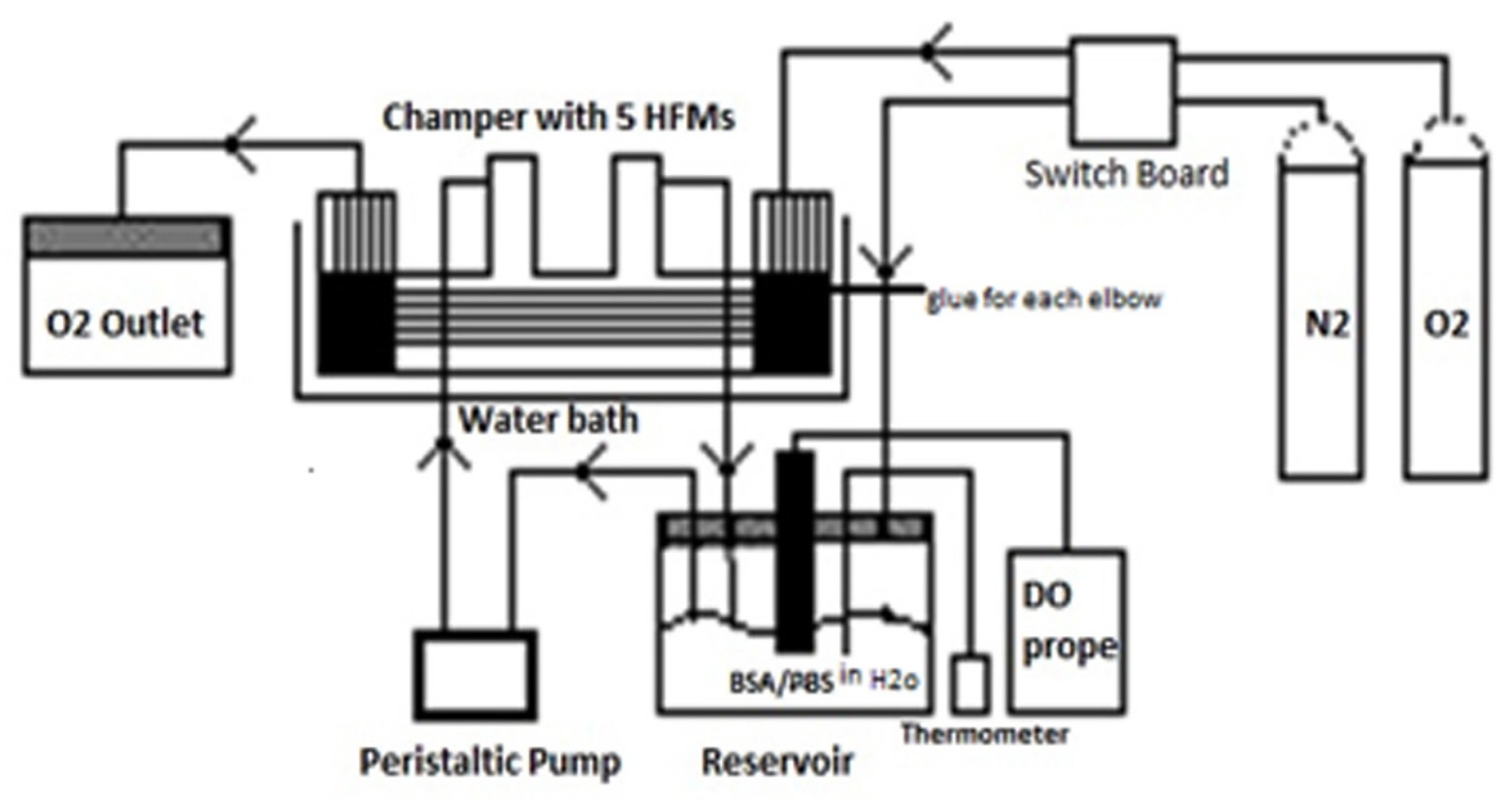
Polysulfone HFMs (30 cm) were prepared in the laboratory. Bovine Serum Albumin (BSA) was procured from VWR amresco (Fayetteville, AR, USA). Phosphate buffered saline (PBS), 20X concentrate, pH 7.5, 3, 4-dihydroxyphenethylamine (dopamine) hydrochloride were procured from Sigma-Aldrich (Milwaukee, WI, USA). A nitrogen cylinder, an oxygen cylinder, a pump (Masterflex L/S Easy Load), a pump head, P/N 07518–10, and Masterflex modular drive (1–100 rpm), P/N 07553–80 (Cole-Parmer Instruments Company, Vernon Hills, IL, USA) were employed to circulate the fluids on both sides. Epikure 3030 and Epon 828 were purchased from Hexian (Houston, TX, USA). Peptoid, oxygen meter, pipe (PVC), clear polyurethane ether based tubing clear (1.5 inch), and silicon tubing (3/8 O.D and ¼ inch I.D) were purchased from MC Master Carr. The ¼" M NPT ¼ " Hose Barb was purchased from the Apex company and the Fastenal company (Fuquay-Varina, NC, USA). A water bath (model 28L-M, AMPS 8, V/HZ 120/60 SERIAL 514280) along with a 229 X 1” BD needle and a syringe were also used in this study. Laptop software 4320 Universal Serial Data Capture (04/09, Traceable, TX, USA) and Oxygen meter (21800-022) were purchased from Control Company (Webster, TX, USA). A lab facility EL-USB-TC-LCD Humidity & Temperature USB data logger was purchased from Omega company (Norwalk, CT, USA). NaCl, glycerol, and sodium benzoate were of laboratory grade and were procured locally. The principal characteristics of the three different groups of fibers evaluated in this study are summarized in Figure 1.
2.2. Fabrication of Hollow Fiber Membrane (HFMs)
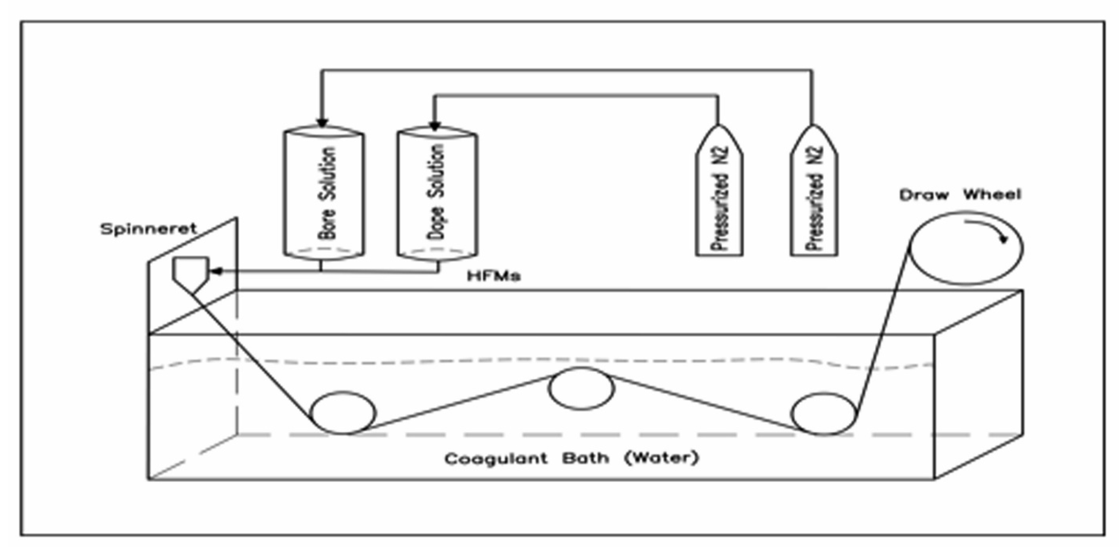
PSU HFMs were prepared by a conventional hollow fiber membrane spinning device. Polysulfone (powder) 17.5% (v/v) in N-Methylpyrrolidone (NMP) was prepared as the spinning dope, while the bore solution was 15% (v/v) NMP in water(mixture). A tube-in-hole spinneret, with 0.8 (mm) and 1.6 (mm) inner and outer diameters, respectively, was applied to gain HFMs precursors [20]. The air gap was adjusted at 8 (cm) between the spinneret and water bath for all spinning. Both dope and pore solutions were extruded through spinning by using different nitrogen pressures for each solution (90, 15 Psi, respectively). HFMs were pushed through a water path to complete the HFMs formation. [21]. Then, the solidified membrane was submerged by hand underneath the tow rollers. The fiber bundles were collected by a draw wheel attached to a small motor with adjustable speeds (2 m/min). After collection, the HFM bundles were stored in bags of 5% aqueous NaCl solution. NaCl was replaced at every 24-h interval for 3 days. Then, the 25% glycerol (v/v) solution and antibiotic (0.2% sodium benzoate) were prepared [22]. Details of the set-up are given in Figure 2.
2.3. Preparation of the Chamber for Gas Exchange Experiments
Epoxy resins Epikure 3030 and Epon 828 were mixed gently in a 2:1 ratio. The resin glue was poured into each elbow of the chamber after placing the fibers inside it, and it was left to dry at room temperature for 24 h. Preparation of each elbow took 1 day. All pieces of the chamber were assembled together as depicted in Figure 1. A new design of gas exchange module was fabricated by inserting HFMs (5 fibers, 30 cm) into a 1/5 in pip clear tubing (PVC) [23].
Both ends of the chambers were secured to the tubing by epoxy, Epikure 3030 and Epon 828, then trimmed to the length of the tubing to expose the submission of the HFMs lumens. Next, 11.3 cm of HFMs was exposed within the module to present a surface area of 11.3 cm. A model system recalculating test loop was utilized to estimate O2 exchange rates using three groups of fibers, namely polydopamine (PSU-PDA) fibers, peptoid fibers (PSU-PDA-NMEG5), and unmodified.
The loop was with a fluid reservoir, peristaltic pump, oxygenator, and gas exchange module. The testing fluid, consisting of 200 mL of BSA (35 mg/mL (w/v) in PBS, flowed from a reservoir to a Master Flex L/S peristaltic pump (Vernon Hills, IL, USA) to a model gas exchange testing the liquid and returned to the reservoir. The inlet partial pressure of O2 (PO2) was adjusted at 2.5 SCFH. The fluid flow rate was maintained at 88 mL/min. Heating was turned on and the fluid temperature was maintained at 37 °C.
The reservoir has five outlet/inlet points for oxygen probes, outlet of liquid, inlet of liquid, BSA with PBS, and inlet of nitrogen gas (N2) by silicon nylon tubbing to test the HFMs. The time was adjusted on the auto controlled panel for each cylinder by valve (N2 for 20 min, and oxygen for 3 h). The experiment was initiated by turning on the pump at level 1. Afterward, the oxygen was turned on in order to provide the membranes with oxygen gas at 15 kpa with an O2 inlet 2.5 Standard cubic feet per hour flow rate inside the hollow fiber membrane. Later, the nitrogen was turned on in order to consume the oxygen generated by BSA/PBS. When the highest level was reached, only minimum levels of dissolved oxygen remained. Both cylinders were operated directly from the valve for switching on and off.
The concentration was estimated every 15 min with an oxygen meter placed in the reservoir [13,24]. The experiment was monitored every hour to ensure proper functioning of the set-ups without any leaks. Kla (30 min−1) was estimated at 15 min or 3 h period using the Kla equation versus time to calculate Kla from the slope’s equation (Equation (1)) as follows:
where:
Kla = −Ln [(C − Co)/(C* − Co)]
Kla is volumetric oxygen transfer coefficient (1/h.)
T is time (min.)
C is the liquid dissolved-oxygen concentration (mg/L).
C* is the equilibrium concentration of oxygen between water and gas phase (mg/L).
Co is the liquid oxygen concentration at start of measurement (mg/L).
All Kla values were measured with three to five replicates in order to guarantee the certainty of values found through standard deviations. In addition, HFM PSU were modified according to Mahmoudi et al. who described the process of a hollow fiber coating [25].
3. Results
Oxygen transfer was estimated with a new design of gas exchange module for both unmodified fibers (PSU) and modified fibers, i.e. either polydopamine fibers (PSU-PDA) or peptoid fibers (PSU-PDA-NMEG5). Oxygen transfer coefficients for unmodified and modified fibers in a solution of BSA/PBS 35% (mg/mL) as a liquid phase over time are depicted in Figure 3. PBS was set as blank solution. As a result of that, the oxygen transfer coefficients of unmodified fibers were blocked over time due to the interaction between fouled protein and membrane surface. Modifying the fibers (PSU-PDA-PSA 35 mg/mL) and (PSU-PDA-NMEG5-BSA-35 mg/mL) resulted in an improvement in the oxygen transfer and in resistance to protein fouling. On the other hand, unmodified hollow fiber membranes were completely blocked after 3 h. However, the peptoid hollow fiber membranes were not blocked over a test time of 6 h. In addition, polydopamine hollow fiber membranes were less resistant to protein fouling than peptoid HFMs because (PSU-PDA-BSA-35 mg/mL) with blocked membranes appearing after 5 h.
Overall, results indicate that modified HFMs, both PSU-PDA and PSU-PDA-NMEG5-BSA-35, have higher oxygen transfer coefficients and undergo less fouling compared to unmodified HFMs (PSU-BSA-35). Oxygen transfer rates were maintained for longer periods of time in peptoid HFMs compared to polydopamine HFMs (Table 1). The highest kLa values were observed in the case of the PSU-PDA-NMEG5-BSA membrane (0.19 ± 0.14 min−1). This was followed by the PSU-PDA-BSA membrane where kLa values of 0.11 ± 0.22 min−1 were achieved. kLA values for PSU-BSA was the minimum (0.088 ± 0.11 min−1). Moreover, both fibers of (PSU-BSA- 35 mg/mL) and (PSU-PDA-BSA35 mg/mL) were blocked and oxygen transfer completely stopped at 3 h for PSU-BSA and 6 h for PSU-PDA-BSA membranes. On the other hand, PSU-PDA-NMEG5 was resistant to protein fouling for 6 h; the kLa value at 6 h was observed to be 0.014 ± 0.008 min−1 Table 1.
4. Discussions
The goal of this study was to create a lung that can function within the body until there is availability from a donor. The design of the artificial lung was going to be implemented on a new gas exchange module which was based on HFMs. HFMs resulted in shortening the low fouling period of the device and therefore did not present as a long-term solution. This shortcoming was overcome by successfully modifying the membrane to extend for longer periods. The principle behind this solution was to increase the oxygen diffusional gradient inside the system.
In order to validate the hypothesis that the membrane fouling is contributed by high concentration of fouled protein, BSA/PBS at 35 mg/mL solution concentration was used in this study.
The hydrophilic modification of porous polysulfone (PSF) membranes can be achieved gradual addition of polydopamine (PSU- PDA) and peptoid (PSU-PDA-NMEG5), as reported in the literature [26]. PSU-BSA-35 mg /ml, PSU-PDA-BSA-35 mg/ml, and PSU-PDA-NMEG5-BSA-35 mg were tested by potting into the new design of gas exchange module with a surface area of 11.3 cm in a recirculation loop with an oxygen inlet of 2.4 standard cubic feet per hour (SCFH) under steady liquid flow, liquid temperature, O2 pressure, and pumping speed. Both surfaces of the modified membranes were found to have high permanence and resisted protein fouling gradually. Similar results have been reported in previous studies on modified membranes. [27]. These properties make the membranes very suitable for use in medical devices due to the decreased need for frequent membrane replacement and decreased contamination rates.
Maximum oxygen transfer rates were achieved in the case of PSU-PDA-NMEG5-BSA followed by PSU-PDA-BSA membrane, which is in accordance with previous literature reports [27]. Changing membrane material and experimenting with polymers varying in side chains, charge, and hydrophobicity is one of the approaches which have been recently employed in developing membranes which minimize fouling and maximize oxygen transfer [28]. In this study, the lowest levels of oxygen transfer along with low resistance to fouling was seen in the case of the PSU-BSA membrane which has the weakest modification. This points to the efficacy offered by the addition of peptoids and novel moieties to standard membrane. This aspect has been pointed out by Rana and Matsura in their review on membrane surface modifications [29]. Findings from our study also show that the addition of NMEG5 to membranes greatly enhanced the permeability of the membrane to oxygen and with low fouling for up to 6 h as compared to 3 h with MMEG5, which is in accordance with previous studies [13]. The increase time to fouling contributes to an increase in the life of the membrane in medical devices, which reduces the economic illness burden on the patients. Improving O2 provided through bioactive HFMs is the next generation strategy to become more effective at resisting fouled protein for a long time, developing O2 that is transported through HFMs and improving paracorporeal respiratory assist devices.
According to our results (Table 1), standard deviation is more than reading because one of the readings among the triplicate is “0”. This strongly emphasizes that the trails should be performed in higher quantities (at least 5 readings) to avoid numerical errors like this.
5. Conclusions
Peptoid can be classified as an effective antifouling polymer with no biodegradability issue. The effect of peptoid-modified hollow fibers on both the biocompatibility and the transport of oxygen gas from bovine solution was evaluated. Using polydopamine PSU, hollow fibers were successfully adjusted with peptoid. Peptoid-modified surfaces had substantially less adsorption in bovine serum relative to unmodified and PDA-coated surfaces. In addition, the data on the mechanical properties showed that peptoid-modified hollow fibers have physical stability. Such results indicate that the modified NMEG5 peptoid hollow fibers showed potential for biomedical use in membrane oxygenators. Further studies are therefore needed to develop the biocompatibility of hollow fibers with a higher exchange rate of gas. With detailed research in line with our results, we propose that peptoid-modified hollow fibers may be used in membrane oxygenators to boost the ability of the respiratory support system and, therefore, to provide better treatment to patients with serious respiratory failure.
Author Contributions
Conceptualization, N.A. and V.N.V.; methodology, N.A.; software, M.A.; validation, N.A., M.A. and V.N.V.; investigation, V.N.V.; resources, N.A.; data curation, M.A.; writing—original draft preparation, N.A.; writing—review and editing, V.N.V.; visualization, N.A.; supervision, N.A.; project administration, M.A. All authors have read and agreed to the published version of the manuscript.
Funding
This research received no external funding.
Data Availability Statement
Exclude this statement.
Acknowledgments
The authors would like to thank Kevin Robert for assistance with fibers preparation and Mahmoudi, N for assistance with fibers modification. We also thank John Moore, Lauren Reed, Alex Moix, and Tammy Lutz-Rechtin for assistance in the laboratory. I would like to thank Jamie Kestekin and Shannon Servoss for facilitating lab work at the University of Arkansas.
Conflicts of Interest
The authors declare no conflict of interest.
References
- Ware, L.B.; Matthay, M.A. The acute respiratory distress syndrome. N. Engl. J. Med. 2000, 342, 1334–1349. [Google Scholar] [CrossRef]
- Cook, K.E.; Makarewicz, A.J.; Backer, C.L.; Mockros, L.F.; Przybylo, H.J.; Crawford, S.E.; Mavroudis, C. Testing of an intrathoracic artificial lung in a pig model. ASAIO J. 1996, 42, M604–M608. [Google Scholar] [CrossRef]
- Lynch, W.R.; Montoya, J.P.; Brant, D.O.; Schreiner, R.J.; Iannettoni, M.D.; Bartlett, R.H. Hemodynamic effect of a low-resistance artificial lung in series with the native lungs of sheep. Ann. Thorac. Surg. 2000, 69, 351–356. [Google Scholar] [CrossRef]
- Beckley, P.D.; Holt, D.W.; Tallman, R.D., Jr. Oxygenators for extracorporeal circulation. In Cardiopulmonary Bypass; Springer: New York, NY, USA, 1995; pp. 199–219. [Google Scholar]
- Iwahashi, H.; Yuri, K.; Nosé, Y. Development of the oxygenator: Past, present, and future. J. Artif. Organs 2004, 7, 111–120. [Google Scholar] [CrossRef]
- Park, J.Y.; Acar, M.H.; Akthakul, A.; Kuhlman, W.; Mayes, A.M. Polysulfone-graft-poly (ethylene glycol) graft copolymers for surface modification of polysulfone membranes. Biomaterials 2006, 27, 856–865. [Google Scholar] [CrossRef]
- Yang, M.C.; Lin, W.C. Protein adsorption and platelet adhesion of polysulfone membrane immobilized with chitosan and heparin conjugate. Polym. Adv. Technol. 2003, 14, 103–113. [Google Scholar] [CrossRef]
- Golob, J.F.; Federspiel, W.J.; Merrill, T.L.; Frankowski, B.J.; Litwak, K.; Russian, H.; Hattler, B.G. Acute in vivo testing of an intravascular respiratory support catheter. ASAIO J. 2001, 47, 432–437. [Google Scholar] [CrossRef] [PubMed]
- Ye, S.H.; Arazawa, D.T.; Zhu, Y.; Shankarraman, V.; Malkin, A.D.; Kimmel, J.D.; Wagner, W.R. Hollow fiber membrane modification with functional zwitterionic macromolecules for improved thromboresistance in artificial lungs. Langmuir 2015, 31, 2463–2471. [Google Scholar] [CrossRef] [Green Version]
- Hattler, B.G.; Lund, L.W.; Golob, J.; Russian, H.; Lann, M.F.; Merrill, T.L.; Federspiel, W.J. A respiratory gas exchange catheter: In vitro and in vivo tests in large animals. J. Thorac. Cardiovasc. Surg. 2002, 124, 520–530. [Google Scholar] [CrossRef] [Green Version]
- Oh, H.I.; Ye, S.H.; Johnson, C.A., Jr.; Woolley, J.R.; Federspiel, W.J.; Wagner, W.R. Hemocompatibility assessment of carbonic anhydrase modified hollow fiber membranes for artificial lungs. Artif. Organs 2010, 34, 439–442. [Google Scholar] [CrossRef] [PubMed] [Green Version]
- Lin, C.C.; Yang, M.C. Urea permeation and hydrolysis through hollow fiber dialyzer immobilized with urease: Storage and operation properties. Biomaterials 2003, 24, 1989–1994. [Google Scholar] [CrossRef]
- Arazawa, D.T.; Oh, H.I.; Ye, S.H.; Johnson, C.A.; Woolley, J.R.; Wagner, W.R.; Federspiel, W.J. Immobilized carbonic anhydrase on hollow fiber membranes accelerates CO2 removal from blood. J. Membr. Sci. 2012, 403, 25–31. [Google Scholar] [CrossRef] [Green Version]
- Jiang, J.; Zhu, L.; Zhu, L.; Zhu, B.; Xu, Y. Surface characteristics of a self-polymerized dopamine coating deposited on hydrophobic polymer films. Langmuir 2011, 27, 14180–14187. [Google Scholar] [CrossRef]
- Shao, L.; Wang, Z.X.; Zhang, Y.L.; Jiang, Z.X.; Liu, Y.Y. A facile strategy to enhance PVDF ultrafiltration membrane performance via self-polymerized polydopamine followed by hydrolysis of ammonium fluotitanate. J. Membr. Sci. 2014, 461, 10–21. [Google Scholar] [CrossRef]
- Li, F.; Meng, J.; Ye, J.; Yang, B.; Tian, Q.; Deng, C. Surface modification of PES ultrafiltration membrane by polydopamine coating and poly (ethylene glycol) grafting: Morphology, stability, and anti-fouling. Desalination 2014, 344, 422–430. [Google Scholar] [CrossRef]
- Statz, A.R.; Meagher, R.J.; Barron, A.E.; Messersmith, P.B. New peptidomimetic polymers for antifouling surfaces. J. Am. Chem. Soc. 2005, 127, 7972–7973. [Google Scholar] [CrossRef]
- Statz, A.R.; Barron, A.E.; Messersmith, P.B. Protein, cell and bacterial fouling resistance of polypeptoid-modified surfaces: Effect of side-chain chemistry. Soft Matter 2008, 4, 131–139. [Google Scholar] [CrossRef]
- Alluri, P.G.; Reddy, M.M.; Bachhawat-Sikder, K.; Olivos, H.J.; Kodadek, T. Isolation of protein ligands from large peptoid libraries. J. Am. Chem. Soc. 2003, 125, 13995–14004. [Google Scholar] [CrossRef]
- Reed, L. Hollow Fibers for Artificial Lung Applications. Bachelor’s Thesis, University of Arkansas, Fayetteville, AR, USA, December 2016. [Google Scholar]
- Deshmukh, S.P.; Li, K. Effect of ethanol composition in water coagulation bath on morphology of PVDF hollow fibre membranes. J. Membr. Sci. 1998, 150, 75–85. [Google Scholar] [CrossRef]
- Chipley, J.R. 2 Sodium Benzoate and Benzoic Acid. In Antimicrobials in Food; CRC Press: Boca Raton, FL, USA, 2005; pp. 41–88. [Google Scholar] [CrossRef]
- Eash, H.J.; Jones, H.M.; Hattler, B.G.; Federspiel, W.J. Evaluation of plasma resistant hollow fiber membranes for artificial lungs. ASAIO J. 2004, 50, 491–497. [Google Scholar] [CrossRef] [Green Version]
- Asgharpour, M.; Mehrnia, M.R.; Mostoufi, N. Effect of surface contaminants on oxygen transfer in bubble column reactors. Biochem. Eng. J. 2010, 49, 351–360. [Google Scholar] [CrossRef]
- Mahmoudi, N.; Reed, L.; Moix, A.; Alshammari, N.; Hestekin, J.; Servoss, S.L. PEG-mimetic peptoid reduces protein fouling of polysulfone hollow fibers. Colloids Surf. B Biointerfaces 2017, 149, 23–29. [Google Scholar] [CrossRef]
- Li, X.-L.; Zhu, L.-P.; Jiang, J.-H.; Yi, Z.; Zhu, B.-K.; Xu, Y.-Y. Hydrophilic nanofiltration membranes with self-polymerized and strongly-adhered polydopamine as separating layer. Chin. J. Polym. Sci. 2011, 30, 152–163. [Google Scholar] [CrossRef]
- Mahmoudi, N.; Roberts, J.; Harrison, G.; Alshammari, N.; Hestekin, J.; Servoss, S.L. Low Fouling, Peptoid-Coated Polysulfone Hollow Fiber Membranes—The Effect of Grafting Density and Number of Side Chains. Appl. Biochem. Biotechnol. 2020, 191, 824–837. [Google Scholar] [CrossRef]
- He, T.; He, J.; Wang, Z.; Cui, Z. Modification strategies to improve the membrane hemocompatibility in extracorporeal membrane oxygenator (ECMO). Adv. Compos. Hybrid Mater. 2021, 1–18. [Google Scholar] [CrossRef]
- Rana, D.; Matsuura, T. Surafce modifications for antifouling membranes. Chem. Rev. 2010, 110, 2448–2471. [Google Scholar] [CrossRef]
Figure 1.
Diagram of a new design of gas exchange module for measuring O2 rates of unmodified and modified HFMS.
Figure 1.
Diagram of a new design of gas exchange module for measuring O2 rates of unmodified and modified HFMS.

Figure 2.
Conventional hollow fiber membrane spinning device.

Figure 3.
Oxygen transfers coefficients for unmodified and modified fibers over time.


Table 1.
Comparison of kLa (min−1) between modified and unmodified fibers for 6 h.
| Time (min.) | PSU-BSA 35 mg/mL | PSU-PDA-BSA 35 mg/mL | PSU-PDA-NMEG5-BSA 35 mg/mL |
|---|---|---|---|
| 30 | 0.029 ± 0.016 | 0.012 ± 0.009 | 0.0166 ± 0.005 |
| 60 | 0.026 ± 0.017 | 0.013 ± 0.010 | 0.015 ± 0.004 |
| 90 | 0.019 ± 0.010 | 0.015 ± 0.012 | 0.018 ± 0.006 |
| 120 | 0.011 ± 0.004 | 0.015 ± 0.011 | 0.019 ± 0.007 |
| 150 | 0.007 ± 0.0102 | 0.014 ± 0.008 | 0.028 ± 0.02 |
| 180 | 0 | 0.019 ± 0.015 | 0.04 ± 0.005 |
| 210 | 0 | 0.013 ± 0.023 | 0.023 ± 0.005 |
| 240 | 0 | 0.006 ± 0.002 | 0.015 ± 0.014 |
| 270 | 0 | 0.001 ± 0.001 | 0.010 ± 0.011 |
| 300 | 0 | 0.002 ± 0.006 | 0.010 ± 0.014 |
| 330 | 0 | 0.001 ± 0.002 | 0.018 ± 0.012 |
| 360 | 0 | 0 | 0.014 ± 0.008 |
Publisher’s Note: MDPI stays neutral with regard to jurisdictional claims in published maps and institutional affiliations. |
© 2021 by the authors. Licensee MDPI, Basel, Switzerland. This article is an open access article distributed under the terms and conditions of the Creative Commons Attribution (CC BY) license (https://creativecommons.org/licenses/by/4.0/).
Share and Cite
MDPI and ACS Style
Alshammari, N.; Alazmi, M.; Veettil, V.N. Applying a Hydrophilic Modified Hollow Fiber Membrane to Reduce Fouling in Artificial Lungs. Separations 2021, 8, 113. https://0-doi-org.brum.beds.ac.uk/10.3390/separations8080113
AMA Style
Alshammari N, Alazmi M, Veettil VN. Applying a Hydrophilic Modified Hollow Fiber Membrane to Reduce Fouling in Artificial Lungs. Separations. 2021; 8(8):113. https://0-doi-org.brum.beds.ac.uk/10.3390/separations8080113
Chicago/Turabian StyleAlshammari, Nawaf, Meshari Alazmi, and Vajid Nettoor Veettil. 2021. "Applying a Hydrophilic Modified Hollow Fiber Membrane to Reduce Fouling in Artificial Lungs" Separations 8, no. 8: 113. https://0-doi-org.brum.beds.ac.uk/10.3390/separations8080113
Note that from the first issue of 2016, this journal uses article numbers instead of page numbers. See further details here.