
Solid State NMR for Nonexperts: An Overview of Simple but General Practical Methods


1
Institute of Organic Chemistry, University of Regensburg, 93053 Regensburg, Germany
2
Institut für Chemie und Biochemie, Freie Universitat Berlin, 14195 Berlin, Germany
*
Author to whom correspondence should be addressed.
Solids 2021, 2(2), 139-154; https://0-doi-org.brum.beds.ac.uk/10.3390/solids2020009
Submission received: 9 March 2021
/
Revised: 23 March 2021
/
Accepted: 30 March 2021
/
Published: 1 April 2021
(This article belongs to the Special Issue Exclusive Papers of the Editorial Board Members (EBMs) of Solids)
Abstract
:There are varieties of methods available for the exploration of solids using nuclear magnetic resonance (NMR) spectroscopy. Some of these methods are quite sophisticated, others require specialized equipment. This review is addressed to those for whom NMR is not the main research method. It discusses simple methods that can be applied to solids with little or no adaptation to a specific system. Despite their technical simplicity and ease of use, these methods are powerful analytical tools that provide unique insights into the structure, dynamics, and noncovalent interactions in homo- and heterogeneous systems. Particular attention is paid to the characterization of porous materials and solids containing phosphorus. 31P NMR of organometallic compounds has been used as an example of how theoretical calculations can help in deeper analysis of experimental data.
Keywords:
1H NMR; 13C NMR; 15N NMR; 29Si NMR; 31P NMR; mesoporous silica; organometallic complexes; surface; hydrogen bonding; DFT
1. Introduction
Science is becoming more and more interdisciplinary [1]. This happens at all scales. Almost all experimental studies use more than one method to characterize the object under study. Even when each particular method is supervised by an expert in the field, the principal investigator should be able to formulate the questions that each method should answer. Commonly used methods do not cause this problem. One of such universal methods is solution-state nuclear magnetic resonance (NMR) when used for structure elucidation in chemistry. In most cases, a given structure can be determined using the usual set of standard methods familiar to every synthetic chemist. On the contrary, when this method is used to study noncovalent interactions, the set of required NMR techniques must be expanded and adapted to the particular system under consideration. Many of these techniques are easy to implement as well. When done correctly, NMR can provide truly unique information [2,3,4,5]. The same is with solid state NMR. Much can be gained by asking clearly. The studied system can be crystalline, amorphous, porous, polymeric, or heterogeneous. The main thing is to find NMR-active nuclei, whose parameters depend on the property that needs to be studied. A deeper understanding of the systems under study can be achieved by calculating the effect of conformation and intermolecular interactions on NMR chemical shifts. Such calculations are not always difficult, but can be very helpful in interpreting experimental results.
In this short review, we demonstrate how the simplest methods of one-dimensional solid-state NMR can be applied to a variety of solid and heterogeneous samples in order to expand the possibilities of their research. These experiments do not require any special hardware or software or much experience in solid state NMR. Only examples nuclei with a spin quantum number of ½ are considered. Neither the general theory of solid-state NMR nor the physical background of these experiments or quadrupolar nuclei specialized NMR experiments are discussed here. A number of books [6,7,8] and reviews are devoted to these topics [9,10,11,12,13,14,15,16,17,18]. Only the basics that are necessary to understand the methods in question are presented. This review includes only a very limited number of examples of simple static calculations of NMR parameters using the density functional theory (DFT). More detailed descriptions of these and advanced methods can be found elsewhere [19,20,21,22,23].
2. Chemical Shift Anisotropy
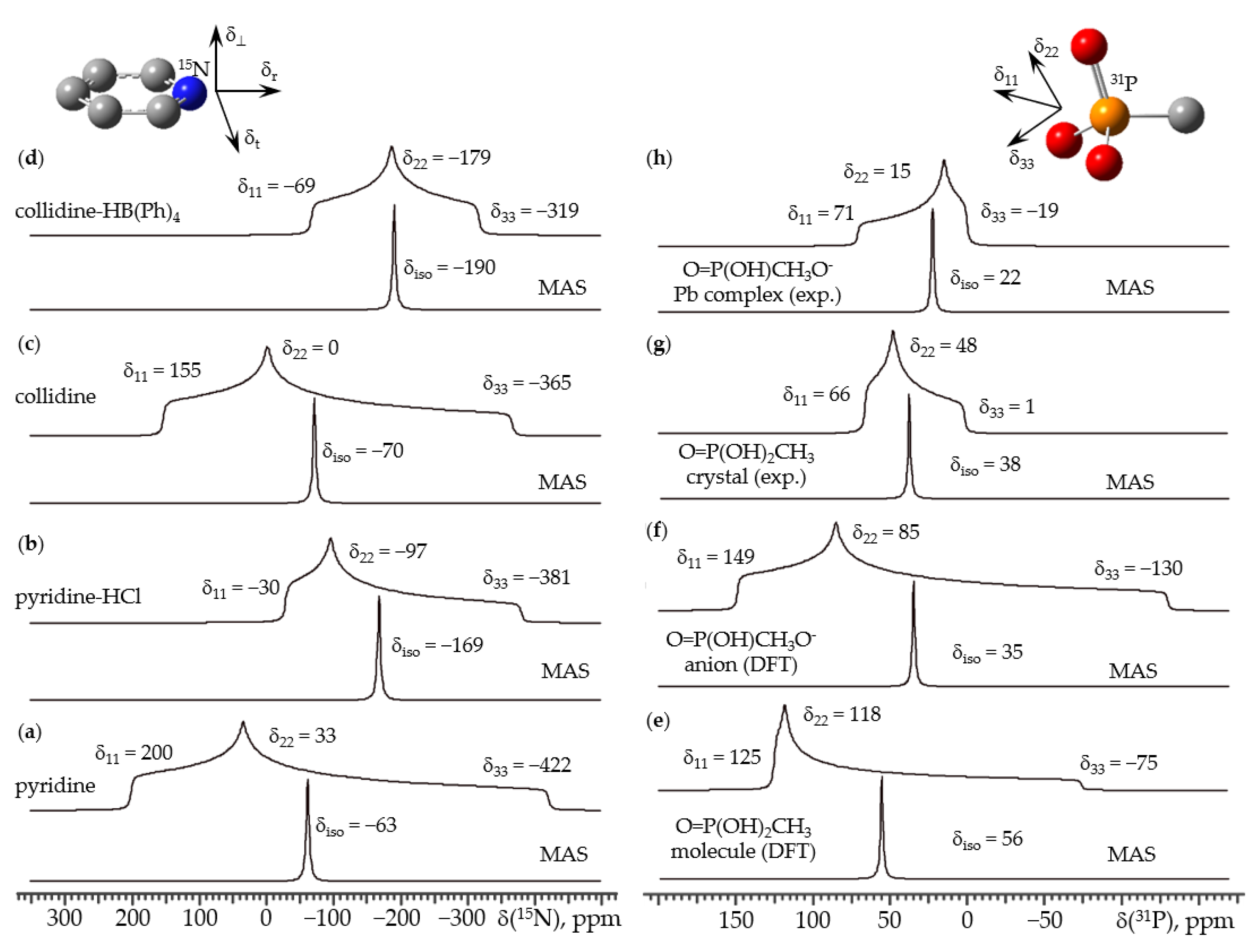
One of the main and easiest to measure spectral characteristics of NMR is a chemical shift. Chemical shift is a tensor quantity, the principal components of which are δ11 ≥ δ22 ≥ δ33. In a powdery sample, the chemical shift of a given molecule depends on its orientation relative to the direction of the applied magnetic field and is ≤δ11 and ≥δ33. For some molecules, these components can be easily represented in the molecular coordinate system. Let us consider 15N chemical shift of pyridine. When the molecular C2 symmetry axis is along the direction of the applied magnetic field, the rotation of the molecule around this axis does not change the shielding of the 15N nucleus. This component of the tensor will be labeled as δr, Figure 1 top left. The same is true when the direction of the applied magnetic field is normal to the molecular plane and the molecule rotates around this axis. This component will be labeled as δ⊥. The third axis, δt, lies in the molecular plane and is orthogonal to δr and δ⊥. For pyridine, δ11 = δt, δ22 = δr, and δ33 = δ⊥, Figure 1a [24].
Hydrogen bonding causes significant change in δt, but only slightly affects δr and δ⊥ [24]. In the pyridinium cation δ11 = δr, δ22 = δt, and δ33 = δ⊥, Figure 1b. Very similar values and changes were observed for 2,4,6-trimethylpyridine (collidine), Figure 1c,d [25]. The fact that the effect of noncovalent interactions on the 15N NMR chemical shift tensor of pyridines is caused by a significant change of the value of only one of its principle components, δt, leads to monotonic and characteristic changes in δiso = (δ11 + δ22 + δ33)/3. For example, for all studied symmetrically substituted pyridines the difference in δiso(15N) of the base and its conjugate acid is about 125 ppm [27].
In solution NMR, the anisotropy of chemical shift tensors is averaged out by fast molecular tumbling and only single isotropic chemical shift values are observed, δiso. For powdered samples, this anisotropy can be averaged out by magic-angle spinning (MAS) [6,15,28]. The signals in MAS spectra become narrow and Gaussian/Lorentzian shaped, Figure 1 bottom spectra. When multiple signals are present in the spectrum, they are either resolved or can be obtained by deconvolution. The signal-to-noise ratio increases significantly since the given integral intensity is distributed in these MAS spectra over a much narrower spectral range.
The direction of the principal components of the chemical shift tensor in the molecular coordinate system is not always user friendly. In the 31P NMR chemical shift tensor of an isolated molecule of methylphosphonic acid, δ22 is directed approximately along the P=O bond while δ33 is in the plane between the hydroxyl groups, Figure 1 top right [26]. Deprotonation or noncovalent interactions change these directions. As a result, the observed changes in the 31P NMR chemical shift tensor are difficult to discuss in the molecular coordinate system. The effect of noncovalent interactions on the 31P NMR chemical shift tensor of this molecule is very large. Compare, for example, the tensors of the isolated, noninteracting molecule and of a molecule in the crystal phase, Figure 1e,g, or the tensors of an isolated molecule of methylphosphonate and of the same molecule in a polycrystalline sample of this anion in the complex with Pt, Figure 1f,h. In contrast, changes in the corresponding δiso are quite moderate. In this case, it is no longer obvious whether the significant loss of information about the system is compensated for by a significant increase in the signal-to-noise ratio.
3. Cross-Polarization
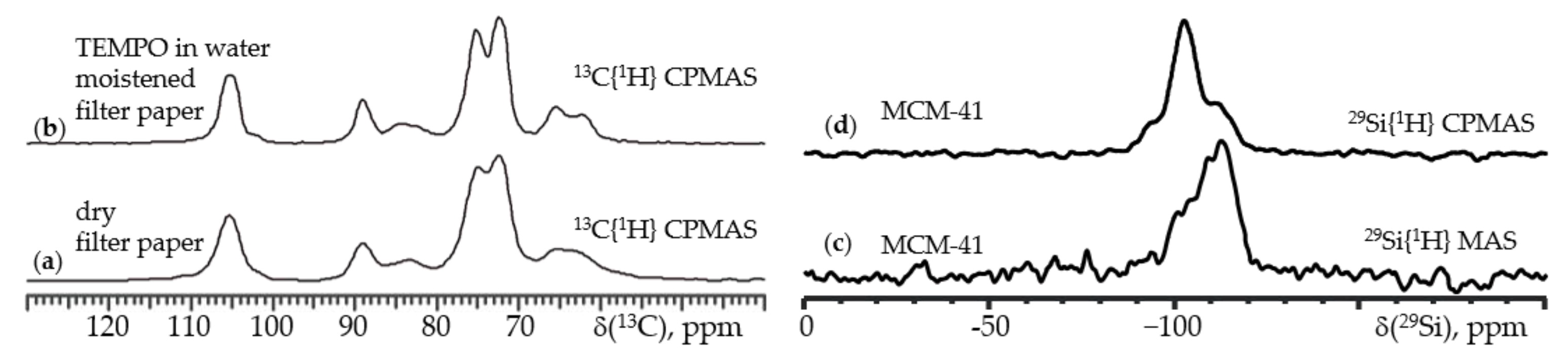
When the nuclei under study are nuclei other than 1H, 19F, and 31P, the sensitivity of the method becomes a bottleneck. In particular, for 13C and 29Si NMR, whose natural abundances are 1% and 5%, one would prefer to use the original samples without laborious isotope enrichment. These dilute spins can borrow magnetization from protons that results in a signal enhancement up to a factor of 4 (13C) and 5 (29Si). This technique is called cross-polarization (CP). Another advantage of this technique that the recycle delay required to avoid signal saturation is controlled by the T1 relaxation time of the 1H nuclei, not that of the dilute spins. The former in most cases is several times shorter than the latter. Note that CP can be combined with other methods that enhance relaxation. Figure 2a,b shows 13C{1H} CPMAS NMR spectra of two filter paper (100% cellulose) samples. The best recycle delays for a dry paper sample and the same paper moistened with an aqueous solution of the 2,2,6,6-tetramethylpiperidinyloxyl radical (TEMPO) were 5.0 and 0.5 s, respectively. In the latter sample, fluctuating local magnetic fields induced by unpaired electron spins return excess of nuclear spin energy in the form of heat to the surroundings. However, everything has a price. First, the presence of the magnetic dipole–dipole interaction between the proton spins and the spins under observation requires the use of a high-power heteronuclear decoupling, which is designated here as {1H}. In many cases, higher decoupling strengths provide better signal-to-noise ratios. However, it is dangerous to use a high-power decoupling for more than a few tens of milliseconds. The rf-coil can be damaged. If you are not sure about the correctness of the parameters, contact a specialist. Second, the gain obtained by CP depends on the magnetic dipole–dipole interaction between the dilute spins and neighboring protons. Therefore, the observed gain is nonuniform for heterogeneous samples. Figure 2c,d shows 29Si{1H} MAS NMR spectra of an MCM-41 mesoporous silica sample collected without and with CP. The relative intensities corresponding to chemically different silicon atoms differ significantly, because CP specifically enhances the magnetization of surface siloxane groups [29]. Be critical when evaluating sample composition using CP NMR spectra.
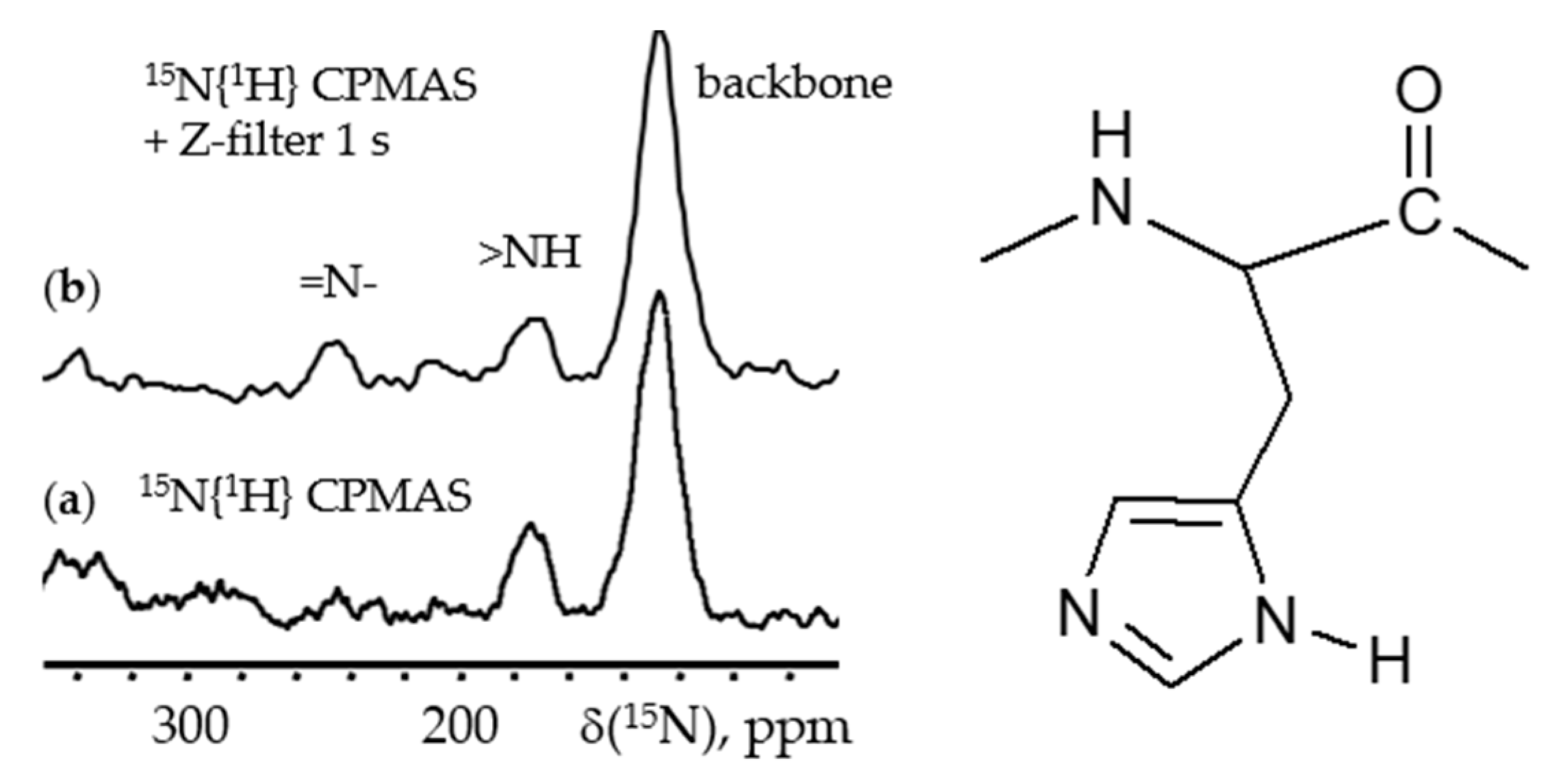
This problem is not specific to 29Si NMR. Figure 3 shows the 15N{1H} CPMAS NMR spectra of a lyophilized sample of the mutant H64A of human carbonic anhydrase II [30]. The natural abundant 15N nuclei of the peptide backbone contribute to a peak at around 120 ppm. This sample contained doubly 15N labeled 4-methylimidazole. These nitrogen nuclei resonate at 253 and 173 ppm, =N- and >NH, respectively. However, only the latter is observed when using the standard CP pulse sequence, Figure 3a. The magnetization transfer to =N- could probably be improved by using a longer cross-polarization contact time. However, this approach has a number of associated problems. Instead, before the FID was acquired, the 15N magnetization was directed for 1 s along the direction of the applied magnetic field (Z-filter sequence). Under these conditions, the magnetization of the >NH nitrogen atoms can be transferred to the =N- nitrogen atoms (spin diffusion). This equilibration process was successful but not complete, and an intensity difference remained, Figure 3b.
4. Calculation of NMR Chemical Shifts
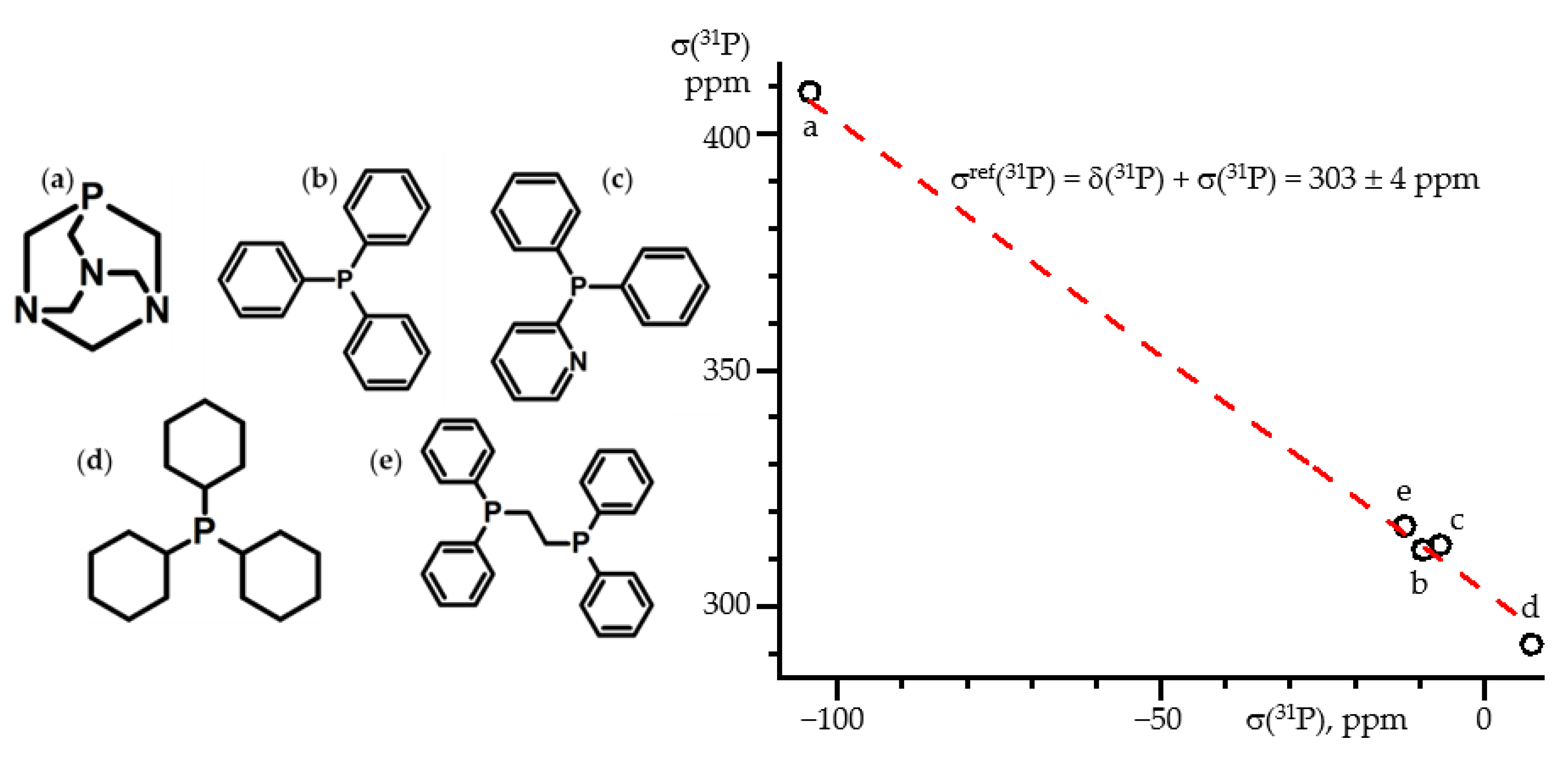
Theoretical calculations provide the absolute chemical shielding tensor, σ, the components of which are σ11 ≤ σ22 ≤ σ33. σiso = (σ11 + σ22 + σ33)/3. The experimentally reported chemical shift, δ, is measured relative to the absolute chemical shielding of a reference, δ = (σref − σ)/(1 − σref). σref is of the order of 10−4 that is δ ≈ (σref − σ). Therefore, in order to compare the values of the experimentally observed δ and calculated σ, a σref value is needed. For example, δ(31P) ≡ 0 ppm for 85% H3PO4 in H2O. This value is easy to measure but not to calculate. Neither the polarizable continuum model (PCM) nor solvation model based on density (SMD) approaches [31,32,33] can satisfactorily simulate the effect of solvation on the chemical shielding of H3PO4. Therefore, δ(31P) is known, but the corresponding σ(31P) cannot be calculated, so σref(31P) remains unknown. For 31P this problem has been solved recently [34]. The values of δ(31P) were measured for a number of solids in which δ(31P) was not influenced by noncovalent interactions, Figure 4. The conformation of the molecules was taken from X-ray diffraction (XRD) data. For these conformations were calculated the values of σ(31P), from which the mean value, σref(31P), was evaluated. The margin of error of σref(31P) ranged from ±9 to ±4 ppm, depending on the level of approximation. The best results were obtained for the specialized pcSseg-3 basis set [35] and the ωB97XD DFT functional [36]. However, smaller basis sets and simpler DFT functionals also provide satisfactory accuracy. The reported molecular structures can be used to determine σref(31P) for any approximation of interest [34].
Note that for molecules with large dipole moments, the measured magnetic shielding of 31P depends on the electric field generated by surrounding molecules [37]. As a result, there is a large deviation between the observed and predicted δ(31P) even if the conformation of the molecules is known and the shielding does not depend on noncovalent intermolecular interactions. The results obtained indicate that the field strength experienced by these molecules in crystals is similar to that in polar solvents. This effect cannot be satisfactorily reproduced within the PCM and SMD approaches. However, it can be successfully accounted for by the adduct under field approach (AuF) [37]. The dependence of the electron density on the applied electric field and the associated changes in chemical and spectral properties were widely studied [38,39,40,41,42,43]. Within the AuF approach, both the structure and NMR properties were calculated in the presence of an external electric field. This external electric field is a tool to put pressure on the electron density of the molecule under investigation in order to simulate changes caused by the environment. A detailed description of this method can be found elsewhere [44,45,46].
5. 1H NMR
In most solids, protons form strongly coupled spin networks. As a result, high resolution 1H NMR in solids requires ultrafast MAS or specific pulse sequences. A detailed description of these methods can be found elsewhere [47,48,49,50,51,52,53]. The problem is absent when protons either are diluted or possess high mobility.
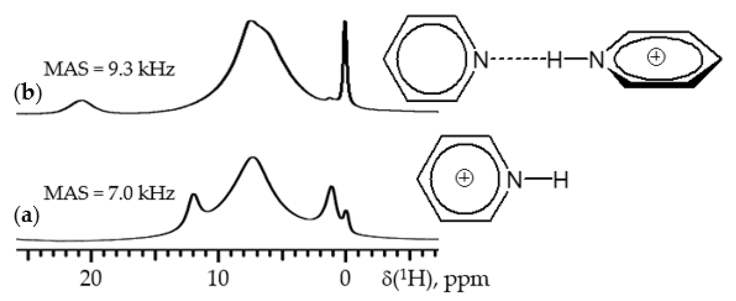
Figure 5 shows 1H MAS NMR spectra of polycrystalline samples of pyridinium tetrakis [3,5-bis(trifluoromethyl)-phenyl]-borate and the proton-bound homodimer of pyridine tetrakis [3,5-bis(trifluoromethyl)-phenyl]-borate [54,55]. The chemical shifts of mobile protons at 12.0 and 20.7 ppm are clearly detected at small MAS rates due to the use of pyridine-d5, Figure 5a,b. In these samples, protons were diluted spins and proton–proton magnetic dipolar interactions were weak.
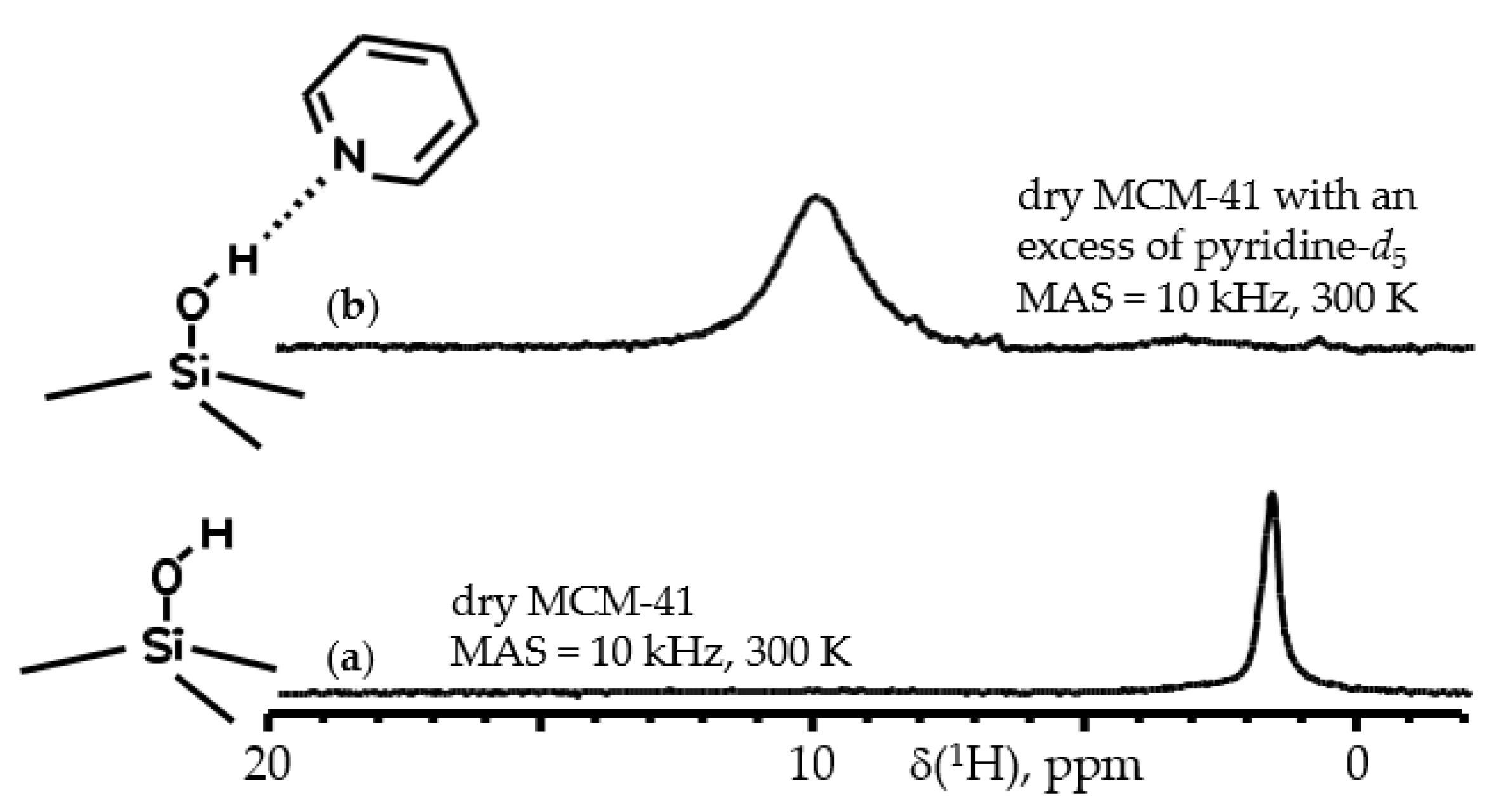
Figure 6a shows 1H MAS NMR spectra of dry mesoporous silica MCM-41. The distance between the surface silanol groups of MCM-41 is about 3–6 Å [29,56] while the rotation around the Si-O bond is most likely not hindered. Therefore, the proton–proton magnetic dipolar interactions are weak and a narrow signal belonging to these surface silanol groups is present at 1.8 ppm in different types of porous silica. When this silica is loaded with pyridine, the chemical shift of these groups increases to the limiting value of 9.9 ppm, Figure 6b [57]. The resonance is broadened because of the hydrogen bonding exchange. The vanishing of the original resonance of the noninteracting silanol groups shows that all these groups are accessible for pyridine. It is not always so. In other samples and for other guests, a gradual redistribution of signal intensity between noninteracting and interacting sites can be observed [58].
An NMR study of fluids in confinement is in demand today. The main attention has been focused on water and water–alcohol mixtures [59,60,61,62,63,64,65]. Some other substances have been studies as well [66,67,68,69,70]. Interaction with the surface causes a change in the melting/freezing point of the adsorbate [71,72,73,74,75]. When the confinement is not very tight and the surface concentration is below monolayer coverage, the reorientation of adsorbed molecules is fast on a millisecond time scale and their 1H NMR linewidths are small [76]. Specific examples are not considered here in detail because the methods, temperature ranges, filling factors, and other parameters used depend on the host, the guest, and the property in question.
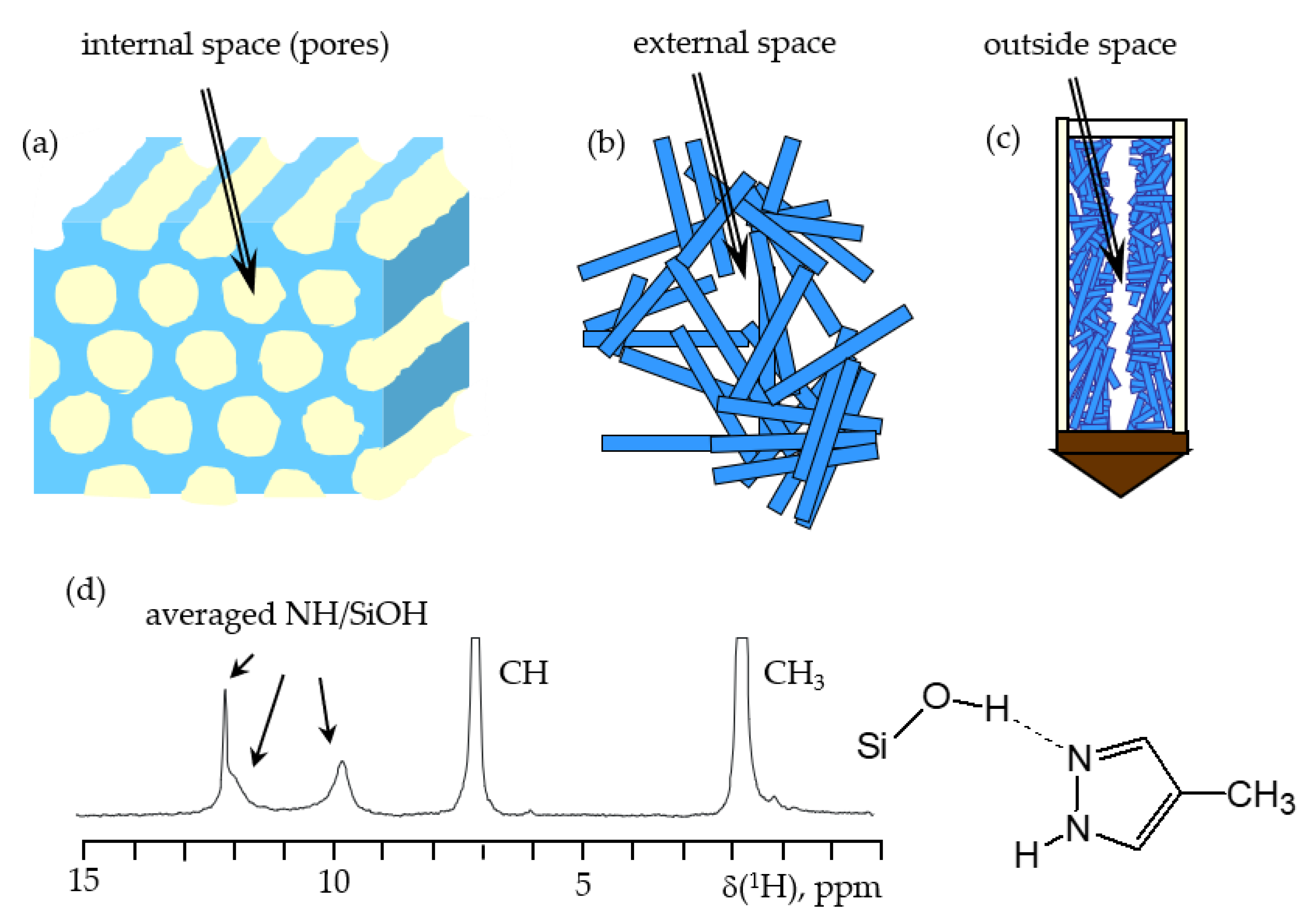
There is a general method for investigating various local spaces of powdery porous solids containing surface OH-groups using 1H NMR [77]. Consider a sample of MCM-41 mesoporous silica loaded with 4-methyl-1H-pyrazole (MPz). Pyrazole molecules are distributed over three local spaces. These spaces are shown in Figure 7: (a) internal pores of a given crystallite, (b) an external space between crystallites, and (c) an axial void space at the center of the NMR rotor because the crystallites are compressed toward the NMR rotor walls due to a fast MAS rotation. MPz is liquid at room temperature and can be easily loaded onto porous materials where it forms hydrogen bonds with surface OH-groups. At room temperature, the coalesced NH/OH signal exhibits some exchange broadening, which disappears at high temperatures. On the other hand, up to at least 400 K, the exchange of MPz molecules between the different local spaces is slow on the NMR time scale. At this temperature, 1H NMR spectra contain three resolved NH/OH signals, one for each of the local spaces, Figure 7d. This is because the OH/NH ratios are different in these local spaces. These ratios can be measured from the relative integral intensity of the respective signals. The OH/NH ratios depend on the MPz/host ratio used. Therefore, they provide insight into the pore filling mechanisms, surface/volume, volume/volume, and surface/surface ratios of the different spaces. Note that the volumes of the external void spaces of the MCM-41 and SBA-15 mesoporous silica studied turned out to be larger than those of the internal pores. The surface areas of the external spaces were much smaller than those of the internal pores, although in the case of SBA-15 this difference was not significant [77].
6. 15N NMR
15N NMR is not among the most popular routine NMR methods. The natural abundance of this isotope is 0.4%, and the strength of its magnetic moment is rather small. However, in some cases, the efforts spent on obtaining isotopically enriched compounds pay off with the results obtained. As explained in Section 2, the isotropic 15N NMR chemical shift of symmetrically substituted pyridines exhibits significant and characteristic changes upon hydrogen bonding. Figure 8a shows average N···H distances obtained by 15N NMR of collidine–15N-acid complexes as a function of δiso(15N) [25]. Here δiso(15N) ≡ 0 ppm for frozen collidine and is about 125 ppm for collidinium [27]. This dependence can be used for any symmetrically substituted pyridine. For a detailed discussion about geometric and NMR parameter correlations, refer to other publications [78,79]. The 15N NMR spectrum of an 15N-enriched pyridine derivative can be easily measured in solution [80,81,82], crystals [55,83], and polymeric materials [84], which provides insight into the hydrogen bond geometry and the protonation state of this base. δiso(15N) is sensitive to other noncovalent interactions as well [85,86]. It is quite probable that δiso(15N) characteristically correlates with some parameters of these interactions. However, no such analysis has been carried out to date.
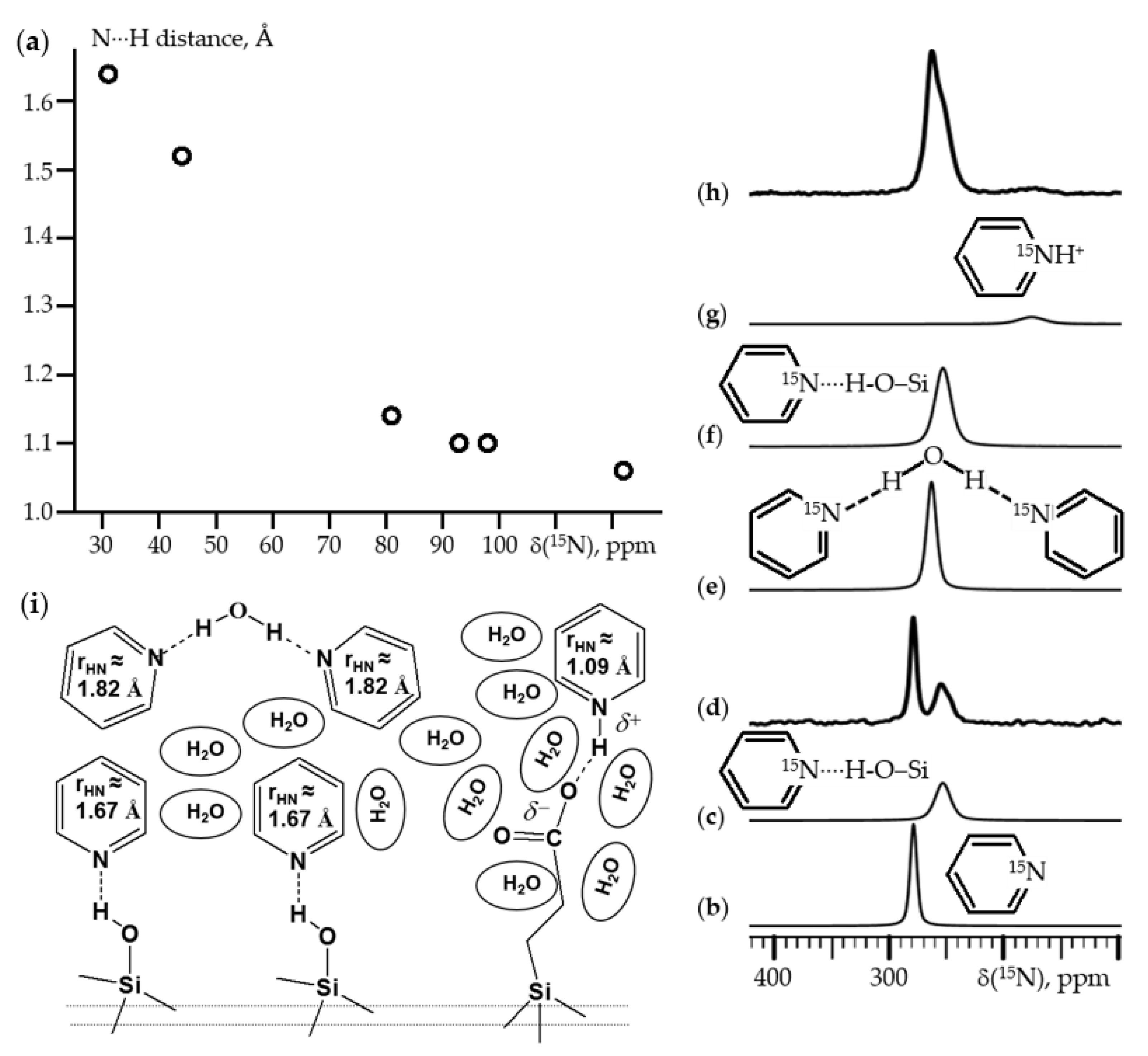
Pyridines-15N appears to be the best NMR probes for studying acidic active sites in porous solids. Figure 8d shows the 15N{1H} CPMAS NMR spectrum of pyridine-15N loaded onto a dry unfunctionalized SBA-15 mesoporous silica sample [87]. The spectrum was measured at 130 K and referenced to 15NH4Cl. In this scale, frozen pyridine resonates at 276 ppm. This spectrum contains the signals of frozen pyridine and of pyridine hydrogen bonded to the surface silanol groups, Figure 8b,c. The surface density of these groups can be measured from the relative integral intensity of these signals. It is about 2.9 nm−2 for MCM-41 and 3.7 nm−2 for SBA-15 [57]. Figure 8h shows the 15N {1H} CPMAS NMR spectrum of pyridine-15N loaded onto a propionic acid functionalized SBA-15 sample in the presence of water. This spectrum contains the signals of 2:1 pyridine:water complex [88], of pyridine-HOSi, and of pyridinium, Figure 8e,f,g. The analysis of these spectra explains the mechanism of water’s effect on the surface acidity. Water does not change the acidity of the surface silanol groups, since it cannot solvate them. When unfunctionalized silica is loaded with a pyridine/water mixture with an excess of pyridine, pyridine molecules are distributed between the pyridine-HOSi and 2:1 pyridine:water phases, Figure 8i. The same phases are present in the case of the propionic acid functionalized SBA-15. In addition, there are pyridine molecules that interact with the carboxyl moieties. These molecules are protonated because these fragments are solvated with water, Figure 8i. Note that in the absence of water, the geometry of hydrogen bonds of the pyridine-HOSi and pyridine-HOOC surface complexes is the same [87]. This means that, in the absence of water, the proton-donating ability of silanol groups is equal to that of propionic acid.
The surface silanol groups can only be deprotonated with very strong bases such as 4-dimethylaminopyridine and 4-diethyl-2,6-di-tert-butylaminopyridine [56]. The protonated molecules remain immobilized on the surface at room temperature, but can freely rotate around their C2 molecular axis. Their concentration is not affected by water, and water does not participate in or coordinate around the protonated species. The situation is different when the silica surface is functionalized with sulfonic or phosphonic acids. These acidic groups always protonate pyridine because these materials contain about six water molecules per acid moiety even after drying at 420 K in high vacuum [89]. Each sulfonic acid moiety interacts with one pyridine molecule, while each phosphonic acid moiety can interact simultaneously with two pyridine molecules. In all considered cases, the geometry of hydrogen bonds can be measured from the experimentally observed 15N NMR chemical shifts.
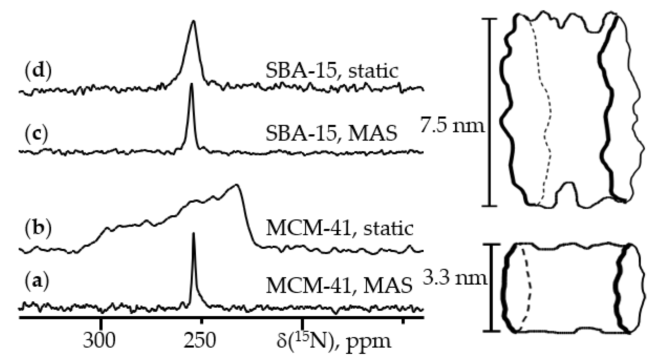
As has been explained in Chapter 2, chemical shift is a tensor quantity. Therefore, the line shapes of NMR signals depend on the rotational diffusion. This effect can be used to study the surface diffusion of adsorbed molecules. Figure 9 shows the 15N{1H} NMR spectra of pyridine-15N loaded onto MCM-41 and SBA-15 mesoporous silica [57]. These spectra were measured at 300 K and are referenced to 15NH4Cl. Less than a monolayer was occupied in both samples. Under these conditions, pyridine does not leave the surface, and its surface diffusion is fast on the NMR time scale. The acidity of the surface silanol groups does not depend on the type of silica. MAS averages out shielding anisotropy, which is weaker than the spinning frequency. As a result, the 15N{1H} MAS NMR spectra are the same for both samples, Figure 9a,c. On the other hand, the static 15N{1H} NMR spectrum of the MCM-41 sample demonstrates the residual shielding anisotropy, which is absent in the case of the SBA-15 sample, Figure 9b,d. The presence of this residual shielding anisotropy indicates the very low degree of roughness of the inner surfaces of this MCM-41 sample. Please refer to the original paper for a detailed analysis [57].
7. 31P NMR
The 31P isotope is present in 100% natural abundance, has a spin quantum number of 1/2 and a chemical shift range of more than 400 ppm. 31P NMR can be routinely used in the evaluation of the structure of organic complexes in solution [90,91] and solids [92,93,94] including organometallic compounds [26,95,96,97,98]. Molecules containing phosphorus are very convenient NMR probes for studying mobility at interfaces [99,100,101]. For example, 31P NMR has been used to elucidate thermal and hydration effects on the mobility of compact, branched, and bulky pharmaceutical molecules loaded in submonolayer amounts onto mesoporous silica [102]. Great progress has been achieved in our understanding of the effects of noncovalent interactions on the 31P chemical shift of P=O groups [26,103,104]. However, P=O groups can form two noncovalent interactions simultaneously [105,106,107]. Since the phosphorus atom of the P=O group is not directly involved in intermolecular interactions, the changes of its isotropic chemical shift is not strictly specific to a certain interaction, and different structures can result in similar values [108]. In addition, the shielding may depend on the conformation of the molecule [26]. Consequently, variations in δiso(31P) caused by noncovalent interactions and conformational changes cannot always be interpreted.
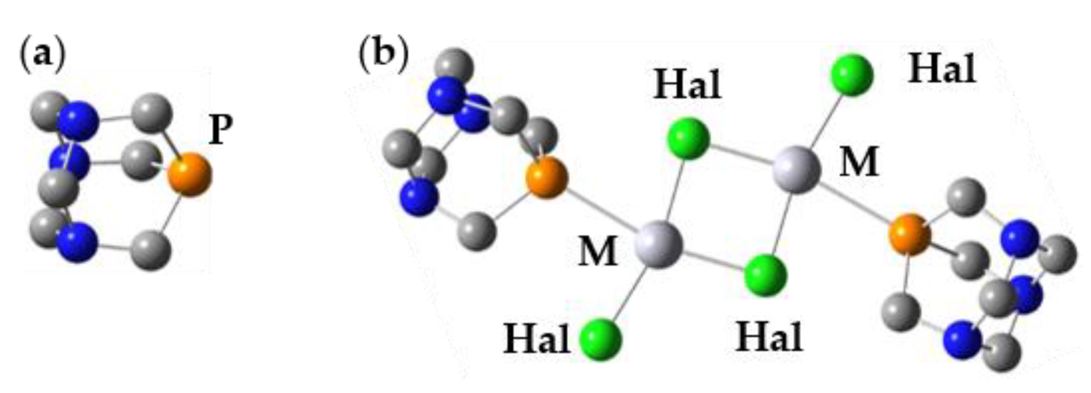
On the other hand, there are molecules in which δ(31P) depends only on interactions of a certain nature. In such cases, NMR allows very accurate analysis of spectral features [109]. For example, 1,3,5-triaza-7-phosphaadamantane (PTA, Figure 10a) is a rigid and relatively chemically inert molecule. In acidic solution, PTA would be protonated at one of its nitrogen atoms. This protonation results in a 6 ppm change in δiso(31P) [110]. In contrast, when PTA is coordinated to transition metals, its chemical shift varies in a wide range [111]. Figure 10b shows the proposed structure of Hal2Hg(PTA) complexes in crystals (Hal = Cl, Br, I). This structure is adapted from [112]. In PTA δiso(31P) = −104.3 ppm [34], while changes to −38.3, −44.1, and −61.4 in Cl2Hg(PTA), Br2Hg(PTA), and I2Hg(PTA) [112]. Therefore, 31P NMR of organometallic complexes of PTA can be very informative. The structures of these Hal2Hg(PTA) complexes are difficult to adapt for calculations. However, there is another important application of 31P NMR. Consider an organometallic complex in which the metal is coordinated with a phosphorus atom. In many cases, the structure of this complex is determined from XRD, while δiso(31P) is measured in solution [113,114,115,116,117]. If this value can be reproduced in calculations using the structure of the crystalline phase, then the transition to solution has no significant effect on the structure. If this is not the case, then the experimentally reported δiso(31P) is either not related to the proposed structure, or the structure is labile. Note that such calculations can be done using moderate basis sets [118].
8. Conclusions
Solid-state NMR can do a lot and does not always require excessive efforts. Its advantages are especially noticeable for polycrystalline, amorphous, and heterogeneous samples. It is these systems that are especially difficult to study by other spectral methods. Only a small set of the simplest but broadly applicable methods are discussed here. There are many other powerful and specialized techniques out there today. You can get acquainted with them using the references listed in the Introduction. Two-dimensional solid-state NMR methods are rapidly developing and becoming available for widespread use [119,120,121]. Since 2008, the annual number of publications on “solid-state NMR” has exceeded 2000 [122]. We hope that this review will help some research to include solid-state NMR in the list of spectral methods they actively use.
Author Contributions
Conceptualization, I.G.S. and H.-H.L.; methodology, I.G.S. and H.-H.L.; software, I.G.S. and H.-H.L.; resources, I.G.S.; data curation, I.G.S.; writing—original draft preparation, I.G.S.; writing—review and editing, I.G.S. and H.-H.L.; visualization, I.G.S. and H.-H.L.; funding acquisition, I.G.S. Both authors have read and agreed to the published version of the manuscript.
Funding
This research received no external funding. The APC was funded by MDPI.
Institutional Review Board Statement
Not applicable.
Informed Consent Statement
Not applicable.
Data Availability Statement
Data sharing is not applicable to this article.
Conflicts of Interest
The authors declare no conflict of interest.
References
- Porter, A.L.; Rafols, I. Is science becoming more interdisciplinary? Measuring and mapping six research fields over time. Scientometrics 2009, 81, 719–745. [Google Scholar] [CrossRef]
- Melnikova, D.L.; Badrieva, Z.F.; Kostin, M.A.; Maller, C.; Stas, M.; Buczek, A.; Broda, M.A.; Kupka, T.; Kelterer, A.-M.; Tolstoy, P.M.; et al. On Complex Formation between 5-Fluorouracil and β-Cyclodextrin in Solution and in the Solid State: IR Markers and Detection of Short-Lived Complexes by Diffusion NMR. Molecules 2020, 25, 5706. [Google Scholar] [CrossRef]
- Antonov, A.S.; Karpov, V.V.; Tupikina, E.Y.; Tolstoy, P.M.; Vovk, M.A. Aggregation Behavior of Lithionaphthalenes in Solution: Experimental and Theoretical Study. Organometallics 2020, 39, 3705–3714. [Google Scholar] [CrossRef]
- Jansen, D.; Gramüller, J.; Niemeyer, F.; Schaller, T.; Letzel, M.C.; Grimme, S.; Zhu, H.; Gschwind, R.M.; Niemeyer, J. What is the role of acid–acid interactions in asymmetric phosphoric acid organocatalysis? A detailed mechanistic study using interlocked and non-interlocked catalysts. Chem. Sci. 2020, 11, 4381–4390. [Google Scholar] [CrossRef] [Green Version]
- Lokesh, N.; Hioe, J.; Gramüller, J.; Gschwind, R.M. Relaxation Dispersion NMR to Reveal Fast Dynamics in Brønsted Acid Catalysis: Influence of Sterics and H-Bond Strength on Conformations and Substrate Hopping. J. Am. Chem. Soc. 2019, 141, 16398–16407. [Google Scholar] [CrossRef] [Green Version]
- Duer, M.J. (Ed.) Solid-State NMR Spectroscopy. Principles and Applications; Blackwell Science Ltd.: Oxford, UK, 2002. [Google Scholar]
- Bachmutov, V.I. Solid-State NMR in Materials Science. Principles and Applications; CRC Press: Boca Raton, FL, USA, 2011. [Google Scholar]
- Hodgkinson, P. (Ed.) Modern Methods in Solid-State NMR: A Practitioner’s Guide; The Royal Society of Chemistry: London, UK, 2018. [Google Scholar]
- Buntkowsky, G.; Vogel, M. Small Molecules, Non-Covalent Interactions, and Confinement. Molecules 2020, 25, 3311. [Google Scholar] [CrossRef]
- Brunner, E.; Rauche, M. Solid-state NMR spectroscopy: An advancing tool to analyse the structure and properties of metal–organic frameworks. Chem. Sci. 2020, 11, 4297–4304. [Google Scholar] [CrossRef] [Green Version]
- Gutmann, T.; Groszewicz, P.B.; Buntkowsky, G. Solid-state NMR of nanocrystals. Annu. Rep. NMR Spectrosc. 2019, 97, 1–82. [Google Scholar] [CrossRef]
- Smith, M.E. Recent progress in solid-state nuclear magnetic resonance of half-integer spin low-γ quadrupolar nuclei applied to inorganic materials. Magn. Reson. Chem. 2020, 1–44. [Google Scholar] [CrossRef]
- Bakhmutov, V.I. Strategies for Solid-State NMR Studies of Materials: From Diamagnetic to Paramagnetic Porous Solids. Chem. Rev. 2011, 111, 530–562. [Google Scholar] [CrossRef] [PubMed]
- Hodgkinson, P.; Wimperis, S. Solid-state NMR spectroscopy. Phys. Chem. Chem. Phys. 2009, 11, 6875. [Google Scholar] [CrossRef] [PubMed]
- Bryce, D.L.; Bernard, G.M.; Gee, M.; Lumsden, M.D.; Eichele, K.; Wasylishen, R.E. Practical Aspects of Modern Routine Solid-State Multinuclear Magnetic Resonance Spectroscopy: One-Dimensional Experiments. Can. J. Anal. Sci. Spectrosc. 2001, 46, 46–82. [Google Scholar] [CrossRef]
- Brown, S.P.; Spiess, W. Advanced Solid-State NMR Methods for the Elucidation of Structure and Dynamics of Molecular, Macromolecular, and Supramolecular Systems. Chem. Rev. 2001, 101, 4125–4156. [Google Scholar] [CrossRef] [PubMed]
- Albert, K. NMR investigations of stationary phases. J. Sep. Sci. 2003, 26, 215–224. [Google Scholar] [CrossRef]
- Gullion, T. Introduction to rotational-echo, double-resonance NMR. Concepts Magn. Reson. 1998, 10, 277–289. [Google Scholar] [CrossRef]
- Kaupp, M.; Bühl, M.; Malkin, V.G. (Eds.) Calculation of NMR and EPR Parameters; Wiley-VCH: Weinheim, Germany, 2004. [Google Scholar]
- Pilar, K.; Deng, Z.; Preefer, M.B.; Cooley, J.A.; Clément, R.; Seshadri, R.; Cheetham, A.K. Ab initio computation for solid-state 31P NMR of inorganic phosphates: Revisiting X-ray structures. Phys. Chem. Chem. Phys. 2019, 21, 10070–10074. [Google Scholar] [CrossRef] [Green Version]
- Grasa, P.; Baker, A.; Combes, C.; Rey, C.; Sarda, S.; Wright, A.J.; Smith, M.E.; Hanna, J.V.; Gervais, C.; Laurencin, D.; et al. From crystalline to amorphous calcium pyrophosphates: A solid state Nuclear Magnetic Resonance perspective. Acta Biomater. 2016, 31, 348–357. [Google Scholar] [CrossRef]
- Medvedev, A.G.; Churakov, A.V.; Prikhodchenko, P.V.; Lev, O.; Vener, M.V. Crystalline Peroxosolvates: Nature of the Coformer, Hydrogen-Bonded Networks and Clusters, Intermolecular Interactions. Molecules 2021, 26, 26. [Google Scholar] [CrossRef]
- Charpentier, T. The PAW/GIPAW approach for computing NMR parameters: A new dimension added to NMR study of solids. Solid State Nucl. Magn. Reson. 2011, 40, 1–20. [Google Scholar] [CrossRef]
- Solum, M.S.; Altmann, K.L.; Strohmeier, M.; Berges, D.A.; Zhang, Y.; Facelli, J.C.; Pugmire, R.J.; Grant, D.M. 15N Chemical Shift Principal Values in Nitrogen Heterocycles. J. Am. Chem. Soc. 1997, 119, 9804–9809. [Google Scholar] [CrossRef]
- Lorente, P.; Shenderovich, I.G.; Golubev, N.S.; Denisov, G.S.; Buntkowsky, G.; Limbach, H.-H. 1H/15N NMR Chemical Shielding, Dipolar 15N,2H Coupling and Hydrogen Bond Geometry Correlations in a Novel Serious of Hydrogen-Bonded Acid-Base Complexes of Collidine with Carboxylic Acids. Magn. Reson. Chem. 2001, 39, S18–S29. [Google Scholar] [CrossRef]
- Shenderovich, I.G. Effect of Noncovalent Interactions on the 31P Chemical Shift Tensor of Phosphine Oxides, Phosphinic, Phosphonic, and Phosphoric Acids, and Their Complexes with Lead(II). J. Phys. Chem. C 2013, 117, 26689–26702. [Google Scholar] [CrossRef]
- Andreeva, D.V.; Ip, B.; Gurinov, A.A.; Tolstoy, P.M.; Denisov, G.S.; Shenderovich, I.G.; Limbach, H.-H. Geometrical Features of Hydrogen Bonded Complexes Involving Sterically Hindered Pyridines. J. Phys. Chem. A 2006, 110, 10872–10879. [Google Scholar] [CrossRef] [PubMed]
- Lowe, I.J. Free Induction Decays of Rotating Solids. Phys. Rev. Lett. 1959, 2, 285–287. [Google Scholar] [CrossRef]
- Shenderovich, I.G.; Mauder, D.; Akcakayiran, D.; Buntkowsky, G.; Limbach, H.-H.; Findenegg, G.H. NMR Provides Checklist of Generic Properties for Atomic-Scale Models of Periodic Mesoporous Silicas. J. Phys. Chem. B 2007, 111, 12088–12096. [Google Scholar] [CrossRef] [PubMed]
- Shenderovich, I.G.; Lesnichin, S.B.; Tu, C.; Silverman, D.N.; Tolstoy, P.M.; Denisov, G.S.; Limbach, H.-H. NMR Studies of Active-Site Properties of Human Carbonic Anhydrase II by using 15N-Labeled 4-Methylimidazole as a Local Probe and Histidine Hydrogen-Bond Correlations. Chem. Eur. J. 2015, 21, 2915–2929. [Google Scholar] [CrossRef]
- Cossi, M.; Barone, V.; Cammi, R.; Tomasi, J. Ab initio study of solvated molecules: A new implementation of the polarizable continuum model. Chem. Phys. Lett. 1996, 255, 327–335. [Google Scholar] [CrossRef]
- Tomasi, J.; Mennucci, B.; Cammi, R. Quantum mechanical continuum solvation models. Chem. Rev. 2005, 105, 2999–3093. [Google Scholar] [CrossRef]
- Marenich, A.V.; Cramer, C.J.; Truhlar, D.G. Universal solvation model based on solute electron density and on a continuum model of the solvent defined by the bulk dielectric constant and atomic surface tensions. J. Phys. Chem. B 2009, 113, 6378–6396. [Google Scholar] [CrossRef]
- Shenderovich, I.G. Experimentally Established Benchmark Calculations of 31P NMR Quantities. Chemistry-Methods 2021, 1, 61–70. [Google Scholar] [CrossRef]
- Jensen, F. Segmented Contracted Basis Sets Optimized for Nuclear Magnetic Shielding. J. Chem. Theory Comput. 2015, 11, 132–138. [Google Scholar] [CrossRef] [PubMed]
- Chai, J.-D.; Head-Gordon, M. Long-range corrected hybrid density functionals with damped atom–atom dispersion corrections. Phys. Chem. Chem. Phys. 2008, 10, 6615–6620. [Google Scholar] [CrossRef] [PubMed] [Green Version]
- Shenderovich, I.G. Electric field effect on 31P NMR magnetic shielding. J. Chem. Phys. 2020, 153, 184501. [Google Scholar] [CrossRef] [PubMed]
- Nardo, V.M.; Cassone, G.; Ponterio, R.C.; Saija, F.; Sponer, J.; Tommasini, M.; Trusso, S. Electric-Field-Induced Effects on the Dipole Moment and Vibrational Modes of the Centrosymmetric Indigo Molecule. J. Phys. Chem. A 2020, 124, 10856–10869. [Google Scholar] [CrossRef] [PubMed]
- Cassone, G.; Sponer, J.; Trusso, S.; Saija, F. Ab initio spectroscopy of water under electric fields. Phys. Chem. Chem. Phys. 2019, 21, 21205–21212. [Google Scholar] [CrossRef]
- Dominikowska, J.; Palusiak, M. Tuning Aromaticity of para-Substituted Benzene Derivatives with an External Electric Field. ChemPhysChem 2018, 19, 590–595. [Google Scholar] [CrossRef]
- Alkorta, I.; Elguero, J.; Provasi, P.F.; Pagola, G.I.; Ferraro, M.B. Electric field effects on nuclear magnetic shielding of the 1:1 and 2:1 (homo and heterochiral) complexes of XOOX′ (X, X′ = H, CH3) with lithium cation and their chiral dis-crimination. J. Chem. Phys. 2011, 135, 104116. [Google Scholar] [CrossRef] [Green Version]
- Del Bene, J.E.; Jordan, M.J.T. To What Extent Do External Fields and Vibrational and Isotopic Effects Influence NMR Coupling Constants Across Hydrogen Bonds? Two-Bond Cl-N Spin-Spin Coupling Constants (2hJCl-N) in Model ClH:NH3 Complexes. J. Phys. Chem. A 2002, 106, 5385–5392. [Google Scholar] [CrossRef]
- Ramos, M.; Alkorta, I.; Elguero, J.; Golubev, N.S.; Denisov, G.S.; Benedict, H.; Limbach, H.-H. Theoretical Study of the Influence of Electric Fields on Hydrogen-Bonded Acid−Base Complexes. J. Phys. Chem. A 1997, 101, 9791–9800. [Google Scholar] [CrossRef]
- Shenderovich, I.G.; Denisov, G.S. Adduct under Field—A Qualitative Approach to Account for Solvent Effect on Hydrogen Bonding. Molecules 2020, 25, 436. [Google Scholar] [CrossRef] [Green Version]
- Shenderovich, I.G.; Denisov, G.S. Modeling of Solute-Solvent Interactions Using an External Electric Field—From Tautomeric Equilibrium in Nonpolar Solvents to the Dissociation of Alkali Metal Halides. Molecules 2021, 26, 1283. [Google Scholar] [CrossRef]
- Shenderovich, I.G.; Denisov, G.S. Solvent effects on acid-base complexes. What is more important: A macroscopic reaction field or solute-solvent interactions? J. Chem. Phys. 2019, 150, 204505. [Google Scholar] [CrossRef] [PubMed]
- Struppe, J.; Quinn, C.M.; Sarkar, S.; Gronenborn, A.M.; Polenova, T. Ultrafast 1H MAS NMR Crystallography for Natural Abundance Pharmaceutical Compounds. Mol. Pharm. 2020, 17, 674–682. [Google Scholar] [CrossRef] [PubMed]
- Brown, S.P. Applications of high-resolution 1H solid-state NMR. Solid State Nucl. Magn. Reson. 2012, 41, 1–27. [Google Scholar] [CrossRef]
- Paruzzo, F.M.; Emsley, L. High-resolution 1H NMR of powdered solids by homonuclear dipolar decoupling. J. Magn. Reson. 2019, 309, 106598. [Google Scholar] [CrossRef]
- Dudek, M.K.; Kazmierski, S.; Kostrzewa, M.; Potrzebowski, M.J. Solid-State NMR Studies of Molecular Crystals. Annu. Rep. NMR Spectrosc. 2018, 95, 1–81. [Google Scholar] [CrossRef]
- Mote, K.R.; Agarwal, V.; Madhu, P.K. Five decades of homonuclear dipolar decoupling in solid-state NMR: Status and outlook. Prog. Nucl. Magn. Reson. Spectrosc. 2016, 97, 1–39. [Google Scholar] [CrossRef]
- Vinogradov, E.; Madhu, P.K.; Vega, S. Strategies for high-resolution proton spectroscopy in solid-state NMR. Top. Curr. Chem. 2004, 246, 33–90. [Google Scholar] [CrossRef]
- Schnell, I.; Spiess, H.W. High-Resolution 1H NMR Spectroscopy in the Solid State: Very Fast Sample Rotation and Multiple-Quantum Coherences. J. Magn. Reson. 2001, 151, 153–227. [Google Scholar] [CrossRef]
- Gurinov, A.A.; Lesnichin, S.B.; Limbach, H.-H.; Shenderovich, I.G. How Short is the Strongest Hydrogen Bond in the Proton-Bound Homodimers of Pyridine Derivatives? J. Phys. Chem. A 2014, 118, 10804–10812. [Google Scholar] [CrossRef]
- Kong, S.; Borissova, A.O.; Lesnichin, S.B.; Hartl, M.; Daemen, L.L.; Eckert, J.; Antipin, M.Y.; Shenderovich, I.G. Geometry and Spectral Properties of the Protonated Homodimer of Pyridine in the Liquid and Solid States. A Combined NMR, X-ray Diffraction and Inelastic Neutron Scattering Study. J. Phys. Chem. A 2011, 115, 8041–8048. [Google Scholar] [CrossRef]
- Ip, B.C.K.; Andreeva, D.V.; Buntkowsky, G.; Akcakayiran, D.; Findenegg, G.H.; Shenderovich, I.G. NMR Study of Proton Transfer to Strong Bases on Inner Surfaces of MCM-41. Micropor. Mesopor. Mater. 2010, 134, 22–28. [Google Scholar] [CrossRef]
- Shenderovich, I.G.; Buntkowsky, G.; Schreiber, A.; Gedat, E.; Sharif, S.; Albrecht, J.; Golubev, N.S.; Findenegg, G.H.; Limbach, H.-H. Pyridine-15N A Mobile NMR Sensor for Surface Acidity and Surface Defects of Mesoporous Silica. J. Phys. Chem. B 2003, 107, 11924–11939. [Google Scholar] [CrossRef]
- Grünberg, B.; Emmler, T.; Gedat, E.; Shenderovich, I.; Findenegg, G.H.; Limbach, H.-H.; Buntkowsky, G. Hydrogen Bonding of Water Confined in Mesoporous Silica MCM-41 and SBA-15 Studied by 1H Solid-State NMR. Chem. Eur. J. 2004, 10, 5689–5696. [Google Scholar] [CrossRef]
- Li, Z.; Rieg, C.; Beurer, A.-K.; Benz, M.; Bender, J.; Schneck, C.; Traa, Y.; Dyballa, M.; Hunger, M. Effect of aluminum and sodium on the sorption of water and methanol in microporous MFI-type zeolites and mesoporous SBA-15 materials. Adsorption 2021, 27, 49–68. [Google Scholar] [CrossRef]
- Breynaert, E.; Houlleberghs, M.; Radhakrishnan, S.; Grübel, G.; Taulelle, F.; Martens, J.A. Water as a tuneable solvent: A perspective. Chem. Soc. Rev. 2020, 49, 2557–2569. [Google Scholar] [CrossRef]
- Ok, S.; Hwang, B.; Liu, T.; Welch, S.; Sheets, J.M.; Cole, D.R.; Liu, K.-H.; Mou, C.-Y. Fluid Behavior in Nanoporous Silica. Front. Chem. 2020, 8, 734. [Google Scholar] [CrossRef] [PubMed]
- Veena, V.S.; Illath, K.; Lazar, A.; Vinod, C.P.; Ajithkumar, T.G.; Jayanthi, S. Distribution of water in the pores of periodic mesoporous organosilicates—A proton solid state MAS NMR study. Phys. Chem. Chem. Phys. 2018, 20, 29351. [Google Scholar] [CrossRef] [PubMed]
- Kumari, B.; Brodrecht, M.; Breitzke, H.; Werner, M.; Grünberg, B.; Limbach, H.-H.; Forg, S.; Sanjon, E.P.; Drossel, B.; Gutmann, T.; et al. Mixtures of Alcohols and Water confined in Mesoporous Silica: A Combined Solid-State NMR and Molecular Dynamics Simulation Study. J. Phys. Chem. C 2018, 122, 19540–19550. [Google Scholar] [CrossRef]
- Sebastiani, D. Ab-Initio Molecular Dynamics Simulations and Calculations of Spectroscopic Parameters in Hydrogen-Bonding Liquids in Confinement. Z. Phys. Chem. 2018, 232, 973–987. [Google Scholar] [CrossRef]
- Kristinaitytė, K.; Dagys, L.; Kausteklis, J.; Klimavicius, V.; Doroshenko, I.; Pogorelov, V.; Valevičienė, N.R.; Balevicius, V. NMR and FTIR studies of clustering of water molecules: From low-temperature matrices to nano-structured materials used in innovative medicine. J. Mol. Liq. 2017, 235, 1–6. [Google Scholar] [CrossRef]
- Shelyapina, M.G.; Silyukov, O.I.; Lushpinskaya, I.P.; Kurnosenko, S.A.; Mazur, A.S.; Shenderovich, I.G.; Zvereva, I.A. NMR Study of Intercalates and Grafted Organic Derivatives of H2La2Ti3O10. Molecules 2020, 25, 5229. [Google Scholar] [CrossRef]
- Klimavicius, V.; Dagys, L.; Chizhik, V.; Balevicius, V. CP MAS Kinetics Study of Ionic Liquids Confined in Mesoporous Silica: Convergence of Non-Classical and Classical Spin Coupling Models. Appl. Magn. Reson. 2017, 48, 673–685. [Google Scholar] [CrossRef]
- Buntkowsky, G.; Breitzke, H.; Adamczyk, A.; Roelofs, F.; Emmler, T.; Gedat, E.; Grünberg, B.; Xu, Y.; Limbach, H.-H.; Shenderovich, I.; et al. Structural and Dynamical Properties of Guest Molecules Confined in Mesoporous Silica Materials Revealed by NMR. Phys. Chem. Chem. Phys. 2007, 9, 4843–4853. [Google Scholar] [CrossRef]
- Vyalikh, A.; Emmler, T.; Gedat, E.; Shenderovich, I.; Findenegg, G.H.; Limbach, H.-H.; Buntkowsky, G. Evidence of Microphase Separation in Controlled Pore Glasses. Solid State Nucl. Magn. Reson. 2005, 28, 117–124. [Google Scholar] [CrossRef]
- Gedat, E.; Schreiber, A.; Findenegg, G.H.; Shenderovich, I.; Limbach, H.-H.; Buntkowsky, G. Stray field gradient NMR reveals effects of hydrogen bonding on diffusion coefficients of pyridine in mesoporous silica. Magn. Reson. Chem. 2001, 39, S149–S157. [Google Scholar] [CrossRef]
- Neffati, R.; Judeinstein, P.; Rault, J. Freezing, melting and dynamics of supercooled water confined in porous glass. J. Phys. Condens. Matter 2020, 32, 465101. [Google Scholar] [CrossRef]
- Enninful, H.R.N.B.; Schneider, D.; Kohns, R.; Enke, D.; Valiullin, R. A novel approach for advanced thermoporometry characterization of mesoporous solids: Transition kernels and the serially connected pore model. Micropor. Mesopor. Mat. 2020, 309, 110534. [Google Scholar] [CrossRef]
- Werner, M.; Rothermel, N.; Breitzke, H.; Gutmann, T.; Buntkowsky, G. Recent Advances in Solid State NMR of Small Molecules in Confinement. Isr. J. Chem. 2014, 54, 60–73. [Google Scholar] [CrossRef]
- Sattig, M.; Reutter, S.; Fujara, F.; Werner, M.; Buntkowsky, G.; Vogel, M. NMR studies on the temperature-dependent dynamics of confined water. Phys. Chem. Chem. Phys. 2014, 16, 19229–19240. [Google Scholar] [CrossRef] [Green Version]
- Grünberg, B.; Grünberg, A.; Limbach, H.-H.; Buntkowsky, G. Melting of Naphthalene Confined in Mesoporous Silica MCM-41. Appl. Marn. Reson. 2013, 44, 189–201. [Google Scholar] [CrossRef]
- Szalontai, G. 1H NMR linewidths of small organic guest molecules physisorbed on different mesoporous silicas. J. Mol. Struct. 2020, 1205, 127646. [Google Scholar] [CrossRef]
- Torres Barthelemy, V.; Pérez-Hernández, N.; Shenderovich, I.G.; Tolstoy, P.M.; Denisov, G.S.; Limbach, H.-H. NMR-Detected Host-Guest Proton Exchange as a Tool to Explore Surface/Volume Ratios and Fluid Filling of Internal and External Spaces of Porous Solids containing Surface-OH-Groups. J. Phys. Chem. C 2020, 124, 22082–22095. [Google Scholar] [CrossRef]
- Gurinov, A.A.; Denisov, G.S.; Borissova, A.O.; Goloveshkin, A.S.; Greindl, J.; Limbach, H.-H.; Shenderovich, I.G. NMR Study of Solvation Effect on the Geometry of Proton-Bound Homodimers of Increasing Size. J. Phys. Chem. A 2017, 121, 8697–8705. [Google Scholar] [CrossRef] [PubMed] [Green Version]
- Limbach, H.-H.; Pietrzak, M.; Sharif, S.; Tolstoy, P.M.; Shenderovich, I.G.; Smirnov, S.N.; Golubev, N.S.; Denisov, G.S. NMR-Parameters and Geometries of OHN- and ODN Hydrogen Bonds of Pyridine-Acid Complexes. Chem. Eur. J. 2004, 10, 5195–5204. [Google Scholar] [CrossRef] [PubMed]
- Lesnichin, S.B.; Tolstoy, P.M.; Limbach, H.-H.; Shenderovich, I.G. Counteranion-Dependent Mechanisms of Intramolecular Proton Transfer in Aprotic Solution. Phys. Chem. Chem. Phys. 2010, 12, 10373–10379. [Google Scholar] [CrossRef]
- Chan-Huot, M.; Dos, A.; Zander, R.; Sharif, S.; Tolstoy, P.M.; Compton, S.; Fogle, E.; Toney, M.D.; Shenderovich, I.; Denisov, G.S.; et al. NMR Studies of Protonation and Hydrogen Bond States of Internal Aldimines of Pyridoxal 5′-Phosphate Acid−Base in Alanine Racemase, Aspartate Aminotransferase, and Poly-L-lysine. J. Am. Chem. Soc. 2013, 135, 18160–18175. [Google Scholar] [CrossRef]
- Limbach, H.-H.; Chan-Huot, M.; Sharif, S.; Tolstoy, P.M.; Shenderovich, I.G.; Denisov, G.S. Critical Hydrogen Bonds and Protonation States of Pyridoxal 5′-phosphate Revealed by NMR. Biochim. Biophys. Acta (BBA) Proteins Proteom. 2011, 1814, 1426–1437. [Google Scholar] [CrossRef]
- Ip, B.C.K.; Shenderovich, I.G.; Tolstoy, P.M.; Frydel, J.; Denisov, G.S.; Buntkowsky, G.; Limbach, H.-H. NMR Studies of Solid Pentachlorophenol-4-Methylpyridine Complexes Exhibiting Strong OHN Hydrogen Bonds: Geometric H/D Isotope Effects and Hydrogen Bond Coupling Cause Isotopic Polymorphism. J. Phys. Chem. A 2012, 116, 11370–11387. [Google Scholar] [CrossRef]
- Bismarck, A.; Aranberri-Askargorta, I.; Springer, J.; Lampke, T.; Wielage, B.; Stamboulis, A.; Shenderovich, I.; Limbach, H.-H. Surface Characterization of Flax, Hemp and Cellulose Fibers; Surface Properties and the Water Uptake Behavior. Polym. Compos. 2002, 23, 872–894. [Google Scholar] [CrossRef]
- Gurinov, A.A.; Rozhkova, Y.A.; Zukal, A.; Čejka, J.; Shenderovich, I.G. Mutable Lewis and Brønsted Acidity of Aluminated SBA-15 as Revealed by NMR of Adsorbed Pyridine-15N. Langmuir 2011, 27, 12115–12123. [Google Scholar] [CrossRef]
- Akcakayiran, D.; Mauder, D.; Hess, C.; Sievers, T.K.; Kurth, D.G.; Shenderovich, I.; Limbach, H.-H.; Findenegg, G.H. Carboxylic Acid-Doped SBA-15 Silica as a Host for Metallo-supramolecular Coordination Polymers. J. Phys. Chem. B 2008, 112, 14637–14647. [Google Scholar] [CrossRef] [PubMed]
- Gurinov, A.A.; Mauder, D.; Akcakayiran, D.; Findenegg, G.H.; Shenderovich, I.G. Does Water Affect the Acidity of Surfaces? The Proton-Donating Ability of Silanol and Carboxylic Acid Groups at Mesoporous Silica. ChemPhysChem 2012, 13, 2282–2285. [Google Scholar] [CrossRef] [PubMed]
- Sharif, S.; Shenderovich, I.G.; González, L.; Denisov, G.S.; Silverman, D.N.; Limbach, H.-H. NMR and Ab initio Studies of Small Complexes Formed between Water and Pyridine Derivatives in Solid and Liquid Phase. J. Phys. Chem. A 2007, 111, 6084–6093. [Google Scholar] [CrossRef] [PubMed]
- Mauder, D.; Akcakayiran, D.; Lesnichin, S.B.; Findenegg, G.H.; Shenderovich, I.G. Acidity of Sulfonic and Phosphonic Acid-Functionalized SBA-15 under Almost Water-Free Conditions. J. Phys. Chem. C 2009, 113, 19185–19192. [Google Scholar] [CrossRef]
- Mulloyarova, V.V.; Ustimchuk, D.O.; Filarowski, A.; Tolstoy, P.M. H/D Isotope Effects on 1H-NMR Chemical Shifts in Cyclic Heterodimers and Heterotrimers of Phosphinic and Phosphoric Acids. Molecules 2020, 25, 1907. [Google Scholar] [CrossRef] [Green Version]
- Mulloyarova, V.V.; Giba, I.S.; Kostin, M.A.; Denisov, G.S.; Shenderovich, I.G.; Tolstoy, P.M. Cyclic Trimers of Phosphinic Acids in Polar Aprotic Solvent: Symmetry, Chirality and H/D Isotope Effects on NMR Chemical Shifts. Phys. Chem. Chem. Phys. 2018, 20, 4901–4910. [Google Scholar] [CrossRef]
- Machida, S.; Sohmiya, M.; Ide, Y.; Sugahara, Y. Solid-State 31P Nuclear Magnetic Resonance Study of Interlayer Hydroxide Surfaces of Kaolinite Probed with an Interlayer Triethylphosphine Oxide Monolayer. Langmuir 2018, 34, 12694–12701. [Google Scholar] [CrossRef] [PubMed]
- Seitz, A.E.; Hippauf, F.; Kremer, W.; Kaskel, S.; Scheer, M. Facile storage and release of white phosphorus and yellow arsenic. Nat. Commun. 2018, 9, 361. [Google Scholar] [CrossRef] [Green Version]
- Begimova, G.U.; Tupikina, E.Y.; Yu, V.K.; Denisov, G.S.; Bodensteiner, M.; Shenderovich, I.G. Effect of Hydrogen Bonding to Water on the 31P Chemical Shift Tensor of Phenyl- and Trialkylphosphine Oxides and a-Amino Phosphonates. J. Phys. Chem. C 2016, 120, 8717–8729. [Google Scholar] [CrossRef]
- Moussa, M.E.; Shelyganov, P.A.; Wegley, B.; Seidl, M.; Scheer, M. The Potential of the Diphosphorus Complex [Cp2W2(CO)4( η2-P2)] as an Organometallic Connecter in Supramolecular Chemistry. Eur. J. Inorg. Chem. 2019, 2019, 4241–4248. [Google Scholar] [CrossRef] [PubMed] [Green Version]
- Guenther, J.; Reibenspies, J.; Blümel, J. Synthesis and characterization of tridentate phosphine ligands incorporating long methylene chains and ethoxysilane groups for immobilizing molecular rhodium catalysts. Mol. Catal. 2019, 479, 110629. [Google Scholar] [CrossRef]
- Cluff, K.J.; Bhuvanesh, N.; Blümel, J. Monometallic Ni-0 and Heterobimetallic Ni-0/Au-I Complexes of Tripodal Phosphine Ligands: Characterization in Solution and in the Solid State and Catalysis. Chem. Eur. J. 2015, 21, 10138–10148. [Google Scholar] [CrossRef] [PubMed]
- Pazderski, L. 15N and 31P NMR Coordination Shifts in Transition Metal Complexes with Nitrogen- and Phosphorus-Containing Heterocycles. Annu. Rep. NMR Spectrosc. 2013, 80, 33–179. [Google Scholar] [CrossRef]
- Hubbard, P.J.; Benzie, J.W.; Bakhmutov, V.I.; Blümel, J. Disentangling Different Modes of Mobility of Triphenylphosphine Oxide Adsorbed on Alumina. J. Chem. Phys. 2020, 152, 054718. [Google Scholar] [CrossRef]
- Kharel, S.; Cluff, K.J.; Bhuvanesh, N.; Gladysz, J.A.; Blümel, J. Structures and Dynamics of Secondary and Tertiary Alkylphosphine Oxides Adsorbed on Silica. Chem. Asian J. 2019, 14, 2704–2711. [Google Scholar] [CrossRef]
- Hilliard, C.R.; Kharel, S.; Cluff, K.; Bhuvanesh, N.; Gladysz, J.; Bluemel, J. Structures and Unexpected Dynamic Properties of Phosphine Oxides Adsorbed on Silica Surfaces. Chem. Eur. J. 2014, 20, 17292–17295. [Google Scholar] [CrossRef]
- Shenderovich, I.G. For Whom a Puddle Is the Sea? Adsorption of Organic Guests on Hydrated MCM-41 Silica. Langmuir 2020, 36, 11383–11392. [Google Scholar] [CrossRef]
- Pires, E.; Fraile, J.M. Study of interactions between Brønsted acids and triethylphosphine oxide in solution by 31P NMR: Evidence for 2:1 species. Phys. Chem. Chem. Phys. 2020, 22, 24351–24358. [Google Scholar] [CrossRef]
- Ostras’, A.S.; Ivanov, D.M.; Novikov, A.S.; Tolstoy, P.M. Phosphine Oxides as Spectroscopic Halogen Bond Descriptors: IR and NMR Correlations with Interatomic Distances and Complexation Energy. Molecules 2020, 25, 1406. [Google Scholar] [CrossRef] [Green Version]
- Tupikina, E.Y.; Bodensteiner, M.; Tolstoy, P.M.; Denisov, G.S.; Shenderovich, I.G. P=O Moiety as an Ambidextrous Hydrogen Bond Acceptor. J. Phys. Chem. C 2018, 122, 1711–1720. [Google Scholar] [CrossRef]
- Arp, F.F.; Bhuvanesh, N.; Blümel, J. Hydrogen peroxide adducts of triarylphosphine oxides. Dalton Trans. 2019, 48, 14312–14325. [Google Scholar] [CrossRef] [PubMed]
- Ahn, S.H.; Lindhardt, D.; Bhuvanesh, N.; Blümel, J. Di(hydroperoxy)cycloalkanes Stabilized via Hydrogen Bonding by Phosphine Oxides: Safe and Efficient Baeyer−Villiger Oxidants. ACS Sustainable Chem. Eng. 2018, 6, 6829–6840. [Google Scholar] [CrossRef]
- Chernyshov, I.Y.; Vener, M.V.; Shenderovich, I.G. Local-structure effects on 31P NMR chemical shift tensors in solid state. J. Chem. Phys. 2019, 150, 144706. [Google Scholar] [CrossRef] [PubMed]
- Golubev, N.S.; Melikova, S.M.; Shchepkin, D.N.; Shenderovich, I.G.; Tolstoy, P.M.; Denisov, G.S. Interpretation of H/D Isotope Effects on NMR Chemical Shifts of [FHF]- Ion Based on Calculations of Nuclear Magnetic Shielding Tensor Surface. Z. Phys. Chem. 2003, 217, 1549–1563. [Google Scholar] [CrossRef]
- Fisher, K.J.; Alyea, E.C.; Shehnazarian, N. A 31P NMR Study of the Water Soluble Derivatives of 1,3,5-triaza-7-phosphaadamantane (PTA). Phosphorus Sulfur Silicon 1990, 48, 37–40. [Google Scholar] [CrossRef]
- Phillips, A.D.; Gonsalvi, L.; Romerosa, A.; Vizza, F.; Peruzzini, M. Coordination chemistry of 1,3,5-triaza-7-phosphaadamantane (PTA) Transition metal complexes and related catalytic, medicinal and photoluminescent applications. Coord. Chem. Rev. 2004, 248, 955–993. [Google Scholar] [CrossRef]
- Alyea, E.C.; Fisher, K.J.; Johnson, S. Synthesis, solid state 31P CP-MAS NMR, infrared and Raman studies of mercury(II) complexes of 1,3,5-triaza-7-phosphaadamantane (PTA). Can. J. Chem. 1989, 67, 1319–1323. [Google Scholar] [CrossRef]
- Smoleński, P.; Kirillov, A.M.; Guedes da Silva, M.F.C.; Pombeiro, A.J.L. Transformations of the Vaska-type complex trans- [RhCl(CO)(PTA)2] (PTA = 1,3,5-triaza-7-phosphaadamantane) during stepwise addition of HCl: Synthesis, characterization and crystal structure of trans-[RhCl2 (PTA)(PTAH)]. Inorg. Chim. Acta 2011, 378, 342–346. [Google Scholar] [CrossRef]
- Kirillov, A.M.; Smoleński, P.; Guedes da Silva, M.F.C.; Pombeiro, A.J.L. The First Copper Complexes Bearing the 1,3,5-Triaza-7- phosphaadamantane (PTA) Ligand. Eur. J. Inorg. Chem. 2007, 2007, 2686–2692. [Google Scholar] [CrossRef]
- Pellei, M.; Alidori, S.; Camalli, M.; Campi, G.; Lobbia, G.G.; Mancini, M.; Papini, G.; Spagna, R.; Santini, C. Copper(I)– organophosphine complexes of bis(3,5-dimethylpyrazol-1-yl)dithioacetate ligand. Inorg. Chim. Acta 2008, 361, 1456–1462. [Google Scholar] [CrossRef]
- Gavara, R.; Pinto, A.; Donamaria, R.; Olmos, M.E.; de Luzuriaga, J.M.L.; Rodriguez, L. Polarized Supramolecular Aggregates Based on Luminescent Perhalogenated Gold Derivatives. Inorg. Chem. 2017, 56, 11946–11955. [Google Scholar] [CrossRef] [Green Version]
- Bertrand, B.; Spreckelmeyer, S.; Bodio, E.; Cocco, F.; Picquet, M.; Richard, P.; Le Gendre, P.; Orvig, C.; Cinellu, M.A.; Casini, A. Exploring the potential of gold (III) cyclometallated compounds as cytotoxic agents: Variations on the CˆN theme. Dalton Trans. 2015, 44, 11911–11918. [Google Scholar] [CrossRef] [PubMed] [Green Version]
- Shenderovich, I.G. 1,3,5-Triaza-7-Phosphaadamantane (PTA) as a 31P NMR Probe for Organometallic Transition Metal Complexes in Solution. Molecules 2021, 26, 1390. [Google Scholar] [CrossRef]
- Straasø, L.A.; Saleem, Q.; Hansen, M.R. A Toolbox of Solid-State NMR Experiments for the Characterization of Soft Organic Nanomaterials. Annu. Rep. NMR Spectrosc. 2016, 88, 307–383. [Google Scholar] [CrossRef] [Green Version]
- Jeziorna, A.; Kazmierski, S.; Paluch, P.; Skorupska, E.; Potrzebowski, M.J. Recent Progress in the Solid-State NMR Studies of Short Peptides: Techniques, Structure and Dynamics. Annu. Rep. NMR Spectrosc. 2014, 83, 67–143. [Google Scholar] [CrossRef]
- Jaeger, M.; Aspers, R.L.E.G. Covariance NMR and Small Molecule Applications. Annu. Rep. NMR Spectrosc. 2014, 83, 271–349. [Google Scholar] [CrossRef]
- Web of Science. Available online: www.webofknowledge.com (accessed on 31 March 2021).
Figure 1.
Static and magic-angle spinning (MAS) 15N and 31P NMR spectra of nitrogen heterocycles (a–d) and methylphosphonic acid (e–h): (a) pyridine, (b) pyridinium chloride, (c) collidine, (d) collidinium tetraphenylborate, (e) methylphosphonic acid, this structure is optimized using DFT, (f) methylphosphonate, DFT optimized, (g) polycrystalline methylphosphonic acid, (h) polycrystalline lead(II) methylphosphonate. 15N NMR chemical shifts are referenced to liquid nitromethane [24,25]. 31P NMR chemical shifts are referenced to H3PO4 (85% in H2O) [26].

Figure 2.
13C{1H} CPMAS NMR spectra of (a) dry filter paper (b) the same paper moistened with an aqueous solution of 2,2,6,6-tetramethylpiperidinyloxyl radical (TEMPO). 29Si{1H} MAS NMR of an MCM-41 mesoporous silica sample (c) without and (d) with cross-polarization (CP). The 29Si spectra are adapted from [29].

Figure 3.
15N{1H} CPMAS NMR spectra of a lyophilized sample of the mutant H64A of human carbonic anhydrase II [30]. (a) CP pulse sequence with a cross-polarization contact time of 3 ms. (b) The 15N magnetizations is equilibrated for 1 s using a Z-filter sequence.
Figure 3.
15N{1H} CPMAS NMR spectra of a lyophilized sample of the mutant H64A of human carbonic anhydrase II [30]. (a) CP pulse sequence with a cross-polarization contact time of 3 ms. (b) The 15N magnetizations is equilibrated for 1 s using a Z-filter sequence.

Figure 4.
σ(31P) calculated under the pcSseg-3/ωB97XD approximation as a function of the experimental δ(31P) of polycrystalline 1,3,5-triaza-7-phosphaadamantane (a), triphenylphosphine (b), diphenyl-2-pyridylphosphine (c), tricyclohexylphosphane (d), and 1,2-bis(diphenylphosphino) ethane (e) [34].
Figure 4.
σ(31P) calculated under the pcSseg-3/ωB97XD approximation as a function of the experimental δ(31P) of polycrystalline 1,3,5-triaza-7-phosphaadamantane (a), triphenylphosphine (b), diphenyl-2-pyridylphosphine (c), tricyclohexylphosphane (d), and 1,2-bis(diphenylphosphino) ethane (e) [34].

Figure 5.
1H MAS NMR spectra of polycrystalline samples of (a) the conjugate acid and (b) the proton-bound homodimer of pyridine-d5 at 300 K [54].
Figure 5.
1H MAS NMR spectra of polycrystalline samples of (a) the conjugate acid and (b) the proton-bound homodimer of pyridine-d5 at 300 K [54].

Figure 6.
1H MAS NMR spectra of (a) dry mesoporous silica MCM-41 and (b) the same sample loaded with an excess of pyridine-d5 at 300 K. The spectra are adapted from [57].
Figure 6.
1H MAS NMR spectra of (a) dry mesoporous silica MCM-41 and (b) the same sample loaded with an excess of pyridine-d5 at 300 K. The spectra are adapted from [57].

Figure 7.
Local spaces in a sample of powdery porous solid [77]. (a) An internal pore of a given crystallite. (b) An external space between crystallites. (c) The packing of the crystallites inside the NMR rotor due to the MAS centrifugal forces. (d) 1H MAS NMR spectrum of 4-methyl-1H-pyrazole embedded MCM-41 mesoporous silica at 398 K.

Figure 8.
(a) Average N···H distances obtained by 15N NMR of collidine-15N-acid complexes as a function of δiso(15N). Here δiso(15N) is 0 ppm for frozen collidine [25]. 15N {1H} CPMAS NMR spectra at 130 K of pyridine-15N loaded onto a dry unfunctionalized SBA-15 mesoporous silica sample (d) and a propionic acid functionalized SBA-15 in the presence of water (h). The observed signals were attributed to frozen pyridine (b), pyridine hydrogen bonded to silanol groups (c,f), pyridinium (g), and pyridine-water-pyridine complex (e). Here δiso(15N) is 0 ppm for 15NH4Cl and 276 ppm for frozen pyridine [87]. (i) Schematic representation of species present in the propionic acid functionalized SBA-15 loaded with pyridine/water [87].

Figure 9.
15N {1H} CP NMR spectra at 300 K of pyridine-15N loaded onto mesoporous silica [57]. (a) MAS spectrum, MCM-41, (b) static spectrum, MCM-41, (c) MAS spectrum, SBA-15, and (d) static spectrum, SBA-15MCM-41. Here δiso(15N) is 0 ppm for 15NH4Cl and 276 ppm for frozen pyridine.
Figure 9.
15N {1H} CP NMR spectra at 300 K of pyridine-15N loaded onto mesoporous silica [57]. (a) MAS spectrum, MCM-41, (b) static spectrum, MCM-41, (c) MAS spectrum, SBA-15, and (d) static spectrum, SBA-15MCM-41. Here δiso(15N) is 0 ppm for 15NH4Cl and 276 ppm for frozen pyridine.

Figure 10.
The structure of (a) 1,3,5-triaza-7-phosphaadamantane (PTA) and (b) the proposed structure of Hal2Hg(PTA) complexes in crystals (Hal = Cl, Br, I) [112].
Figure 10.
The structure of (a) 1,3,5-triaza-7-phosphaadamantane (PTA) and (b) the proposed structure of Hal2Hg(PTA) complexes in crystals (Hal = Cl, Br, I) [112].

Publisher’s Note: MDPI stays neutral with regard to jurisdictional claims in published maps and institutional affiliations. |
© 2021 by the authors. Licensee MDPI, Basel, Switzerland. This article is an open access article distributed under the terms and conditions of the Creative Commons Attribution (CC BY) license (https://creativecommons.org/licenses/by/4.0/).
Share and Cite
MDPI and ACS Style
Shenderovich, I.G.; Limbach, H.-H. Solid State NMR for Nonexperts: An Overview of Simple but General Practical Methods. Solids 2021, 2, 139-154. https://0-doi-org.brum.beds.ac.uk/10.3390/solids2020009
AMA Style
Shenderovich IG, Limbach H-H. Solid State NMR for Nonexperts: An Overview of Simple but General Practical Methods. Solids. 2021; 2(2):139-154. https://0-doi-org.brum.beds.ac.uk/10.3390/solids2020009
Chicago/Turabian StyleShenderovich, Ilya G., and Hans-Heinrich Limbach. 2021. "Solid State NMR for Nonexperts: An Overview of Simple but General Practical Methods" Solids 2, no. 2: 139-154. https://0-doi-org.brum.beds.ac.uk/10.3390/solids2020009