Article

Effects of Different Nutrient and Trace Metal Concentrations on Growth of the Toxic Dinoflagellate Gymnodinium catenatum Isolated from Korean Coastal Waters

1
Library of Marine Samples, Korea Institute of Ocean Science and Technology, Geoje 656-830, Korea
2
Biological Resource Center/Korean Collection for Type Cultures (KCTC), Korea Research Institute of Bioscience and Biotechnology, Jeonbuk 56212, Korea
3
Marine Ecology Research Center, Yeosu 59697, Korea
4
Environment and Resource Convergence Center, Advanced Institute of Convergence Technology, Suwon 16229, Korea
5
Nakdonggang National Institute of Biological Resources (NNIBR), Sangju 37242, Korea
6
Department of Oceanography, Pukyong National University, Busan 608-705, Korea
7
National Institute of Fisheries Science, Busan 619-705, Korea
*
Author to whom correspondence should be addressed.
Sustainability 2020, 12(12), 4992; https://0-doi-org.brum.beds.ac.uk/10.3390/su12124992
Submission received: 29 May 2020 / Revised: 16 June 2020 / Accepted: 17 June 2020 / Published: 18 June 2020
(This article belongs to the Special Issue Harmful Organisms and their Management for Sustainable Environment)

Abstract

:
The effects of the addition of nutrients (nitrate: N; phosphate: P; and vitamin B1) and trace metals (iron: Fe; Copper: Cu; and selenium: Se) on the growth of Gymnodinium catenatum, which was isolated from Korean coastal waters, were investigated. The Korean isolate of G. catenatum grew under a wide range of concentrations of N and P. Whilst high concentrations of N (> N: P ratio of 23.5) did not stimulate the growth rate, an enhanced growth rate and cell density were observed with the addition of P. The experimental addition of vitamin B1 revealed that G. catenatum is not dependent on vitamin B1 for growth. Moreover, the addition of Fe and Cu resulted in no significant differences in the growth patterns and rates of G. catenatum between the controls and treatments. It is thus possible that growth of the Korean isolate of G. catenatum does not require high concentrations of Fe and Cu. However, the cell densities were enhanced in the stationary phases of treatments upon addition of Se, and the maximum cell densities were higher than those in the culture experiments upon additions of other nutrient and trace metals. Our findings indicate that G. catenatum prefers P and Se for proliferation, rather than other nutritional sources.

1. Introduction

Paralytic shellfish toxins, which are produced by some marine toxic dinoflagellates and freshwater cyanobacteria [1,2,3], can be accumulated in a wide variety of marine organisms through the food web, and this accumulation can lead to outbreaks of paralytic shellfish poisoning (PSP) [2,4,5,6]. PSP outbreaks have been reported globally and caused human intoxications and death, serious economic losses in fisheries industries, and negative impacts on marine ecosystems (e.g., [2,4,7,8]). Due to such outbreaks, the toxicity, geological distribution, nutrient ecophysiology and classification of the causative organisms have been studied intensively (e.g., [2,3]).
To date, about 12 species of Alexandrium, Pyrodinium bahamense and Gymnodinium catenatum have been reported as the main toxic dinoflagellates responsible for PSP [3,9,10,11,12,13,14,15,16]. Of these, the PSP outbreaks caused by G. catenatum have been frequently reported in Australia [17], Portugal [18], Spain [19], Morocco [20], Japan [21] and Latin America [3]. According to Band-Schmidt et al. [3], blooms of G. catenatum have been associated with outbreaks of PSP. The blooms of G. catenatum and related PSP incidents have not been recorded to date in Korean coastal area; however, toxicity was reported from isolates of G. catenatum collected from the southern coast of Korea [22]. Nevertheless, little is known about the effects of environmental factors related to the growth of G. catenatum isolates in Korean coastal waters.
Nutrients, together with light and temperature, are important factors in phytoplankton growth and distribution in marine ecosystems [23,24]. In particular, there is evidence of strong relationships between anthropogenic nutrient loading and harmful algal blooms [2,4,25,26]. An understanding of environmental factors, such as nutrients, that can lead to the proliferation of harmful dinoflagellates is important in predicting harmful algal blooms, as well as for developing management strategies to deal with this threat. Despite extensive study of the effects of nutrients on the growth of some harmful dinoflagellates through laboratory and field experiments, our knowledge of the effects of nutrients on harmful dinoflagellates is quite poor (e.g., [27]). In addition, as most of the studies of harmful algal blooms focusing on nutrients have primarily investigated the importance of macronutrients, such as nitrate and phosphate [28,29], the effects of micronutrients, such as vitamins and trace metals, in relation to the growth of harmful species has rarely been considered.
Early studies revealed that trace metals such as iron [30,31] and selenium [32] are important factors in the development of some harmful algal blooms, and selenium, in particular, has been considered to stimulate the growth of G. catenatum [33,34]. Couet et al. [35] also reported the over-production of toxins of harmful dinoflagellates exposed to copper stress, and Tang et al. [36] discussed the significant ecological role of B-vitamins in regulating the dynamics of harmful algal blooms. However, in Korean coastal waters, the effects of nutritional factors including macronutrients on harmful species have rarely been investigated, and most studies have focused on Alexandrium species. According to Lee et al. [37], G. catenatum is distributed widely along the south coast of Korea, indicating that this species can be regarded as potentially harmful species in Korean coastal areas [22]. In this study, to improve our understanding of the ecophysiology of G. catenatum in Korean coastal waters, we aim to investigate the growth of the Korean isolate of G. catenatum under different concentrations of nutrients and trace metals.

2. Materials and Methods

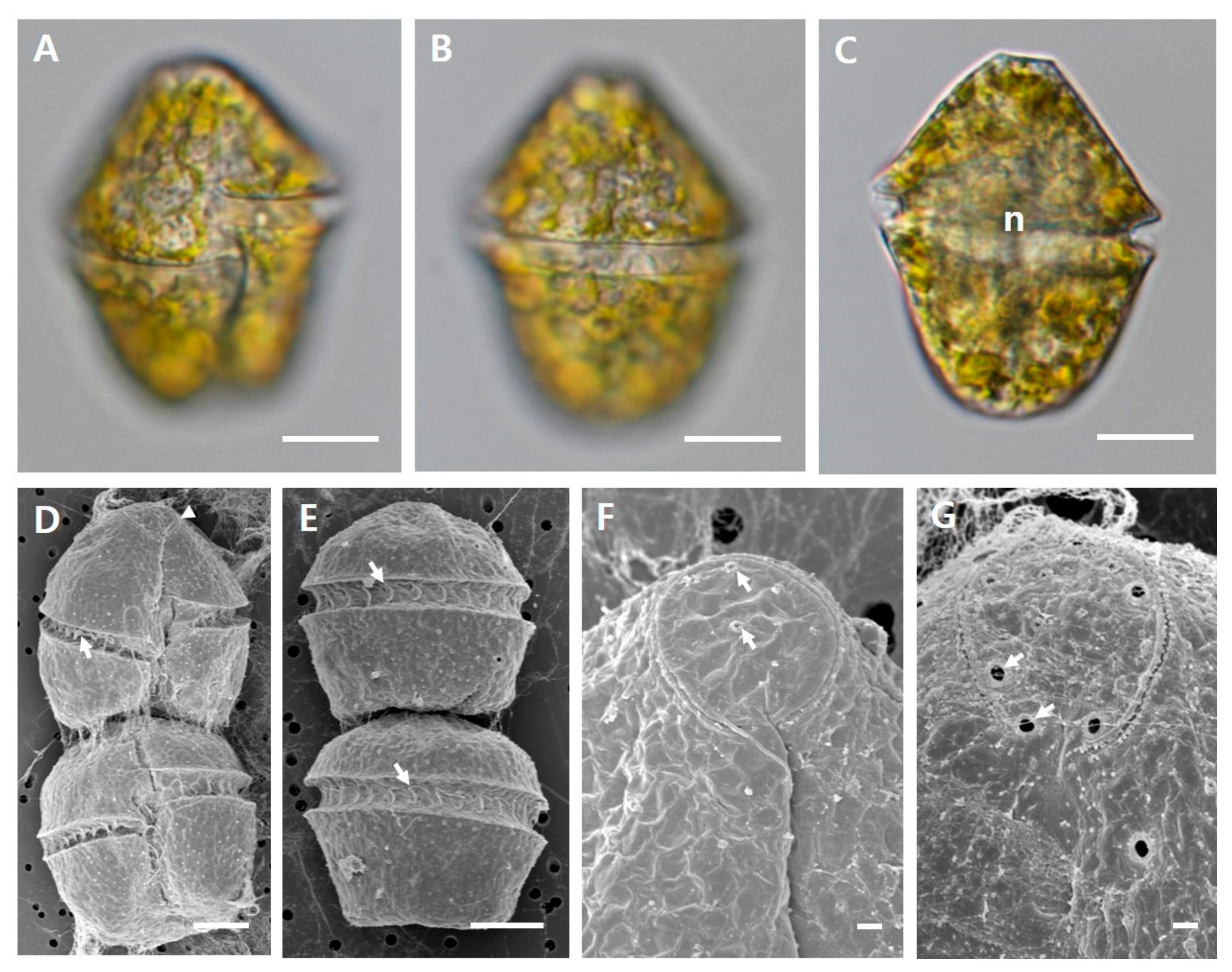
2.1. Culture of Gymnodinium catenatum

A strain of Gymnodinium catenatum (LIMS-PS-2604) was obtained from the Library of Marine Samples, Korea Institute of Ocean Science and Technology. This strain was established from phytoplankton samples collected in the South Sea of Korea (34°29’7.68” N, 128°28’54.54” E) and has been maintained at 20 °C and ca 100 µmol photons m−2 s−1 cool-white illumination (fluorescent lamp) under a 12L:12D photo-cycle. The morphological features of G. catenatum were photographed using an AxiCam MRc digital camera on an upright microscope (Axio Imager 2, Zeiss, Germany) and a field emission scanning electron microscope (JSM 7600F, JEOL, Tokyo, Japan), which are shown in Figure 1. The phylogeny of the strain was reported in a previous study [38].
Experimental cultures were established in 2 L culture bottles (SPL, Pocheon, Korea) containing f/2 culture medium (Marine Water Enrichment Solution, Sigma Aldrich, St. Louis, MO, USA) without silicate, prepared with sterile sea water at 20 °C and ca 100 µmol photons m−2 s−1 cool-white illumination (fluorescent lamp) under a 24L:0D photo-cycle on a roller apparatus (Wheaton, IL, USA). Cultures from the exponential growth phase were used for the experiments.

2.2. Growth Experiment

Surface seawater collected from the East China Sea (32°00’ N, 127°00’ E) was used to prepare the culture medium with controlled concentrations of nitrate (N), phosphate (P), vitamin B1, Iron (Fe) and Copper (Cu). The concentrations of dissolved inorganic nitrogen (sum of NH4+, NO2, NO3) and phosphorus (PO43−) in the seawater were 7.4 and 0.6 µM, respectively, as measured by a nutrient auto analyzer (QuickChem 8000, Lachat, Loveland, CO, USA). For the selenium (Se) addition, seawater was collected from a location off the Korean coast (37°32’26.62” N, 130°50’43.31” E). The seawaters were filtered through a 47 mm membrane filter and autoclaved for all experiments.
In this study, we carried out two major experiments involving nutrients (N, P and vitamin B1) and trace metals (Fe, Cu and Se). The basal culture medium used to control the concentrations of nutritional factors was the f/2 culture medium without silicate (f/2-Si culture medium), and the N:P ratio in the stock solution with this culture medium was 24.2 (the values of nitrate and phosphate calculated from the collected seawater and the culture medium). Treatments were made by adding or reducing the concentrations of the nutrients and trace metals, and the treatment without addition of nutritional factors was used as a control (Table 1). However, as the original recipe of the f/2 culture medium does not include Se, the Se concentration was varied through the addition of selenous acid to a solution of the basal culture medium (Table 1).
Growths of Gymnodinium catenatum were tested in stock solutions supplied with different concentrations of nutrients and trace metals and filtered and autoclaved seawater was used to make the stock solutions. Twenty-six stock solutions with target concentrations of nutrients and trace metals were made (Table 1), and 30 mL of each solution was added to separate 50 mL Pyrex tubes. The subcultures of G. catenatum for the experiments were established in 2 L culture bottles (SPL, Pocheon, Korea) containing individual nutritional source-poor media, and these were used for inoculation into experimental tubes. G. catenatum in concentrations ranging from 100 to 200 cells/mL was inoculated into the 50 mL Pyrex tubes. The tubes were placed in incubators and incubated at 20 °C and ca 100 µmol photons m2 s−1 cool-white illumination (fluorescent lamp) under a 14L:10D photo-cycle. All experiments were conducted in triplicate.

2.3. Calculation of Growth Rate

Culture growth was monitored at 2 day intervals for 32 days and determined using an in vivo fluorometer (Turner Designs Model 10-AU, Sunnyvale, USA), and the fluorescence data were used to calculate specific growth rates. The regression equation for in vivo fluorescence values provided a good fit to the observed cell densities; the adjusted r2 value for Gymnodinium catenatum was >0.99 (data not shown). To estimate the specific growth rate (µ) of G. catenatum, we used the following equation:
µ = log2 (NtN0) / t1t0
where N0 and Nt are the initial (t0) and final (t1) in vivo fluorescence values during the incubation experiments, respectively. The in vivo fluorescence values estimated during the logarithmic growth phase were used to obtain the specific growth rate.

3. Results and Discussion

3.1. Growth of Gymnodinium catenatum under Different Concentrations of N, P and Vitamin B1

The growth curves and rates of Gymnodinium catenatum cultures exposed to different concentrations of nutrients (N, P and vitamin B1) are shown in Figure 2 and Table 2. In all the nutrient treatments, G. catenatum culture grew consistently during days 2–20 (Figure 2). In the N control, and the treatments 1 and 2, G. catenatum entered the senescence phase from day 24, following an stationary phase for 8 days; however, in treatment 3, with the addition of 1.76 × 10³ µM nitrate, the stationary phase continued after an incubation of 14 days (the exponential growth phase). The pattern in treatment 3 was observed with low cell density, and the growth rate (0.11 day−1) was also the lowest. The maximum cell density (1389 cells mL−1) was observed in treatment 2 (Table 2), and the control and treatments 1 and 2 had similar growth rates (0.18–0.21 day−1). Compared with the N control and the treatments, the high growth rates of G. catenatum in the P addition experiments were observed in treatments 2 (addition of 3.62 × 101 µM phosphate) and 3 (addition of 7.24 × 101 µM phosphate), with high cell densities (764 and 909 cells mL−1) (Table 2); however, the maximum cell density was lower than that for the N treatments. Low growth rates were observed in the control (0.10 day−1) and treatment 1 (0.08 day−1), with low cell densities (Table 2), and the growth rate in treatment 3 with P addition was higher than that in N treatment 3. G. catenatum grew consistently until day 22 and then entered a stationary phase, and the senescence phase was not observed until 32 days of incubation. This growth pattern was similar to that observed in N treatment 3.
Band-Schmidt et al. [39] investigated the growth rates of G. catenatum strains under different growth conditions, and reported the growth rates of a strain (established from Bahía Concepción Gulf of California, Mexico) tested in f/2 medium at 20°C and ca 100 µmol photons m−2 s−1 cool-white illumination (fluorescent lamp) under a 12L:10D photo-cycle, which is similar to the conditions described here, ranging from 0.28 to 0.32 day−1. These growth rates are higher than those obtained for f/2-Si culture medium (referred to as treatment 2) in the present study. However, Bustillos-Guzmán et al. [40] reported that the growth rate of G. catenatum culture established from Concepción Bay, Gulf of California is 0.24 day−1 (the N:P ratio of 23.5), which is similar to own observations. This finding indicates that the growth rate of G. catenatum can vary within strains, or can depend on different geographical strains. Bustillos-Guzmán et al. [40] also found that the maximum cell density for treatment with a N:P ratio of 23.5 is higher than those for treatments with low N:P ratios (5.4 and 9.2), and that higher N:P ratios than 23.5 can produce intermediate densities. This result accords with our observation that treatment 2 with a N:P ratio of 24.2 produce a maximum cell density. However, in this case, treatment 3, with the addition of a relatively high N concentration (a N:P ratio of 48.1), had the lowest maximum cell density (485 cells mL−1) among the control and all treatments, indicating that higher N:P ratios than 24.2 do not increase the growth rate and cell density of Korean isolates of G. catenatum.
Interestingly, although the cell density in treatment 3 was lower than those in the control and other treatments, no senescence phase was observed in treatment 3. The senescence phase is observed when nutritional sources such as nitrate and phosphate in the culture medium are lower. Consequently, while the addition of a high N concentration (or >N:P ratio of 24.2) did not stimulate growth rate or cell density in the Korean isolate of G. catenatum, it can nevertheless have a positive effect on the maintenance of cell density. According to Bustillos-Guzmán et al. [40], G. catenatum may have a high storage capacity for phosphorus and nitrogen. This indicates that under N sufficient condition, G. catenatum utilizes a N storage strategy for maintaining cell density.
In contrast to the additions of N, additions of P stimulated the growth rate and cell density of G. catenatum (Figure 2). According to Oh et al. [41], under P-limited conditions the growth of G. catenatum can be stimulated by the addition of dissolved organic phosphorus (DOP). DOP can act as an additional source of P for microorganisms in a marine ecosystem in which dissolved inorganic phosphorus is seasonally depleted [42], and G. catenatum can take advantage for the uptake of P from DOP [41]. In addition, Yamamoto et al. [43] concluded that in the P-depleted condition, G. catenatum may be able to form blooms in Hiroshima Bay, Japan, with a higher affinity for DOP [43]. These observations indicate that sources of P are an important nutritional factor in enhancing the growth of G. catenatum. However, as in this study the additions of both N and P resulted in similar maximum cell densities (Table 2), sources of P alone certainly favor neither the enhancement of cell density of G. catenatum nor bloom formation. In the growth curve for the P control and treatments, no senescence phase was observed for the 32-day incubation, which differs from the growth pattern seen for the N control, and for treatments 1 and 2 with moderate concentrations of N. This may be related to the storage strategy for P, as well as for a high N concentration, suggesting that G. catenatum may be able to make use of one of two nutritional sources for maintaining cell density, when either N or P is depleted or sufficient.
Tang et al. [36] reported that vitamins are specifically important to the occurrence of harmful algal blooms, and Gobler et al. [44] observed the enhancement of large dinoflagellates by the enrichment of coastal waters with vitamin B1 and B12. However, in this study the addition of vitamin B1 did not stimulate the growth rate of G. catenatum, and the highest growth rate of G. catenatum was observed in the control without addition of vitamin B1 (Figure 2), and the maximum cell density was observed for the treatment 1 (addition of 2.96 × 10⁻² µM vitamin B1) (Figure 2). Although we did not measure the concentration of vitamin B1 in the seawater used to make the solutions, neither the increased growth rate nor maximum cell density seem to be related to high concentration of vitamin B1. In addition, despite the fact that the culture medium for the controls and treatments in this experiment contained N, P, and vitamin B12, significant increases in growth and cell density of G. catenatum were not observed, reflecting the fact that G. catenatum does not require these vitamins for growth. According to Croft et al. [45], more than 20% of the 306 microalgal species surveyed are auxotrophs for the vitamin B1, and Tang et al. [36] reported that, among 45 species of dinoflagellates investigated, the numbers of auxotrophs for the vitamin B1 are 49%, which is lower than the percentage of auxotrophs for the vitamin B12 (91%). In addition, according to Gobler et al. [44], vitamin B1 is present in coastal waters at concentrations greater than vitamin B12, suggesting a possible preference of some dinoflagellates for vitamin B12 over vitamin B1 for growth. In that case, given that the vitamin B12-dependent species must be able to obtain the vitamin from an external source such as bacteria [46], more detailed studies are required to clarify the relationship between vitamin uptake of G. catenatum and the presence of bacteria. Nevertheless, in the addition experiments of vitamin B1, an interesting observation was that the growth pattern of G. catenatum was similar to those of the N control, and, in treatments 1 and 2, that the senescence phase was observed from day 22 following the stationary phase. This indicates that even in a culture in which N and P are both depleted by uptake, G. catenatum does not require vitamin B1 for growth. However, further studies are still needed to clarify the relationship between the vitamin requirement of G. catenatum and the concentration of N under conditions that other nutritional sources are depleted.

3.2. Growth of Gymnodinium catenatum under Different Concentrations of Fe, Cu and Se

The growth curves and rates of Gymnodinium catenatum culture under different concentrations of trace metals (Fe, Cu and Se) are shown in Figure 3 and Table 2. On additions of Fe and Cu, no significant differences were observed in the growth patterns and rates between control and treatments. For the addition of Fe, G. catenatum grew consistently in the control and all treatments for days 1–22 and then entered a stationary phase. A senescence phase was not observed. The growth rates in this experiment were between 0.14 and 0.16 day−1. For Cu addition, both the growth curves and the rates (0.14–0.17 day−1) were similar to those seen for Fe addition, although G. catenatum grew until day 18 before entering a stationary phase. The senescence phase was not observed in this experiment. The growth patterns for the Fe and Cu addition were similar to that for P addition. The maximum cell densities for Fe and Cu addition were 1310 cells mL−1 and 1071 cells mL−1, respectively (Table 2), and both were observed in treatment 2 with additions of 1.17 × 101 µM Fe and of 3.93 × 10⁻² Cu.
The culture media for the control and treatments in these experiments contained N, P, and vitamins, all of which can affect growth and cell density in G. catenatum. In this culture, additions of Fe and Cu did not seem to enhance the growth rates of G. catenatum, given that there were no significant differences in growth curves or rates between the treatments and controls without the addition of Fe and Cu (Figure 3). Fe and Cu are essential elements for phytoplankton, and play an important role in many of the biochemical and metabolic processes involved in cell growth [47,48,49]. Some studies recorded that the growth of Protoceratium reticulatum and Alexandrium tamarense increased with increasing Fe concentrations [50,51], although in contrast the cell growth of Scrippsiella trochoidea was inhibited by an increase in Fe [52]. Other studies reported a significant negative effect of Cu on the growth of dinoflagellates such as A. catenella, Ostreopsis cf. ovata [35,53]. These findings indicate that Fe and Cu requirements can vary greatly among phytoplankton. The basic knowledge of the Fe and Cu requirements of G. catenatum is lacking to date; however, our results indicate that elevated trace metal (Fe and Cu) concentrations do not increase the growth and cell density of G. catenatum in nutrient rich waters.
There has been considerable interest in the Se requirements of harmful algal species (e.g., [32,33,50,54]), suggesting that the addition of Se to a culture medium stimulates growth. In particular, culture experiments show a clear requirement for Se by G. catenatum [54]. In our experiment, Korean isolates of G. catenatum could grow in an f/2 culture medium without Se addition (referred to as the control), however the growth was more elevated when Se was added (Figure 3). The maximum cell densities in treatments 3, 4 and 5 were twice as high as that of the control, which were also higher than those of other nutrient and trace metal sources in this study (Table 2). There were no significant differences in growth rates (0.15–0.17 day−1) between the control and the treatments. In both the control and the treatments, G. catenatum grew until day 16 or 20 before entering a stationary phase, and a senescence phase was not observed. This growth pattern was similar to those seen in the P, Fe and Cu addition experiments.
According to Boisson et al. [55] and Price et al. [56], Se is either essential for some phytoplankton in low concentrations or it cannot enhance growth of phytoplankton. This indicates that Se requirements can vary among phytoplankton, and that phytoplankton can have an optimal range of Se for growth. In Se addition, no obvious differences in cell densities of G. catenatum were observed until the end of the exponential growth phase, resulting in similar growth rates between the control and treatments. However, cell densities were increased in the stationary phases of treatments 3, 4 and 5 with addition of Se. This result suggests that concentrations greater than 10−3 µM of Se can enhance cell densities or biomass yield in culture experiments, although we did not measure the concentration of Se of the seawaters used for making the solutions. According to Doblin et al. [32], the Se requirement of G. catenatum varies between strains obtained from Australia, Spain and Japan, with addition of selenium at concentrations in the range 10−9–10−7 M causing a variable increase in growth and biomass yield. These Se requirements also differ from that of the Korean isolate of G. catenatum. It is thus possible that the growth of G. catenatum can be enhanced for a wide range of concentrations of Se.

3.3. Proliferation Potential of Gymnodinium catenatum and Nutritional Conditions in Korean Coastal Waters

According to Band-Schmidt et al. [57], blooms caused by G. catenatum have been associated with an increase in nutrients, mainly by nitrogen compounds in the water column. In our study, N seems to be one of the most important nutritional factors in the growth of the Korean isolate of G. catenatum, however the G. catenatum prefers high concentrations of P and Se, rather than preference to N. In a previous study, it was reported that the maximum cell density of the Korean isolate of G. catenatum in laboratory experiments was observed at 20 °C, which is commonly recorded in summer in Korea [38]. According to Koo [58], in the southern coastal area of Korea from which the isolate of G. catenatum was obtained, average N and P concentrations were 0.8 and 0.1 µM L−1 in summer, respectively. This indicates that P can be a limiting factor for phytoplankton growth in Korean coastal waters, and because of this, in summer G. catenatum may prefer high concentration of P for the growth.
According to Koo [58], Cho [59] and Jang et al. [60], low levels of N and P in summer could be a result of the uptake of dinoflagellates, among which Margalefidinium polykrikoides (= Cochlodinium polykrikoides), Prorocentrum obtusidens (= P. donghaiense), and Alexandrium species are well known to dominate in Korean coastal waters in summer, causing dense blooms (e.g., [61,62,63,64]). The maximum growth rates of these species in the laboratory are higher than that of G. catenatum (e.g., [65,66,67]), indicating that G. catenatum may be at a disadvantage beside the more the dominant species in terms of nutrient competition. Consequently, proliferation of G. catenatum is not expected in summer of Korean coastal area. However, as during summer, after rainfall events, riverine input of inorganic Se and its interaction with dissolved organic matter (DOM) can be a critical factor for bloom initiation of G. catenatum [34], this species may have a competitive advantage if Se, P and DOM loading after rainfall is increased in Korean coastal waters.

Author Contributions

Data curation, formal analysis, writing-original draft and writing-review & editing, K.H.H. and H.H.S.; Data curation and formal analysis, H.J.K.; Data curation and writing-review & editing, Z.L.; Data curation and investigation, J.Y.Y. and K.Y.K.; Formal analysis, M.H.S., J.H., S.D.L. and S.M.Y.; Writing-review & editing, S.J.O., J.W.P. and W.-A.L.; Conceptualization and funding acquisition, H.H.S. All authors have read and agree to the published version of the manuscript.

Funding

This work was supported by grants from NIFS (R2020041), KIOST (PE99821) projects, and NNIBR project (202001103).

Conflicts of Interest

The authors declare no conflict of interest.

References

  1. Lagos, N.; Onodera, H.; Zagatto, P.A.; Andrinolo, D.; Azevedo, S.M.F.Q.; Oshima, Y. The first evidence of paralytic shellfish toxins in the freshwater cyanobacterium Cylindrospermopsis raciborskii, isolated from Brazil. Toxicon 1999, 37, 1359–1373. [Google Scholar] [CrossRef]
  2. Anderson, D.M.; Alpermann, T.J.; Cembella, A.D.; Collos, Y.; Masseret, E.; Montresor, M. The globally distributed genus Alexandrium: Multifaceted roles in marine ecosystems and impacts on human health. Harmful Algae 2012, 14, 10–35. [Google Scholar] [CrossRef] [PubMed] [Green Version]
  3. Band–Schmidt, C.J.; Durán–Riveroll, L.M.; Bustillos–Guzmán, J.J.; Leyva–Valencia, I.; López–Cortés, D.J.; Núñez–Vázquez, E.J.; Hernández–Sandoval, F.E.; Ramirez–Rodriguez, D.V. Paralytic toxin producing dinoflagellates in Latin America: Ecology and physiology. Front. Mar. Sci. 2019, 6, 42. [Google Scholar] [CrossRef]
  4. Hallegraeff, G.M. A review of harmful algal blooms and their apparent global increase. Phycologia 1993, 32, 79–99. [Google Scholar] [CrossRef] [Green Version]
  5. Landsberg, J.H. The Effects of Harmful Algal Blooms on Aquatic Organisms. Rev. Fish. Sci. 2002, 10, 113–390. [Google Scholar] [CrossRef]
  6. Yñiguez, A.T.; Lim, P.T.; Leaw, C.P.; Jipanin, S.J.; Iwataki, M.; Benico, G.; Azanza, R.V. Over 30 years of HABs in the Philippines and Malaysia: What have we learned? Harmful Algae 2020, 101776. [Google Scholar] [CrossRef]
  7. Chang, D.S.; Shin, I.S.; Pyeun, J.H.; Park, Y.H. A study on paralytic shellfish poison of sea mussel Mytilus edulis. Korean J. Fish. Aquat. Sci. 1987, 20, 293–299. [Google Scholar]
  8. Azanza, R.V.; Taylor, F.J. Are Pyrodinium blooms in the Southeast Asian region recurring and spreading? A review at the end of the millennium. Ambio 2001, 39, 356–364. [Google Scholar] [CrossRef]
  9. Harada, T.; Oshima, Y.; Kamiya, H.; Yasumoto, T. Confirmation of paralytic shellfish toxins in the dinoflagellate Pyrodinium bahamense var. compressa and bivalves in Palau. Nippon Suisan Gakkaishi 1982, 48, 821–825. [Google Scholar] [CrossRef]
  10. Oshima, Y.; Hasegawa, M.; Yasumoto, T.; Hallegraeff, G.; Blackburn, S. Dinoflagellate Gymnodinium catenatum as the source of paralytic shellfish toxins in Tasmanian shellfish. Toxicon 1987, 25, 1105–1111. [Google Scholar] [CrossRef]
  11. Anderson, D.M.; Kulis, D.M.; Doucette, G.J.; Gallagher, J.C.; Balech, E. Biogeography of toxic dinoflagellates in the genus Alexandrium from the northeastern United States and Canada. Mar. Biol. 1994, 120, 467–478. [Google Scholar] [CrossRef]
  12. Taylor, F.J.R.; Fukuyo, Y.; Larsen, J.; Hallegraeff, G.M. Taxonomy of Harmful Dinoflagellates. In Manual on Harmful Marine Microalgae. Monographs on Oceanographic Methodology, 11th ed.; Hallegraeff, G.M., Anderson, D.M., Cembella, A.D., Eds.; Amsterdam: Paris, France, 2003; pp. 389–432. [Google Scholar]
  13. Moestrup, Ø.; Akselmann-Cardella, R.; Churro, C.; Fraga, S.; Hoppenrath, M.; Iwataki, M.; Larsen, J.; Lundholm, N.; Zingone, A. IOC-UNESCO Taxonomic Reference List of Harmful Micro Algae. Available online: http://www.marinespecies.org/hab (accessed on 20 February 2020).
  14. John, U.; Litaker, R.W.; Montresor, M.; Murray, S.; Brosnahan, M.L.; Anderson, D.M. Formal revision of the Alexandrium tamarense species complex (Dinophyceae) taxonomy: The Introduction of five species with emphasis on molecular–based (rDNA) classification. Protist 2014, 165, 779–804. [Google Scholar] [CrossRef] [Green Version]
  15. Murray, S.A.; Diwan, R.; Orr, R.J.; Kohli, G.S.; John, U. Gene duplication, loss and selection in the evolution of saxitoxin biosynthesis in alveolates. Mol. Phylogenetics Evol. 2015, 92, 165–180. [Google Scholar] [CrossRef] [Green Version]
  16. Lassus, P.; Chomérat, N.; Hess, P.; Nézan, E. Toxic and Harmful Microalgae of the World Ocean (Micro-Algues Toxiques et Nuisibles de l’océan Mondial; IOC Manuals and Guides 68 (Bilingual English/French); International Society for the Study of Harmful Algae; Intergovernmental Oceanographic Commission of UNESCO: Copenhagen, Denmark, 2016; p. 524. [Google Scholar]
  17. Hallegraeff, G.M.; Stanley, S.O.; Bolch, C.J.; Blackburn, S.I. Gymnodinium catenatum blooms and shellfish toxicity in Southern Tasmania, Australia. In Red Tide: Biology, Environmental Science and Toxicology; Okaichi, T., Anderson, D.M., Nemato, T., Eds.; Elsevier: New York, NY, USA, 1989; pp. 77–80. [Google Scholar]
  18. Franca, S.; Almeida, J.K. Paralytic shellfish poisons in bivalve molluscs on the Portuguess coast caused by a bloom of the dinoflagellate Gymnodinium catenatum. In Red Tide: Biology, Environmental Science and Toxicology; Okaichi, T., Anderson, D.M., Nemato, T., Eds.; Elsevier: New York, NY, USA, 1989; pp. 93–96. [Google Scholar]
  19. Anderson, D.M.; Sullivan, J.J.; Reguera, B. Paralytic shellfish poisoning in northwest Spain: The toxicity of the Gymnodinium catenatum. Toxicon 1989, 27, 665–674. [Google Scholar] [CrossRef]
  20. Amanhir, R.; Benhadouch, A.; Taleb, H.; Leblad, R.; Vale, P.; Blaghen, M. Weekly occurrence of Gymnodinium catenatum and paralytic shellfish poisoning in the Mediterranean shore of Morocco. Int. J. Biochem. Res. Rev. 2017, 17, 1–11. [Google Scholar] [CrossRef] [Green Version]
  21. Ikeda, T.; Matsuno, S.; Sato, S. First report on paralytic shellfish poisoning caused by Gymnodinium catenatum Graham (Dinophyceae) in Japan. In Red Tide: Biology, Environmental Science and Toxicology; Okaichi, T., Anderson, D.M., Nemato, T., Eds.; Elsevier: New York, NY, USA, 1989; pp. 411–414. [Google Scholar]
  22. Park, T.G.; Kim, C.H.; Oshima, Y. Paralytic shellfish toxin profiles of different geographic population of Gymnodinium catenatum (Dinophyceae) in Korean coastal waters. Phycol. Res. 2004, 52, 300–305. [Google Scholar] [CrossRef]
  23. Smayda, T.J. Ecological features of harmful algal blooms in coastal upwelling ecosystems. Afr. J. Mar. Sci. 2000, 22, 219–253. [Google Scholar] [CrossRef] [Green Version]
  24. Smayda, T.J. Harmful algal blooms: Their ecophysiology and general relevance to phytoplankton bloom in the sea. Limnol. Oceanogr. 1997, 42, 1137–1153. [Google Scholar] [CrossRef]
  25. Hallegraeff, G.M. Harmful algal blooms: A global overview. In Manual on Harmful Marine Microalgae, 2nd ed.; Hallegraeff, G.M., Anderson, D.M., Cembella, A.D., Eds.; UNESCO: Paris, France, 1995; pp. 25–50. [Google Scholar]
  26. Global Ecology and Oceanography of Harmful Algal Blooms (GEOHAB). Global Ecology and Oceanography of Harmful Algal Blooms. In Global Ecology and Oceanography of Harmful Algal Blooms: Eutrophication and HABs; Gilbert, P., Ed.; Scientific Committee on Oceanic Research (SCOR: Paris, France; Intergovernmental Oceanographic Commission (IOC): Baltimore, MD, USA, 2006; p. 74. [Google Scholar]
  27. Kudela, R.M.; Seeyave, S.; Cochlan, W.P. The role of nutrients in regulation and promotion of harmful algal blooms in upwelling systems. Prog. Oceanogr. 2010, 85, 122–135. [Google Scholar] [CrossRef]
  28. Anderson, D.M.; Burkholder, J.M.; Cochlan, W.P.; Gilbert, P.M.; Gobler, C.J.; Hell, C.A.; Kudela, R.M.; Parsons, M.L.; Rensel, J.E.J.; Townsend, D.W.; et al. Harmful algal blooms and eutrophication: Examining linkages from selected coastal regions of the United States. Harmful Algae 2008, 8, 39–53. [Google Scholar] [CrossRef] [Green Version]
  29. Heisler, J.P.; Gilbert, J.; Burkholder, J.; Anderson, D.; Cochlan, W.; Dennison, W.; Dortch, Q.; Gobler, C.J.; Heil, C.; Humphries, E.; et al. Eutrophication and harmful algal blooms: A scientific consensus. Harmful Algae 2008, 8, 3–13. [Google Scholar] [CrossRef] [Green Version]
  30. Bruland, K.W.; Donat, J.R.; Hutchins, D.A. Interactive influences of bioactive trace metals on biological production in oceanic waters. Limnol. Oceanogr. 1991, 36, 1555–1577. [Google Scholar] [CrossRef] [Green Version]
  31. Maldonado, M.T.; Allen, A.E.; Chong, J.S.; Lin, K.; Leus, D.; Karpenko, N.; Harris, S.L. Copper-dependent iron transport in coastal and oceanic diatoms. Limnol. Oceanogr. 2006, 51, 1729–1743. [Google Scholar] [CrossRef] [Green Version]
  32. Doblin, M.A.; Blackburn, S.I.; Hallegraeff, G.M. Intraspecific variation in the selenium requirement of different geographic strains of the toxic dinoflagellate Gymnodinium catenatum. J. Plankton Res. 2000, 22, 421–432. [Google Scholar] [CrossRef] [Green Version]
  33. Boyer, G.L.; Brand, L.E. Trace metals and harmful algal blooms. In Physiological Ecology of Harmful Algal Blooms; NATO-ASI Series 41; Anderson, D.M., Cembella, A.D., Hallegraeff, G.M., Eds.; Springer: Berlin, Germany, 1998; pp. 489–508. [Google Scholar]
  34. Doblin, M.A.; Blackburn, S.I.; Hallegraeff, G.M. Growth and biomass stimulation of the toxic dinoflagellate Gymnodinium catenatum (Graham) by dissolved organic substances. J. Exp. Mar. Biol. Ecol. 1999, 236, 33–47. [Google Scholar] [CrossRef]
  35. Couet, D.; Pringault, O.; Bancon-Montigny, C.; Briant, N.; Poulichet, F.E.; Delpoux, S.; Yahia, O.K.; Hela, B.; Charaf, M.; Hervé, F.; et al. Effects of cooper and butyltin compounds on the growth, photosynthetic activity and toxin production of two HAB dinoflagellates: The planktonic Alexandrium catenella and the benthic Ostreopsis cf. ovata. Aquat. Toxicol. 2018, 196, 154–167. [Google Scholar] [CrossRef] [Green Version]
  36. Tang, Y.Z.; Koch, F.; Gobler, C.J. Most harmful algal bloom species are vitamin B1 and B12 auxotrophs. Proc. Natl. Acad. Sci. USA 2010, 107, 20756–20761. [Google Scholar] [CrossRef] [Green Version]
  37. Lee, C.K.; Kim, H.C.; Lee, S.G.; Jung, C.S.; Kim, H.G.; Lim, W. Abundance of harmful algae, Cochlodinium polykrikoides, Gyrodinium impudicum and Gymnodinium catenatum in the coastal area of South Sea of Korea and their effects of temperature, salinity, irradiance and nutrient on the growth in culture. Korean J. Fish. Aquat. Sci. 2001, 34, 536–544. [Google Scholar]
  38. Han, K.H.; Li, Z.; Kang, B.J.; Youn, J.Y.; Shin, H.H. Toxic dinoflagellate Gymnodinium catenatum Graham (Dinophyceae) from the southern coast of Korea: Morphology, phylogeny and effects of temperature and salinity on growth. Korean J. Environ. Biol. 2019, 37, 31–41. [Google Scholar] [CrossRef]
  39. Band–Schmidt, C.J.; Morquecho, L.; Lechuga-Devéze, C.H.; Anderson, D.M. Effects of growth medium, temperature, salinity and seawater source on the growth of Gymnodinium catenatum (Dinophyceae) from Bahía Concepción, Gulf of California, Mexico. J. Plankton Res. 2004, 26, 1459–1470. [Google Scholar] [CrossRef]
  40. Bustillos-Guzmán, J.J.; Band-Schmidt, C.J.; López-Cortés, D.J.; Gárate-Lizárraga, I.; Núñez-Vázquez, E.J.; Hernández-Sandoval, F.E. Variations in growth and toxicity in Gymnodinium catenatum Graham from the Gulf of Calinifornia under different ratios of nitrogen and phosphorus. Cienc. Mar. 2012, 38, 101–117. [Google Scholar] [CrossRef]
  41. Oh, S.J.; Yamamoto, T.; Kataoka, Y.; Matsuda, O.; Matsuyama, Y.; Kotani, Y. Utilization of dissolved organic phosphorus by the two toxic dinoflagellates, Alexandrium tamarense and Gymnodinium catenatum (Dinophyceae). Fish. Sci. 2002, 68, 416–424. [Google Scholar] [CrossRef] [Green Version]
  42. Mather, R.L.; Reynolds, S.E.; Wolff, G.A.; Williams, R.G.; Torres-Valdes, S.; Woodward, E.M.S.; Landolfi, A.; Pan, X.; Sanders, R.; Achterberg, E.P. Phosphorus cycling in the north and South Atlantic Ocean subtropical gyres. Nat. Geosci. 2008, 1, 439–443. [Google Scholar] [CrossRef]
  43. Yamamoto, T.; Oh, S.J.; Kataoka, Y. Growth and uptake kinetics for nitrate, ammonium and phosphate by the toxic dinoflagellate Gymnodinium catenatum isolated from Hiroshima Bay, Japan. Fish. Sci. 2004, 70, 108–115. [Google Scholar] [CrossRef]
  44. Gobler, C.J.; Norman, C.; Panzeca, C.; Taylor, G.T.; Sañudo-Wilhelmy, S.A. Effect of B-Vitamins (B1, B12) and inorganic nutrients on algal bloom dynamics in a coastal ecosystem. Aquat. Microb. Ecol. 2007, 49, 181–194. [Google Scholar] [CrossRef]
  45. Croft, M.T.; Warren, M.J.; Smith, A.G. Algae need their vitamins. Eukaryot. Cell 2006, 5, 1175–1183. [Google Scholar] [CrossRef] [Green Version]
  46. Croft, M.T.; Lawrence, A.D.; Raux-Deery, E.; Warren, M.J.; Smith, A.G. Algae acquire vitamin B12 through a symbiotic relationship with bacteria. Nature 2005, 438, 90–93. [Google Scholar] [CrossRef] [PubMed]
  47. Reuter, J.G.; Ades, D.R. The role of iron nutrition in photosynthesis and nitrogen assimilation in Scenedemus quadricauda (Chlorophyceae). J. Phycol. 1987, 23, 452–457. [Google Scholar] [CrossRef]
  48. Geider, R.J.; La Roche, J. The role of iron in phytoplankton photosynthesis, and the potential for iron limitation of primary productivity in the sea, Mini review. Photosynth. Res. 1994, 39, 275–301. [Google Scholar] [CrossRef]
  49. Hänsch, R.; Mendel, R.R. Physiological functions of mineral micronutrients (Cu, Zn, Mn, Fe, Ni, Mo, B, Cl). Curr. Opin. Plant Biol. 2009, 12, 259–266. [Google Scholar] [CrossRef]
  50. Mitrovic, S.M.; Amandi, M.F.; Mckenzie, L.; Furey, A.; James, K.J. Effects of selenium, iron and cobalt addition to growth and yessotoxin production of the toxic marine dinoflagellate Protoceratium reticulatum in culture. J. Exp. Mar. Biol. Ecol. 2004, 313, 337–351. [Google Scholar] [CrossRef]
  51. He, H.; Chen, F.; Li, H.; Xiang, W.; Li, Y.; Jiang, Y. Effect of iron on growth, biochemical composition and paralytic shellfish poisoning toxins production of Alexandrium tamarense. Harmful Algae 2010, 9, 98–104. [Google Scholar] [CrossRef]
  52. Qin, X.M.; Zhou, J.Z.; Qian, P.Y. Effects of Fe and Mn on the growth of a red tide dinoflagellate Scrippsiella trochoidea (Stein) Loeblch III. Chin. J. Oceanol. Limnol. 1997, 15, 173–180. [Google Scholar] [CrossRef]
  53. Herzi, F.; Jean, N.; Zhao, H.; Mounier, S.; Mabrouk, H.H.; Hlaili, A.S. Cooper and cadmium effects on growth and extracellular exudation of the marine toxic dinoflagellate Alexandrium catenaella: 3D-fluorescence spectroscopy approach. Chemosphere 2013, 93, 1230–1239. [Google Scholar] [CrossRef]
  54. Doblin, M.A.; Blackburn, S.I.; Hallegraeff, G.M. Comparative study of selenium requirements of three species: Gymnodinium catenatum, Alexandrium minutum (Dinophyta) and Chaetoceros cf. tenuissimus (Bacillariophyta). J. Plankton Res. 1999, 21, 1153–1169. [Google Scholar] [CrossRef] [Green Version]
  55. Boisson, F.; Gnassia-Barelli, M.; Romeo, M. Toxicity and accumulation of selenite and selenate in the unicellular marine alga Cricosphaera elongata. Arch. Environ. Contam. Toxicol. 1995, 28, 487–493. [Google Scholar] [CrossRef]
  56. Price, N.M.; Thompson, P.A.; Harrison, P.J. Selenium: An essential element for growth of the coastal marine diatom Thalassiosira pseudonana (bacillariophyceae) 1, 2. J. Phycol. 1987, 23, 1–9. [Google Scholar] [CrossRef]
  57. Band-Schmidt, C.J.; Bustillos-Guzmán, J.J.; López-Cortés, D.J.; Gárate-Lizárraga, I.; Núñez-Vázquez, E.J.; Hernández-Sandoval, F.E. Ecological and physiological studies of Gymnodinium catenatum in the Mexican Pacific: A review. Mar. Drugs 2010, 8, 1935–1961. [Google Scholar] [CrossRef] [Green Version]
  58. Koo, J.S. A Study on the Relation between Nutrient and HAB in the South Sea of Korea. Master’s Thesis, Pusan National University, Busan, Korea, 2004. [Google Scholar]
  59. Cho, E.S. A comparative study on outbreak and non-outbreak of Cochlodinium polykrikoides Margalef in South Sea of Korea in 2007–2009. Korean Soc. Mar. Environ. Saf. 2010, 16, 31–41. [Google Scholar]
  60. Jang, P.G.; Shin, H.H.; Baek, S.H.; Jang, M.C.; Lee, T.S.; Shin, K. Nutrient distribution and effects on phytoplankton assemblages in the western Korea/Tsushima Strait. N. Z. J. Mar. Freshw. Res. 2013, 47, 21–37. [Google Scholar] [CrossRef]
  61. Lee, Y.S. Factors affecting outbreaks of high-density Cochlodinium polykrikoides red tides in the coastal seawaters around Yeosu and Tongyeong, Korea. Mar. Pollut. Bull. 2006, 52, 1249–1259. [Google Scholar] [CrossRef]
  62. Wang, H.; Lu, D.; Huang, H.; Xia, P.; Dai, X. Comparison of morphological structure and ITS sequence between two Prorocentrum strains from the East China Sea and Masan Bay of Korea. J. Mar. Sci. 2011, 29, 42–48. [Google Scholar]
  63. Shin, H.H.; Li, Z.; Kim, E.S.; Park, J.W.; Lim, W.A. Which species, Alexandrium catenella (Group I) or A. pacificum (Group IV), is really responsible for past paralytic shellfish poisoning outbreaks in Jinhae-Masan Bay, Korea? Harmful Algae 2017, 68, 31–39. [Google Scholar] [CrossRef]
  64. Shin, H.H.; Li, Z.; Mertens, K.N.; Seo, M.H.; Gu, H.; Lim, W.A.; Yoon, Y.H.; Soh, H.Y.; Matsuoka, K. Prorocentrum shikokuense Hada and P. donghaiense Lu are junior synonyms of P. obtusidens Schiller, but not of P. dentatum Stein (Prorocentrales, Dinophyceae). Harmful Algae 2019, 89, 101686. [Google Scholar] [CrossRef]
  65. Kim, D.I.; Matsuyama, Y.; Nagasoe, S.; Yamaguchi, M.; Yoon, Y.H.; Oshima, Y.; Imada, N.; Honjo, T. Effects of temperature, salinity and irradiance on the growth of the harmful red tide dinoflagellate Cochlodinium polykrikoides Margalef (Dinophyceae). J. Plankton Res. 2004, 26, 61–66. [Google Scholar] [CrossRef]
  66. Hu, Z.; Mulholland, M.R.; Duan, S.; Xu, N. Effects of nitrogen supply and its composition on the growth of Prorocentrum donghaiense. Harmful Algae 2012, 13, 72–82. [Google Scholar] [CrossRef]
  67. Oh, S.J.; Park, J.A.; Kwon, H.K.; Yang, H.S.; Lim, W.A. Ecophysiological studies on the population dynamics of two toxic dinoflagellates Alexandrium tamarense and Alexandrium catenella isolated from the southern coast of Korea. I. Effects of temperature and salinity on the growth. J. Korean Soc. Mar. Environ. Energy 2012, 15, 133–141. [Google Scholar] [CrossRef]
Figure 1. Light and scanning electron micrographs of a Korean isolate of Gymnodinium catenatum (culture strain LIMS-PS-2604). (A) Surface focus of ventral view showing the cingulum. (B) Surface focus of dorsal view showing the cingulum. (C) Deeper focus of dorsal view showing the nucleus (n). (D) ventral view of a two-celled chain showing apical groove (arrowhead) and transverse flagella (arrow). (E) dorsal view of a two-celled chain showing cingulum and transverse flagella (arrows). (F,G) Details of apical groove, with small pores (arrows). Scale bars: A–E = 10 µm; F–G = 1 µm.
Sustainability 12 04992 g001
Figure 2. Growth curves and rates of Gymnodinium catenatum culture exposed to different nitrate (N), phosphate (P) and vitamin B1 concentrations.
Sustainability 12 04992 g002
Figure 3. Growth curves and rates of Gymnodinium catenatum culture exposed to different iron (Fe), copper (Cu) and selenium (Se) concentrations.
Sustainability 12 04992 g003
Table 1. Nutrient and trace metal concentrations (µM) used in the experimental design. N:P ratio in parentheses.
ControlTreatment 1Treatment 2Treatment 3Treatment 4Treatment 5
Nutrient
NNone added8.82 × 101 (2.6)8.82 × 10² (24.2)1.76 × 10³ (48.1)--
PNone added3.62 × 1 (210.9)3.62 × 101 (24.2)7.24 × 101 (12.2)--
Vitamin B1None added2.96 × 10−22.96 × 10−15.92 × 10−1--
Trace Metal
FeNone added1.17 × 11.17 × 1012.34 × 101--
CuNone added3.93 × 10−33.93 × 10−27.86 × 10−2--
SeNone added10−510−410−310−210−1
Table 2. Growth rates (day−1) and maximum cell densities (cells mL−1) of Gymnodinium catenatum in the controls and treatments with nutrients and trace metals. Maximum cell density in parentheses.
ControlTreatment 1Treatment 2Treatment 3Treatment 4Treatment 5
Nutrient
N0.18 (862)0.21 (1214)0.20 (1389)0.11 (485)--
P0.10 (464)0.08 (455)0.16 (764)0.14 (909)--
Vitamin B10.17 (545)0.12 (699)0.12 (561)0.12 (540)--
Trace Metal
Fe0.14 (851)0.15 (1125)0.16 (1310)0.14 (852)--
Cu0.17 (1055)0.14 (986)0.15 (1071)0.17 (1058)--
Se0.17 (993)0.16 (1391)0.16 (1388)0.15 (1811)0.16 (2021)0.15 (1862)

