
Occurrence and Effects of Antimicrobials Drugs in Aquatic Ecosystems
by
 , , and
Ronield Fernandez
1,*, 
Nieves R. Colás-Ruiz
2,
Hernando José Bolívar-Anillo
1,
Giorgio Anfuso
3,* and
Miriam Hampel
4 1
Facultad de Ciencias Básicas y Biomédicas, Universidad Simón Bolívar, Carrera 59 No. 59-65, Barranquilla 080002, Colombia
2
Facultad de Ciencias Marinas y Ambientales (CASEM), Universidad de Cádiz, 11510 Puerto Real, Spain
3
Departamento de Ciencias de la Tierra, Facultad de Ciencias del Mar y Ambientales, Universidad de Cádiz, 11510 Puerto Real, Spain
4
Instituto Universitario de Investigación Marina (INMAR), Departamento de Química Física, Universidad de Cádiz, 11510 Puerto Real, Spain
*
Authors to whom correspondence should be addressed.
Sustainability 2021, 13(23), 13428; https://0-doi-org.brum.beds.ac.uk/10.3390/su132313428
Submission received: 4 November 2021
/
Revised: 1 December 2021
/
Accepted: 2 December 2021
/
Published: 4 December 2021
(This article belongs to the Topic From Coastal Engineering to Integrated Coastal Zone Management)
Abstract
:Currently, thanks to the development of sensitive analytical techniques, the presence of different emerging pollutants in aquatic ecosystems has been evidenced; however, most of them have not been submitted to any regulation so far. Among emerging contaminants, antimicrobials have received particular attention in recent decades, mainly due to the concerning development of antibiotic resistance observed in bacteria, but little is known about the toxicological and ecological impact that antimicrobials can have on aquatic ecosystems. Their high consumption in human and veterinary medicine, food-producing animals and aquaculture, as well as persistence and poor absorption have caused antimicrobials to be discharged into receiving waters, with or without prior treatment, where they have been detected at ng-mg L−1 levels with the potential to cause effects on the various organisms living within aquatic systems. This review presents the current knowledge on the occurrence of antimicrobials in aquatic ecosystems, emphasizing their occurrence in different environmental matrixes and the effects on aquatic organisms (cyanobacteria, microalgae, invertebrates and vertebrates).
1. Introduction
The continuous production and consumption of synthetic substances, together with the low efficiency of conventional wastewater treatment technologies, have caused the discharge of thousands of substances into aquatic ecosystems, threatening the health of the organisms living in them. Among the synthetic substances, there is a growing interest in the so-called emerging contaminants (ECs), the use of which is, in many cases, unregulated. Furthermore, such substances, which have gone unnoticed for a long time because of the difficulties that existed in their detection and quantification, can now be easily detected and monitored thanks to the development of analytical techniques [1,2,3].
Within ECs, antimicrobials drugs are substances capable to kill or inhibit the growth of bacteria (antibacterials), fungi (antifungals), parasites (antiparasitics) and viruses (antivirals) [4]. Antimicrobial drugs are frequently used for the treatment of infectious diseases in human and veterinary medicine [5,6]. In addition, they are used in terrestrial animal farms and aquaculture [7].
There are currently no regulations for the consumption of antimicrobials worldwide [8]. The easy access to such substances, the increment in the human population and the rise in animal protein production, has led to an increase in their consumption [9,10]. In this context, Bortone et al. [11] reported an increase in antimicrobial consumption of 65% between 2000 and 2015. Recently, the pandemic due to COVID-19 has dramatically favored the large-scale use of a group of antimicrobials for therapeutic purposes (azithromycin, ivermectin, remdesivir, favipiravir, among others) [12]. Therefore, monitoring of antimicrobial drugs is necessary for the development of guidelines to enable their regulation.
The high consumption of antimicrobials together with their low absorption by humans and animals [13] has caused them to be continuously discharged into wastewater treatment plants (WWTP) [14] or directly into surface waters. Conventional treatments applied in WWTPs are ineffective in removing many of these antimicrobial compounds [15], allowing their entry into multiple aquatic ecosystems at concentrations in the range of ng to mg L−1 [16,17].
The presence and effects of antimicrobial compounds have been studied extensively in terms of the phenomenon of antibacterial resistance in both aquatic and human environments [18]. However, there is currently very little knowledge of the effects of antimicrobials on aquatic organisms (cyanobacteria, microalgae, invertebrates and vertebrates) [10]. Most of the available data are reduced to acute toxicity tests in which instantaneous alarming responses such as death and histopathologies are evaluated after exposure to elevated levels (mg L−1). However, due to the continuous exposure, environmental concentrations of antimicrobials, in the range of ng to µg L−1, could induce more subtle negative effects on living organisms which may reduce fitness or reproductive potential and thence have knock-on ecological effects [10,19,20].
Therefore, the aim of the present review paper is to show the current knowledge on consumption and concentrations of antimicrobial drugs (antibacterial, antifungal, antiparasitic and antiviral) in different environmental matrices (influents and effluents from WWTP, surface water, sediment and seawater) and especially, to review the current knowledge on the toxic effects of antimicrobial drugs on aquatic organisms (cyanobacteria, microalgae, invertebrates and vertebrates). This review brings together data on the concentrations and effects of all groups of antimicrobials, thus allowing comparisons among them.
2. Use and Consumption of Antimicrobials
Antimicrobials have been widely used in human [21] and veterinary medicine [22,23], food-producing animals [24] and aquaculture [25,26]. Some examples of antimicrobials and their mechanisms of action are shown in Table 1.
2.1. Human Medicine
In human medicine, a large number of antimicrobials are used for the prevention and treatment of infectious diseases, e.g., antibacterials are frequently used for the treatment of urinary tract infections [33], antifungals for the treatment of dermatophytosis [34] antiparasitics for the treatment of trichomoniasis [35], antivirals for the treatment of Human Immunodeficiency Virus (HIV) and, recently, for the Severe Acute Respiratory Syndrome Coronavirus 2 (SARS-CoV-2) [36]. Presently, there are no worldwide regulations on the consumption of antimicrobials in human medicine; however, campaigns are carried out in different countries to reduce their consumption. For example, since 2002, Spain has been part of the European project ESAC (European Surveillance of Antimicrobial Consumption), which compiles data on antimicrobial consumption in European Union countries [37].
In 2010, countries such as India, China and the United States were the largest consumers of antibacterials globally [10]. Klein et al. [38] analyzed the yearly global consumption of antibacterials between 2000 and 2015 and found that it increased by 65% (from 21.1 to 34.8 billion defined daily doses—DDDs), while the consumption rate increased by 39% (from 11.3 to 15.7 DDDs per 1000 inhabitants per day). Among all antibacterials studied, broad-spectrum penicillins evidenced the highest global consumption in 2015. Bruyndonckx et al. [39] collected data on antibacterial consumption in Europe between the years 1997 and 2017 where the highest consumption was evidenced in Greece (32.15 DDD per 1000 inhabitants per day), followed by Cyprus and Spain with 28.8 and 25.0 DDD per 1000 inhabitants per day, respectively. The antibacterials with the highest consumption were beta-lactams (penicillins), macrolides and lincosamides and tetracyclines [39].
The consumption or prescription of antifungal compounds in humans has been little studied and reports are based on country-specific behaviors. For example, Belgium is the country with the highest consumption of systemic antifungals in Europe. In the United Kingdom, approximately 1.5 tons of azole antifungals and 85 and 0.5 kg of griseofulvin and amphotericin B were prescribed, respectively in 2018 [29]. On the other hand, in a hospital in Valencia, Spain, as a consequence of the COVID-19 pandemic, fungal infections increased and consequently the consumption of antifungals in intensive care units increased by 75% in 2020 compared to 2019 [40].
The consumption of antivirals and antiparasitics in human medicine has not been analyzed globally and information on their consumption in continents or countries is very scarce. Adriaenssens et al. [31] and Nannou et al. [41] provided data on the consumption of antivirals in Europe in 2008 and 2018 respectively. France and Italy were the countries with the highest consumption in 2008, while Portugal and Estonia showed higher consumption in 2018 over France and Italy. In both studies, antivirals against HIV and Hepatitis B virus were the most widely used antivirals. On the other hand, the pandemic due to COVID-19 increased the consumption of antivirals such as remdesivir, oseltamivir, favipiravir, remdesivir among others and antiparasitics such as ivermectin [12,42].
2.2. Veterinary Medicine and Food-Producing Animals
In particular, antibacterial compounds are widely used in animal husbandry to promote growth and prevent disease [43]. Pigs, poultry and cattle represent the main sources of animal protein for human consumption [44]. Regulations on antimicrobial consumption in animals range from country to country. For example, in January 2017, the U.S. Food and Drug Administration (FDA) finished implementing Guidance for Industry #213, eliminating the use of medically important antibiotics for growth promotion. In the European Union, the implementation of new guidelines, such as Regulation (EU) 2019/6, are focused to promote the rational use of antibacterials in the veterinary field [45].
According to Kovalakova et al. [10] in 2010, a total of 63,200 tons of antibacterials were consumed for livestock worldwide. Tiseo et al. [9] estimated a global consumption in 2017 of 93,309 tons of antibacterials for livestock and projected an increase of 11.5% by 2030 to 104,079 tons. These authors also evidenced that Asia was the continent with the highest consumption of antibacterials in 2017, followed by South America, Europe, North America, Africa and Oceania, while China, Brazil and the United States were the main consuming countries worldwide with a percentage of 45%, 7.9% and 7.0%, respectively.
Antibacterials for human use such as tetracyclines and penicillins are frequently used in food producing animals [27]. For example, in 2018, they represented 78% of the sales of antibacterials for livestock in the United States [24]. Similarly, Davies et al. [46] in a study on 207 sheep farms, found that the most common antibacterials were tetracyclines (57.4%) and penicillins (23.7%). Other antibacterials in animal use are shown in Table 1.
Antiparasitics represent 23% of the worldwide costs for animal health [28]. North America represents 43% of deworming costs above Europe and Latin America. Likewise, companion animals and livestock represent 86% of deworming costs above swine and poultry [28]. The antiparasitics praziquantel and niclosamide are frequently used for treatment of pets (dogs and cats) [23] and benzimidazoles (albendazole) and macrocyclic lactones (ivermectin) for treatment of horses and cattle [27,28]. Other antiparasitics for animal use are shown in Table 1. The consumption of antivirals and antifungals in animals is not widespread and no concrete data on their consumption and future estimates are available [30,32,47]. The compounds used in animals are the same as those used in humans, for example, acyclovir is used to treat feline herpes [32], while ketoconazole is used for the treatment of mycoses in dogs [30]. Other antivirals and antifungals for animal use are shown in Table 1.
2.3. Aquaculture
In aquaculture, as in livestock and poultry farming, antimicrobials are used to prevent and control infections by eliminating pathogens that can affect populations [25]. It is difficult to determine the global figures of antimicrobial use in aquaculture, as it is subject to variations in each country, authorized compounds, diversity of species and their mode of farming [48]. For example, Vietnam is the country with the highest number of authorized antibacterials (30), followed by Chile (19) and South Korea (17). The antimicrobials used in aquaculture are mostly antibacterial compounds (Table 1). Within this group, oxytetracycline, florfenicol and sulfadiazine are the most commonly used [25,48].
Schar et al. [26] estimated a global consumption in 2017 of 10,259 tons and estimated an increase in consumption of 33% to 13,600 tons by 2030. China is the largest producer and consumer of antibacterials worldwide, followed by Vietnam [48]. In Latin America, Brazil and Chile are the largest consumers and in Europe is Norway [48].
3. Sources of Antimicrobials in Aquatic Environments
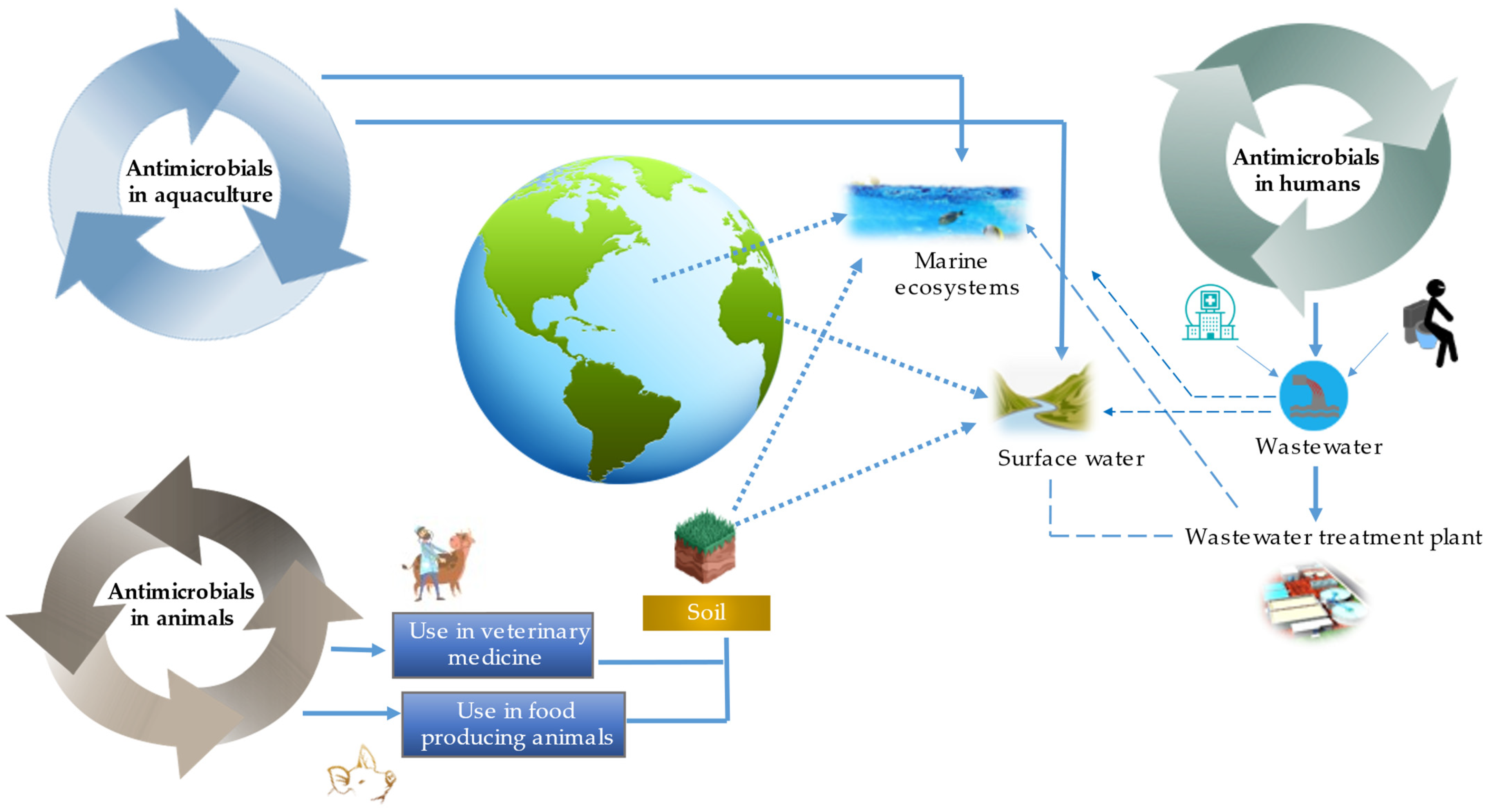
The presence of antimicrobials in aquatic ecosystems (Figure 1) is essentially linked to the excretion of unmetabolized antimicrobials by humans and animals [49], direct introduction by aquaculture [50] and inadequate disposal at the hospital level [49].
Domestic and hospital wastewaters are the first destination of antimicrobials used in human medicine (Figure 1). These can be discharged directly into surface waters and marine ecosystems or after being treated in WWTPs [51]. Conventional purification processes used in WWTPs, such as activated sludge, sequencing batch reactors and anaerobic digestion process have low removal rates for antimicrobials [52].
The implementation of advanced treatment processes such as membrane bioreactors, bioelectrochemical systems and constructed wetlands showed a higher removal efficiency respect to conventional treatments of different antimicrobial compounds [15,52]. In this sense, bioelectrochemical systems have the highest removal rate (>90%), followed by membrane bioreactors (>67%) and constructed wetlands (>63%). Other current alternatives, such as the use of Lemna minor systems, have reported elimination percentages of 100, 96 and 86.5% of antibacterials such as cefadroxil, metronidazole and cephalexin, respectively [53,54].
Soil, like wastewater, is one of the main destinations for antimicrobials used in veterinary medicine and food animals (Figure 1). This is because a high percentage of antimicrobials used in animals are excreted unchanged and reach the soil directly [55]. In addition to their presence, antimicrobials have the particularity of persisting in the soil, contributing to their release into the biosphere and their subsequent arrival in aquatic ecosystems through lixiviation [51].
In contrast to human and veterinary use of antimicrobials and their discharge into sewage systems, current aquaculture practices are responsible for the direct introduction of antimicrobials into aquatic (marine and freshwater) ecosystems [50]. The direct and untargeted introduction of these compounds into aquaculture cages in rivers and oceans leads to dispersion in adjacent waters and thus exposure of surrounding aquatic organisms [48,50].
4. Antimicrobial Levels in Aquatic Ecosystems
4.1. Influents and Effluents from WWTPs
The concentrations of antimicrobials present in WWTP influents and effluents have been compiled from studies conducted worldwide (Table 2). The maximum concentrations of antimicrobials reported in influents are generally higher than in effluents [14,17]. Likewise, the maximum concentrations of antibacterials reported in influents and effluents were higher relative to the other antimicrobial groups [14,17].
Antibacterials are reported more frequently compared to the other antimicrobials [3,56]. Most common antibacterials include beta-lactams, tetracyclines, quinolones, macrolides, sulfonamides and diaminopyrimidines [14,57]. Groups of antiparasitics, antifungals and antivirals include benzimidazoles, azoles and antiretrovirals against HIV (Table 2).
Antibacterials for human and animal use such as sulfamethoxazole and oxytetracycline have been detected in WWTP influents at concentrations up to 59.28 mg L−1 and 1487 ng L−1, respectively [14,17]. Likewise, concentrations of antiparasitics, antifungals and antivirals have been detected at concentrations up to 5,000,960 and 500,000 ng L−1, respectively [3,8].
Antibacterial concentrations of up to 14 mg L−1 have been detected in WWTP effluents [17]. Similarly, concentrations of antiparasitics such as albendazole (129 ng L−1), antifungals such as fluconazole (950 ng L−1) and antivirals such as zidovudine (50,000 ng L−1) have been reported [3,8,58].

Table 2.
Antimicrobial concentrations in aquatic environments.
| Antimicrobial | WWTP Influent (ng L−1) | WWTP Effluent (ng L−1) | Surface Water (ng L−1) | River Water (ng L−1) | Seawater (ng L−1) | Sediment (µg kg −1) | Reference |
|---|---|---|---|---|---|---|---|
| Antibacterial | |||||||
| Penicillin | 160 | 20 | 235 | 115 | 0.4 | - | [13,14,59] |
| Amoxicillin | 33,800 | 116,400 | 1620−4950 | 940–3190 | 5–127.8 | - | [14,60,61] |
| Oxytetracycline | 75–1487 | 2.4–24 | 230 | 51.5 | 25.1 | 652 1 | [7,10,14,62] |
| Tetracycline | 45 * | 3.2 * | 68.90 | 31.4 | 2.4–313 | 135 1 | [7,10,17,63] |
| Doxycycline | 24–120 | 14–49 | 9.4–25 | 1.9–68 | 103 | 7.0 1 | [7,14,62] |
| Erythromycin | 200–300 | 30–350 | 913 | 2070 | 0.13–6.7 | 67.7 1 | [7,10,57,64] |
| Azithromycin | 80–860 | 8–190 | 235 | 455 | 168 | - | [60,64,65] |
| Clarithromycin | 122 | 8–460 | 75–91 | 250 | 0.2–9.4 | - | [7,14,62] |
| Clindamycin | 14–37 | 18–57 | 20 | - | 4.2 | - | [7,57,62] |
| Gentamicin | 14,400–19,100 | 500 | 1400 | - | - | - | [14] |
| Amikacin | 2300 | 1000 | 1200 | - | - | - | [14] |
| Ciprofloxacin | 27 * | 14 * | 990 | 641.3 | 6.9 | 1290 1 | [7,17,60,66] |
| Norfloxacin | 450–2200 | 0.2–628 | 3–518 | 39 | 207.5 | 5770 1 | [7,14,62] |
| Enrofloxacin | 58 | 52 | - | 5681 | 122 | 2.34 1 | [7,57,62] |
| Sulfamethoxazole | 59.28 * | 80,000 | 585 | 1090 | 4.4 | 0.73 1 0.011 2 | [7,17,60,67] |
| Sulfadiazine | 13–26 | 10–21 | 739.20 | 580 | 8.3 | 22.0 1 | [7,13,57] |
| Trimethoprim | 31.7–1866 | 66.6–299 | 710 | 380 | 55.8 | 9.84 1 0.002 2 | [7,10,57,64,67] |
| Chloramphenicol | 13–24 | 6–21 | 91.80 | 5.8 | 8.1 | 700 * | [7,13,57,62,68,69] |
| Antiparasitic | |||||||
| Metronidazole | 5–5000 | 1–70 | 1–5000 | - | - | 6–2000 1 | [3] |
| Albendazole | 464 | 129 | - | 0.4–10.92 | 4–10 | 0.59 1 | [58,65,70] |
| Thiabendazole | 2–80 | 10–80 | - | 1.53 | 9 | 0.01 1 | [58,65,70] |
| Ivermectina | - | - | - | 2.54 | - | 0.013 1 | [65] |
| Antifungal | |||||||
| Clotrimazole | 16 | 0.2 | 6 | 5 | - | 2.5 1 | [8,71,72] |
| Ketoconazole | 22 | 6.7 | 11 | 1 | - | 0.49 1 | [8,71,72] |
| Miconazole | 16 | 7.9 | 30 | 2–30 | - | 1.25–2.06 1 | [8,71,72] |
| Fluconazole | 960 | 950 | 133 | 109 | - | 0.057 1 | [8,71,72] |
| Antivirals | |||||||
| Oseltamivir | 59–2700 | 33–159 | 0.3–17 | 12–58 | - | - | [41] |
| Zidovudine | 9000–85,000 | 95–50,000 | 70–2500 | 1–94 | - | 118 1 | [3,41,73] |
| Acyclovir | 1100–2500 | 12–50 | 730–1500 | 58–750 | - | - | [74] |
| Neviparine | 80–5000 | 95–3000 | 8.5–7000 | 4859 | - | 15–85 1 | [3,41] |
| Efavirenz | 700–50,000 | 50–50,000 | 0.1–900 | 134–354 | - | 4–5 1 | [3,41] |
| Lamivudine | 900–500,000 | 110–95,000 | 70–250,000 | 20 | - | 0.6–0.7 1 | [3,41] |
(-): unreported; (*): mg L−1; (1): river sediments; (2): marine sediments.
4.2. Surface Water, River Water, Seawater and Sediments
The concentrations of antimicrobials present in surface water, river water, seawater and sediments are presented in Table 2. Antivirals showed the highest concentrations in surface waters and rivers, while antibacterials and antiparasitics in sediments. Little information is available on antimicrobial concentrations in seawater.
Antibacterials such as oxytetracycline (230 ng L−1), erythromycin (913 ng L−1) and trimethoprim (710 ng L−1) have been respectively reported in surface waters in China, France and the United States [10]. In addition, the presence of antiparasitics, antifungals and antivirals has been reported worldwide at concentrations up to 5000, 133 and 250,000 ng L−1, respectively [3,71].
Among antimicrobials, only antibacterials (0.2–313 ng L−1) and antiparasitics (4–10 ng L−1) have been detected in seawater [62,63,70]. In rivers, concentrations of antibacterials (1.9–5681 ng L−1), antiparasitics (0.4–10.92 ng L−1), antifungals (1–109 ng L−1) and antivirals (1–4859 ng L−1) were reported [7,14,41,65,72].
Based on their physicochemical characteristics, antimicrobials have often been reported in river and ocean sediments (Table 2) that frequently act as temporary traps of polar con-taminants. Accumulated antimicrobials can eventually be released back into rivers and oceans [7]. In sediments from rivers, sulfadiazine, albendazole, clotrimazole and zidovudine have been detected at concentrations of 22, 0.59, 2.5 and 118 µg kg−1 respectively [7,65,71,73], while sulfamethoxazole (0.011 µg kg−1) and trimethoprim (0.002 µg kg−1) have been recorded in marine sediments [67].
5. Toxicological Effects of Antimicrobials on Aquatic Organisms
The presence of antimicrobials in aquatic environments results in chronic low-level exposure and potential effects in different organisms (cyanobacteria, microalgae, invertebrates and vertebrates) [75]. During many years, research on the effects of exposure to antimicrobial compounds has predominantly focused on antibacterial resistance in bacteria (Kovalakova et al. [10]). Therefore, for many compounds, comprehensive ecotoxicological data are not available [76]. Most research on the effects of antimicrobials in aquatic systems is reduced to toxicity tests on freshwater model organisms such as Microcystis aeruginosa, Daphnia magna and Danio rerio [77,78,79] and only a few studies have evaluated the effect of antimicrobials on marine organisms [80]. In addition, most studies evaluate the effect of individual antimicrobials [14,71,75,81,82] but this is not the case in natural environments, where a complex cocktail is usually observed and can produce different effects on organisms [20,83]. Furthermore, up to 90% of antimicrobials are excreted in the environment unchanged or as active metabolites [13] and also undergo natural transformation processes such as adsorption in sediments or degradation (e.g., photodegradation, hydroxylation), which can give rise to new potentially toxic compounds [84]. The effects observed in aquatic organisms exposed to antimicrobials include molecular, cellular and physiological changes (Figure 2) at exposure concentrations in the range of ng L−1-mg L−1.
5.1. Fresh Water Organisms
5.1.1. Microalgae and Cyanobacteria
Within the cyanobacteria group, probably the most studied species is Microcystis aeruginosa. Ye et al. [85] evaluated the effect of tetracycline (TE), chlortetracycline (CTC) and oxytetracycline (OTC) on the growth of M. aeruginosa. The results showed mean effective concentration 10 (EC10) values at 96 h in the range of 0.63–3.02 mg L−1 and mean effective concentration 20 (EC20) 96 h of 1.58–4.86 mg L−1 (Table 3). Similarly, Xu et al. [86] reported mean effective concentration 50 (EC50) values of TE (2.2 mg L−1), CTC (3.1 mg L−1) and OTC (4.5 mg L−1) for Selenastrum capricornutum and EC50 and EC10 values of OTC for Anabaena sp. of 2.7 and 1.5 mg L−1, respectively.
Carusso et al. [87], by growth inhibition experiments, determined the inhibitory concentration 10 (IC10) and 50 (IC50) of CTC, OTC and enrofloxacin (ENR) on Pseudokirchneriella subcapitata and Ankistrodesmus fusiformis. The results showed a higher sensitivity of P. subcapitata exposed to OTC and 50% inhibitory concentrations were always lower in P. subcapitata compared to A. fusiformis (Table 3).
In addition to the evaluation of instantaneous responses such as growth inhibition in microalgae and cyanobacteria, several authors have studied the effects at concentrations similar to those reported in the environments, e.g., Liu et al. [88] analyzed the proteomic response of the cyanobacterium Microcystis aeruginosa exposed to amoxicillin for 30 days at a concentration of 100 ng L−1. In total, 35 up-regulated proteins (superoxide dismutase (SOD), glutathione reductase (GR), among others) and 27 down-regulated proteins (glucose-6-phosphate isomerase, glutamine synthetase, among others) were identified. All of them are closely related to photosynthesis. Similarly, Chen et al. [89] evaluated the protein expression of M. aeruginosa exposed to spiramycin at 50 and 200 ng L−1 in combination with different levels of nitrogen and phosphorus. The results showed changes in the expression of proteins related to processes such as photosynthesis, stress and cell division, such as SOD, enolase, RNA polymerase alpha and serine protease at both 50 and 200 ng L−1 and at low and high levels of nitrogen and phosphorus.
The effect of antiparasitics such as flubendazole and fenbendazole has been evaluated in microalgae and cyanobacteria. Wagil et al. [90] reported decreased reproduction of Scenedesmus vacuolatus exposed to concentrations >1 mg L−1. As for antivirals. Almeida et al. [91] and Silva et al. [92] reported growth inhibition of Raphidocelis subcapitata and Microcystis novacekii after exposure to acyclovir, efavirenz, lamivudine, zidovudine and tenofovir (Table 3). The effect of the antifungals propiconazole and tebuconazole on the growth and antioxidant response of the microalga Chlorella pyrenoidosa was evaluated by Nong et al. [93], identifying that doses of 100, 200 and 1000 μg L−1 were enough to inhibit growth. These authors also observed a gradual increase of the activity of the enzyme biomarkers SOD and catalase (CAT) with concentrations ranging from 100 to 20,000 µg L−1.
5.1.2. Invertebrates
Daphnia magna is commonly used in standard toxicity tests for crustaceans, such as the acute immobilization test (OECD 202) and reproduction test (OECD 211) [94]. Zhang et al. [95] evaluated the growth inhibition rate of D. magna with three antibacterials, chloramphenicol, thiamphenicol and florfenicol and their mixtures, by EC50 acute toxicity tests, at two temperatures. A significant increase in toxicity was observed with increasing exposure temperature, for example, the EC50 of chloramphenicol at 20 °C was 283.86 mg L−1, while at 25 °C it was 85.18 mg L−1. Similarly, the mixture of chloramphenicol and thiamphenicol showed a significant increase in toxicity at 25 °C, being the most toxic combination with an EC50 of 42.11 mg L−1.
Luo et al. [96] evaluated the effect of lomefloxacin on the antioxidant response of D. magna under simulated solar radiation as a variable, observing an increase in reactive oxygen species (ROS) and decrease in oxidative stress biomarkers such as SOD (Table 3).
The effects of antibacterial compounds have also been evaluated in the bivalves Mytilus edulis, Ruditapes philippinarum and Dreissena polymorpha (Table 3).
The effect of antiparasitic, antifungal and antiviral drugs on D. magna has also been evaluated. For example, Bundschuh et al. [97] and Wagil et al. [90] evidenced alterations in motility and growth of D. magna due to the effect of three antiparasitics, e.g., flubendazole, fenbendazole and ivermectin, at environmentally relevant concentrations in the range of ng to µg L−1 (Table 3) and Viera et al. [98] and Omotola et al. [81] reported concentrations of clotrimarzole (5143 mg L−1) and lamivudine (0.1 mg L−1) capable of causing death to D. magna.
Table 3.
Main ecotoxicological effects reported for freshwater and marine species exposed to various molecules of different classes of antimicrobials at selected exposure doses.
Table 3.
Main ecotoxicological effects reported for freshwater and marine species exposed to various molecules of different classes of antimicrobials at selected exposure doses.
| Antimicrobial Class | Organism | Species | Molecule | Type of Assay | Effect | Exposure Doses (mg L−1) | Reference |
|---|---|---|---|---|---|---|---|
| Antibacterial | Freshwater cianobacteria | Microcystis aeruginosa | Tetracycline | Acute toxicity test, growth rate. EC10-96 h and EC20-96 h | Inhibition of growth | 0.63 1.58 | [85] |
| Chlortetracycline | 1.86 4.09 | ||||||
| Oxytetracycline | 3.02 4.86 | ||||||
| Freshwater cianobacteria | Chlorella pyrenoidesa | Tigecycline | Acute toxicity test, growth rate. EC50-144 h | Inhibition of growth | 6.20 | [82] | |
| Spiramycin | 4.58 | ||||||
| Amoxicillin | >2 1 | ||||||
| Freshwater cianobacteria | Anabaena cylindrica | Tigecycline | Acute toxicity test, growth rate. EC50-144 h | Inhibition of growth | 0.062 | [82] | |
| Spiramycin | 0.038 | ||||||
| Amoxicillin | 7.6 | ||||||
| Fresh water microalgae | Pseudokirchneriella subcapitata | Oxytetracycline | Acute toxicity test, growth rate. IC10-96 h and IC50-96 h | Inhibition of growth | 0.07 ± 0.03 0.64 ± 0.38 | [87] | |
| Ankistrodesmus fusiformis | 0.05 ± 0.01 4.17 ± 3.79 | ||||||
| Fresh water microalgae | Pseudokirchneriella subcapitata | Penicillin g | Acute toxicity test, growth rate. EC50-72 h | Inhibition of growth | 7114 | [14] | |
| Vancomycin | 371 | ||||||
| Fresh water crustacean | Daphnia magna | Lomefloxacin | Biomarkers assay | Decreased CAT and SOD activity, induction of LPO and ROS activity. | 100 2 | [96] | |
| Streptomycin | Acute immobilization test. EC50-48 h | immobilization | 487 | [14] | |||
| Penicillin g | 1496 | ||||||
| Vancomycin | 687 | ||||||
| Marine bivalve | Mytilus edulis | Trimetoprim | In vitro haemocyte assay | DNA damage decreased phagocytic efficiency | 20 | [99] | |
| Erythromycin | Biomarkers assay | Induction of CAT and GST activity | 200 | [100] | |||
| Aquatic plant | Lemna minor | Oxytetracycline | Decreased plant growth | 0.001 | [101] | ||
| Ciprofloxacin | Biomarkers assay | Increased hydrogen peroxide | ≥1.05 | [102] | |||
| Bivalve | Mytilus galloprovincialis | Sulfamethoxazole | Osmoregulation alteration | 0.01 | [103] | ||
| Bivalve | Dreissena polymorpha | Trimethoprim | SCGE (single cell gel electrophoresis) assay | Genotoxicity | 5 2 | [104] | |
| Bivalve | Ruditapes philippinarum | Amoxicillin | Biomarkers assay | Increased CAT and decreased SOD activity | 0.4 | [105] | |
| Fresh water Vertebrate, fish | Carassius auratus | Erythromycin | Biomarkers assay | Decreased AChE activity, induction of SOD activity. | 0.004–0.1 | [75,106] | |
| Altered activity of anti-oxidant enzyme | 0.002 | ||||||
| Roxithromycin | Biomarkers assay | Increase in AChE and SOD activity, induction of EROD activity, | 0.004–0.1 | [107] | |||
| Fresh water Vertebrate, fish | Danio rerio | Mixture (Ciprofloxacin, ofloxacin, norfloxacin, and enrofloxacin) | Abnormal development and histopathological changes | >37.5 | [75] | ||
| Oxytetracycline | Delayed hatching, inflammatory response | 0.0001–10 | [75] | ||||
| Nitrofurantoin | Biomarkers assay | Increase CAT and GST | ≥0.32 and ≥0.02 | [108] | |||
| Sulfamethoxazole | Increased mortality and inflammatory response | 0.260 | [75] | ||||
| Mixture (Sulfamethoxazole, sulfadiazine, sulfadimidine) | Abnormal swimming and heart rate | 1–10 | [75] | ||||
| Clarithromycin | Metabolic changes | 0.1 | [75] | ||||
| Erythromycin | Biomarkers assay | Significant increase of SOD, GPX and LPO activity and significant decrease of CAT | 0.01–0.1 | [109] | |||
| Oxytetracycline | Haematological parameters | Haematological alteration | 20–200 | [75] | |||
| Euryhalines Vertebrates, fish | Oncorhynchus mykiss | Oxytetracycline | Biomarkers assay | DNA damage and altered activity of anti-oxidant enzyme | 50 | [75] | |
| Erythromycin | Biomarkers assay | Oxidative stress and genotoxicity | 0.0008 | [75] | |||
| Oxytetracycline | Haematological and liver parameters | Increased ALT, AST and decreases WBC | 2500 3 | [7] | |||
| Fresh water Vertebrate, fish | Oreochromis niloticus | Florfenicol | Liver parameters | Decreased AST, creatinine | 5 3 | [7] | |
| Fresh water Vertebrate, fish | Lepomis gibbosus | Mixture (Ciprofloxacin, ibuprofen and fluoxetine) | Mortality | 0.01 | [75] | ||
| Fresh water Vertebrate, fish | Xiphophorus Helleri | Norfloxacin | RT—qPCR | Genotoxicity | 0.24–6 | [75] | |
| Fresh water Vertebrate, fish | Oryzias latipes | Erythromycin | Biomarkers assay | Oxidative stress and genotoxicity | >1000 | [75] | |
| Fresh water Vertebrate, fish | Cyprinus carpio | Oxytetracycline | Haematological parameters | Increased Glu, WBC and Ht | 75 3 | [7] | |
| Marine Vertebrate, fish | Sparus aurata | Oxytetracycline | Haematological parameters | Increased of WBC | 75 3 | [7] | |
| Antiparasitic | Fresh water microalgae | Scenedesmus vacuolatus | Flubendazole | Acute toxicity test, reproduction | Decrease in reproduction | >1 | [90] |
| Fenbendazole | >1 | ||||||
| Fresh water crustacean | Daphnia magna | Metronidazol | Acute and chronic toxicity | Decrease in reproduction | 1000 | [110] | |
| Fresh water crustacean | Daphnia magna | Flubendazole | Acute toxicity test, growth rate. EC50-48 h | Inhibition of growth | 0.043–0.046 | [90] | |
| Fenbendazole | 0.018–0.020 | ||||||
| Fresh water crustacean | Daphnia magna | Fenbendazole | Acute immobilisation test. EC50-48 h | immobilization | 0.012–0.02 | ||
| Flubendazole | 0.057–0.086 | [97] | |||||
| Ivermectin | 0.00049–0.00072 | ||||||
| Fresh water crustacean | Gammarus pulex | Fenbendazole | Acute immobilisation test. EC50-96 h | immobilization | 0.123–0.174 | [97] | |
| Flubendazole | 0.087–0.127 | ||||||
| Ivermectin | 0.001–0.0016 | ||||||
| Fresh water crustacean | Asellus aquaticus | Fenbendazole | Acute immobilisation test. EC50-96 h | immobilization | >1 | [97] | |
| Flubendazole | >1 | ||||||
| Ivermectin | 0.315–0.482 | ||||||
| Fresh water Vertebrate, fish | Danio rerio | Doramectin | Abnormal swimming | 0.58 | [111] | ||
| Antifungals | Fresh water Vertebrate, fish | Carassius auratus | Ketoconazol | Biomarkers assay | Increase in SOD activity and decrease in GST, EROD and AChE. | 0.002–0.02 | [71,112] |
| qPCR | Increased of cyp1a | 0.025–0.1 | |||||
| Fresh water crustacean | Daphnia magna | Clotrimazole | Acute toxicity test. LC50-48 h | Mortality | 5143 | [98] | |
| Fresh water Vertebrate, fish | Cyprinus carpio | Clotrimazole | RT—qPCR | High mdr1 and mrp2 gene expression | 0.00287–0.034 | [71] | |
| Decrease of cyp2k and cyp3a | 0.00001–0.1 | ||||||
| Euryhalines Vertebrates, fish | Salmo salar | Ketoconazole | Decrease of p-nitrophenol hydroxylase | 0.2–80 2 | [71] | ||
| Miconazole | Decrease of Ethinyl estradiol | 0.1–10 | |||||
| Fresh water Vertebrate, fish | Danio rerio | Clotrimazole | RT—qPCR | Increased of fshr and fshβ | 0.03–0.197 | [71] | |
| RT—qPCR | Increased of cyp17a1 and cyp11c1 | 0.071–0.258 | |||||
| Propiconazole | qPCR | Increased of cyp51 and cyp7a1 | 0.42–17.57 | ||||
| Propiconazole | Acute toxicity test. LC50-96 h | Mortality | 12.90 | ||||
| Difenoconazole | Acute toxicity test. LC50-96 h | Mortality | 2.34 | ||||
| Euryhalines Vertebrates, fish | Oncorhynchus mykiss | Clotrimazole | Biomarkers assay | Increase in EROD activity | 0.0001–0.01 | [71] | |
| Biomarkers assay | Decrease in EROD activity | 0.34–17.24 | |||||
| Propiconazole | Acute toxicity test. LC50-96 h | Mortality | 5.04 | ||||
| Fresh water Vertebrate, fish | Oryzias latipes | Fluconazole | Acute toxicity test. LC50-96 h | Mortality | >100 | [71] | |
| Pimephales promelas | Ketoconazole | qPCR | Increase of cyp11a | 0.1, 0.3, 0.9 | [2,71] | ||
| Propiconazole | qPCR | Increase of cyp19, cyp17 and cyp11a | 0.005–1 | ||||
| Antivirals | Freshwater cianobacteria | Microcystis novacekii | Tenofovir | Acute toxicity test, growth rate. EC50-96 h | Inhibition of growth | 156.81–165.21 | [92] |
| Fresh water crustacean | Daphnia magna | Lamivudine | Acute Mortality test 48 h | Mortality | 0.1 | [81] | |
| Fresh water crustacean | Ceriodaphnia dubia | Acyclovir | Acute toxicity test, growth rate. EC50-8 days | Inhibition of growth | 2529–3707 | [91] | |
| Efavirenz | 0.024–0.027 | ||||||
| Lamivudine | 1242–1456 | ||||||
| Zidovudine | 5370–5989 | ||||||
| Fresh water algae | Raphidocelis subcapitata | Acyclovir | Acute toxicity test, growth rate. IC50-96 h | Inhibition of growth | 3249–4016 | [91] | |
| Efavirenz | 0.031–0.038 | ||||||
| Lamivudine | 2753–3297 | ||||||
| Zidovudine | 4969–5962 |
(1): g L−1; (2): µM; (3): mg kg−1; (CAT): Catalase; GST: Glutathione S-transferase; SOD: Superoxide dismutase; EROD: 7-Ethoxy thiopheneoxazolonedeethylase; AChE: Acetylcholine esterase; mdr1: Multi-Drug Resistance gene; mrp2: Multidrug resistance- associated protein 2 gene; GPX: Glutathione peroxidase; LPO: lipid peroxidation; ROS: Reactive oxygen species; (ALT): Alanina transaminasa; (AST): Aspartato transaminasa; (WBC): White blood cell; (Glu): Glucose; (Ht): Hematocrit; (cyp1a): Cytochrome P450 1a gene; (cyp2k):Cytochrome P450 2k gene; (cyp3a): Citocromo P450 3a gene; (fshr): Follicle Stimulating Hormone Receptor gene; (fshβ): Follicle-stimulating hormone β gene; (cyp17a1): Cytochrome P450 17A1; (cyp11c1): Cytochrome P450 11c1; (cyp51): Cytochrome P450 51; (cyp7a1): Cytochrome P450 7a1; (cyp11a): Cytochrome P450 11a; (cyp19): Cytochrome P450 19; (cyp17): Cytochrome P450 17.
5.1.3. Vertebrates
There are three main freshwater vertebrate organisms used as models in the ecotoxicological tests, all of them fish: goldfish (Carassius auratus), medaka (Oryzias latipes) and zebrafish (Danio rerio). Specific toxicological studies have also been conducted on other fish, such as rainbow trout (Oncorhynchus mykiss), flathead minnow (Pimephales promelas) or Indian carp (Catla catla).
Several antibacterials have been tested on freshwater fish, at different life stages and at decreasing concentrations from sublethal (mg L−1) to environmentally relevant (µg L−1). Using sublethal concentrations, Mattioli et al. [113] evaluated the risk of Nile tilapia (Oreochromis niloticus) exposed to florfenicol concentrations (58.73–381.8 mg L−1) and obtained a mean lethal concentration value (LC50–96 h) of 349.94 mg L−1. De Oliveira et al. [108] evaluated the effects of nitrofurantoin on Danio rerio embryos, using sublethal concentrations (0–100 mg L−1) for the analysis of some enzymatic biomarkers. Cholinesterase, lactate dehydrogenase and glutathione S-transferase activity was induced at concentrations of 0.02 mg L−1. Ma et al. [114] analyzed the proteomic profile of the liver of Ctenopharyngodon idellus fish exposed to ENR (40 mg kg−1): they identified 3082 proteins and 103 of them were differentially abundant, 49 up-regulated and 54 down-regulated. Some of them were extremely significantly related to translation.
Using concentrations similar to environmental levels, Qiu et al. [115] studied the effects of four antibacterials, sulfamonomethoxine (SMM), cefotaxime sodium (CFT), TC and ENR at 0.01, 1, and 100 μg L−1 on the transcriptome of D. rerio larvae observing that 692 (260 up-regulated and 432 down-regulated), 713 (239 up-regulated and 474 down-regulated), 592 (241 up-regulated and 351 down-regulated) and 567 (208 up-regulated and 359 down-regulated) genes were differentially expressed for SMM, CFT, TC and ENR, respectively. The genes are mainly related to steroid biosynthesis and other metabolic pathways.
Gene expression has also been evaluated in D. rerio, Carassius auratus and Cyprinus carpio fish after exposure to antifungals such as clotrimazole and ketoconazole [71]. Some of the differentially expressed genes were cyp1a, mdr1 and fshr (Table 3). As for antiparasitics, a decrease in D. rerio swimming behavior was evidenced after exposure to doramectin (0.58 mg L−1) [111].
5.2. Marine Organisms
Although antibacterials tend to bioaccumulate in marine organisms [116,117,118] and are nowadays a recognized threat to the marine environment [119], data on their toxicity on marine organisms are scarce, as they are usually discharged in rivers and other freshwater bodies [120].
In this context, Rodrigues et al. [121] evaluated the histopathologic effects of the antibacterials erythromycin (ERY) and OTC in the sea bream (Sparus aurata). S. aurata were exposed acutely (96 h) and chronically (28 days) to concentrations of ERY (0.0002–200 μg L−1) and OTC (0.0004–400 μg L−1). The results showed various alterations (circulatory, regressive, progressive and inflammatory), as well as an increase in the histopathological index of the gills of acutely exposed organisms to ERY and those chronically exposed to OTC. Similarly, Rodrigues et al. [122] evaluated the effect of ERY in S. aurata on some biomarker activity such as glutathione peroxidase (GPX) and glutathione reductase (GR) after exposure to concentrations of 0.3–323 µg L−1 for 96 h and 0.7–8.8 µg L−1 for 28 days. The results showed a decrease in GPX activity in the liver after acute exposure and an increase in the gills after chronic exposure.
Hoseini et al. [123] treated Oncorhynchus mykiss specimens with OTC (0 and 2.5 g kg−1) for 2 weeks and evaluated the effects on immunological parameters, oxidative stress and enzymatic activity, recording a significant increase in serum alanine aminotransferase (ALT) and aspartate aminotransferase (AST) activities, a decrease in SOD activity and an increase in intestinal glutathione transferase (GST). Similarly, Nakano et al. [124] evaluated the effect of OTC in Oncorhynchus kisutch after a treatment of 100 mg kg−1 body weight/day orally for 2 weeks. The results showed an increase in ALT activity and total glutathione (tGSH) levels in the liver.
Other effects of antifungal compounds (azoles) on marine bivalves such as Mytilus edulis and fish such as rainbow trout and Salmo salar are presented in Table 3.
5.3. Toxicity of Antimicrobial Mixtures
Most research on the effects of antimicrobials in aquatic organisms has been conducted using only one compound at a time (Table 3). However, study on the effects of mixtures of antimicrobial compounds on these organisms is increasing. In this regard, Trombini et al. [20] evaluated the effect of the mixture of ciprofloxacin, flumequine and ibuprofen on the crayfish Procambarus clarkii at concentrations of 10 and 100 μg L−1, obtaining alterations in immune responses and the abundance of proteins associated with biotransformation and detoxification processes in the cell (CAT and GST), as well as an increase in the expression of genes encoding antioxidant enzymes such as SOD and GPX. Jiang et al. [83] evaluated the effect of a mixture of 5 antibacterials (amoxicillin, ciprofloxacin, spiramycin, sulfamethoxazole and TE) at concentrations between 50 and 500 ng L−1 on the biochemical, transcriptomic and proteomic responses of Microcystis aeruginosa. The biochemical responses showed an increase in the growth rate of M. aeruginosa at levels between 50–400 ng L−1. The transcriptomic analysis revealed 206 up-regulated and 114 down-regulated genes in organisms exposed to 200 ng L−1 and proteomic analysis identified 61 up-regulated and 25 down-regulated proteins. Differentially expressed genes and proteins were closely related to processes such as photosynthesis and carbon metabolism.
Other studies on the effects of antimicrobial compound mixtures on aquatic organisms are presented in Table 3.
6. Conclusions
Studies on the presence of antimicrobials in aquatic ecosystems and their effects on aquatic organisms have focused mainly on antibacterials; however, the effects of antiparasitic, antifungal and antiviral compounds in these ecosystems need to be further studied and determined.
The types of antimicrobials and the levels detected are related to the low efficiency of their removal in WWTPs, mainly in developing countries. Trade and endemic diseases also play an important role, for example, antivirals and antiparasitics are rarely detected in Europe, however, in Africa they have been detected at concentrations very close to mg L−1, which could be related to a higher consumption of these antimicrobials in malaria and AIDS endemic countries in Africa. As for trade, it is difficult to establish a relationship between the consumption of antimicrobials and their presence in aquatic ecosystems; however, the highest concentrations detected are reported in countries with high consumption, such as China, India and the United States. In South America, there are few studies that provide information on the presence of antimicrobials in different aquatic ecosystems.
On the other hand, most of the effects are usually measured at concentrations that are not relevant from an environmental point of view (mg L−1), and do not reflect the real behavior in aquatic scenarios. The use of molecular tools and chronic exposure tests at concentrations similar to environmental levels (ng-µg L−1) need to be performed more frequently.
Undoubtedly, the continuous introduction of antimicrobial compounds into aquatic ecosystems is a global problem that paints a bleak picture for the future. There is an imminent need for different countries to establish standards that allow greater control over the consumption of antimicrobials (e.g., human and veterinary medicine and food-producing animals), and to implement new technologies in WWTPs and/or new wastewater treatment systems to eliminate these compounds, thus preventing their entry into aquatic ecosystems.
Author Contributions
Conceptualization, R.F. and M.H.; methodology, R.F., H.J.B.-A. and G.A., writing—original draft preparation, R.F., H.J.B.-A., N.R.C.-R., M.H. and G.A., writing—review and editing, G.A., M.H. and N.R.C.-R. All authors have read and agreed to the published version of the manuscript.
Funding
This study was funded by the Spanish Ramón y Cajal funding scheme supporting MH (contract reference RYC-2012-12217) and the Spanish Ministry of Economy, Industry and Competitiveness (MINECO) within the framework of the project: Integration of Omics Tools for the Environmental Risk Assessment of Emerging Pollutants in Marine Species of Commercial Interest, HORACIO (CTM2015-70731-R).
Institutional Review Board Statement
Not applicable.
Informed Consent Statement
Not applicable. The study did not involve humans or animals.
Data Availability Statement
Data supporting reported results can be found asking directly of the first author.
Acknowledgments
This work was supported by a fellowship of the Latin American Association of Postgraduates (AUIP) in agreement with the Simón Bolívar University of Colombia, the PROPLAYAS Network, the Caribbean Marine and Limnological Research Center CICMAR and the University of Cadiz in Spain. This work is a contribution to the PAI Research Group RNM-328 (Junta de Andalucía, Spain).
Conflicts of Interest
The authors declare no conflict of interest.
References
- Valdez-Carrillo, M.; Abrell, L.; Ramírez-Hernández, J.; Reyes-López, J.A.; Carreón-Diazconti, C. Pharmaceuticals as Emerging Contaminants in the Aquatic Environment of Latin America: A Review. Environ. Sci. Pollut. Res. 2020, 27, 44863–44891. [Google Scholar] [CrossRef]
- Rathi, B.S.; Kumar, P.S.; Show, P.-L. A review on effective removal of emerging contaminants from aquatic systems: Current trends and scope for further research. J. Hazard. Mater. 2020, 409, 124413. [Google Scholar] [CrossRef] [PubMed]
- K’Oreje, K.O.; Okoth, M.; Van Langenhove, H.; Demeestere, K. Occurrence and treatment of contaminants of emerging concern in the African aquatic environment: Literature review and a look ahead. J. Environ. Manag. 2019, 254, 109752. [Google Scholar] [CrossRef] [PubMed]
- Ryan, K.J.; Ray, C. Sherris Microbiología Médica, 6th ed.; McGraw-Hill: New York, NY, USA, 2010. [Google Scholar]
- Adeapena, W.; Afari-Asiedu, S.; Najjemba, R.; Griensven, J.; Delamou, A.; Buabeng, K.O.; Asante, K.P. Antibiotic Use in a Municipal Veterinary Clinic in Ghana. Trop. Med. Infect. Dis. 2021, 6, 138. [Google Scholar] [CrossRef] [PubMed]
- Bungau, S.; Tit, D.M.; Behl, T.; Aleya, L.; Zaha, D.C. Aspects of excessive antibiotic consumption and environmental influences correlated with the occurrence of resistance to antimicrobial agents. Curr. Opin. Environ. Sci. Health 2020, 19, 100224. [Google Scholar] [CrossRef]
- Bojarski, B.; Kot, B.; Witeska, M. Antibacterials in Aquatic Environment and Their Toxicity to Fish. Pharmaceuticals 2020, 13, 189. [Google Scholar] [CrossRef]
- Assress, H.A.; Nyoni, H.; Mamba, B.; Msagati, T.A. Occurrence and risk assessment of azole antifungal drugs in water and wastewater. Ecotoxicol. Environ. Saf. 2019, 187, 109868. [Google Scholar] [CrossRef] [PubMed]
- Tiseo, K.; Huber, L.; Gilbert, M.; Robinson, T.P.; Van Boeckel, T.P. Global Trends in Antimicrobial Use in Food Animals from 2017 to 2030. Antibiotics 2020, 9, 918. [Google Scholar] [CrossRef]
- Kovaláková, P.; Cizmas, L.; McDonald, T.J.; Marsalek, B.; Feng, M.; Sharma, V.K. Occurrence and toxicity of antibiotics in the aquatic environment: A review. Chemosphere 2020, 251, 126351. [Google Scholar] [CrossRef]
- Bortone, B.; Jackson, C.; Hsia, Y.; Bielicki, J.; Magrini, N.; Sharland, M. High global consumption of potentially inappropriate fixed dose combination antibiotics: Analysis of data from 75 countries. PLoS ONE 2021, 16, e0241899. [Google Scholar] [CrossRef]
- Nippes, R.P.; Macruz, P.D.; da Silva, G.N.; Scaliante, M.H.N.O. A critical review on environmental presence of pharmaceutical drugs tested for the covid-19 treatment. Process Saf. Environ. Prot. 2021, 152, 568–582. [Google Scholar] [CrossRef]
- Li, Z.; Li, M.; Zhang, Z.; Li, P.; Zang, Y.; Liu, X. Antibiotics in aquatic environments of China: A review and meta-analysis. Ecotoxicol. Environ. Saf. 2020, 199, 110668. [Google Scholar] [CrossRef] [PubMed]
- Felis, E.; Kalka, J.; Sochacki, A.; Kowalska, K.; Bajkacz, S.; Harnisz, M.; Korzeniewska, E. Antimicrobial pharmaceuticals in the aquatic environment—occurrence and environmental implications. Eur. J. Pharmacol. 2019, 866, 172813. [Google Scholar] [CrossRef] [PubMed]
- Ávila, C.; García-Galán, M.J.; Borrego, C.M.; Rodríguez-Mozaz, S.; García, J.; Barceló, D. New insights on the combined removal of antibiotics and ARGs in urban wastewater through the use of two configurations of vertical subsurface flow constructed wetlands. Sci. Total Environ. 2020, 755, 142554. [Google Scholar] [CrossRef] [PubMed]
- Bilal, M.; Mehmood, S.; Rasheed, T.; Iqbal, H.M. Antibiotics traces in the aquatic environment: Persistence and adverse environmental impact. Curr. Opin. Environ. Sci. Health 2019, 13, 68–74. [Google Scholar] [CrossRef]
- Ngqwala, N.P.; Muchesa, P. Occurrence of pharmaceuticals in aquatic environments: A review and potential impacts in South Africa. S. Afr. J. Sci. 2020, 116. [Google Scholar] [CrossRef]
- Fernandez, R.E.R.; Bolívar-Anillo, H.; Turcios, C.H.; García, L.C.; Hernández, M.S.; Abdellah, E. Antibiotic Resistance: The Role of Man, Animals and the Environment. Rev. Salud Uninorte 2020, 36, 298–324. [Google Scholar] [CrossRef]
- Hampel, M.; Blasco, J.; Martín Díaz, M.L. Biomarkers and Effects. In Marine Ecotoxicology; Elsevier: Amsterdam, The Netherlands, 2016; pp. 121–165. [Google Scholar]
- Trombini, C.; Kazakova, J.; Montilla-López, A.; Fernández-Cisnal, R.; Hampel, M.; Fernández-Torres, R.; Bello-López, M.; Abril, N.; Blasco, J. Assessment of pharmaceutical mixture (ibuprofen, ciprofloxacin and flumequine) effects to the crayfish Procambarus clarkii: A multilevel analysis (biochemical, transcriptional and proteomic approaches). Environ. Res. 2021, 200, 111396. [Google Scholar] [CrossRef]
- Grenni, P.; Ancona, V.; Caracciolo, A. Ecological effects of antibiotics on natural ecosystems: A review. Microchem. J. 2018, 136, 25–39. [Google Scholar] [CrossRef]
- Aidara-Kane, A.; Angulo, F.J.; Conly, J.M.; Minato, Y.; Silbergeld, E.K.; McEwen, S.A.; Collignon, P.J.; WHO Guideline Development Group. World Health Organization (WHO) guidelines on use of medically important antimicrobials in food-producing animals. Antimicrob. Resist. Infect. Control. 2018, 7, 1–8. [Google Scholar] [CrossRef] [Green Version]
- Paliy, A.P.; Petrov, R.V.; Kovalenko, L.M.; Livoshchenko, L.P.; Livoshchenko, Y.M.; Klishchova, Z.E.; Bula, L.V.; Ostapenko, V.I.; Doletskyi, S.P.; Palii, A.P. Effectiveness of a Modern Antiparasitic Agent for Deworming in Domestic Animals. Ukr. J. Ecol. 2021, 11, 10–17. [Google Scholar] [CrossRef]
- Patel, S.J.; Wellington, M.; Shah, R.M.; Ferreira, M.J. Antibiotic Stewardship in Food-producing Animals: Challenges, Progress, and Opportunities. Clin. Ther. 2020, 42, 1649–1658. [Google Scholar] [CrossRef] [PubMed]
- Lulijwa, R.; Rupia, E.J.; Alfaro, A.C. Antibiotic use in aquaculture, policies and regulation, health and environmental risks: A review of the top 15 major producers. Rev. Aquac. 2019, 12, 640–663. [Google Scholar] [CrossRef]
- Schar, D.; Klein, E.Y.; Laxminarayan, R.; Gilbert, M.; Van Boeckel, T.P. Global trends in antimicrobial use in aquaculture. Sci. Rep. 2020, 10, 21878. [Google Scholar] [CrossRef] [PubMed]
- Hennessey, M.; Whatford, L.; Payne-Gifford, S.; Johnson, K.F.; Van Winden, S.; Barling, D.; Häsler, B. Antimicrobial & antiparasitic use and resistance in British sheep and cattle: A systematic review. Prev. Vet. Med. 2020, 185, 105174. [Google Scholar] [CrossRef]
- Selzer, P.M.; Epe, C. Antiparasitics in Animal Health: Quo Vadis? Trends Parasitol. 2020, 37, 77–89. [Google Scholar] [CrossRef] [PubMed]
- Sallach, J.B.; Thirkell, T.J.; Field, K.J.; Carter, L.J. The emerging threat of human-use antifungals in sustainable and circular agriculture schemes. Plants People Planet 2021, 3, 685–693. [Google Scholar] [CrossRef]
- Álvarez-Pérez, S.; García, M.E.; Anega, B.; Blanco, J.L. Antifungal Resistance in Animal Medicine: Current State and Future Challenges. In Fungal Diseases in Animals; Gupta, A., Singh, N.P., Eds.; Springer: Cham, Switzerland, 2021; pp. 163–179. [Google Scholar]
- Adriaenssens, N.; Coenen, S.; Kroes, A.C.M.; Versporten, A.; Vankerckhoven, V.; Muller, A.; Blix, H.S.; Goossens, H.; Mittermayer, H.; Vaerenberg, S.; et al. European Surveillance of Antimicrobial Consumption (ESAC): Systemic antiviral use in Europe. J. Antimicrob. Chemother. 2011, 66, 1897–1905. [Google Scholar] [CrossRef] [Green Version]
- Bule, M.; Khan, F.; Niaz, K. Antivirals: Past, Present and Future. In Recent Advances in Animal Virology; Springer: Singapore, 2019. [Google Scholar]
- Girijan, S.K.; Paul, R.; Kumar, V.J.R.; Pillai, D. Investigating the impact of hospital antibiotic usage on aquatic environment and aquaculture systems: A molecular study of quinolone resistance in Escherichia coli. Sci. Total Environ. 2020, 748, 141538. [Google Scholar] [CrossRef]
- Becker, P.; Lecerf, P.; Claereboudt, J.; Devleesschauwer, B.; Packeu, A.; Hendrickx, M. Superficial mycoses in Belgium: Burden, costs and antifungal drugs consumption. Mycoses 2020, 63, 500–508. [Google Scholar] [CrossRef]
- Das, P. Management and Control of Trichomonas vaginalis Infection: A Recent Study. In Highlights on Medicine and Medical Research; Book Publisher International: London, UK, 2021; Volume 10. [Google Scholar]
- Trivedi, J.; Mohan, M.; Byrareddy, S.N. Drug Repurposing Approaches to Combating Viral Infections. J. Clin. Med. 2020, 9, 3777. [Google Scholar] [CrossRef]
- Lázaro-Bengoa, E.; Iglesias, F.J.D.A.; López-Navas, A.; Fernández-Cortizo, M.J. Uso de antibióticos en España y marco regulador para su desarrollo clínico en la Unión Europea. Enferm. Infecc. Microbiol. Clínica 2010, 28, 10–16. [Google Scholar] [CrossRef]
- Klein, E.Y.; Van Boeckel, T.P.; Martinez, E.M.; Pant, S.; Gandra, S.; Levin, S.A.; Goossens, H.; Laxminarayan, R. Global increase and geographic convergence in antibiotic consumption between 2000 and 2015. Proc. Natl. Acad. Sci. USA 2018, 115, E3463–E3470. [Google Scholar] [CrossRef] [Green Version]
- Bruyndonckx, R.; Adriaenssens, N.; Versporten, A.; Hens, N.; Monnet, D.L.; Molenberghs, G.; Goossens, H.; Weist, K.; Coenen, S.; Strauss, R.; et al. Consumption of antibiotics in the community, European Union/European Economic Area, 1997–2017. J. Antimicrob. Chemother. 2021, 76, 7–13. [Google Scholar] [CrossRef]
- Bayona, J.M.; Palop, N.T.; García, C.S.; Escrivá, B.F.; Aviñó, M.C.; García, P.O.; Cardona, C.G. Impact of the SARS-CoV-2 Pandemic in Candidaemia, Invasive Aspergillosis and Antifungal Consumption in a Tertiary Hospital. J. Fungi 2021, 7, 440. [Google Scholar] [CrossRef] [PubMed]
- Nannou, C.; Ofrydopoulou, A.; Evgenidou, E.; Heath, D.; Heath, E.; Lambropoulou, D. Antiviral drugs in aquatic environment and wastewater treatment plants: A review on occurrence, fate, removal and ecotoxicity. Sci. Total Environ. 2019, 699, 134322. [Google Scholar] [CrossRef] [PubMed]
- Frediansyah, A.; Tiwari, R.; Sharun, K.; Dhama, K.; Harapan, H. Antivirals for COVID-19: A critical review. Clin. Epidemiol. Glob. Health 2020, 9, 90–98. [Google Scholar] [CrossRef]
- Sivagami, K.; Vignesh, V.J.; Srinivasan, R.; Divyapriya, G.; Nambi, I.M. Antibiotic usage, residues and resistance genes from food animals to human and environment: An Indian scenario. J. Environ. Chem. Eng. 2020, 8, 102221. [Google Scholar] [CrossRef]
- FAO’s Animal Production and Health Division Meat & Meat Products. Available online: http://www.fao.org/ag/againfo/themes/en/meat/backgr_sources.html (accessed on 26 October 2021).
- Llanos-Soto, S.G.; Vezeau, N.; Wemette, M.; Bulut, E.; Safi, A.G.; Moroni, P.; Shapiro, M.A.; Ivanek, R. Survey of perceptions and attitudes of an international group of veterinarians regarding antibiotic use and resistance on dairy cattle farms. Prev. Veter Med. 2021, 188, 105253. [Google Scholar] [CrossRef]
- Davies, P.; Remnant, J.G.; Green, M.; Gascoigne, E.; Gibbon, N.; Hyde, R.; Porteous, J.R.; Schubert, K.; Lovatt, F.; Corbishley, A. Quantitative analysis of antibiotic usage in British sheep flocks. Vet. Rec. 2017, 181, 511. [Google Scholar] [CrossRef] [PubMed]
- Goodyear, K.L.; Threlfall, E.J. Use of veterinary antimycotic products in the UK, 1998–2002. Int. J. Antimicrob. Agents 2004, 24, 311–314. [Google Scholar] [CrossRef]
- Chen, J.; Sun, R.; Pan, C.-G.; Sun, Y.; Mai, B.-X.; Li, Q.X. Antibiotics and Food Safety in Aquaculture. J. Agric. Food Chem. 2020, 68, 11908–11919. [Google Scholar] [CrossRef] [PubMed]
- Frascaroli, G.; Reid, D.; Hunter, C.; Roberts, J.; Helwig, K.; Spencer, J.; Escudero, A. Pharmaceuticals in Wastewater Treatment Plants: A Systematic Review on the Substances of Greatest Concern Responsible for the Development of Antimicrobial Resistance. Appl. Sci. 2021, 11, 6670. [Google Scholar] [CrossRef]
- de la Casa-Resino, I.; Empl, M.T.; Villa, S.; Kolar, B.; Fabrega, J.; Lillicrap, A.D.; Karamanlis, X.N.; Carapeto-García, R. Environmental risk assessment of veterinary medicinal products intended for use in aquaculture in Europe: The need for developing a harmonised approach. Environ. Sci. Eur. 2021, 33, 1–17. [Google Scholar] [CrossRef]
- Martínez-Alcalá, I.; Guillén-Navarro, J.M.; Lahora, A. Occurrence and fate of pharmaceuticals in a wastewater treatment plant from southeast of Spain and risk assessment. J. Environ. Manag. 2020, 279, 111565. [Google Scholar] [CrossRef]
- Zhu, T.-T.; Su, Z.-X.; Lai, W.-X.; Zhang, Y.-B.; Liu, Y.-W. Insights into the fate and removal of antibiotics and antibiotic resistance genes using biological wastewater treatment technology. Sci. Total Environ. 2021, 776, 145906. [Google Scholar] [CrossRef]
- Iatrou, E.I.; Gatidou, G.; Damalas, D.; Thomaidis, N.S.; Stasinakis, A.S. Fate of antimicrobials in duckweed Lemna minor wastewater treatment systems. J. Hazard. Mater. 2017, 330, 116–126. [Google Scholar] [CrossRef] [PubMed]
- Alquzweeni, S.S.; Al-Zubaidi, H.A.M.; Samaka, I.S.; Albahadily, A.R. Development of a grau model for simulating cephalexin residue removal from wastewater by using Lemna minor. Cogent Eng. 2021, 8. [Google Scholar] [CrossRef]
- Starling, M.C.V.M.; Neto, R.P.d.M.; Pires, G.F.; Vilela, P.B.; Amorim, C.C. Combat of antimicrobial resistance in municipal wastewater treatment plant effluent via solar advanced oxidation processes: Achievements and perspectives. Sci. Total Environ. 2021, 786, 147448. [Google Scholar] [CrossRef]
- Fekadu, S.; Alemayehu, E.; Dewil, R.; Van der Bruggen, B. Pharmaceuticals in freshwater aquatic environments: A comparison of the African and European challenge. Sci. Total Environ. 2018, 654, 324–337. [Google Scholar] [CrossRef]
- Szymańska, U.; Wiergowski, M.; Soltyszewski, I.; Kuzemko, J.; Wiergowska, G.; Woźniak, M.K. Presence of antibiotics in the aquatic environment in Europe and their analytical monitoring: Recent trends and perspectives. Microchem. J. 2019, 147, 729–740. [Google Scholar] [CrossRef]
- Porto, R.S.; Rodrigues-Silva, C.; Schneider, J.; Rath, S. Benzimidazoles in wastewater: Analytical method development, monitoring and degradation by photolysis and ozonation. J. Environ. Manag. 2018, 232, 729–737. [Google Scholar] [CrossRef] [PubMed]
- Du, J.; Zhao, H.; Liu, S.; Xie, H.; Wang, Y.; Chen, J. Antibiotics in the coastal water of the South Yellow Sea in China: Occurrence, distribution and ecological risks. Sci. Total Environ. 2017, 595, 521–527. [Google Scholar] [CrossRef] [PubMed]
- Da Le, N.; Hoang, A.Q.; Hoang, T.T.H.; Nguyen, T.A.H.; Duong, T.T.; Pham, T.M.H.; Nguyen, T.D.; Hoang, V.C.; Phung, T.X.B.; Le, H.T.; et al. Antibiotic and antiparasitic residues in surface water of urban rivers in the Red River Delta (Hanoi, Vietnam): Concentrations, profiles, source estimation, and risk assessment. Environ. Sci. Pollut. Res. 2020, 28, 10622–10632. [Google Scholar] [CrossRef] [PubMed]
- Alygizakis, N.; Gago-Ferrero, P.; Borova, V.L.; Pavlidou, A.; Hatzianestis, I.; Thomaidis, N.S. Occurrence and spatial distribution of 158 pharmaceuticals, drugs of abuse and related metabolites in offshore seawater. Sci. Total Environ. 2016, 541, 1097–1105. [Google Scholar] [CrossRef] [Green Version]
- Biel-Maeso, M.; Baena-Nogueras, R.M.; Fernández, M.D.C.C.; Lara-Martín, P.A. Occurrence, distribution and environmental risk of pharmaceutically active compounds (PhACs) in coastal and ocean waters from the Gulf of Cadiz (SW Spain). Sci. Total Environ. 2018, 612, 649–659. [Google Scholar] [CrossRef]
- Gaw, S.; Thomas, K.V.; Hutchinson, T. Sources, impacts and trends of pharmaceuticals in the marine and coastal environment. Philos. Trans. R. Soc. B Biol. Sci. 2014, 369, 20130572. [Google Scholar] [CrossRef] [Green Version]
- Chia, M.A.; Lorenzi, A.S.; Ameh, I.; Dauda, S.; Cordeiro-Araújo, M.K.; Agee, J.T.; Okpanachi, I.Y.; Adesalu, A.T. Susceptibility of phytoplankton to the increasing presence of active pharmaceutical ingredients (APIs) in the aquatic environment: A review. Aquat. Toxicol. 2021, 234, 105809. [Google Scholar] [CrossRef]
- Chen, S.; Gan, Z.; Li, Z.; Li, Y.; Ma, X.; Chen, M.; Qu, B.; Ding, S.; Su, S. Occurrence and risk assessment of anthelmintics in Tuojiang River in Sichuan, China. Ecotoxicol. Environ. Saf. 2021, 220, 112360. [Google Scholar] [CrossRef]
- Du, J.; Zhao, H.; Wang, Y.; Xie, H.; Zhu, M.; Chen, J. Presence and environmental risk assessment of selected antibiotics in coastal water adjacent to mariculture areas in the Bohai Sea. Ecotoxicol. Environ. Saf. 2019, 177, 117–123. [Google Scholar] [CrossRef]
- Siedlewicz, G.; Borecka, M.; Białk-Bielińska, A.; Sikora, K.; Stepnowski, P.; Pazdro, K. Determination of antibiotic residues in southern Baltic Sea sediments using tandem solid-phase extraction and liquid chromatography coupled with tandem mass spectrometry. Oceanologia 2016, 58, 221–234. [Google Scholar] [CrossRef] [Green Version]
- Petrie, B.; Barden, R.; Kasprzyk-Hordern, B. A review on emerging contaminants in wastewaters and the environment: Current knowledge, understudied areas and recommendations for future monitoring. Water Res. 2015, 72, 3–27. [Google Scholar] [CrossRef]
- Chen, K.; Zhou, J. Occurrence and behavior of antibiotics in water and sediments from the Huangpu River, Shanghai, China. Chemosphere 2014, 95, 604–612. [Google Scholar] [CrossRef] [PubMed]
- Sim, W.-J.; Kim, H.-Y.; Choi, S.-D.; Kwon, J.-H.; Oh, J.-E. Evaluation of pharmaceuticals and personal care products with emphasis on anthelmintics in human sanitary waste, sewage, hospital wastewater, livestock wastewater and receiving water. J. Hazard. Mater. 2013, 248-249, 219–227. [Google Scholar] [CrossRef]
- Bhagat, J.; Singh, N.; Nishimura, N.; Shimada, Y. A comprehensive review on environmental toxicity of azole compounds to fish. Chemosphere 2020, 262, 128335. [Google Scholar] [CrossRef] [PubMed]
- Huang, Q.; Yu, Y.; Tang, C.; Peng, X. Determination of commonly used azole antifungals in various waters and sewage sludge using ultra-high performance liquid chromatography–tandem mass spectrometry. J. Chromatogr. A 2010, 1217, 3481–3488. [Google Scholar] [CrossRef]
- Kairigo, P.; Ngumba, E.; Sundberg, L.-R.; Gachanja, A.; Tuhkanen, T. Contamination of Surface Water and River Sediments by Antibiotic and Antiretroviral Drug Cocktails in Low and Middle-Income Countries: Occurrence, Risk and Mitigation Strategies. Water 2020, 12, 1376. [Google Scholar] [CrossRef]
- Gupta, A.; Vyas, R.K. Occurrence of acyclovir in the aquatic environment, its removal and research perspectives: A review. J. Water Process. Eng. 2020, 39, 101855. [Google Scholar] [CrossRef]
- Yang, C.; Song, G.; Lim, W. A review of the toxicity in fish exposed to antibiotics. Comp. Biochem. Physiol. Part C Toxicol. Pharmacol. 2020, 237, 108840. [Google Scholar] [CrossRef] [PubMed]
- Välitalo, P.; Kruglova, A.; Mikola, A.; Vahala, R. Toxicological impacts of antibiotics on aquatic micro-organisms: A mini-review. Int. J. Hyg. Environ. Health 2017, 220, 558–569. [Google Scholar] [CrossRef] [Green Version]
- Cruces, E.; Barrios, A.C.; Cahue, Y.P.; Januszewski, B.; Gilbertson, L.M.; Perreault, F. Similar toxicity mechanisms between graphene oxide and oxidized multi-walled carbon nanotubes in Microcystis aeruginosa. Chemosphere 2020, 265, 129137. [Google Scholar] [CrossRef] [PubMed]
- Tkaczyk, A.; Bownik, A.; Dudka, J.; Kowal, K.; Ślaska, B. Daphnia magna model in the toxicity assessment of pharmaceuticals: A review. Sci. Total Environ. 2020, 763, 143038. [Google Scholar] [CrossRef]
- Padilla, S.; Glaberman, S. The Zebrafish (Danio rerio) Model in Toxicity Testing. In An Introduction to Interdisciplinary Toxi-Cology; Elsevier: Amsterdam, The Netherlands, 2020. [Google Scholar]
- Rodríguez-Romero, A.; Viguri, J.R.; Calosi, P. Acquiring an evolutionary perspective in marine ecotoxicology to tackle emerging concerns in a rapidly changing ocean. Sci. Total Environ. 2020, 764, 142816. [Google Scholar] [CrossRef]
- Omotola, E.; Genthe, B.; Ndlela, L.; Olatunji, O. Environmental Risk Characterization of an Antiretroviral (ARV) Lamivudine in Ecosystems. Int. J. Environ. Res. Public Health 2021, 18, 8358. [Google Scholar] [CrossRef]
- Zhong, X.; Zhu, Y.; Wang, Y.; Zhao, Q.; Huang, H. Effects of three antibiotics on growth and antioxidant response of Chlorella pyrenoidosa and Anabaena cylindrica. Ecotoxicol. Environ. Saf. 2021, 211, 111954. [Google Scholar] [CrossRef] [PubMed]
- Jiang, Y.; Liu, Y.; Zhang, J. Mechanisms for the stimulatory effects of a five-component mixture of antibiotics in Microcystis aeruginosa at transcriptomic and proteomic levels. J. Hazard. Mater. 2020, 406, 124722. [Google Scholar] [CrossRef] [PubMed]
- Fu, L.; Huang, T.; Wang, S.; Wang, X.; Su, L.; Li, C.; Zhao, Y. Toxicity of 13 different antibiotics towards freshwater green algae Pseudokirchneriella subcapitata and their modes of action. Chemosphere 2017, 168, 217–222. [Google Scholar] [CrossRef]
- Ye, J.; Du, Y.; Wang, L.; Qian, J.; Chen, J.; Wu, Q.; Hu, X. Toxin Release of Cyanobacterium Microcystis aeruginosa after Exposure to Typical Tetracycline Antibiotic Contaminants. Toxins 2017, 9, 53. [Google Scholar] [CrossRef]
- Xu, L.; Zhang, H.; Xiong, P.; Zhu, Q.; Liao, C.; Jiang, G. Occurrence, fate, and risk assessment of typical tetracycline antibiotics in the aquatic environment: A review. Sci. Total Environ. 2020, 753, 141975. [Google Scholar] [CrossRef]
- Carusso, S.; Juárez, A.; Moretton, J.; Magdaleno, A. Effects of three veterinary antibiotics and their binary mixtures on two green alga species. Chemosphere 2017, 194, 821–827. [Google Scholar] [CrossRef]
- Liu, Y.; Chen, S.; Zhang, J.; Gao, B. Growth, microcystin-production and proteomic responses of Microcystis aeruginosa under long-term exposure to amoxicillin. Water Res. 2016, 93, 141–152. [Google Scholar] [CrossRef]
- Chen, S.; Liu, Y.; Zhang, J.; Gao, B. iTRAQ-based quantitative proteomic analysis of Microcystis aeruginosa exposed to spiramycin at different nutrient levels. Aquat. Toxicol. 2017, 185, 193–200. [Google Scholar] [CrossRef]
- Wagil, M.; Białk-Bielińska, A.; Puckowski, A.; Wychodnik, K.; Maszkowska, J.; Mulkiewicz, E.; Kumirska, J.; Stepnowski, P.; Stolte, S. Toxicity of anthelmintic drugs (fenbendazole and flubendazole) to aquatic organisms. Environ. Sci. Pollut. Res. 2014, 22, 2566–2573. [Google Scholar] [CrossRef] [PubMed] [Green Version]
- Almeida, L.C.; Mattos, A.C.; Dinamarco, C.P.G.; Figueiredo, N.G.; Bila, D.M. Chronic toxicity and environmental risk assessment of antivirals in Ceriodaphnia dubia and Raphidocelis subcapitata. Water Sci. Technol. 2021, 84, 1623–1634. [Google Scholar] [CrossRef] [PubMed]
- Silva, S.R.; Barbosa, F.A.R.; Mol, M.P.; Magalhães, S.M.S. Toxicity for Aquatic Organisms of Antiretroviral Tenofovir Disoproxil. J. Environ. Prot. 2019, 10, 1565–1577. [Google Scholar] [CrossRef] [Green Version]
- Nong, Q.-Y.; Liu, Y.-A.; Qin, L.-T.; Liu, M.; Mo, L.-Y.; Liang, Y.-P.; Zeng, H.-H. Toxic mechanism of three azole fungicides and their mixture to green alga Chlorella pyrenoidosa. Chemosphere 2020, 262, 127793. [Google Scholar] [CrossRef]
- Liu, L.; Wu, W.; Zhang, J.; Lv, P.; Xu, L.; Yan, Y. Progress of research on the toxicology of antibiotic pollution in aquatic organisms. Acta Ecol. Sin. 2018, 38, 36–41. [Google Scholar] [CrossRef]
- Zhang, Y.; Guo, P.; Wang, M.; Wu, Y.; Sun, Y.; Su, H.; Deng, J. Mixture toxicity effects of chloramphenicol, thiamphenicol, florfenicol in Daphnia magna under different temperatures. Ecotoxicology 2020, 30, 31–42. [Google Scholar] [CrossRef]
- Luo, T.; Chen, J.; Li, X.; Zhang, S.; Yao, H.; Peijnenburg, W.J. Effects of lomefloxacin on survival, growth and reproduction of Daphnia magna under simulated sunlight radiation. Ecotoxicol. Environ. Saf. 2018, 166, 63–70. [Google Scholar] [CrossRef]
- Bundschuh, M.; Hahn, T.; Ehrlich, B.; Höltge, S.; Kreuzig, R.; Schulz, R. Acute Toxicity and Environmental Risks of Five Veterinary Pharmaceuticals for Aquatic Macroinvertebrates. Bull. Environ. Contam. Toxicol. 2015, 96, 139–143. [Google Scholar] [CrossRef]
- Vieira, M.; Soares, A.M.; Nunes, B. Biomarker-based assessment of the toxicity of the antifungal clotrimazol to the microcrustacean Daphnia magna. Environ. Toxicol. Pharmacol. 2019, 71, 103210. [Google Scholar] [CrossRef]
- Lacaze, E.; Pédelucq, J.; Fortier, M.; Brousseau, P.; Auffret, M.; Budzinski, H.; Fournier, M. Genotoxic and immunotoxic potential effects of selected psychotropic drugs and antibiotics on blue mussel (Mytilus edulis) hemocytes. Environ. Pollut. 2015, 202, 177–186. [Google Scholar] [CrossRef]
- Liang, R.; Shao, X.; Shi, Y.; Jiang, L.; Han, G. Antioxidant defenses and metabolic responses of blue mussels (Mytilus edulis) exposed to various concentrations of erythromycin. Sci. Total Environ. 2019, 698, 134221. [Google Scholar] [CrossRef]
- Gomes, M.P.; de Brito, J.C.M.; Rocha, D.C.; Navarro-Silva, M.A.; Juneau, P. Individual and combined effects of amoxicillin, enrofloxacin, and oxytetracycline on Lemna minor physiology. Ecotoxicol. Environ. Saf. 2020, 203, 111025. [Google Scholar] [CrossRef]
- Gomes, M.P.; Gonçalves, C.A.; de Brito, J.C.M.; Souza, A.M.; Cruz, F.V.D.S.; Bicalho, E.M.; Figueredo, C.; Garcia, Q.S. Ciprofloxacin induces oxidative stress in duckweed (Lemna minor L.): Implications for energy metabolism and antibiotic-uptake ability. J. Hazard. Mater. 2017, 328, 140–149. [Google Scholar] [CrossRef] [PubMed]
- Serra-Compte, A.; Alvarez-Muñoz, D.; Solé, M.; Cáceres, N.; Barceló, D.; Rodríguez-Mozaz, S. Comprehensive study of sulfamethoxazole effects in marine mussels: Bioconcentration, enzymatic activities and metabolomics. Environ. Res. 2019, 173, 12–22. [Google Scholar] [CrossRef] [Green Version]
- Binelli, A.; Cogni, D.; Parolini, M.; Riva, C.; Provini, A. Cytotoxic and genotoxic effects of in vitro exposure to Triclosan and Trimethoprim on zebra mussel (Dreissena polymorpha) hemocytes. Comp. Biochem. Physiol. Part C Toxicol. Pharmacol. 2009, 150, 50–56. [Google Scholar] [CrossRef] [PubMed]
- Matozzo, V.; Battistara, M.; Marisa, I.; Bertin, V.; Orsetti, A. Assessing the Effects of Amoxicillin on Antioxidant Enzyme Activities, Lipid Peroxidation and Protein Carbonyl Content in the Clam Ruditapes philippinarum and the Mussel Mytilus galloprovincialis. Bull. Environ. Contam. Toxicol. 2016, 97, 521–527. [Google Scholar] [CrossRef] [PubMed]
- Liu, J.; Lu, G.; Ding, J.; Zhang, Z.; Wang, Y. Tissue distribution, bioconcentration, metabolism, and effects of erythromycin in crucian carp (Carassius auratus). Sci. Total Environ. 2014, 490, 914–920. [Google Scholar] [CrossRef]
- Liu, J.; Lu, G.; Wang, Y.; Yan, Z.; Yang, X.; Ding, J.; Jiang, Z. Bioconcentration, metabolism, and biomarker responses in freshwater fish Carassius auratus exposed to roxithromycin. Chemosphere 2014, 99, 102–108. [Google Scholar] [CrossRef] [PubMed]
- De Oliveira, R.C.S.; Oliveira, R.; Rodrigues, M.A.C.; de Farias, N.; Sousa-Moura, D.; Nunes, N.A.; Andrade, T.S.; Grisolia, C.K. Lethal and Sub-lethal Effects of Nitrofurantoin on Zebrafish Early-Life Stages. Water Air Soil Pollut. 2020, 231, 54. [Google Scholar] [CrossRef]
- Renuka, S.; Umamaheswari, S.; Shobana, C.; Ramesh, M.; Poopal, R.K. Response of antioxidants to semisynthetic bacteriostatic antibiotic (erythromycin) concentrations: A study on freshwater fish. Acta Ecol. Sin. 2018, 39, 166–172. [Google Scholar] [CrossRef]
- Wollenberger, L.; Halling-Sørensen, B.; Kusk, K. Acute and chronic toxicity of veterinary antibiotics to Daphnia magna. Chemosphere 2000, 40, 723–730. [Google Scholar] [CrossRef]
- Carlsson, G.; Blomberg, M.; Pohl, J.; Örn, S. Swimming activity in zebrafish larvae exposed to veterinary antiparasitic pharmaceuticals. Environ. Toxicol. Pharmacol. 2018, 63, 74–77. [Google Scholar] [CrossRef] [PubMed]
- Liu, J.; Lu, G.; Yang, H.; Yan, Z.; Wang, Y.; Wang, P. Bioconcentration and metabolism of ketoconazole and effects on multi-biomarkers in crucian carp (Carassius auratus). Chemosphere 2016, 150, 145–151. [Google Scholar] [CrossRef]
- Mattioli, C.C.; Chiste, B.M.; Takeshita, N.A.; Jonsson, C.M.; Ferracini, V.L.; Hisano, H. Acute Toxicity and Risk Assessment of Florfenicol for Nile Tilapia Larvae. Bull. Environ. Contam. Toxicol. 2020, 105, 721–727. [Google Scholar] [CrossRef]
- Ma, R.; Fang, W.; Yang, Z.; Hu, K. Liver proteome analysis of grass carp (Ctenopharyngodon idellus) following treatment with enrofloxacin. Fish Physiol. Biochem. 2019, 45, 1941–1952. [Google Scholar] [CrossRef] [PubMed]
- Qiu, W.; Liu, X.; Yang, F.; Li, R.; Xiong, Y.; Fu, C.; Li, G.; Liu, S.; Zheng, C. Single and joint toxic effects of four antibiotics on some metabolic pathways of zebrafish (Danio rerio) larvae. Sci. Total Environ. 2020, 716, 137062. [Google Scholar] [CrossRef]
- Chen, H.; Liu, S.; Xu, X.-R.; Liu, S.-S.; Zhou, G.; Sun, K.-F.; Zhao, J.-L.; Ying, G.-G. Antibiotics in typical marine aquaculture farms surrounding Hailing Island, South China: Occurrence, bioaccumulation and human dietary exposure. Mar. Pollut. Bull. 2015, 90, 181–187. [Google Scholar] [CrossRef]
- Liu, S.; Bekele, T.G.; Zhao, H.; Cai, X.; Chen, J. Bioaccumulation and tissue distribution of antibiotics in wild marine fish from Laizhou Bay, North China. Sci. Total Environ. 2018, 631–632, 1398–1405. [Google Scholar] [CrossRef]
- Price, D.; Sánchez, J.; Ibarra, R.; St-Hilaire, S. Variation in the concentration of antibiotics in tissue during oral antibiotic treatments in farmed salmonids. Aquaculture 2018, 498, 587–593. [Google Scholar] [CrossRef]
- Mezzelani, M.; Gorbi, S.; Regoli, F. Pharmaceuticals in the aquatic environments: Evidence of emerged threat and future challenges for marine organisms. Mar. Environ. Res. 2018, 140, 41–60. [Google Scholar] [CrossRef] [PubMed]
- Zuccato, E.; Castiglioni, S.; Bagnati, R.; Melis, M.; Fanelli, R. Source, occurrence and fate of antibiotics in the Italian aquatic environment. J. Hazard. Mater. 2010, 179, 1042–1048. [Google Scholar] [CrossRef]
- Rodrigues, S.; Antunes, S.C.; Nunes, B.; Correia, A.T. Histopathological effects in gills and liver of Sparus aurata following acute and chronic exposures to erythromycin and oxytetracycline. Environ. Sci. Pollut. Res. 2019, 26, 15481–15495. [Google Scholar] [CrossRef] [PubMed]
- Rodrigues, S.; Antunes, S.; Correia, A.T.; Golovko, O.; Žlábek, V.; Nunes, B. Assessment of toxic effects of the antibiotic erythromycin on the marine fish gilthead seabream (Sparus aurata L.) by a multi-biomarker approach. Chemosphere 2018, 216, 234–247. [Google Scholar] [CrossRef] [PubMed]
- Hoseini, S.M.; Yousefi, M. Beneficial effects of thyme (Thymus vulgaris) extract on oxytetracycline-induced stress response, immunosuppression, oxidative stress and enzymatic changes in rainbow trout (Oncorhynchus mykiss). Aquac. Nutr. 2018, 25, 298–309. [Google Scholar] [CrossRef]
- Nakano, T.; Hayashi, S.; Nagamine, N. Effect of excessive doses of oxytetracycline on stress-related biomarker expression in Coho salmon. Environ. Sci. Pollut. Res. 2015, 25, 7121–7128. [Google Scholar] [CrossRef]
Figure 1.
Sources of antimicrobial compounds in the environment. Human and veterinary medicine and food-producing animals such as livestock and fish are the main sources of antimicrobials in aquatic ecosystems.
Figure 1.
Sources of antimicrobial compounds in the environment. Human and veterinary medicine and food-producing animals such as livestock and fish are the main sources of antimicrobials in aquatic ecosystems.

Figure 2.
General toxicological outcome pathway in contaminant exposed organisms.

Table 1.
Summary examples, mode of action and use of antimicrobials in human and veterinary medicine, food producing animals and aquaculture.
Table 1.
Summary examples, mode of action and use of antimicrobials in human and veterinary medicine, food producing animals and aquaculture.
| Mode of Action | Example | Main Use | Reference |
|---|---|---|---|
| Antibacterial | |||
| Inhibits cell wall synthesis | Penicillin G | Human and animal growth promoter. | [21,25,27] |
| Amoxicillin | Veterinary and aquaculture | ||
| Inhibits protein synthesis | Gentamicin | Human, veterinary and aquaculture | [21,25,27] |
| Oxitetracycline | Human, animal growth promoter, veterinary and aquaculture | ||
| Florfenicol | Aquaculture | ||
| Inhibits synthesis of nucleic acids acting on DNA girase | Ciprofloxacin | Human | [21,25] |
| Interferes with folic acid synthesis. | Sulfamethoxazole | Human, veterinary and aquaculture | [21,25,27] |
| Sulfadiazine | Aquaculture | ||
| Antiparasitic | |||
| Connection to GABA and direct activatión of chlorine channels | Ivermectin | Human and veterinary | [4,21,27,28] |
| Inhibits DNA synthesis | Metronidazole | Human and veterinary | [4,21,27,28] |
| Inhibition of polymerization of microtubules | Albendazole | Human and veterinary | [4,21,27,28] |
| Increased permeability of parasite to calcium | Prazicuantel | Human and veterinary | [4,21,27,28] |
| Antifungals | |||
| Inhibits ergosterol synthesis | Ketoconazole | Human and veterinary | [29,30] |
| Binds to ergosterol and alteration of cytoplasmic membrane | Amphotericin B | Human and veterinary | [29,30] |
| Alteration of microtubules | Griseofulvin | Human and veterinary | [29,30] |
| Antivirals | |||
| Blocks the active site of neurominidase of the influenza virus | Oseltamivir | Human and veterinary | [31,32] |
| Inhibits viral DNA Polymerase | Acyclovir | Human and veterinary | [31,32] |
| Inhibit reverse transcriptase | Lamivudine Zidovudine | Human | [31] |
Publisher’s Note: MDPI stays neutral with regard to jurisdictional claims in published maps and institutional affiliations. |
© 2021 by the authors. Licensee MDPI, Basel, Switzerland. This article is an open access article distributed under the terms and conditions of the Creative Commons Attribution (CC BY) license (https://creativecommons.org/licenses/by/4.0/).
Share and Cite
MDPI and ACS Style
Fernandez, R.; Colás-Ruiz, N.R.; Bolívar-Anillo, H.J.; Anfuso, G.; Hampel, M. Occurrence and Effects of Antimicrobials Drugs in Aquatic Ecosystems. Sustainability 2021, 13, 13428. https://0-doi-org.brum.beds.ac.uk/10.3390/su132313428
AMA Style
Fernandez R, Colás-Ruiz NR, Bolívar-Anillo HJ, Anfuso G, Hampel M. Occurrence and Effects of Antimicrobials Drugs in Aquatic Ecosystems. Sustainability. 2021; 13(23):13428. https://0-doi-org.brum.beds.ac.uk/10.3390/su132313428
Chicago/Turabian StyleFernandez, Ronield, Nieves R. Colás-Ruiz, Hernando José Bolívar-Anillo, Giorgio Anfuso, and Miriam Hampel. 2021. "Occurrence and Effects of Antimicrobials Drugs in Aquatic Ecosystems" Sustainability 13, no. 23: 13428. https://0-doi-org.brum.beds.ac.uk/10.3390/su132313428
Note that from the first issue of 2016, this journal uses article numbers instead of page numbers. See further details here.