
Successful Treatment of Embolic Aortic Valve Endocarditis in a Patient Affected by COVID-19 Pneumonia

Abstract
:1. Introduction
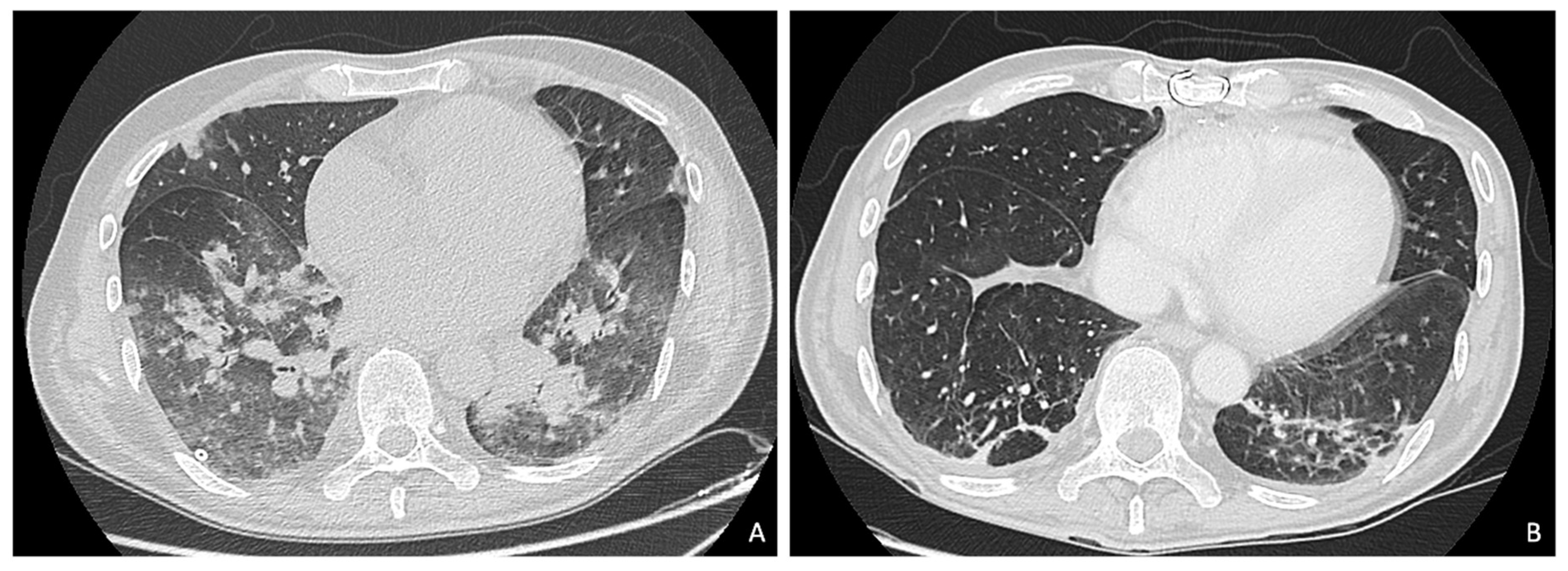
2. Case Report
3. Discussion
4. Conclusions
Supplementary Materials
Author Contributions
Funding
Institutional Review Board Statement
Informed Consent Statement
Data Availability Statement
Conflicts of Interest
Abbreviations
| AR | Aortic regurgitation |
| ARDS | Acute respiratory distress syndrome |
| CDC | Center for Disease Control |
| COVID-19 | COrona VIrus Disease 19 |
| CPAP | Continuous positive airway pressure |
| CT | Computed tomography |
| ICU | Intensive care unit |
| LVEF | Left ventricle ejection fraction |
| MR | Mitral regurgitation |
| NIV | Non-invasive ventilation |
| OR | Operative room |
| OTI | Orotracheal Intubation |
| PCR | Polymerase chain reaction |
| TOE | Trans-oesophageal echocardiography |
| TTE | Transthoracic echocardiography |
References
- Belluschi, I.; De Bonis, M.; Alfieri, O.; Del Forno, B.; Alamanni, F.; Polvani, G.; Pompilio, G.; Roberto, M.; Merlino, L.G.; Troise, G.; et al. First reorganization in Europe of a regional cardiac surgery system to deal with the coronavirus-2019 pandemic. Eur. J. Cardio-Thorac. Surg. 2020, 58, 25–29. [Google Scholar] [CrossRef] [PubMed]
- Elrod, J.K.; Fortenberry, J.L. The Hub-and-spoke organization design: An avenue for serving patients well. BMC Health Serv. Res. 2017, 17, 457. [Google Scholar] [CrossRef] [PubMed]
- Lauer, S.A.; Grantz, K.H.; Bi, Q.; Jones, F.K.; Zheng, Q.; Meredith, H.R.; Azman, A.S.; Reich, N.G.; Lessler, J. The incubation period of coronavirus disease 2019 (COVID-19) from publicly reported con-firmed cases: Estimation and application. Ann. Intern. Med. 2020, 172, 577–582. [Google Scholar] [CrossRef] [PubMed] [Green Version]
- Huang, C.; Wang, Y.; Li, X.; Ren, L.; Zhao, J.; Hu, Y.; Zhang, L.; Fan, G.; Xu, J.; Gu, X.; et al. Clinical features of patients infected with 2019 novel coronavirus in Wuhan, China. Lancet 2020, 395, 497–506. [Google Scholar] [CrossRef] [Green Version]
- Cabrini, L.; Landoni, G.; Bocchino, S.; Lembo, R.; Monti, G.; Greco, M.; Zambon, M.; Colombo, S.; Pasin, L.; Beretta, L.; et al. Long-term survival rate in patients with acute respiratory failure treated with noninvasive ventilation in ordinary wards. Crit. Care Med. 2016, 44, 2139–2144. [Google Scholar] [CrossRef] [PubMed]
- Sartini, C.; Tresoldi, M.; Scarpellini, P.; Tettamanti, A.; Carcò, F.; Landoni, G.; Zangrillo, A. Respiratory Parameters in Patients With COVID-19 After Using Noninvasive Ventilation in the Prone Position Outside the Intensive Care Unit. JAMA 2020, 323, 2338–2340. [Google Scholar] [CrossRef] [PubMed]
- Prendergast, B.; Habib, G.; Lancellotti, P.; Antunes, M.J.; Bonginorni, M.G.; Casalta, J.P.; Del Zotti, F.; Dulgheru, R.; El Khoury, G.; Erba, P.A.; et al. Faculty Opinions recommendation of 2015 ESC Guidelines for the management of infective endocarditis: The Task Force for the Management of Infective Endocarditis of the European Society of Cardiology (ESC). Endorsed by: European Association for Cardio-Thoracic Surgery (EACTS), the European Association of Nuclear Medicine (EANM). Fac. Opin. 2015, 36, 3075–3123. [Google Scholar] [CrossRef]
- Mussini, C.; Falcone, M.; Nozza, S.; Sagnelli, C.; Parrella, R.; Meschiari, M.; Petrosillo, N.; Mastroianni, C.; Cascio, A.; Iaria, C.; et al. Therapeutic strategies for severe COVID-19: A position paper from the Italian Society of Infectious and Tropical Diseases (SIMIT). Clin. Microbiol. Infect. 2020, 12. [Google Scholar] [CrossRef]


Publisher’s Note: MDPI stays neutral with regard to jurisdictional claims in published maps and institutional affiliations. |
© 2021 by the authors. Licensee MDPI, Basel, Switzerland. This article is an open access article distributed under the terms and conditions of the Creative Commons Attribution (CC BY) license (http://creativecommons.org/licenses/by/4.0/).
Share and Cite
Belluschi, I.; Melisurgo, G.; Campana, C.; Castiglioni, A. Successful Treatment of Embolic Aortic Valve Endocarditis in a Patient Affected by COVID-19 Pneumonia. Surgeries 2021, 2, 113-118. https://0-doi-org.brum.beds.ac.uk/10.3390/surgeries2010010
Belluschi I, Melisurgo G, Campana C, Castiglioni A. Successful Treatment of Embolic Aortic Valve Endocarditis in a Patient Affected by COVID-19 Pneumonia. Surgeries. 2021; 2(1):113-118. https://0-doi-org.brum.beds.ac.uk/10.3390/surgeries2010010
Chicago/Turabian StyleBelluschi, Igor, Giulio Melisurgo, Carlo Campana, and Alessandro Castiglioni. 2021. "Successful Treatment of Embolic Aortic Valve Endocarditis in a Patient Affected by COVID-19 Pneumonia" Surgeries 2, no. 1: 113-118. https://0-doi-org.brum.beds.ac.uk/10.3390/surgeries2010010