Article

Histological and Histomorphometric Analyses of Two Bovine Bone Blocks Implanted in Rabbit Calvaria

by
Sergio Alexandre Gehrke
1,2,
Patricia Mazón
3,
Massimo Del Fabbro
4,5,
Margherita Tumedei
6,
Jaime Aramburú Júnior
1,7,
Leticia Pérez-Díaz
8 and
Piedad N. De Aza
2,*
1
Research Department, Biotecnos–Technology and Science, Cuareim 1483, 11100-Montevideo, Uruguay
2
Instituto de Bioingenieria, Universidad Miguel Hernandez, Avda. Ferrocarril s/n. 03202-Elche, (Alicante), Spain
3
Departamento de Materiales, Óptica y Tecnologia Electrónica, Universidad Miguel Hernández, Avda. Universidad s/n, 03202-Elche (Alicante), Spain
4
Department of Biomedical, Surgical and Dental Sciences, Università Degli Studi di Milano, via Commenda 10, 20122-Milan, Italy
5
IRCCS Orthopedic Institute Galeazzi, via R. Galeazzi 4. 20161-Milan, Italy
6
Department of Medical, Oral and Biotechnological Sciences, University of Chieti-Pescara, 66100-Chieti, Italy
7
Department of Medicine Veterinary, Faculty of Itapiranga, Andradas s/n, 89896000-Itapiranga, Brazil
8
Laboratorio de Interacciones Moleculares, Facultad de Ciencias, Universidad de la Republica, 11400-Montevideo, Uruguay
*
Author to whom correspondence should be addressed.
Submission received: 12 February 2019 / Revised: 1 May 2019 / Accepted: 5 May 2019 / Published: 7 May 2019
(This article belongs to the Special Issue Biomaterials and Symmetry)

Abstract

:
This study compared the osteogenic potential of two types of bovine bone blocks. Blocks were obtained by either sintered or a nonsintered process. Calvaria were surgically exposed in 20 rabbits. In each animal, six 0.5-mm-diameter cortical microperforations were drilled with a carbide bur before grafting to promote graft irrigation. The sintered (group 1) and nonsintered (group 2) bovine bone blocks (6 mm diameter, 5 mm high) were bilaterally screwed onto calvarial bone. Blocks were previously prepared from a larger block using a trephine bur. Rabbits were sacrificed after 6 and 8 weeks for the histological and histomorphometric analyses. Samples were processed using the historesin technique. The quantitative and qualitative analyses of the newly formed bone were undertaken using light microscopy. Both groups showed modest new bone formation and remodeling. At the 8-week follow-up, the sintered group displayed significantly lower bone resorption (average of 10% in group 1 and 25% in group 2) and neo-formation (12.86 ± 1.52%) compared to the nonsintered group (16.10 ± 1.29%) at both follow-ups (p < 0.05). One limitation of the present animal model is that the study demonstrates that variations in the physico-chemical properties of the bone substitute material clearly influence the in vivo behavior.

1. Introduction

Bone grafting procedures to correct defects that result from trauma or diseases often require using materials in the block form, which can be shaped and fixed to the receiving defect site [1,2]. Although an autologous graft is considered the gold standard [3,4], certain disadvantages are involved, such as quantity limitation, morbidity, and double surgical intervention, among others [5,6]. In order to address such drawbacks, materials of different origins (allogenic, xenogeneic, or synthetic) have been investigated and used as bone substitutes [7,8,9,10].
These alternative materials have been increasingly used for bone reconstruction, especially for their osteoconductive properties as they provide mechanical support to the bone formation process [11]. Bone substitutes should exhibit a similar macromorphology to the recipient bone structure in terms of internal trabecular architecture, porosity and surface chemistry to provide an adequate environment for osteogenesis-related cellular activity [12]. The material can simply osseointegrate and undergo minimal or no resorption, or can be totally absorbed and replaced with new vital bone tissue. The latter, in addition to adequate mechanical characteristics, offers a microvascular network capable of supplying oxygen and nutrients for tissue metabolism, and also warrants effective immune defense [13,14].
A frequently used alternative for bone grafts is bone of bovine origin. Its structure and chemical composition are similar to human bone tissue, and it also offers the possibility of being produced in large quantities at a relatively low cost [15]. Bovine bone effectively supports new bone formation by achieving osseointegration, although it undergoes very limited resorption. There have been several attempts taken to improve the osteogenic potential of graft materials from biological origin for clinical use [13,16,17,18]. The biological reactions produced using biological graft materials depend, to a significant extent, on its chemical composition, phase purity, and morphology (e.g., particle size, shape, and porosity). Thus, phase modulation of biological graft materials has been attempted through various techniques, e.g., solid state sintering or electrospun [19,20,21].
Though some sintering protocols for bovine bone can preserve the morphology and improve hydrophilicity of the grafts [22], some protocols of production exclude the use of high temperatures (sintering) [23,24]. High-temperature sintering has been demonstrated to increase the density and to reduce the nanoporosity of materials [19,25]. After sintering, a solid scaffold remains and all the remaining proteins are denatured. Sintering bovine bone is a chemico-physical process through which the crystalline size of the material particles can increase, which may further improve the volume stability at the implantation site [13,26].
This study aimed to compare sintered and nonsintered bovine bone blocks by means of histological and histomorphmetrical analyses after 6 and 8 weeks of implantation. The used model was an appositional graft in rabbit calvaria.

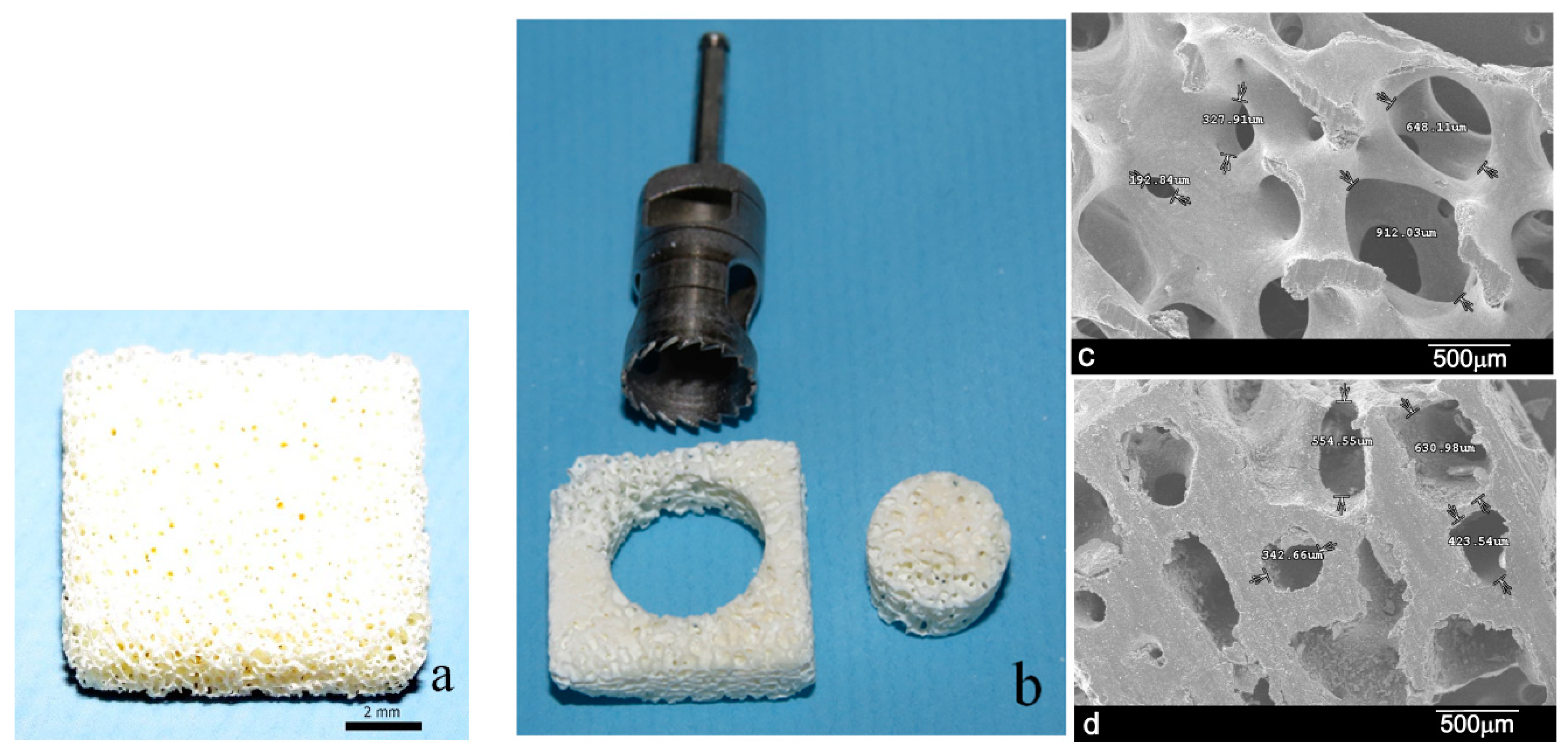
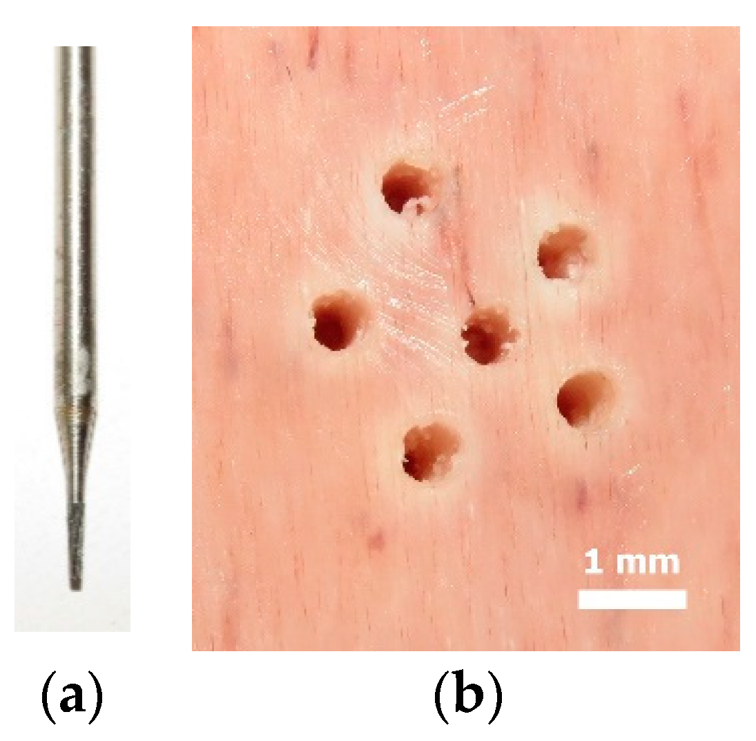
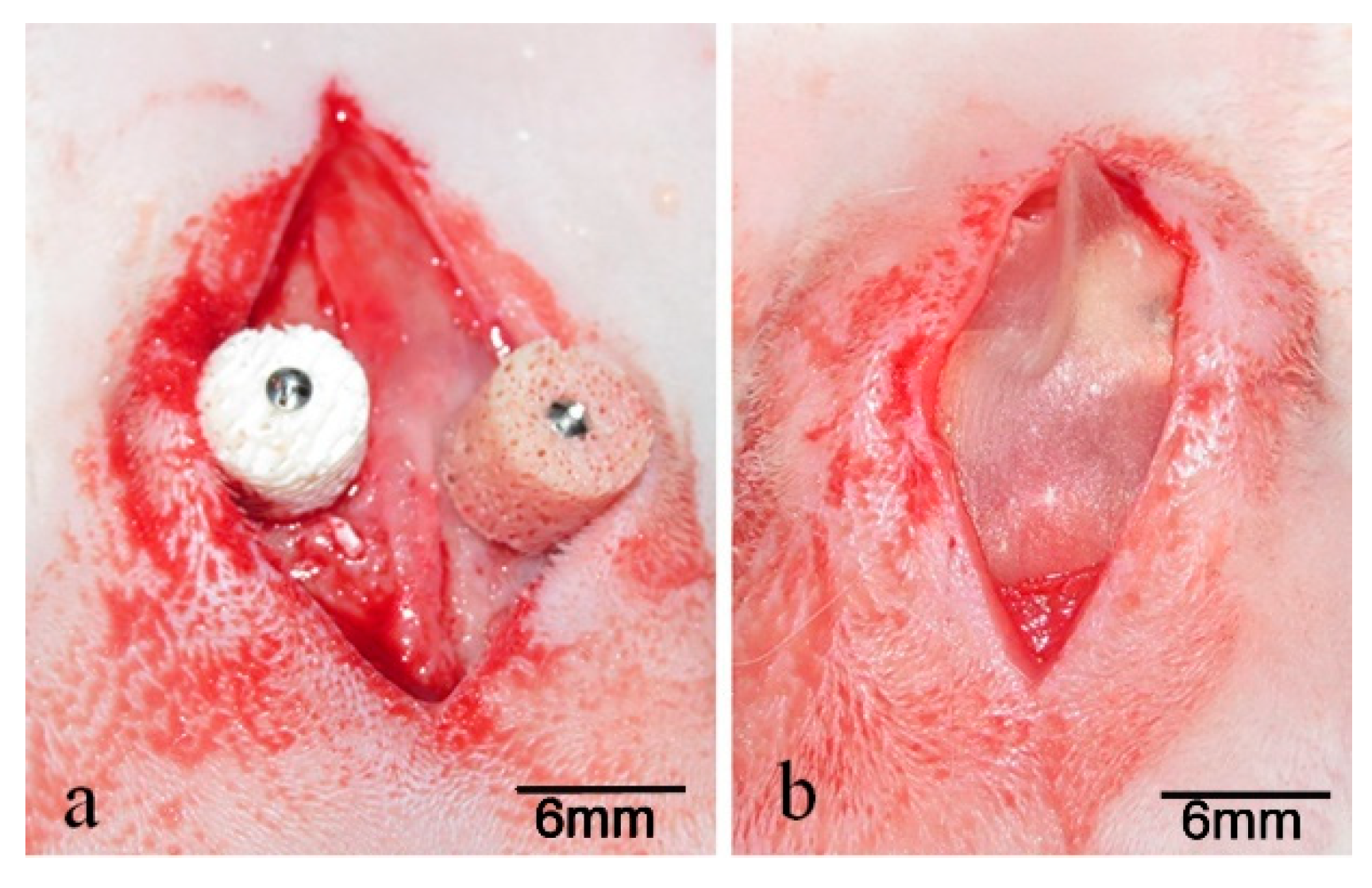
2. Materials and Methods
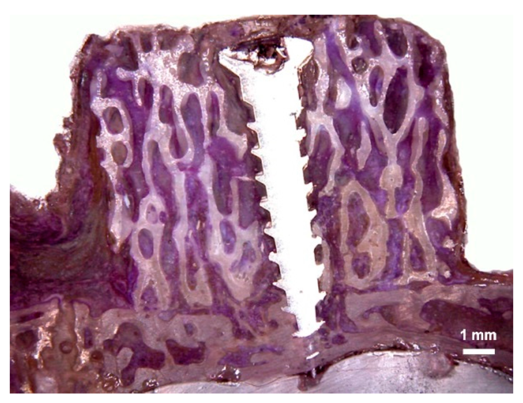
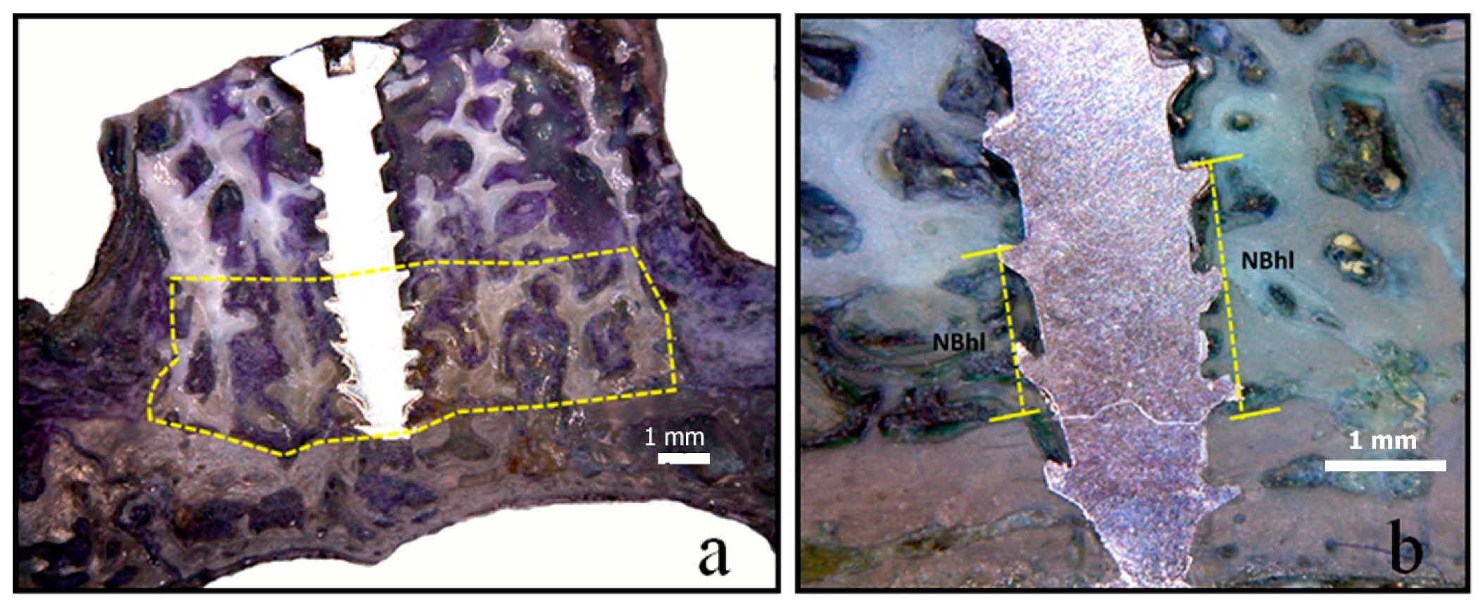
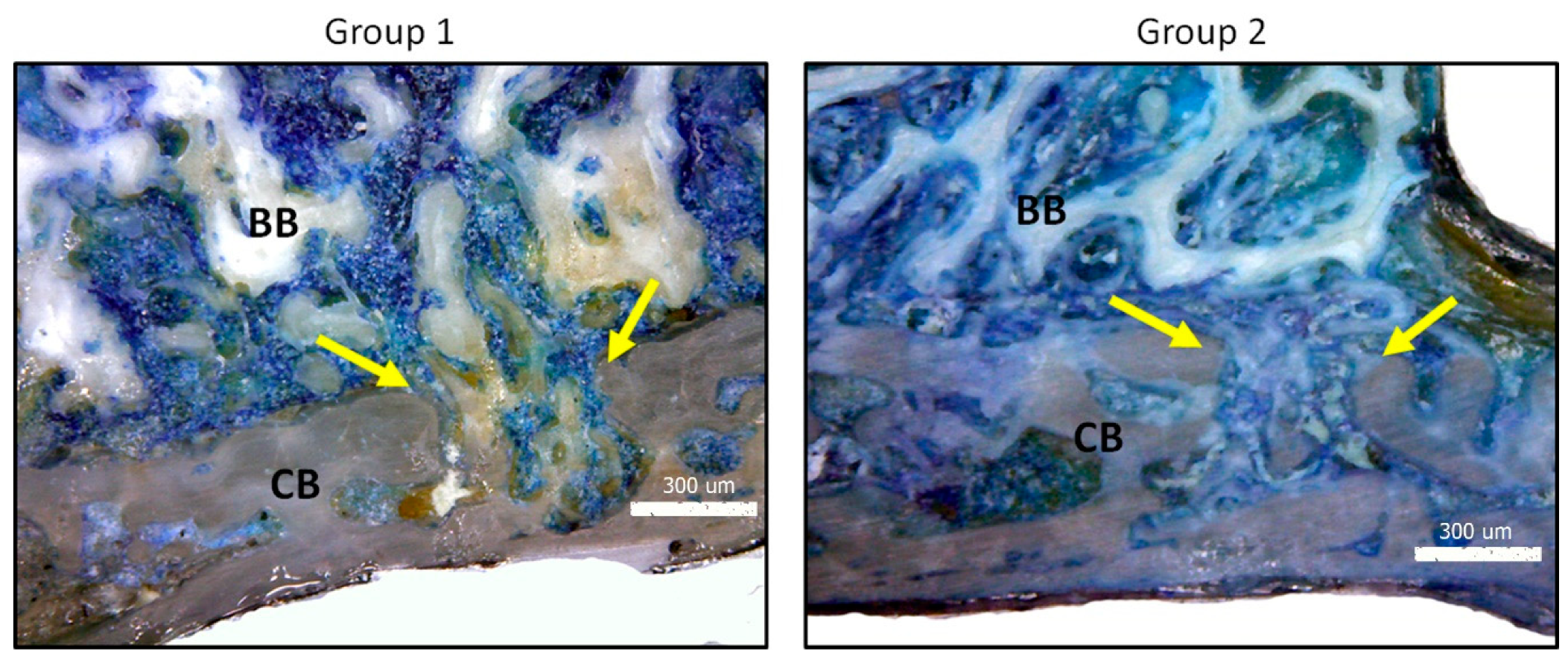
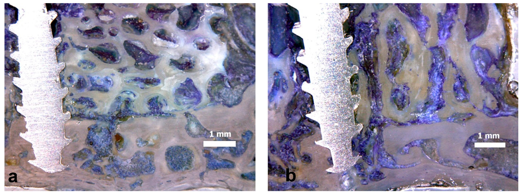
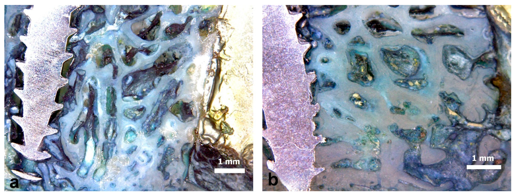
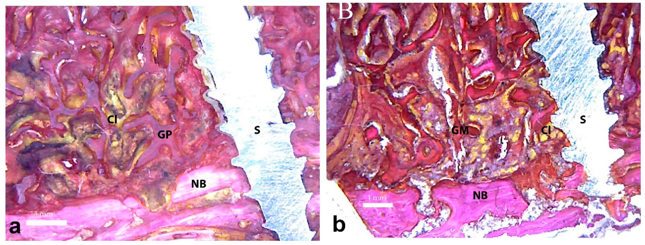
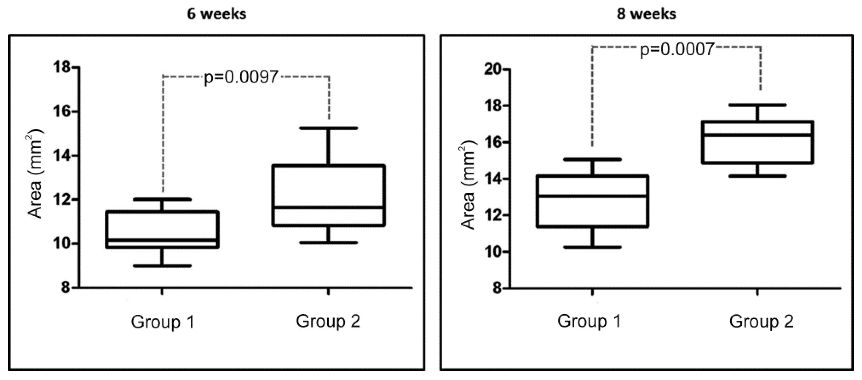
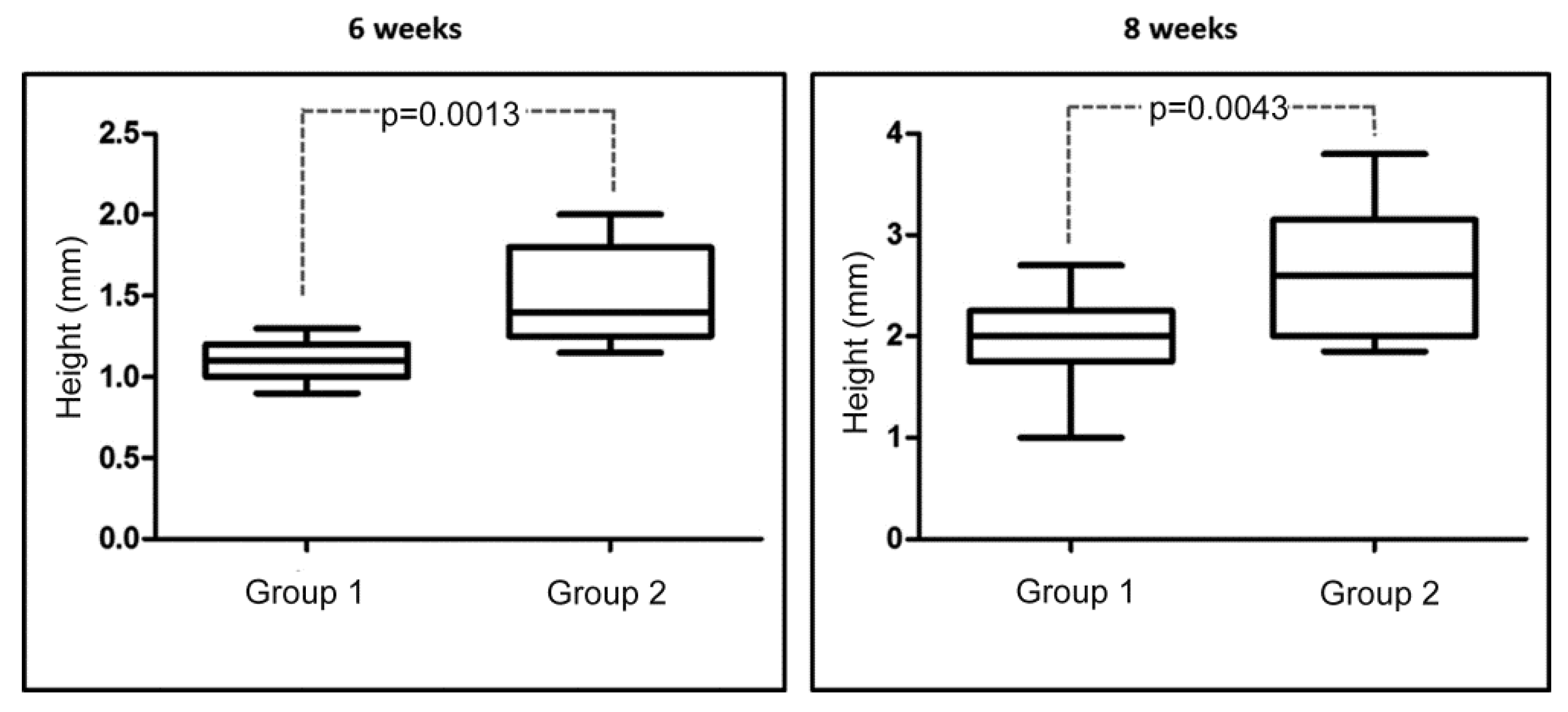
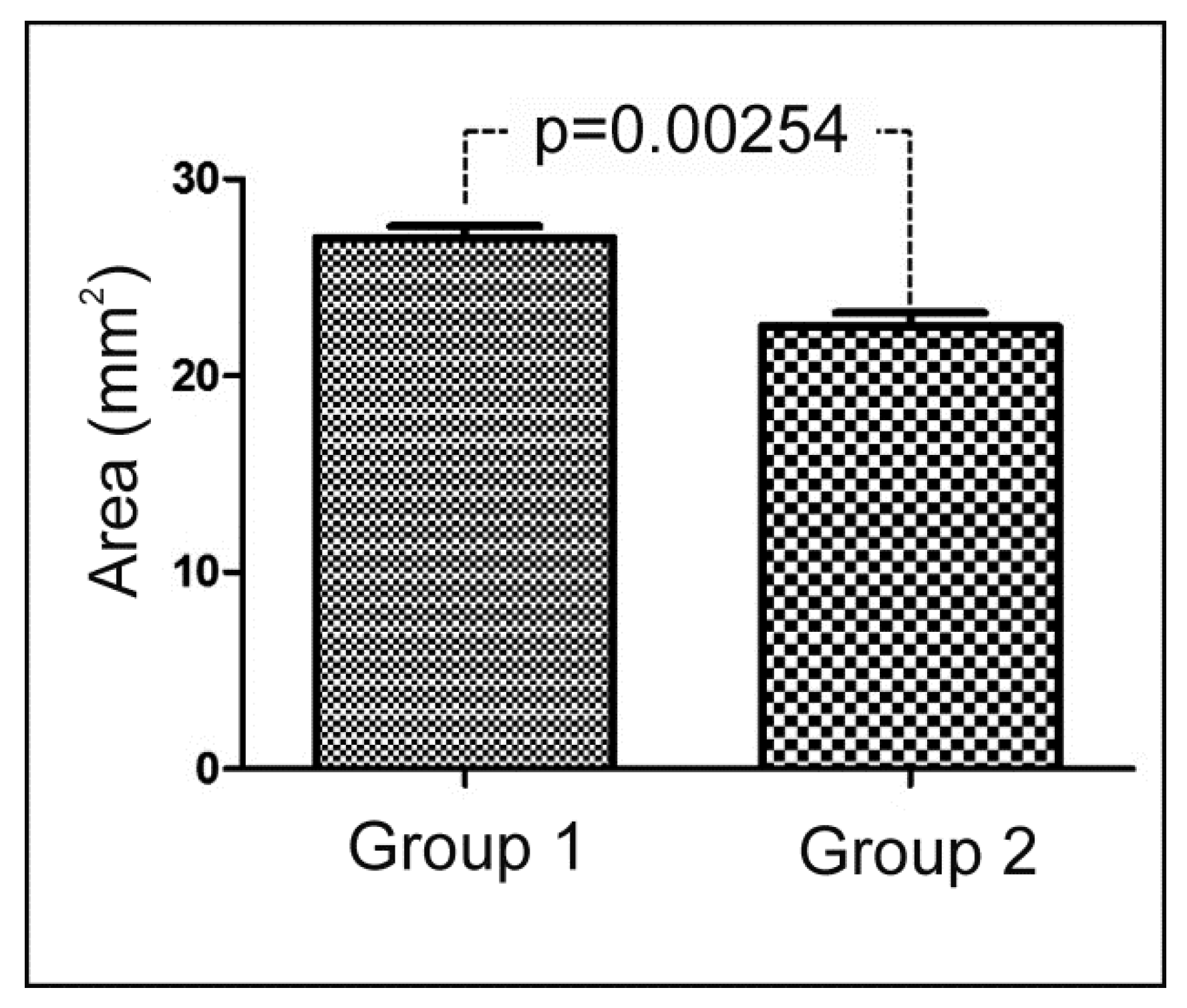
Materials: Two bovine bone blocks were utilized for the appositional bone graft. Group 1 (sintered): the material was submitted to a high temperature for sintering (950 °C), and was then treated with organic solvent and sterilized [20] (Orthogen Bone®, Baumer SA, Mogi-Mirim, Brazil). Specifically, this process removes the organic elements contained in intratrabecular spaces (deproteinization). Group 2 (nonsintered): the material was treated via chemical processing, which removed the vascular elements and adipose tissue contained in intratrabecular spaces, and maintained the content of collagenous proteins (20–25%), and in the mineral part composed of calcium and phosphorus (Lumina Bone®, Critéria Indústria e Comércio de Produtos Medicinais e Odontologicos Ltda, São Carlos, Brazil). Both graft materials were obtained from the femoral epiphysis.
Figure 1a shows the samples’ original form. After opening the blister of each sample, blocks were prepared using a trephine bur, 6 mm in diameter and 5 mm high (Figure 1b). The images obtained using scanning electronic microscopy show how similar the structures of the sintered and nonsintered bone blocks were (Figure 1c,d). The physical characteristics of both materials were evaluated using scanning electron microscopy (SEM, Hitachi S-3500N, Tokyo, Japan). Regarding the physico-chemical structural features, crystallinity, chemical composition, mechanical resistance, porosity, trabecular density, gas and fluid intrusion, cell adhesion, viability, and proliferation of the two employed materials were similar, as a previous study published by our group has shown [25]. The biodegradability test suggested a higher dissolution rate of the nonsintered versus the sintered bovine blocks by day 14.
Animals: Twenty adult white New Zealand (Oryctolagus cuniculus) female rabbits weighing between 3.5 and 4 kg were used. The experiment was performed in accordance with the relevant guidelines and regulations of the Brazilian College of Animal Experimentation (COBEA). The routine management of the Department of Surgery of Small Animals was applied to the animals. This study was approved by the Research Committee at the Faculty of Medicine Veterinary of the University of Itapiranga, Itapiranga, Brazil (#002-09-2015).
Animal surgical management: Before surgery commenced, an intramuscular injection of ketamine (35 mg/kg; Agener Pharmaceutica, São Paulo, Brazil) was utilized as general anesthesia. Rompum muscle relaxant (5 mg/kg; Bayer, Leverkusen, Germany), the Acepran tranquilizer (0.75 mg/kg, Univet, São Paulo, Brazil) and local anesthetic (3% Prilocaine-Felipressine, Astra, Mexico D.F., Mexico) were subcutaneously injected at the surgery site to diminish bleeding. A single dose of (600 000 IU Benzetacil, Bayer, São Paulo, Brazil) was postoperatively administered. In order to control both postoperative swelling and pain, 0.1 mL of ketoprofen was administered daily for 3 days running. Following surgery, animals were individually housed at 21 °C in a 12-h light/dark cycle. They were fed ad libitum standard laboratory diet.
Insertion of graft material: The graft material was positioned on the calvaria bone of each animal. A periosteal elevator was used to elevate subcutaneous and skin tissues. Using an appropriate angled handpiece motor and a 0.5-mm-diameter carbide bur under external irrigation with normal saline solution, in each local block insertion at the receptor site of the calvaria, six cortical microperforations were made (Figure 2) to promote the blood irrigation of grafts.
Each animal received one sintered bovine bone block on the right side and one nonsintered block on the left side of calvaria. After positioning blocks on the perforated calvaria regions, they were fixed (Figure 3a) with a graft screw, which was 7 mm long and 1.5 mm in diameter (Fixation Screw Kit, Salvin Dental Specialties, Charlotte, NC, USA). Then an absorbable collagen membrane, 20 x 30 mm (R.T.R. (resorbable tissue replacement) Membrane, Septodont Inc., Louisville, KY, USA), was used to cover the blocks (Figure 3b). According to the manufacturer, the resorption time of this membrane is 4–8 weeks. Skin was sutured with a 5-0 nylon monofilament. For each group, 20 blocks were positioned (10 blocks per evaluation time). Ten rabbits were sacrificed 6 weeks after surgery, and the other 10 after 8 weeks, with an intravenous injection of a ketamine overdose (2 mL). The block sections of grafts were then taken and immediately processed.
Histological preparation: First, samples were processed after excision for the histological analyses. They were photographed at high resolution using a digital camera (DSC H7, Sony, Tokyo, Japan) for the macroscopic visual bone formation analysis. Then, samples were fixed in 10% formaldehyde solution (72 h), washed under tap water (12 h), and progressively dehydrated in a series of increasing ethanol solution concentrations (60%, 70%, 80%, and 99%) for 24–56 h. Historesin Technovit 7200 VLC (Kultzer & Co, Wehrhein, city, Germany) was used to embed the dehydrated samples, which is a glycol methacrylate solution. Following the polymerization step, a sawing and grinding technique was used to process samples, as previously reported [1]. A cut from each sample (one per block sample) was made in the central region of blocks that corresponded to the center of the screw using a metallographical cutter (Isomet 1000; Buehler, Esslingen am Neckar, Germany) (Figure 4). Then, samples were polished with an abrasive paper (180 to 1200 mesh) sequence (Metaserv 3000; Buehler, Esslingen am Neckar, Germany). Samples were stained using the Toluidine blue staining technique to analyze the new bone formation. Then, the samples 8-week slides were discolored (ethanol solution 99%–50%) and stained again with picrosirius-hematoxylin to analyze the collagen and the vacuolization signals.
Microscopy images (bone tissue sections of ≈30 µm thickness) were visualized using light microscopy (E200, Nikon, Tokyo, Japan,) to investigate the material resorption, bone formation, and bone organization in the cortical portion. The digital image obtained using a camera connected directly to the microscope (Nikon, Tokyo, Japan) was analyzed using version 5.02 of the software Image Tool for Microsoft Windows™. Two authors (SAG and JAJ) took measurements at several times. Next, the average of these values was computed. If measured values were very different from each other in the same slide (difference > 20%), measurements were repeated by both examiners to confirm data.
Histometric analysis: The measured target defect area was located in the center of bone blocks, at a height corresponding to the apical half of the screw, as shown in Figure 5a. The following histomorphometric measurements were taken of the new bone area: the percentage of the newly formed bone area in relation to the block original area (≈30 mm2) and the height of new bone in relation to the screw (Figure 5b).
Finally, a total area of the blocks 30 mm2 (blocks were 5 mm in height and 6 mm in diameter) was considered as 100%, and then, the residual block area after the 8-week period was measured and calculated proportionally from this value.
Histological observations were performed regarding the bone coming into contact with the graft material.
Statistical analysis: The statistical analysis was undertaken using the software GraphPad Prism 5.0 for Windows (GraphPad Software Inc., San Diego, CA, USA). Differences among groups were assessed using Student’s t-test. Differences were considered significant when p < 0.05. The results are expressed as the mean ± one standard deviation.

3. Results

During the postoperative period, no animal presented complications or infection in the operated region. The grafted biomaterials were not exposed in any animal during the healing period until their euthanasia. The periosteum, connective tissue, and musculature were firmly attached to the grafted blocks.
Descriptive histological analysis: An analysis of the histological sections in both groups showed a low resorption of blocks during the 6-week period, and moderate resorption after 8 weeks, by examining from the receptor bed toward the coronal portion of the block. In all the samples, bone regeneration was more intense at the sites where the receptor bed perforations were made (Figure 6).
Analysis of samples after 6 weeks: In the samples from group 1, the osteogenesis process was identified at the edges of the grafted block that came into contact with the receptor bed where the bone matrix was deposited between the trabecular spaces of blocks (Figure 7a). In the samples from group 2, a slightly more intense intra-trabecular filling by the bone matrix was observed compared to group 1, but its proportion was still small in relation to the block size (Figure 7b). On average, the nonsintered material structure showed greater resorption compared to the sintered blocks.
Analysis of samples after 8 weeks: In the samples of group 1, the intra-trabecular bone filling of approximately half the grafted blocks was observed, but the resorption of the original structure of the grafted material was slight (Figure 8a). Conversely in the group 2 samples, the intra-trabecular bone filling of approximately half the grafted block was observed, although the resorption of the original structure of the grafted material was greater compared to the group 1 samples (Figure 8b).
The analyses of the collagen and graft vascularization with the second staining slides in the samples at 8 weeks showed a low presence of these components (collagen and vassels) inside bone blocks in both groups (Figure 9).
Histomorphometric results: The box plot in Figure 10 shows the values measured for the neoformation area measured in both groups at the two follow-up times, and the comparative p-values of the statistical analysis. The box plot in Figure 11 shows the values measured for the bone neoformation height in relation to the fixation screw used to stabilize blocks.
Table 1 reports the mean values, standard deviations, and statistical comparisons between both groups at each time point. Significantly greater new bone formation was observed in the group 2 samples at each follow-up time.
At the end of the study (8 weeks), there was more bone resorption in group 2 with an area mean and standard deviation of 22.5 ± 2.19 mm2 (reduction average of 25%) than in group 1 with 27.0 ± 1.87 mm2 (reduction average of 10%), showing a significative difference between the groups (p = 0.0254). The values measured of the total area of the blocks after 8 weeks are summarized in the graph bar of Figure 12.

4. Discussion

Alternative materials of different origins (allogenic, xenogeneic, and synthetic) have been used in reconstructive surgery [1,2,3]. Bone substitutes overcome the main autogenous grafting drawback, i.e., morbidity. Unless treated properly, they face the potential risks of inducing an immune response and transmitting infectious diseases to patients [27,28,29]. Most bone substitute materials undergo preparation processes to optimize physico-chemical features and to improve biocompatibility. Deproteinization in xenografts is the most important factor for eliminating immunogenic components (organic elements) by thereby reducing the risk of pro-inflammatory reactions.
In veterinary orthopedic surgery, the follow-up clinical evaluations are generally recommended 6 to 8 weeks after surgery [28]. Other authors showed that the period of eight weeks was appropriate to assess late repair, including new bone tissue formation, resorption of the graft material, bone remodeling and bone regeneration [30]. Then, in our study, two times were used (6 and 8 weeks) to verify the evolution of the healing between the different periods. However, other evaluations with greater time periods are necessary for these materials.
The use of xenogeneic bone block grafts for treating atrophic areas has emerged to overcome the complications associated with autologous bone graft surgery [31,32]. Xenografts are obtained from nonhuman species and have osteoconductive properties and limited resorption over time [33,34]. Most organic/antigenic components are removed from xenografts by heating and chemical processes. After these treatments, the inorganic bovine bone phase is comprised chiefly of hydroxyapatite (HA), which retains the porous architecture [19,35]. Sinterization is a process during which the application of higher temperatures than in the chemical process for purification can provide distinct material characteristics (porosity, grain size, mechanical behavior), which might influence both tissue’s reactions to these materials and the associated bone healing process [13,27,29]. However, we underline that not all sintering processes developed by different manufacturers give rise to products with similar features. Therefore, the tissue response to sintering products may differ according to the physico-chemical features of the product itself. In other words, the clinical success depends on many factors (the amount of newly formed bone being only one of them), and can be better, worse, or the same as nonsintered products
The sintered bovine biomaterial regenerative properties in different clinical situations, such as sinus augmentation and containment bone defects, have been extensively investigated in recent years [35,36]. Creating a critical-size defect in healthy bone and then filling it with biomaterial is a conventional model to evaluate the osteogenic potential [37,38,39,40]. The extensive bleeding induced by defect creation causes the graft to be embedded with blood, which is an excellent way to trigger the healing process [41]. A study by Rocha and coworkers evaluated the expressions of both ascular endothelial growth factor (VEGF) and metalloproteinase (MMP-2 and MMP-9) during the healing period of critical-bone size defects that had been treated with sintered anorganic bone (sAB). Sintered anorganic bone generated continuous bone formation among particles throughout all the periods as the peaks of MMP-9, MMP-2, and VEGF took place at 7 and 14 days in fibroblasts and osteoblasts. The physico-chemical properties of sAB enhanced the autocrine expressions of MMP-9, MMP-2, and VEGF, led to bone formation/remodeling, and cranial defects healed well [42].
In the present study, sintered and nonsintered bovine bone grafts were compared by placing the block directly on the cortical bone surface in a rabbit calvaria model without creating a defect at the receiving site, but simply making small cortical microperforations to stimulate blood irrigation to the graft. With this model, which can simulate appositional block grafts in atrophic jaws, the performances of two materials of the same origin were compared, but which underwent different preparation processes. The present results suggest that the type of sinterization process of xenogeneic bone graft used in this study caused relatively modest graft resorption and new bone formation during the study period compared to nonsintered material.
Bovine blocks have a low degradation rate, and therefore, they are osseointegrated by new bone formation, which takes a long time compared to allografts that degrade faster via complete remodeling into patients’ own bones because variations in the production process of bovine bone can cause morphological, chemical, crystallinity, and impurity differences [43].
This fact was confirmed in the present study, where samples displayed low reabsorption during the evaluated time in both groups. However, the sintered group demonstrated lower bone reabsorption (a mean of 10%) and neoformation compared to nonsintered bovine bone (a mean of 25%). Other authors have evidenced the lesser osteogenic capacity of xenogeneic bovine blocks and have also reported articles scientific with evidence for short follow-up times and diversified methodologies, which makes comparing their results difficult [32].
A similar study to the present one, which compared both nonsintered and sintered bovine bone substitute materials in the sinuses of 33 patients, gave comparable new bone formation results [36]. Six months after healing, the new bone area was larger (30.57 ± 16.07) in the non-sintered group versus the sintered group (29.71 ± 13.67). Differences between groups were statistically significant (p = 0.0137). Both the bovine bone substitute materials gave comparable new bone formation results. Another similar study by Panagiotou et al. obtained comparable new bone formation results in the sinuses of eight patients after 8 months of healing [43]. These studies suggest the potential of nonsintered bovine biomaterial as a scaffold in sinus lift surgery. In fact, the new bone area was bigger (29.13 ± 13.81) in the sintered bovine group than in the nonsintered group (24.63 ± 19.76).
The temperatures applied during the sintering process can alter a material’s physical properties, such as pore morphology and size, density, particle size, compressive strength, and torsional force [12]. Moreover, with the change in specific surface area, density, and porosity, a material’s metabolic features (e.g., dissolution and/or resorption) can also be affected [20,25].
In line with Pripatnanont et al.’s study, it is possible to hypothesize that a minimum temperature during the sintering process is needed to improve a material’s features. The lower amount of newly formed bone observed with sintered material compared to nonsintered material in the present study can be attributed to an insufficient temperature used to process the material. In the aforementioned studies, in which the sintered material proved superior or the equivalent to the nonsintered control, the temperatures used for sintering were >1200 °C [36] and 1250 °C [44], while the material used in the present study was sintered at 950 °C. However, more studies are needed to clarify this theme.
The results of this study demonstrate that bone formation inside both the tested bovine bone blocks was lower than expected for the healing time in this animal model. Thus, in a clinical scenario, the professional who would use these materials should wait longer before placing such materials after grafting.

5. Conclusions

Despite some limitations, this study displayed major differences between sintered and nonsintered bovine bone blocks as sintered blocks showed less bone regeneration than nonsintered blocks, although the sintered blocks had higher volume stability. Based on this result, the type of sintering process can be critical for the tissue response to graft materials and needs to be cautiously considered when choosing a sintered material as an alternative to autogenous bone grafts for clinical application

Author Contributions

Data curation, M.D.F.; Formal analysis, S.A.G., M.T., J.A.-J., L.P.-D., and P.N.D.A.; Funding acquisition, P.N.D.A.; Investigation, S.A.G., P.M., M.T., J.A.-J., L.P.-D., and P.N.D.A.; Methodology, S.A.G., M.T., J.A.-J., and L.P.-D.; Supervision, P.M.; Validation, J.A.-J.; Writing–original draft, S.A.G. and M.D.F.

Funding

This research was funded by the Spanish Ministry of Economy and Competitiveness (MINECO), grant number MAT2013-48426-C2-2-R.

Conflicts of Interest

The authors declare that they have no conflict of interest.

References

  1. Mate-Sanchez de Val, J.E.; Calvo-Guirado, J.L.; Delgado-Ruiz, R.A.; Ramirez-Fernandez, M.P.; Martinez, I.M.; Granero-Marin, J.M.; Negri, B.; Chiva-Garcia, F.; Martinez-Gonzalez, J.M.; De Aza, P.N. New block graft of α-TCP with silicon in critical size defects in rabbits: Chemical characterization, histological, histomorphometric and micro-CT study. Ceram. Int. 2012, 38, 1563–1570. [Google Scholar] [CrossRef]
  2. Velasquez, P.; Luklinska, Z.B.; Meseguer-Olmo, L.; Mate-Sanchez de Val, J.E.; Delgado-Ruiz, R.A.; Calvo-Guirado, J.L.; Ramirez-Fernandez, M.P.; De Aza, P.N. αTCP ceramic doped with Dicalcium Silicate for bone regeneration applications prepared by powder metallurgy method. In vitro and in vivo studies. J. Biomed. Mater. Res. A 2013, 101, 1943–1954. [Google Scholar] [CrossRef] [PubMed]
  3. Samartzis, D.; Shen, F.H.; Goldberg, E.J.; An, H.S. Is autograft the gold standard in achieving radiographic fusion in one-level anterior cervical discectomy and fusion with rigid anterior plate fixation? Spine 2005, 30, 1756–1761. [Google Scholar] [CrossRef] [PubMed]
  4. Bauer, T.W.; Muschler, G.F. Bone graft materials: An overview of the basic science. Clin. Orthop. Relat. Res. 2000, 371, 10–27. [Google Scholar] [CrossRef]
  5. Parrilla-Almansa, A.; García-Carrillo, N.; Ros-Tárraga, P.; Martínez, C.M.; Martínez-Martínez, F.; Meseguer-Olmo, L.; De Aza, P.N. Demineralized Bone Matrix Coating Si-Ca-P Ceramic Does Not Improve the Osseointegration of the Scaffold. Materials 2018, 11, 1580. [Google Scholar] [CrossRef]
  6. Lei, P.; Sun, R.; Wang, L.; Zhou, J.; Wan, L.; Zhou, T.; Hu, Y. A New Method for Xenogeneic Bone Graft Deproteinization: Comparative Study of Radius Defects in a Rabbit Model. PLoS ONE 2015, 10, e0146005. [Google Scholar] [CrossRef]
  7. Calvo-Guirado, J.L.; Ramírez-Fernández, M.P.; Delgado-Ruíz, R.; Maté-Sánchez, J.E.; Velasquez, P.; De Aza, P.N. Influence of Biphasic β-TCP with and without the use of collagen membranes on bone healing of surgically critical size defects. A radiological, histological, and histomorphometric study. Clin. Oral Implants Res. 2014, 25, 1228–1238. [Google Scholar] [CrossRef]
  8. Tomford, W.W. Transmission of disease through transplantation of musculoskeletal allografts. JBJS 1995, 77, 1742–1754. [Google Scholar] [CrossRef]
  9. Carrodeguas, R.G.; De Aza, A.H.; De Aza, P.N.; Baudin, C.; Jiménez, J.; Lopez-Bravo, A.; Pena, P.; De Aza, S. Assessment of natural and synthetic wollastonite as source for bioceramics preparation. J. Biomed. Mater. Res. A 2007, 83, 484–495. [Google Scholar] [CrossRef]
  10. Roberts, T.T.; Rosenbaum, A.J. Bone grafts, bone substitutes and orthobiologics: The bridge between basic science and clinical advancements in fracture healing. Organogenesis 2012, 8, 114–124. [Google Scholar] [CrossRef]
  11. Mate-Sanchez de Val, J.E.; Calvo-Guirado, J.L.; Delgado-Ruiz, R.A.; Ramirez-Fernandez, M.P.; Negri, B.; Abboud, M.; Martinez, I.M.; De Aza, P.N. Physical properties, mechanical behavior, and electron microscopy study of a new α-tcp block graft with silicon in an animal model. J. Biomed. Mater. Res. A 2012, 100, 3446–3454. [Google Scholar] [CrossRef]
  12. Meyer, U.; Joos, U.; Wiesmann, H.P. Biological and biophysical principles in extracorporal bone tissue engineering. Part I. Int. J. Oral Maxillofac. Surg. 2004, 33, 325–332. [Google Scholar] [CrossRef]
  13. Ramírez Fernández, M.P.; Mazón, P.; Gehrke, S.A.; Calvo Guirado, J.L.; De Aza, P.N. Comparison of two xenograft materials used in sinus lift procedures. Material characterization and in vivo behavior. Materials 2017, 10, 623. [Google Scholar] [CrossRef]
  14. Yildirim, M.; Spiekermann, H.; Biesterfeld, S.; Edelhoff, D. Maxillary sinus augmentation using xenogenic bone substitute material bio-oss in combination with venous blood. A histologic and histomorphometric study in humans. Clin. Oral Implant. Res. 2000, 11, 217–229. [Google Scholar] [CrossRef]
  15. Salama, R. Xenogeneic bone grafting in humans. Clin. Orthop. Relat. Res. 1983, 174, 113–121. [Google Scholar] [CrossRef]
  16. Ramirez-Fernandez, M.P.; Gehrke, S.A.; Mazon, P.; Calvo-Guirado, J.L.; De Aza, P.N. Implant stability of biological hydroxyapatites used in dentistry. Materials 2017, 10, 644. [Google Scholar] [CrossRef] [PubMed]
  17. Guarnieri, R.; Belleggia, F.; De Villier, P.; Testarelli, L. Histologic and Histomorphometric Analysis of Bone Regeneration with Bovine Grafting Material after 24 Months of Healing. A Case Report. J. Funct. Biomater. 2018, 9, 48. [Google Scholar] [CrossRef]
  18. Scarano, A.; Inchingolo, F.; Murmura, G.; Traini, T.; Piattelli, A.; Lorusso, F. Three-Dimensional Architecture and Mechanical Properties of Bovine Bone Mixed with Autologous Platelet Liquid, Blood, or Physiological Water: An In Vitro Study. Int. J. Mol. Sci. 2018, 19, 1230. [Google Scholar] [CrossRef]
  19. Maté Sánchez de Val, J.; Mazón, P.; Piattelli, A.; Calvo-Guirado, J.L.; Mareque Bueno, J.; Granero Marín, J.; De Aza, P.N. Comparison among the physical properties of calcium phosphate-based bone substitutes of natural or synthetic origin. Int. J. Appl. Ceram. Technol. 2018, 15, 930–937. [Google Scholar] [CrossRef]
  20. Cestari, T.M.; Granjeiro, J.M.; de Assis, G.F.; Garlet, G.P.; Taga, R. Bone repair and augmentation using block of sintered bovine-derived anorganic bone graft in cranial bone defect model. Clin. Oral Implant. Res. 2009, 20, 340–350. [Google Scholar] [CrossRef] [PubMed]
  21. Taylor, B.L.; Limaye, A.; Yarborough, J.; Freeman, J.W. Investigating processing techniques for bovine gelatin electrospun scaffolds for bone tissue regeneration. J. Biomed. Mater. Res. Part B Appl. Biomater. 2017, 105, 1131–1140. [Google Scholar] [CrossRef]
  22. Trajkovski, B.; Jaunich, M.; Müller, W.-D.; Beuer, F.; Zafiropoulos, G.-G.; Housmand, A. Hydrophilicity, Viscoelastic, and Physicochemical Properties Variations in Dental Bone Grafting Substitutes. Materials 2018, 11, 215. [Google Scholar] [CrossRef]
  23. Sheikh, Z.; Sima, C.; Glogauer, M. Bone Replacement Materials and Techniques Used for Achieving Vertical Alveolar Bone Augmentation. Materials 2015, 8, 2953–2993. [Google Scholar] [CrossRef]
  24. Kačarević, Z.P.; Kavehei, F.; Houshmand, A.; Franke, J.; Smeets, R.; Rimashevskiy, D.; Wenisch, S.; Schnettler, R.; Barbeck, O. Purification processes of xenogeneic bone substitutes and their impact on tissue reactions and regeneration. Int. J. Artif. Organs 2018, 41, 789–800. [Google Scholar] [CrossRef]
  25. Gehrke, S.A.; Mazón, P.; Pérez-Díaz, L.; Calvo Guirado, J.L.; Velásquez, P.; Aragoneses, J.M.; Fernández-Domínguez, M.; De Aza, P.M. Study of Two Bovine Bone Blocks (Sintered and Not-Sintered) Used for Bone Grafts: Physico-Chemical Characterization and In Vitro bioactivity and Cellular Analysis. Materials 2018, 12, 452. [Google Scholar] [CrossRef]
  26. Ramirez-Fernandez, M.P.; Gehrke, S.A.; Pérez Albacete Martinez, C.; Calvo-Guirado, J.L.; De Aza, P.N. SEM-EDX study of the degradation process of two xenograft materials used in sinus lift procedures. Materials 2017, 10, 542. [Google Scholar] [CrossRef]
  27. Chappard, D.; Fressonnet, C.; Genty, C.; Baslé, M.F.; Rebel, A. Fat in bone xenografts: Importance of the purification procedures on cleanliness, wettability and biocompatibility. Biomaterials 1993, 14, 507–512. [Google Scholar] [CrossRef]
  28. Robinson, D.A. Orthopedic Follow-Up Evaluations: Identifying Complications. Today’s Vet. Pract. 2014, 9–10, 71–79. [Google Scholar]
  29. De Aza, P.N.; De Aza, A.H.; Herrera, A.; Lopez-Prats, F.A.; Pena, P. Influence of sterilization techniques on the in vitro bioactivity of pseudowollastonite. J. Am. Ceram. Soc. 2016, 89, 2619–2624. [Google Scholar] [CrossRef]
  30. Sohn, J.-Y.; Park, J.-C.; Um, Y.-J.; Jung, U.W.; Kim, C.S.; Cho, K.S.; Choi, S.H. Spontaneous healing capacity of rabbit cranial defects of various sizes. J. Periodont. Implant Sci. 2010, 40, 180–187. [Google Scholar] [CrossRef]
  31. Felice, P.; Marchetti, C.; Iezzi, G.; Piattelli, A.; Worthington, H.; Pellegrino, G.; Esposito, M. Vertical ridge augmentation of the atrophic posterior mandible with interpositional block graft: Bone from the iliac crest vs bovine anorganic bone. Clinical and histological result up to one year after loading from a randomized-controlled clinical trial. Clin. Oral Implant. Res. 2009, 20, 1386–1393. [Google Scholar] [CrossRef] [PubMed]
  32. Araújo, P.P.; Oliveira, K.P.; Montenegro, S.C.; Carreiro, A.F.; Silva, J.S.; Germano, A.R. Block allograft for reconstruction of alveolar bone ridge in implantology: A systematic review. Implant Dent. 2013, 22, 304–308. [Google Scholar] [CrossRef]
  33. Thaller, S.R.; Hoyt, J.; Borjeson, K.; Dart, A.; Tesluk, H. Reconstruction of calvarial defects with anorganic bovine bone mineral (Bio-Oss) in a rabbit model. J. Craniofac. Surg. 1993, 4, 79–84. [Google Scholar] [CrossRef] [PubMed]
  34. McAllister, B.S.; Margolin, M.D.; Cogan, A.G.; Buck, D.; Hollinger, J.O.; Lynch, S.E. Eighteen-month radiographic and histologic evaluation of sinus grafting with anorganic bovine bone in the chimpanzee. Int. J. Oral Maxillofac. Implant. 1999, 14, 361–368. [Google Scholar]
  35. Mate-Sanchez de Val, J.E.; Calvo-Guirado, J.L.; Gomez Moreno, G.; Perez Albacete-Martinez, C.; Mazón, P.; de Aza, P.N. Influence of hydroxyapatite granule size, porosity and crystallinity on tissue reaction in vivo. Part A: Synthesis, characterization of the materials and SEM analysis. Clin. Oral Implant. Res. 2016, 27, 1331–1338. [Google Scholar] [CrossRef] [PubMed]
  36. Fienitz, T.; Moses, O.; Klemm, C.; Happe, A.; Ferrari, D.; Kreppel, M.; Ormianer, Z.; Gal, M.; Rothamel, D. Histological and radiological evaluation of sintered and non-sintered deproteinized bovine bone substitute materials in sinus augmentation procedures. A prospective, randomized-controlled, clinical multicenter study. Clin. Oral Investig. 2017, 21, 787–794. [Google Scholar] [CrossRef] [PubMed]
  37. Stacchi, C.; Lombardi, T.; Oreglia, F.; Alberghini Maltoni, A.; Traini, T. Histologic and Histomorphometric Comparison between Sintered Nanohydroxyapatite and Anorganic Bovine Xenograft in Maxillary Sinus Grafting: A Split-Mouth Randomized Controlled Clinical Trial. Biomed Res. Int. 2017, 1, 9489825. [Google Scholar] [CrossRef]
  38. Muschler, G.F.; Raut, V.P.; Patterson, T.E.; Wenke, J.C.; Hollinger, J.O. The design and use of animal models for translational research in bone tissue engineering and regenerative medicine. Tissue Eng. Part B Rev. 2010, 16, 123–145. [Google Scholar] [CrossRef] [PubMed]
  39. Gomes, P.S.; Fernandes, M.H. Rodent models in bone-related research: The relevance of calvarial defects in the assessment of bone regeneration strategies. Lab. Anim. 2011, 45, 14–24. [Google Scholar] [CrossRef]
  40. Peric, M.; Dumic-Cule, I.; Grcevic, D.; Matijasic, M.; Verbanac, D.; Paul, R.; Grgurevic, L.; Trkulja, V.; Bagi, C.M.; Vukicevic, S. The rational use of animal models in the evaluation of novel bone regenerative therapies. Bone 2015, 70, 73–86. [Google Scholar] [CrossRef]
  41. Hassanein, A.H.; Clune, J.E.; Mulliken, J.B.; Arany, P.R.; Rogers, G.F.; Kulungowski, A.M.; Greene, A.K. Effect of calvarial burring on resorption of onlay cranial bone graft. J. Craniofac. Surg. 2012, 23, 1495–1498. [Google Scholar] [CrossRef]
  42. Rocha, C.A.; Cestari, T.M.; Vidotti, H.A.; de Assis, G.F.; Garlet, G.P.; Taga, R. Sintered anorganic bone graft increases autocrine expression of VEGF, MMP-2 and MMP-9 during repair of critical size bone defects. J. Mol. Histol. 2014, 45, 447–461. [Google Scholar] [CrossRef] [PubMed]
  43. Panagiotou, D.; Ozkan, K.E.; Dirikan, I.S.; Cakar, G.; Olgac, V.; Yilmaz, S. Comparison of two different xenografts in bilateral sinus augmentation: Radiographic and histologic findings. Quintessence Int. 2015, 46, 611–619. [Google Scholar] [PubMed]
  44. Pripatnanont, P.; Nuntanaranont, T.; Vongvatcharanon, S.; Limlertmongkol, S. Osteoconductive Effects of 3 Heat-Treated Hydroxyapatites in Rabbit Calvarial Defects. J. Oral Maxillofac. Surg. 2007, 65, 2418–2424. [Google Scholar] [CrossRef] [PubMed]
Figure 1. (a) The original form of the used samples, and (b) block preparation using a trephine bur (6 mm in diameter). Scanning electronic microscopy shows the similar structure of the two bone blocks: (c) group 1 (sintered) and (d) group 2 (nonsintered).
Symmetry 11 00641 g001
Figure 2. (a) The carbide bur used to make the microperforations, and (b) the six microperforations performed in calvaria bone to promote the irrigation of blocks.
Symmetry 11 00641 g002
Figure 3. (a) Fixation of blocks in calvaria, and (b) the absorbable collagen membrane positioned to cover blocks.
Symmetry 11 00641 g003
Figure 4. Image of the sample after making a cut in the center of the screw, which corresponded to the center of the block.
Symmetry 11 00641 g004
Figure 5. Images of the new bone area (a) and the new bone growth height measurement in relation to the screw (b).
Symmetry 11 00641 g005
Figure 6. Images of the group samples at 6 weeks showing the more intense bone reaction in the areas where perforations (yellow arrows) were made in cortical bone (CB) to promote irrigation to bone blocks (BB).
Symmetry 11 00641 g006
Figure 7. Images of samples at 6 weeks: (a) group 1, slight block structure resorption can be seen; and (b) group 2, somewhat greater block structure resorption is present versus group 1.
Symmetry 11 00641 g007
Figure 8. Images of samples at 8 weeks: (a) group 1, the image shows minor bone neoformation and resorption of the block structure; and (b) group 2, the image displays more intense new bone formation and resorption of the block structure.
Symmetry 11 00641 g008
Figure 9. Images of samples at 8 weeks: (a) group 1 and (b) group 2. Both images showing a low quantity of collagen, and consequently, poor vascularization. S = screw; NB = native bone; GP = graft material; Cl = collagen.
Symmetry 11 00641 g009
Figure 10. Comparative graph of the measured new bone formation area in blocks, with the p-value of the comparison made between both groups at 6 and 8 weeks, respectively. The limits of the boxes correspond to the 25th and 75th percentiles, and the horizontal line in the box represents the mean value. The minimum and maximum values are also indicated by horizontal bars.
Symmetry 11 00641 g010
Figure 11. Comparative graph of the measured height of the new bone formation around the screw, with the p-value of the comparison made between both groups at 6 and 8 weeks, respectively. The limits of the boxes correspond to the 25th and 75th percentiles, and the horizontal line in the box represents the mean value. The minimum and maximum values are also indicated by horizontal bars.
Symmetry 11 00641 g011
Figure 12. Comparative graph of the measured residual block area after 8-weeks, with the p-value of the comparison made between both groups.
Symmetry 11 00641 g012
Table 1. Collected data of the area and height of the new bone formation in both groups at the two times points.
hNBgmANBf
6 Weeks8 Weeks6 Weeks8 Weeks
Group 11.12 ± 0.132.07 ± 0.3310.42 ± 1.0212.86 ± 1.52
Group 21.48 ± 0.292.76 ± 0.6112.06 ± 1.6716.10 ± 1.29
p value0.0097 *0.0007 *0.0013 *0.0043 *
CI 95% −0.6 to −0.2−1.1 to −0.3−2.8 to −0.5−4.7 to −1.8
mANBf = measured new bone formation area; hNBg = height of new bone growth; CI = confidence interval; * = statistically significant (p < 0.05).

