Article

Anti-Biofilm Activity of the Fungal Phytotoxin Sphaeropsidin A against Clinical Isolates of Antibiotic-Resistant Bacteria

1
Dipartimento di Medicina Molecolare e Biotecnologie Mediche, Università di Napoli Federico II, Via Pansini 5, 80131 Naples, Italy
2
Dipartimento di Scienze Chimiche, Università di Napoli Federico II, Complesso Universitario Monte S. Angelo, Via Cintia 4, 80126 Napoli, Italy
3
Dipartimento di Biologia, Università di Napoli Federico II, Complesso Universitario Monte S. Angelo, Via Cintia 4, 80126 Napoli, Italy
4
Dipartimento di Agraria, Sezione di Patologia Vegetale ed Entomologia, Università degli Studi di Sassari, Viale Italia 39, 07100 Sassari, Italy
*
Authors to whom correspondence should be addressed.
Submission received: 27 May 2020 / Revised: 2 July 2020 / Accepted: 6 July 2020 / Published: 8 July 2020
(This article belongs to the Special Issue Microbial and Plant Phytotoxins)

Abstract

:
Many pathogens involved in human infection have rapidly increased their antibiotic resistance, reducing the effectiveness of therapies in recent decades. Most of them can form biofilms and effective drugs are not available to treat these formations. Natural products could represent an efficient solution in discovering and developing new drugs to overcome antimicrobial resistance and treat biofilm-related infections. In this study, 20 secondary metabolites produced by pathogenic fungi of forest plants and belonging to diverse classes of naturally occurring compounds were evaluated for the first time against clinical isolates of antibiotic-resistant Gram-negative and Gram-positive bacteria. epi-Epoformin, sphaeropsidone, and sphaeropsidin A showed antimicrobial activity on all test strains. In particular, sphaeropsidin A was effective at low concentrations with Minimum Inhibitory Concentration (MIC) values ranging from 6.25 μg/mL to 12.5 μg/mL against all reference and clinical test strains. Furthermore, sphaeropsidin A at sub-inhibitory concentrations decreased methicillin-resistant S. aureus (MRSA) and P. aeruginosa biofilm formation, as quantified by crystal violet staining. Interestingly, mixtures of sphaeropsidin A and epi-epoformin have shown antimicrobial synergistic effects with a concomitant reduction of cytotoxicity against human immortalized keratinocytes. Our data show that sphaeropsidin A and epi-epoformin possess promising antimicrobial properties.
Key Contribution: Twenty secondary metabolites produced by pathogenic fungi of forest plants were evaluated for the first time against clinical isolates of antibiotic-resistant Gram-negative and Gram-positive bacteria. Among them, the phytotoxic compounds epi-epoformin, sphaeropsidone, and sphaeropsidin A showed antimicrobial activity on all test strains.

1. Introduction

In the last century, the use of antibiotics has played a fundamental role in extending the average human life. They allow us to treat trivial or serious infections and to carry out complex medical and surgical procedures that would otherwise result in very high mortality. However, the excessive or inappropriate use of antibiotics, in human medicine but also zootechnics and agriculture, results in bacterial strains possessing increased antibiotic resistance and isolation frequency.
The ability to transfer resistance determinants horizontally makes the bacterium frequently acquire resistance to multiple classes of drugs and consequently infection treatment becomes complicated. Indeed, multidrug-resistant bacteria are currently considered a global health challenge.
On the other hand, the development of new drugs is very slow due to lack of investment. The alarming epidemiological data prompted the WHO to predict that after 2050 deaths from previously treatable infections will be 10 million per year if no action is taken [1]. Increased antibiotic resistance has reduced the effectiveness of therapies by promoting the persistence of infections. The chronicity of many infections is also promoted by the ability of multiple pathogens to form biofilms on biotic or abiotic surfaces [2,3,4]. A biofilm is a mono- or polymicrobial community of cells embedded in an exopolysaccharide matrix. Within the biofilm, bacteria may be less susceptible to both the effectors of the immune response and antimicrobial drugs, so persistence of the infection is favored. The WHO has estimated that about 80% of chronic infections are related to the formation of biofilms. Many of the drugs currently available hardly penetrate the biofilm and the bacteria in the biofilm are 10–1000 times more resistant than the planktonic counterpart [5].
In this scenario, alternative strategies to conventional antimicrobial therapies are necessary and urgent. Among them, photodynamic therapy (PDT), first considered for the treatment of specific types of cancer [6], has been paid increasing attention as an innovative treatment to eradicate localized infections, supported by antibiotic-resistant biofilm-producing bacteria. [7,8]. Various studies report strategies to prevent biofilm formation on medical devices such as catheters, sutures, stents and bone cement [9,10,11,12]. Several researchers are involved in the design and evaluation of specific molecules with potential therapeutic use in chronic and biofilm-related infections [13,14]. In particular, antimicrobial peptides (AMPs), a small bioactive protein consisting of 12–50 amino acids, seem to have emerged as promising active agents against bacteria, viruses, fungi, and also as potential chemotherapeutic agents [15,16,17,18].
Plants and microorganisms have always been an invaluable source of secondary metabolites (SM) that could represent an efficient solution to this problem. SM are usually low molecular weight organic compounds produced by various organisms through the action of different enzymes. These specialized metabolites are often not essential for the growth, development, or reproduction of those organisms producing them, but they could be very important for functions such as protection, competition, and species interactions. Most of the SM isolated from microorganisms and plants have been shown to possess a broad spectrum of biological activities including antimicrobial properties. Furthermore, SM belong to diverse structural classes of naturally occurring compounds and have different mechanisms of action, a potential for the development of new drugs to overcome antimicrobial resistance and to treat biofilm-related infections [19,20,21,22,23].
Among the terrestrial ecosystems, forests represent an enormous reservoir of pathogenic and endophytic fungi, which have been studied for several years to evaluate their ability to biosynthesize phytotoxic metabolites. However, several SM produced by these organisms also possess other biological activities (including antibacterial properties) and most of them have shown potential applications in other fields such as medicine and agriculture [24].
Thus, in our continuing effort to find new natural antibacterial metabolites, 20 secondary metabolites produced by pathogenic fungi of forest plants and belonging to different classes of naturally occurring compounds, such as butenolides, cyclohexen oxides, diterpenes, isobenzofuranones, isocoumarins, macrolides, etc., were evaluated for the first time against reference and clinical strains of antibiotic-resistant staphylococci and P. aeruginosa.

2. Results and Discussion

The secondary metabolites assayed in this study (120, Figure 1) were isolated as phytotoxins produced by different fungal genera responsible for forest plant diseases such as Diplodia, Seiridium, Biscogniauxia, Sardiniella and Hymenoscyphus (Table 1) and are potentially involved in plant pathogenesis [25,26,27,28,29,30,31,32,33,34,35,36].
However, most of these compounds also showed other interesting biological activities as reported in detail in a recent review [24]. In particular, some of them have already been reported for their antifungal activity, such as cyclopaldic acid, epi-epoformin, sphaerosidins A–C, sphaeropsidone and (R)-mellein [24,25,26,30,37,38]. In addition, 1 and 2 were able to inhibit the development of two major rust fungi in agrarian crops P. triticina and U. pisi [39,40]. Compounds 2, 10 and 20 had larvicidal and biting deterrent activity against Aedes aegypti (Diptera: Culicidae), the arboviruses vector responsible for dengue fever [41]. Compound 7 induced haustorium development in radicles of the parasitic weeds Striga and Orobanche [42]. Compound 20 exhibited in vitro antibacterial activity towards Xanthomonas oryzae pv. oryzae, the causal agent of rice bacterial blight [43] and showed promising anticancer activity against drug-resistant melanoma cells [44,45]. Given that the absolute configuration (AC) is strictly linked to biological activity [46,47], the AC of 1214, 19 and 20 was determined using different methods [48,49,50].
Compounds 120 were tested at a single concentration of 100 μg/mL against reference and clinical strains of antibiotic-resistant Gram positive and Gram negative bacteria. epi-Epoformin (1), sphaeropsidone (7), and sphaeropsidin A (20) showed antimicrobial activity on all test strains; the growth inhibition rates were higher than 90% for Gram-positive bacteria and ranged from 50 to 100% for Gram-negative bacteria (Table 2). Dimethylsulphoxide (DMSO), used to dissolve the tested compounds, was simultaneously assayed at increasing concentrations (ranging from 0.1% to the maximum concentration used of 1%) to evaluate a possible effect on bacterial growth. The results showed no inhibition of the test strains in the presence of any of the DMSO concentrations used (data not shown). Therefore, the Minimum Inhibitory Concentration (MIC) and Minimum Bactericidal Concentration (MBC) values of substances 1, 7, 20 were determined (Table 3).
Sphaeropsidin A (20) was effective at low concentrations with MIC values ranging from 6.25 μg/mL to 12.5 μg/mL against all reference and clinical strains of both Gram-negative and Gram-positive bacteria. This result appears to be relevant because multi-drug resistant clinical strains were used as test strains. MBCs of compound 20 ranged from 25 to 100 μg/mL against Gram-positive bacteria, while MBC values were higher than 200 μg/mL for P. aeruginosa strains. MIC values of compound 1 were 100 μg/mL against all tested Gram-positive bacteria, with MBCs of 100 μg/mL, suggesting a bactericidal action. MIC values of 50 μg/mL were obtained for compound 1 against P. aeruginosa strains, while MBC values were higher than 200 μg/mL. For compound 7, a MIC at concentrations below 100 μg/mL was not found for any of the test strains; the MBC values were all above 200 μg/mL.
In order to obtain increased antimicrobial activity using lower concentrations of the more active compounds, we evaluated the potential synergistic effect of sphaeropsidin A (20) in combination with epi-epoformin (1) or sphaeropsidone (7).
Compound 7 showed no synergistic effect in combination with sphaeropsidin A (Fractional Inhibitory Concentration (FIC) index = 2, data not shown). For the combination of epi-epoformin with sphaeropsidin A, we obtained the highest synergistic interaction against Gram-positive bacteria at a concentration of 6.25 μg/mL sphaeropsidin A (1/2 MIC) and 3.12 μg/mL epi-epoformin (1/32 MIC). The highest synergistic interaction against Gram-negative bacteria was obtained at 3.12 μg/mL epi-epoformin (1/16 MIC) and 3.12 μg/mL sphaeropsidin A (1/4 MIC) (Figure 2).
The FIC index showed a synergistic effect of epi-epoformin and sphaeropsidin A against Gram-negative strains (FIC index < 0.5), and an additive effect against Gram-positive strains (0.5 ≤ FIC index ≤ 1.0).
The cytotoxic activity of metabolites 1, 7, and 20 was evaluated on human spontaneously immortalized HaCat keratinocytes. HaCat cells were treated at a concentration ranging from 3.12 μg/mL to 100 μg/mL of metabolites 1, 7, and 20 for 24 h and then subjected to the 3-(4,5-dimethylthiazol-2-yl)-2,5-diphenyl tetrazolium bromide (MTT) assay. At a concentration of 100 μg/mL, all tested metabolites dramatically reduced HaCat cell viability. At 12.5 and 6.25 μg/mL, sphaeropsidin A reduced cell viability to 38% and 43%, respectively. However, when HaCat cells were treated with a mixture of 3.12 μg/mL of sphaeropsidin A and 3.12 μg/mL of epi-epoformin, cell viability was around 60%. This combination was substantially less cytotoxic than 6.25 μg/mL of each compound alone (Figure 3). This result is encouraging given that the combination of these two metabolites, at 3.12 μg/mL each, was shown to synergize against Gram-negative strains. A mixture of 6.25 μg/mL of sphaeropsidin A and 3.12 μg/mL of epi-epoformin showed an additive effect against Gram-positive bacteria. This combination reduced HaCat cell viability to 54% and was slightly less cytotoxic than 9.37 μg/mL of each compound alone (Figure 3).
The persistence of infections is also frequently favored by the ability of bacteria to grow in the form of biofilm, within which the microorganism is protected from the host’s response as well as from many antimicrobial agents. Sphaeropsidin A (20) was also tested for its potential ability to inhibit biofilm formation, starting from sub-MIC concentrations that had shown no influence on the planktonic growth of the test strains (data not shown). In comparison with the untreated control, compound 20 was able to reduce the adhesion of P. aeruginosa clinical and reference strains by 62% and 50%, respectively, at a concentration of 3.12 μg/mL, corresponding to 1/4 MIC. The biofilm formation of MRSA clinical and reference strains was inhibited by 53% and 60% at the concentration of 1.56 μg/mL and 3. 12 μg/mL, respectively, corresponding to 1/4 MIC of compound 20 (Figure 4).
The formation of biofilm by S. haemolyticus strains did not appear to be inhibited by sphaeropsidin A. This latter, tested in synergy with epi-epoformin in combinations lower than the synergistic concentrations inhibiting planktonic growth, did not influence the anti-biofilm effect showed by sphaeropsidin A alone (data not shown).
S. aureus, as commensal of the skin, and P. aeruginosa, as an environmental saprophyte, are the most frequent opportunistic pathogens causing infections of surgical and traumatic wounds and burns [51,52,53,54]. The isolation of antibiotic resistant strains is continuously increasing [55,56,57]. Moreover, their attachment to host tissues, as well as to medical implants and the production of biofilm, play an important role in the persistence of these infections [58,59]. The establishment of a mature biofilm, which is significantly less sensitive to antimicrobial agents than genetically identical non-adherent planktonic cells, considerably delays the healing process [4,60,61]. A biofilm-focused therapeutic approach, that reduces the ability of these pathogens to form biofilms, would decrease the antibiotic recalcitrance of these infections, thus allowing treatment with the antibiotics in use, and faster and more effective healing. Therefore, our data seems to be interesting since sphaeropsidin A appears capable of reducing biofilm formation by all the test strains. To our knowledge, this is the first report on the anti-biofilm activity of sphaeropsidin A. This compound was able to inhibit the biofilm of clinical MRSA at a concentration of 1.56 μg/mL; this result encourages further studies on a greater number of clinical strains and on other bacterial species. In addition, epi-epoformin and sphaeropsidin A synergized against Gram-negative and showed an additive effect against Gram-positive bacteria. However, the clinical value of a drug is strictly dependent on the evaluation of its cytotoxicity for the host cells, but sphaeropsidin A and epi-epoformin did not show sufficient selectivity between bacteria and eukaryotic cells. Nevertheless, in our opinion, the screening performed represents a promising basis to identify scaffolds with antimicrobial potential. epi-Epoformin, spheropsidone and sphaeropsidin A (1, 7 and 20) contain structural features known to be responsible for such activity in naturally occurring compounds [62]. The functionalities are the epoxy group in 1 and 7 and the α, β-unsaturated ketone group in 1 and 20, which could react with a nucleophilic group of the receptor (such as -NH2 or -SH, etc.). In fact, the epoxide, through a bimolecular nucleophilic substitution (SN2), and the α, β-unsaturated carbonyl group, through a Michael addition, could yield conjugates with a stable covalent bond, which could also be shown using spectroscopic techniques such as MS-TOF [63]. The knowledge of the action mechanism of these compounds could suggest chemical modifications of their structure to synthesize derivatives with acceptable biocompatibility and improved antimicrobial properties against multi-resistant and biofilm-producing bacteria.

3. Conclusions

Our results represent preliminary data on the antimicrobial activity of fungal secondary metabolites evaluated for the first time against clinical isolates of P. aeruginosa and S. aureus, considered as common opportunistic pathogens inducing severe human infections. Sphaeropsidin A activity appears noteworthy for its ability to inhibit biofilm formation, preventing a growth mode that results in particular resistance to antibiotic treatment. The results obtained are preliminary to further experiments aimed at developing biocompatible formulations of epi-epoformin and sphaeropsidin A, suitable for wound treatment to prevent the development of serious infections.

4. Materials and Methods

4.1. General Experimental Procedures

The secondary metabolites used in this study (120, Figure 1) have been isolated from pathogenic fungifollowing procedures previously reported and listed in Table 1. All the data regarding their source, their chemical family and literature are reported in Table 1. The purity of each compound was >98%, as ascertained by TLC, ESI-MS and NMR using well-established methods. Analytical and preparative thin-layer chromatography (TLC) was performed on silica gel (Kieselgel 60, F254, 0.25 and 0.5 mm respectively) plates (Merck, Darmstadt, Germany); the spots were visualized by exposure to UV light or by spraying with 10% H2SO4 in CH3OH and then 5% phosphomolybdic acid in EtOH, followed by heating at 110 °C for 10 min. ESI-MS spectra were recorded on Agilent Technologies 6120 quadrupole LC/MS instrument (Agilent instruments, Milan, Italy); 1H NMR spectra were recorded at 400 MHz, on Bruker spectrometer (Bruker BioSpin GmbH., Karlsruhe, Germany), using the same solvent as an internal standard.

4.2. Microbial Strains and Culture Conditions

Bacterial strains used in this study were methicillin-resistant Staphylococcus aureus ATCC 43300, Staphylococcus haemolyticus ATCC 29970 and Pseudomonas aeruginosa PAO1 as reference strains, and three multi-drug resistant clinical isolates: methicillin-resistant S. aureus (MRSA) 1118-116, methicillin and vancomycin-resistant S. haemolythicus (VRSH) 1219-118, and extended-spectrum beta-lactamase (ESBL) producing P. aeruginosa 0418-925. The strains were obtained from a collection previously established at the Department of Molecular Medicine and Medical Biotechnologies (University of Naples Federico II). No ethical approval was required for the study because there was no access to patients’ data. All strains were stored as 15% (v/v) glycerol stocks at −80 °C. Before each experiment, cells were sub-cultured from the stocks onto TSA plates at 37 °C for 24 h. Identification was performed by biochemical characterization using the Vitek2 (Biomerieux, Mercy-l’Etoile, France) and Phoenix (Becton Dickinson, Sparks, MD, USA) systems and confirmed by MS MALDI-TOF (Bruker Daltonics, Bremen, Germany). Susceptibility to antibiotics was assessed using automatic (Vitek2; Phoenix) and Kirby Bauer disk diffusion (Thermo Fisher Scientific, Basingstoke, UK) antibiotic sensitivity testing.

4.3. Antimicrobial Assays
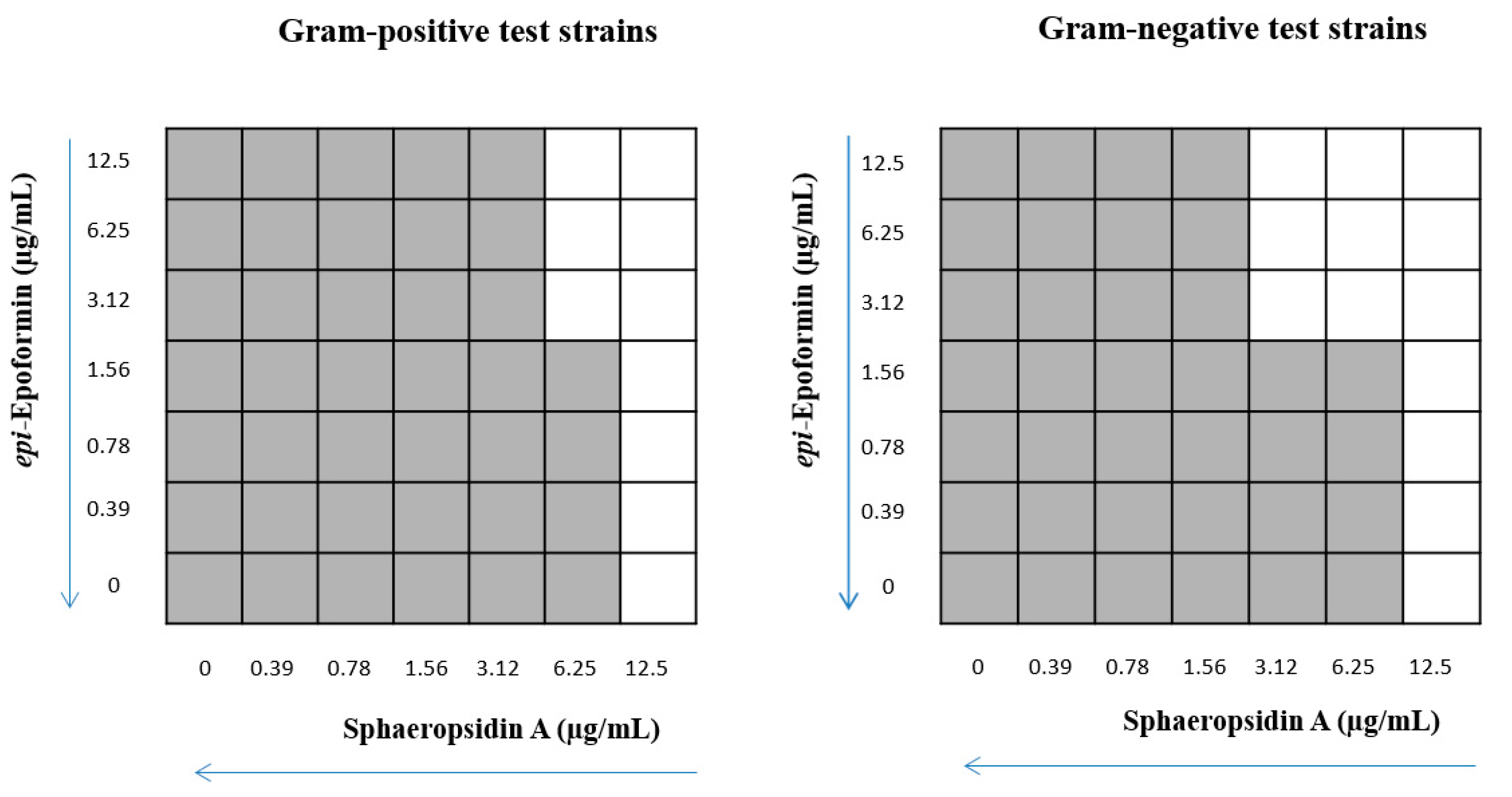
The initial screening of 20 fungal metabolites was performed by standard broth micro-dilution assay in 96-wells polystyrene plates using Mueller-Hinton Broth 2 (MHB2) to test a single high concentration against all test strains. Briefly, 2 × stock solutions of all compounds were made by dissolving them in 2% DMSO. For each strain, the cell suspension was prepared at 0.5 McFarland standard (corresponding to approximately 108 CFU/mL) and subsequently adjusted to approximately 5 × 10⁶ CFU/mL−1. One hundred µL aliquots (5 × 10⁵ CFU) of these bacterial suspensions were treated with 100 µL of 100 µg/mL solution of the compound under investigation; wells with only MHB2 were used as negative control and wells with no compounds as positive growth control. The effect of serial dilutions of DMSO starting from 1% on the growth of test strains was separately tested. Plates were incubated at 37 °C for 19 h under shaking (300 rpm). Then the medium turbidity was measured by a microtiter plate reader at 595 nm (Bio-rad Laboratories s.r.l.). Antimicrobial activity was expressed as a percentage of microbial growth inhibition. Each compound was tested in triplicate and each experiment was performed twice. Minimal inhibitory concentration (MIC) and Minimal Bactericidal Concentration (MBC) of selected compounds were determined by the broth micro-dilution assay. The starting inoculum was 5 × 10⁶ CFU mL−1, and the concentrations of the metabolites ranged from 200 to 0.78 µg/mL (twofold dilutions). As positive controls, conventional antimicrobials, selected depending on antibiotic-susceptibility profiles of the test strains, were included: amikacin (ranged from 32 to 2 µg/mL) was used for Gram-negative strains and teicoplanin (ranged from 0.5 to 4 µg/mL) for Gram-positive strains. Medium turbidity was measured by a microtiter plate reader at 595 nm. The MIC was defined as the lowest concentration of compound that caused ≥90% inhibition of bacterial growth. The MBCs were determined by transferring 200 µL of each sample, previously treated with compound concentrations equal to or higher than the MIC, onto TSA plates and incubating the plates at 37 °C for 24/48 h. The lowest compound concentration that yields no microbial growth on agar plates will be defined as the MBC. Each compound was tested in triplicate; each experiment was performed twice.

4.4. Synergy Assays

The interactions between selected metabolites were evaluated by the checkerboard method in 96-well microtiter plates [64]. The compounds to be tested in combination were serially diluted, one along with the x-axis and the other along with the y-axis. The final compounds’ concentrations (after two-fold dilutions) varied from 0.19 µg/mL up to the 12.5 µg/mL for each one. The checkerboard plates were inoculated with test strains at a concentration of 5 × 10⁶ CFU/mL and incubated at 37 °C for 19 h, then the microbial growth was visually assessed, and the turbidity measured by the microplate reader at 595 nm. To evaluate the effect of the combination treatment, the fractional inhibitory concentration (FIC) index for each combination was calculated as follows: FIC index = FIC of compound A + FIC of compound B, where FIC of compound A (or compound B) will be defined as the ratio of MIC of compound A (or compound B) in combination and MIC of compound A (or of compound B) alone. The FIC index values are interpreted as follows: ≤0.5, synergistic; >0.5 to ≤1.0, additive; >1.0 to ≤2.0, indifferent; and >2.0, antagonistic effects [64].

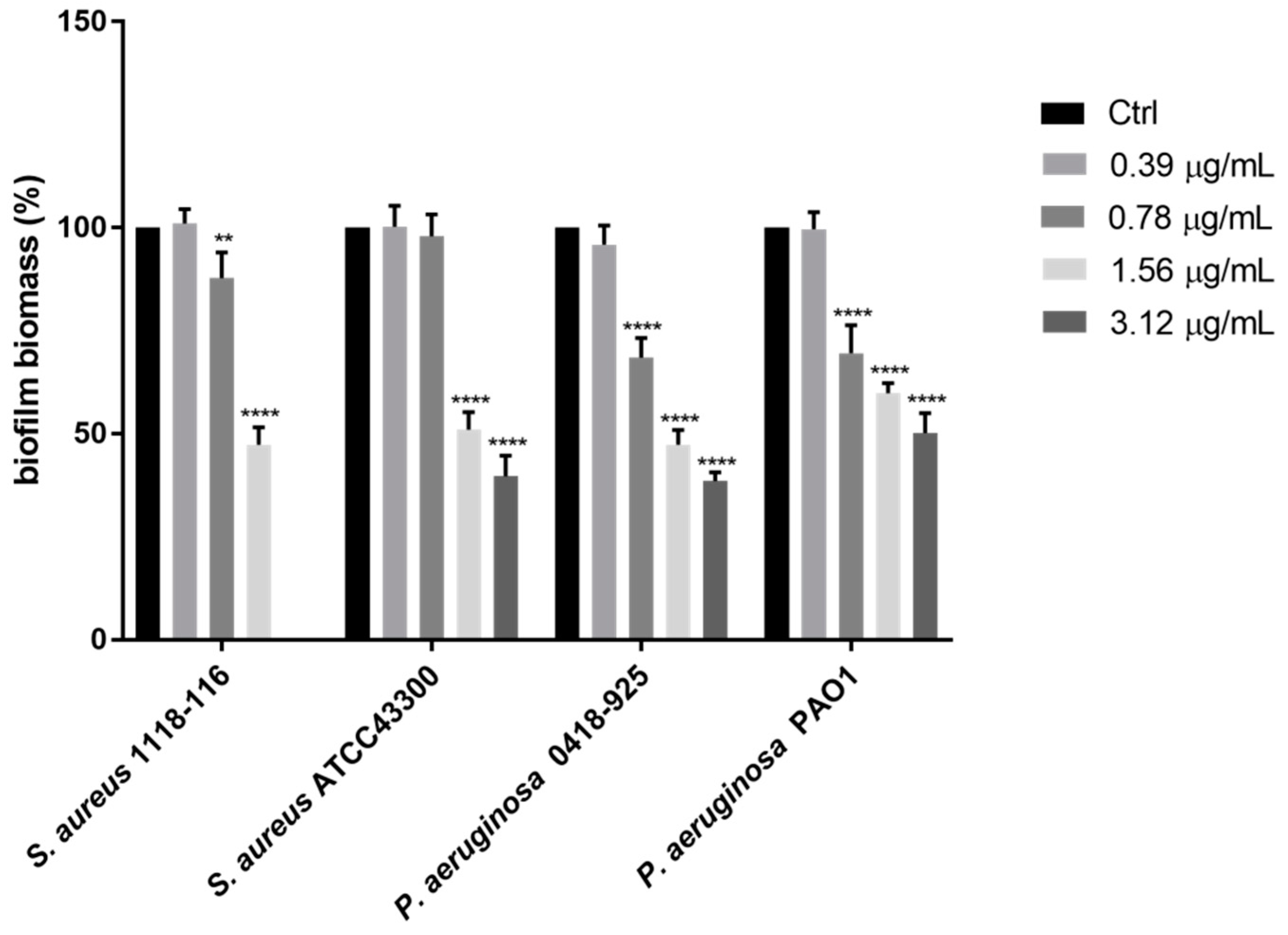
4.5. Biofilm Formation Inhibition Assay

The total biomass of the biofilm was analyzed using the Crystal Violet (CV) staining method in flat-bottomed 96-well microplates as described by Stepanović et al. [65]. For each strain, a cell suspension in MHB2 supplemented with 10% (w/v) glucose was prepared for turbidity of 0.5 McFarland. This suspension was further diluted at 1:100 and 100 µL of the suspension (1 × 10⁶ CFU/mL) were incubated with 100 µL of MHB2 containing the selected compound at serial dilutions of sub-MIC concentrations. The negative control was prepared by inoculating 200 µL of a microbial suspension inactivated by boiling. The positive controls were compound-free wells. To assess biofilm formation, the culture broth was gently aspirated, and each well was washed twice with PBS to remove exclusively non-adherent cells and dried at 60 °C for 45 min. The biofilm was stained by incubation for 30 min with 100 µL of a 0.1% (w/v) crystal violet solution. Any excess of crystal violet was removed by washing with PBS before adding 200 µL of absolute ethanol to release the dye from the biofilm. The absorbance was measured at 595 nm by a microplate reader and was related to the amount of biofilm produced. The percentage of biofilm mass reduction was calculated using the formula: [(Ac-At)/Ac] × 100, where Ac is the OD595 for control wells and At is the OD595 in the presence of the tested compound.

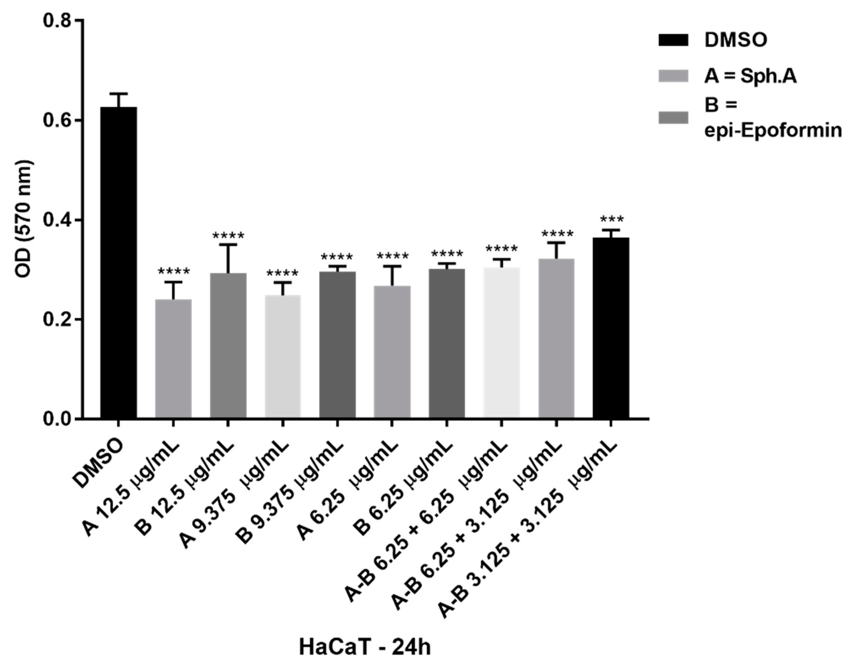
4.6. Cytotoxicity Test

HaCaT (human spontaneously immortalized keratinocytes from adult skin) were purchased from Cell Line Service (CLS, Hattersheim am Main, Germany). Cells were cultured in DMEM High Glucose (Gibco BRL Thermo Fisher, Milan, Italy) supplemented with 10% Fetal Bovine Serum (Gibco BRL, Thermo Fisher, Milan, Italy), 1% L-Glutamine (Gibco BRL) and 1% Pen-Strep solution (Gibco BRL) in a humidified incubator at 37 °C and 5% CO2. Cells were routinely checked for mycoplasma contamination, using a mycoplasma detection kit (ABM, Vancouver, Canada). Cytotoxicity was determined by the MTT 3-(4,5-dimethylthiazol-2-yl)-2,5-diphenyl tetrazolium bromide assay (Sigma-Aldrich, St Louis, MO, USA). HaCaT were seeded in 24-well plates at 3.0 × 10⁴ per well and treated with sphaeropsidin A, sphaeropsidone, and epi-epoformin at concentrations between 3.125 µg/mL and 100 µg/mL for 24 h. The assay was performed according to the manufacturer’s instructions. The optical absorbance was determined at 570 nm and 630 nm using an iMark microplate reader (Bio-Rad, Milan, Italy). Each value shown in the plot is mean ± SD of triplicate determinations. Asterisks represent significant results (*** p > 0.001; **** p < 0.0001).

4.7. Statistical Analysis

Statistical analyses were carried out using the GraphPad Prism 8 software (San Diego, CA). Data were represented as the mean ± standard deviation and analyzed for statistical significance using ordinary one-way or two-way analysis of variance (ANOVA) and multiple comparisons. For all tests, p < 0.005 was considered to indicate a statistically significant difference.

Author Contributions

All the authors contributed to the design of the study, the analysis of the data and writing of the manuscript. provided the fungal cultures and their organic extracts, L.M.; purified the metabolites used in this study R.D.L.; conducted the experiments, E.R., M.E., M.M. and A.D.; writing—original draft preparation, M.M. and E.R.; writing—review and editing, V.C., A.E. and M.R.C.; funding acquisition, M.M. All authors have read and agreed to the published version of the manuscript.

Funding

This research was funded by Programme STAR 2017, financially supported by UniNA and Compagnia di San Paolo, grant number E62F16001250003 to M.M. and DAAD Mobility Program 2018–2020 to V.C.

Acknowledgments

A.E. is associated with “Istituto di Chimica Biomolecolare del CNR”, Pozzuoli, Italy.

Conflicts of Interest

The authors declare no conflict of interest.

References

  1. World Health Organization (WHO); Antimicrobial Resistance. No Time to Wait: Securing the Future from Drug-Resistant Infections. Report to the Secretary-General of the United Nations. Available online: https://www.who.int/antimicrobial-resistance/interagency-coordination-group/IACG_final_report_EN.pdf?ua=1 (accessed on 8 April 2020).
  2. Ciofu, O.; Mandsberg, L.F.; Wang, H.; Høiby, N. Phenotypes selected during chronic lung infection in cystic fibrosis patients: Implications for the treatment of Pseudomonas aeruginosa biofilm infections. FEMS Immunol. Med. Microbiol. 2012, 65, 215–225. [Google Scholar] [CrossRef] [Green Version]
  3. Cantone, E.; Negri, R.; Roscetto, E.; Grassia, R.; Catania, M.R.; Capasso, P.; Maffei, M.; Soriano, A.A.; Leone, C.A.; Iengo, M.; et al. In vivo biofilm formation, Gram-negative infections and TAS2R38 polymorphisms in CRSw NP patients. Laryngoscope 2018, 128, E339–E345. [Google Scholar] [CrossRef] [PubMed]
  4. Gebreyohannes, G.; Nyerere, A.; Bii, C.; Sbhatu, D.B. Challenges of intervention, treatment, and antibiotic resistance of biofilm-forming microorganisms. Heliyon 2019, 5, e02192. [Google Scholar] [CrossRef] [PubMed] [Green Version]
  5. Hall, C.W.; Mah, T.F. Molecular mechanisms of biofilm-based antibiotic resistance and tolerance in pathogenic bacteria. FEMS Microbiol. Rev. 2017, 41, 276–301. [Google Scholar] [CrossRef] [PubMed]
  6. Zhang, Q.; Li, L. Photodynamic combinational therapy in cancer treatment. J. Buon. 2018, 23, 561–567. [Google Scholar]
  7. Barra, F.; Roscetto, E.; Soriano, A.A.; Vollaro, A.; Postiglione, I.; Pierantoni, G.M.; Palumbo, G.; Catania, M.R. Photodynamic and antibiotic therapy in combination to fight biofilms and resistant surface bacterial infections. Int. J. Mol. Sci. 2015, 16, 20417–20430. [Google Scholar] [CrossRef]
  8. Hu, X.; Huang, Y.Y.; Wang, Y.; Wang, X.; Hamblin, M.R. Antimicrobial photodynamic therapy to control clinically relevant biofilm infections. Front. Microbiol. 2018, 9, 1299. [Google Scholar] [CrossRef] [Green Version]
  9. Leaper, D.; Wilson, P.; Assadian, O.; Edmiston, C.; Kiernan, M.; Miller, A.; Bond-Smith, G.; Yap, J. The role of antimicrobial sutures in preventing surgical site infection. Ann. R. Coll. Surg. Engl. 2017, 99, 439–443. [Google Scholar] [CrossRef] [PubMed]
  10. Balato, G.; Roscetto, E.; Vollaro, A.; Galasso, O.; Gasparini, G.; Ascione, T.; Catania, M.R.; Mariconda, M. Bacterial biofilm formation is variably inhibited by different formulations of antibiotic-loaded bone cement in vitro. Knee Surg. Sports Traumatol. Arthrosc. 2019, 27, 1943–1952. [Google Scholar] [CrossRef] [PubMed]
  11. Malek, A.E.; Raad, I.I. Preventing catheter-related infections in cancer patients: A review of current strategies. Expert Rev. Anti. Infect. Ther. 2020. [Google Scholar] [CrossRef] [PubMed]
  12. Artini, M.; Cicatiello, P.; Ricciardelli, A.; Papa, R.; Selan, L.; Dardano, P.; Tilotta, M.; Vrenna, G.; Tutino, M.L.; Giardina, P.; et al. Hydrophobin coating prevents Staphylococcus epidermidis biofilm formation on different surfaces. Biofouling 2017, 33, 601–611. [Google Scholar] [CrossRef] [PubMed]
  13. Vila, J.; Moreno-Morales, J.; Ballesté-Delpierre, C. Current landscape in the discovery of novel antibacterial agents. Clin. Microbiol. Infect. 2019, 26, 596–603. [Google Scholar] [CrossRef]
  14. Ricciardelli, A.; Casillo, A.; Papa, R.; Monti, D.M.; Imbimbo, P.; Vrenna, G.; Artini, M.; Selan, L.; Corsaro, M.M.; Tutino, M.L.; et al. Pentadecanal inspired molecules as new antibiofilm agents against Staphylococcus epidermidis. Biofouling 2018, 34, 1110–1120. [Google Scholar] [CrossRef] [PubMed]
  15. Chung, P.Y.; Khanum, R. Antimicrobial peptides as potential anti-biofilm agents against multidrug-resistant bacteria. J. Microbiol. Immunol. Infect. 2017, 50, 405–410. [Google Scholar] [CrossRef] [PubMed]
  16. Roscetto, E.; Contursi, P.; Vollaro, A.; Fusco, S.; Notomista, E.; Catania, M.R. Antifungal and anti-biofilm activity of the first cryptic antimicrobial peptide from an archaeal protein against Candida spp. clinical isolates. Sci. Rep. 2018, 8, 1–11. [Google Scholar] [CrossRef]
  17. He, M.; Zhang, H.; Li, Y.; Wang, G.; Tang, B.; Zhao, J.; Huang, Y.; Zheng, J. Cathelicidin-derived antimicrobial peptides inhibit Zika virus through direct inactivation and interferon pathway. Front. Immunol. 2018, 9, 722. [Google Scholar] [CrossRef] [Green Version]
  18. Gaglione, R.; Pirone, L.; Farina, B.; Fusco, S.; Smaldone, G.; Aulitto, M.; Dell’Olmo, E.; Roscetto, E.; Del Gatto, A.; Fattorusso, R.; et al. Insights into the anticancer properties of the first antimicrobial peptide from Archaea. Biochim. Biophys. Acta Gen. Subj. 2017, 1861, 2155–2164. [Google Scholar] [CrossRef] [Green Version]
  19. Li, J.W.H.; Vederas, J.C. Drug discovery and natural products: End of an era or an endless frontier? Science 2009, 325, 161–165. [Google Scholar] [CrossRef] [Green Version]
  20. Cragg, G.M.; Newman, D.J. Natural products: A continuing source of novel drug leads. Biochim. Biochim. Biophys. Acta Gen. Subj. 2013, 1830, 3670–3695. [Google Scholar] [CrossRef] [Green Version]
  21. Schueffler, A.; Anke, T. Fungal natural products in research and development. Nat. Prod. Rep. 2014, 31, 1425–1448. [Google Scholar] [CrossRef]
  22. Thomford, N.E.; Senthebane, D.A.; Rowe, A.; Munro, D.; Seele, P.; Maroyi, A.; Dzobo, K. Natural products for drug discovery in the 21st century: Innovations for novel drug discovery. Int. J. Mol. Sci. 2018, 19, 1578. [Google Scholar] [CrossRef] [Green Version]
  23. Wright, G.D. Unlocking the potential of natural products in drug discovery. Microb. Biotechnol. 2019, 12, 55–57. [Google Scholar] [CrossRef] [PubMed] [Green Version]
  24. Masi, M.; Maddau, L.; Linaldeddu, B.T.; Scanu, B.; Evidente, A.; Cimmino, A. Bioactive metabolites from pathogenic and endophytic fungi of forest trees. Curr. Med. Chem. 2018, 25, 208–252. [Google Scholar] [CrossRef] [PubMed]
  25. Andolfi, A.; Maddau, L.; Basso, S.; Linaldeddu, B.T.; Cimmino, A.; Scanu, B.; Deidda, A.; Tuzi, A.; Evidente, A. Diplopimarane, a 20-nor-ent-pimarane produced by the oak pathogen Diplodia quercivora. J. Nat. Prod. 2014, 77, 2352–2360. [Google Scholar] [CrossRef] [PubMed]
  26. Graniti, A.; Sparapano, L.; Evidente, A. Cyclopaldic acid, a major phytotoxic metabolite of Seiridium cupressi, the pathogen of a canker disease of cypress. Plant Pathol. 1992, 41, 563–568. [Google Scholar] [CrossRef]
  27. Evidente, A.; Andolfi, A.; Maddau, L.; Franceschini, A.; Marras, F. Biscopyran, a phytotoxic hexasubstituted pyranopyran produced by Biscogniauxia mediterranea, a fungus pathogen of cork oak. J. Nat. Prod. 2005, 68, 568–571. [Google Scholar] [CrossRef]
  28. Evidente, A.; Sparapano, L.; Fierro, O.; Bruno, G.; Giordano, F.; Motta, A. Sphaeropsidins B and C, phytotoxic pimarane diterpenes from Sphaeropsis sapinea f. sp. cupressi and Diplodia mutila. Phytochemistry 1997, 45, 705–713. [Google Scholar] [CrossRef]
  29. Cimmino, A.; Maddau, L.; Masi, M.; Evidente, M.; Linaldeddu, B.T.; Evidente, A. Further secondary metabolites produced by Diplodia corticola, a fungal pathogen involved in cork oak decline. Tetrahedron 2016, 72, 6788–6793. [Google Scholar] [CrossRef]
  30. Evidente, A.; Maddau, L.; Scanu, B.; Andolfi, A.; Masi, M.; Motta, A.; Tuzi, A. Sphaeropsidones, phytotoxic dimedone methyl ethers produced by Diplodia cupressi: A structure-activity relationship study. J. Nat. Prod. 2011, 74, 757–763. [Google Scholar] [CrossRef]
  31. Masi, M.; Maddau, L.; Linaldeddu, B.T.; Cimmino, A.; D’Amico, W.; Scanu, B.; Evidente, M.; Tuzi, A.; Evidente, A. Bioactive secondary metabolites produced by the oak pathogen Diplodia corticola. J. Agric. Food Chem. 2016, 64, 217–225. [Google Scholar] [CrossRef] [PubMed]
  32. Evidente, A.; Randazzo, G.; Ballio, A. Structure determination of seiridin and isoseiridin, phytotoxic butenolides from culture filtrate of Seiridium cardinale. J. Nat. Prod. 1986, 49, 593–601. [Google Scholar] [CrossRef]
  33. Ballio, A.; Evidente, A.; Graniti, A.; Randazzo, G.; Sparapano, L. Seiricuprolide, a new phytotoxic macrolide from a strain of Seiridium cupressi infecting cypress. Phytochemistry 1988, 27, 3117–3121. [Google Scholar] [CrossRef]
  34. Cimmino, A.; Maddau, L.; Masi, M.; Linaldeddu, B.T.; Evidente, A. Secondary metabolites produced by Sardiniella urbana, a new emerging pathogen on European hackberry. Nat. Prod. Res. 2019, 33, 1862–1869. [Google Scholar] [CrossRef]
  35. Masi, M.; Di Lecce, R.; Tuzi, A.; Linaldeddu, B.T.; Montecchio, L.; Maddau, L.; Evidente, A. Hyfraxinic acid, a phytotoxic tetrasubstituted octanoic Acid, produced by the ash (Fraxinus excelsior L.) pathogen Hymenoscyphus fraxineus together with viridiol and some of its analogues. J. Agric. Food Chem. 2019, 67, 13617–13623. [Google Scholar] [CrossRef] [PubMed]
  36. Evidente, A.; Maddau, L.; Spanu, E.; Franceschini, A.; Lazzaroni, S.; Motta, A. Diplopyrone, a new phytotoxic tetrahydropyranpyran-2-one produced by Diplodia mutila, a fungus pathogen of cork oak. J. Nat. Prod. 2003, 66, 313–315. [Google Scholar] [CrossRef] [PubMed]
  37. Sparapano, L.; Bruno, G.; Fierro, O.; Evidente, A. Studies on structure–activity relationship of sphaeropsidins A–F, phytotoxins produced by Sphaeropsis sapinea f. sp. cupressi. Phytochemistry 2004, 65, 189–198. [Google Scholar] [CrossRef]
  38. Reveglia, P.; Masi, M.; Evidente, A. Melleins—Intriguing natural compounds. Biomolecules 2020, 10, 772. [Google Scholar] [CrossRef] [PubMed]
  39. Barilli, E.; Cimmino, A.; Masi, M.; Evidente, M.; Rubiales, D.; Evidente, A. Inhibition of spore germination and appressorium formation of rust species by plant and fungal metabolites. Nat. Prod. Commun. 2016, 11, 1934578X1601100940. [Google Scholar] [CrossRef] [Green Version]
  40. Barilli, E.; Cimmino, A.; Masi, M.; Evidente, M.; Rubiales, D.; Evidente, A. Inhibition of early development stages of rust fungi by the two fungal metabolites cyclopaldic acid and epi-epoformin. Pest Manag. Sci. 2017, 73, 1161–1168. [Google Scholar] [CrossRef] [PubMed]
  41. Cimmino, A.; Andolfi, A.; Avolio, F.; Ali, A.; Tabanca, N.; Khan, I.A.; Evidente, A. Cyclopaldic Acid, seiridin, and sphaeropsidin a as fungal phytotoxins, and larvicidal and biting deterrents against Aedes aegypti (Diptera: Culicidae): Structure-activity relationships. Chem. Biodivers. 2013, 10, 1239–1251. [Google Scholar] [CrossRef]
  42. Fernández-Aparicio, M.; Masi, M.; Maddau, L.; Cimmino, A.; Evidente, M.; Rubiales, D.; Evidente, A. Induction of haustorium development by sphaeropsidones in radicles of the parasitic weeds Striga and Orobanche. A structure–activity relationship study. J. Agric. Food Chem. 2016, 64, 5188–5196. [Google Scholar] [CrossRef] [PubMed] [Green Version]
  43. Evidente, A.; Venturi, V.; Masi, M.; Degrassi, G.; Cimmino, A.; Maddau, L.; Andolfi, A. In vitro antibacterial activity of sphaeropsidins and chemical derivatives toward Xanthomonas oryzae pv. oryzae, the causal agent of rice bacterial blight. J. Nat. Prod. 2011, 74, 2520–2525. [Google Scholar] [CrossRef] [PubMed]
  44. Mathieu, V.; Chantôme, A.; Lefranc, F.; Cimmino, A.; Miklos, W.; Paulitschke, V.; Mohr, T.; Maddau, L.; Kornienko, A.; Berger, W.; et al. Sphaeropsidin A shows promising activity against drug-resistant cancer cells by targeting regulatory volume increase. Cell. Mol. Life Sci. 2015, 72, 3731–3746. [Google Scholar] [CrossRef] [PubMed] [Green Version]
  45. Ingels, A.; Dinhof, C.; Garg, A.D.; Maddau, L.; Masi, M.; Evidente, A.; Berger, W.; Dejaegher, B.; Mathieu, V. Computed determination of the in vitro optimal chemocombinations of sphaeropsidin A with chemotherapeutic agents to combat melanomas. Cancer Chemoth. Pharmacol. 2017, 79, 971–983. [Google Scholar] [CrossRef]
  46. Evidente, A.; Andolfi, A.; Cimmino, A. Relationships between the stereochemistry and biological activity of fungal phytotoxins. Chirality 2011, 23, 674–693. [Google Scholar] [CrossRef]
  47. Evidente, A.; Cimmino, A.; Andolfi, A. The effect of stereochemistry on the biological activity of natural phytotoxins, fungicides, insecticides and herbicides. Chirality 2013, 25, 59–78. [Google Scholar] [CrossRef]
  48. Masi, M.; Cimmino, A.; Maddau, L.; Kornienko, A.; Tuzi, A.; Evidente, A. Crystal structure and absolute configuration of sphaeropsidin A and its 6-o-p-bromobenzoate. Tetrahedron Lett. 2016, 57, 4592–4594. [Google Scholar] [CrossRef]
  49. Mazzeo, G.; Cimmino, A.; Masi, M.; Longhi, G.; Maddau, L.; Memo, M.; Evidente, A.; Abbate, S. Importance and difficulties in the use of chiroptical methods to assign the absolute configuration of natural products: The case of phytotoxic pyrones and furanones produced by Diplodia corticola. J. Nat. Prod. 2017, 80, 2406–2415. [Google Scholar] [CrossRef]
  50. Fusè, M.; Mazzeo, G.; Longhi, G.; Abbate, S.; Masi, M.; Evidente, A.; Puzzarini, C.; Barone, V. Unbiased determination of absolute configurations by vis-à-vis comparison of experimental and simulated spectra: The challenging case of diplopyrone. J. Phys. Chem. B 2019, 123, 9230–9237. [Google Scholar] [CrossRef]
  51. Fazli, M.; Bjarnsholt, T.; Kirketerp-Møller, K.; Jørgensen, B.; Andersen, A.S.; Krogfelt, K.A.; Givskov, M.; Tolker-Nielsen, T. Nonrandom distribution of Pseudomonas aeruginosa and Staphylococcus aureus in chronic wounds. J. Clin. Microbiol. 2009, 47, 4084–4089. [Google Scholar] [CrossRef] [Green Version]
  52. Martin, J.M.; Zenilman, J.M.; Lazarus, G.S. Molecular microbiology: New dimensions for cutaneous biology and wound healing. J. Investig. Dermatol. 2010, 130, 38–48. [Google Scholar] [CrossRef] [PubMed] [Green Version]
  53. Lachiewicz, A.M.; Hauck, C.G.; Weber, D.J.; Cairns, B.A.; van Duin, D. Bacterial infections after burn injuries: Impact of multidrug resistance. Clin. Infect. Dis. 2017, 65, 2130–2136. [Google Scholar] [CrossRef] [PubMed]
  54. Eisner, R.; Lippmann, N.; Josten, C.; Rodloff, A.C.; Behrendt, D. Development of the bacterial spectrum and antimicrobial resistance in surgical site infections of trauma patients. Surg. Infect. 2020. [Google Scholar] [CrossRef] [PubMed]
  55. Kaur, A.; Singh, S. Prevalence of extended spectrum betalactamase (ESBL) and metallobetalactamase (MBL) producing Pseudomonas aeruginosa and Acinetobacter baumannii isolated from various clinical samples. J. Pathog. 2018, 2018, 6845985. [Google Scholar] [CrossRef] [PubMed] [Green Version]
  56. Royer, G.; Fourreau, F.; Boulanger, B.; Mercier-Darty, M.; Ducellier, D.; Cizeau, F.; Potron, A.; Podglajen, I.; Mongardon, N.; Decousser, J.W. Local outbreak of extended-spectrum β-lactamase SHV2a-producing Pseudomonas aeruginosa reveals the emergence of a new specific sub-lineage of the international ST235 high-risk clone. J. Hosp. Infect. 2020, 104, 33–39. [Google Scholar] [CrossRef] [PubMed]
  57. Vestergaard, M.; Frees, D.; Ingmer, H. Antibiotic resistance and the MRSA problem. Gram Posit. Pathog. 2019, 7, 747–765. [Google Scholar]
  58. Serra, R.; Grande, R.; Butrico, L.; Rossi, A.; Settimio, U.F.; Caroleo, B.; Amato, B.; Gallelli, L.; de Franciscis, S. Chronic wound infections: The role of Pseudomonas aeruginosa and Staphylococcus aureus. Expert. Rev. Anti. Infect. Ther. 2015, 13, 605–613. [Google Scholar] [CrossRef] [PubMed]
  59. De Vor, L.; Rooijakkers, S.H.M.; van Strijp, J.A.G. Staphylococci evade the innate immune response by disarming neutrophils and forming biofilms. FEBS Lett. 2020. [Google Scholar] [CrossRef] [Green Version]
  60. Wolcott, R.D. Biofilms cause chronic infections. J. Wound Care 2017, 26, 423–425. [Google Scholar] [CrossRef] [Green Version]
  61. Roy, S.; Santra, S.; Das, A.; Dixith, S.; Sinha, M.; Ghatak, S.; Ghosh, N.; Banerjee, P.; Khanna, S.; Mathew-Steiner, S.; et al. Staphylococcus aureus biofilm infection compromises wound healing by causing deficiencies in granulation tissue collagen. Ann. Surg. 2019. [Google Scholar] [CrossRef]
  62. Dewick, P.M. Medicinal Natural Products: A Biosynthetic Approach, 3rd ed.; John Wiley & Sons: Chichester, UK, 2009. [Google Scholar]
  63. Le Calve, B.; Lallemand, B.; Perrone, C.; Lenglet, G.; Depauw, S.; Van Goietsenoven, G.; Bury, M.; Vurro, M.; Herphelin, F.; Andolfi, A.; et al. In vitro anticancer activity, toxicity and structure–activity relationships of phyllostictine A, a natural oxazatricycloalkenone produced by the fungus Phyllosticta cirsii. Toxicol. Appl. Pharmacol. 2011, 254, 8–17. [Google Scholar] [CrossRef] [PubMed]
  64. Pillai, S.K.; Moellering, R.C.; Eliopoulos, G.M. Antimicrobial combinations. In Antibiotics in Laboratory. Medicine, 5th ed.; Lorian, V., Ed.; The Lippincott Williams & Wilkins Co.: Philadelphia, PA, USA, 2005; pp. 365–440. [Google Scholar]
  65. Stepanović, S.; Vuković, D.; Hola, V.; di Bonaventura, G.; Djukić, S.; Cirković, I.; Ruzicka, F. Quantification of biofilm in microtiter plates: Overview of testing conditions and practical recommendations for assessment of biofilm production by Staphylococci. Apmis 2007, 115, 891–899. [Google Scholar] [CrossRef] [PubMed]
Figure 1. The structures of compounds 1–20.
Toxins 12 00444 g001
Figure 2. Multi-wells model of checkerboard assay to test the interaction between epi-epoformin and sphaeropsidin A. Turbidity was reported in grey and no bacterial growth in white.
Toxins 12 00444 g002
Figure 3. MTT viability test. Hacat cells were incubated with the indicated amount of spheropsidin A (compound A) and epi-epoformin (compound B) alone or in combination (AB) for 24 h. The MTT viability test was performed as described in Material. The values were the mean’s three values for each experimental point of two biological replicates. Each pair of means were compared using a Tukey’s multiple comparisons test p-value < 0.05, *** p < 0.001; **** p < 0.0001)
Toxins 12 00444 g003
Figure 4. In vitro biofilm formation of test strains following overnight treatment with compound 20 at serial dilutions of sub-MIC concentrations. Biofilm formation was determined by cristal violet assay. Values are presented as mean percentage ± SD. ** p-value = 0.009, **** p-value < 0.001.
Toxins 12 00444 g004
Table 1. Fungal metabolites used in this study.
NumberNameChemical FamilyFungal SourceRef.
1epi-EpoforminCyclohexene oxideDiplodia quercivora[25]
2Cyclopaldic acid IsobenzofuranoneSeiridium cupressi[26]
3BiscopyranPyranopyranBiscogniauxia mediterranea[27]
4Sphaeropsidin BDiterpenoidDiplodia cupressi[28]
5Sphaeropsidin CDiterpenoidD. cupressi[28]
6Sphaeropsidin GDiterpenoidDiplodia corticola[29]
7SphaeropsidoneCyclohexene oxideD. cupressi[30]
8Sapinofuranone CFuranoneD. corticola[31]
9(S,S)-Sapinofuranone BFuranoneD. corticola[31]
10SeiridinButenolideS. cupressi[32]
11SeiricuprolideMacrolideS. cupressi[33]
12Diplobifuranylone AFuranoneD. corticola[31]
13Diplobifuranylone BFuranoneD. corticola[31]
14Diplobifuranylone CFuranoneD. corticola[31]
15(R)-Mellein 3,4-DihydroisocoumarinSardiniella urbana[34]
16cis-4-Hydroxymellein3,4-DihydroisocoumarinS. urbana[34]
17trans-4-Hydroxymellein3,4-DihydroisocoumarinS. urbana[34]
18ViridiolFuranosteroidHymenoscyphus fraxineus[35]
19DiplopyronePyranopyroneD. corticola[36]
20Sphaeropsidin ADiterpenoidD. corticola[31]
Table 2. Antibacterial activity, expressed as the percentage of growth inhibition, of 120 at the 100 μg/mL concentration against Gram-positive and Gram-negative test strains 1,2.
CompoundBacterial Strain
S. aureus ATCC
43300
MRSA1118-116S. haemolyticus
ATCC 29970
S. haemolyticus
VR 1219-118
P. aeruginosa
PAO1
P. aeruginosa
0418-925
1≥90≥90≥90≥90≥90≥90
2≥90≥90≥90≥90-≥90
3----5060
4≥90≥90≥90≥90-60
5----6060
6≥90≥90≥90≥90--
7≥90≥90≥90≥9050≥90
8----6060
9----6060
10----5060
11----6050
12----5050
13----5060
14----5060
15----6060
16----6070
17----6060
18≥90≥90≥90≥90--
19------
20≥90≥90≥90≥90≥90≥90
AKntntntnt>90>90
TE>90>90>90>90ntnt
1 For inhibition values below 50%, no data have been reported (-). AK = Amikacin; TE = Teicoplanin; nt = not tested. 2 Amikacin (32 μg/mL) and teicoplanin (4 μg/mL) were used as positive controls.
Table 3. MIC (μg/mL) and MBC (μg/mL) of compounds 1, 7, 20 against Gram-positive and Gram-negative test strains 1.
Bacterial StrainCompound 1Compound 7Compound 20AmikacinTeicoplanin
MICMBCMICMBCMICMBCMICMBCMICMBC
S. aureus ATCC 43300100100100>20012.5100ntnt14
MRSA 1118-116100100100>2006.2525ntnt0.54
S. haemolyticus ATCC 29970100100100>20012.550ntnt2>4
S. haemolyticus VR 1219-118100100100>20012.550ntnt2>4
P. aeruginosa PAO150>200>100>20012.5>200432ntnt
P. aeruginosa 0418-92550>200100>20012.5>20016>32ntnt
1 Amikacin and teicoplanin were used as positive controls; nt = not tested.

