Article

Molecular Characterization of the Enterohemolysin Gene (ehxA) in Clinical Shiga Toxin-Producing Escherichia coli Isolates

1
Department of Microbiology, School of Public Health, Southern Medical University, Guangzhou 510515, China
2
Department of Laboratory Medicine, Division of Clinical Microbiology, Karolinska Institutet, 141 52 Stockholm, Sweden
3
Molecular Epidemiology and Public Health Laboratory, School of Veterinary Sciences, Massey University, Palmerston North 4100, New Zealand
4
The Public Health Agency of Sweden, 171 82 Solna, Sweden
5
Division of Pediatrics, Department of Clinical Science, Intervention and Technology, Karolinska Institutet and Karolinska University Hospital, 141 86 Stockholm, Sweden
6
Queen Silvia Children’s Hospital, Sahlgrenska University Hospital, 413 45 Gothenburg, Sweden
7
Department of Pediatrics, Department of Pediatrics, Sahlgrenska Academy, Sahlgrenska Academy, University of Gothenburg, 416 85 Gothenburg, Sweden
8
State Key Laboratory of Infectious Disease Prevention and Control, National Institute for Communicable Disease Control and Prevention, Chinese Center for Disease Control and Prevention, Beijing 102206, China
9
Laboratory Medicine, Jönköping Region County, Department of Clinical and Experimental Medicine, Linköping University, 551 85 Jönköping, Sweden
10
Oslo University Hospital, 0372 Oslo, Norway
11
Division of Laboratory Medicine, Institute of Clinical Medicine, University of Oslo, 0372 Oslo, Norway
12
Division of Infectious Diseases, Department of Medicine Huddinge, Karolinska Institutet, 141 86 Stockholm, Sweden
*
Authors to whom correspondence should be addressed.
Submission received: 15 December 2020 / Revised: 11 January 2021 / Accepted: 15 January 2021 / Published: 19 January 2021

Abstract

:
Shiga toxin (Stx)-producing Escherichia coli (STEC) is an important foodborne pathogen with the ability to cause bloody diarrhea (BD) and hemolytic uremic syndrome (HUS). Little is known about enterohemolysin-encoded by ehxA. Here we investigated the prevalence and diversity of ehxA in 239 STEC isolates from human clinical samples. In total, 199 out of 239 isolates (83.26%) were ehxA positive, and ehxA was significantly overrepresented in isolates carrying stx2a + stx2c (p < 0.001) and eae (p < 0.001). The presence of ehxA was significantly associated with BD and serotype O157:H7. Five ehxA subtypes were identified, among which, ehxA subtypes B, C, and F were overrepresented in eae-positive isolates. All O157:H7 isolates carried ehxA subtype B, which was related to BD and HUS. Three ehxA groups were observed in the phylogenetic analysis, namely, group Ⅰ (ehxA subtype A), group Ⅱ (ehxA subtype B, C, and F), and group Ⅲ (ehxA subtype D). Most BD- and HUS-associated isolates were clustered into ehxA group Ⅱ, while ehxA group Ⅰ was associated with non-bloody stool and individuals ≥10 years of age. The presence of ehxA + eae and ehxA + eae + stx2 was significantly associated with HUS and O157:H7 isolates. In summary, this study showed a high prevalence and the considerable genetic diversity of ehxA among clinical STEC isolates. The ehxA genotypes (subtype B and phylogenetic group Ⅱ) could be used as risk predictors, as they were associated with severe clinical symptoms, such as BD and HUS. Furthermore, ehxA, together with stx and eae, can be used as a risk predictor for HUS in STEC infections.
Key Contribution: This study showed a high prevalence and considerable genetic diversity of enterohemolysin-encoding gene ehxA among clinical STEC isolates. The ehxA genotypes could be used as risk predictors, ehxA subtype B and phylogenetic group II were found to be associated with severe clinical symptoms, such as bloody diarrhea (BD) and hemolytic uremic syndrome (HUS).

1. Introduction

Shiga toxin-producing Escherichia coli (STEC) is an important enteric foodborne pathogen that can cause bloody diarrhea (BD), hemorrhagic colitis, and potentially fatal hemolytic uremic syndrome (HUS) in infected humans [1]. STEC is estimated to cause 2.8 million cases of enteric disease in humans per year globally [2]. Over 400 STEC serotypes have been identified, among which, O157:H7 is the most prevalent serotype and is linked to severe human illness, such as HUS [3,4]. Nevertheless, since the early 2010s, non-O157 STEC, especially the so-called “top six” serogroups (O26, O45, O103, O111, O121, and O145), have been associated with continuously increasing numbers of STEC outbreaks and may account for up to 80% of STEC infections [2,5,6]. Humans are infected through contact with infected animals or the consumption of STEC-contaminated water, vegetables, milk, or meat.
Shiga toxins (Stx1 and Stx2) are the main virulence factors of STEC, which can mediate a significant cytotoxic effect in human vascular endothelial cells [7]. stx genes are located in the genomes of Shiga-toxin-converting bacteriophages [8]. At least 15 stx1 and stx2 gene subtypes have been identified, among which, stx2a, stx2c, and stx2d are more virulent than other subtypes as they are highly associated with severe clinical outcomes, such as HUS [9]. Besides Stx, eae-encoding intimin, which is responsible for the intimate adherence of STEC, is also a significant virulence trait of pathogenic STEC [10,11]. In addition, hemolysin-encoding genes have been regarded as STEC virulence markers [7]. So far, four different types of hemolysins have been identified, namely, alpha-hemolysin (hlyA), silent hemolysin (sheA), bacteriophage-associated enterohemolysin (e-hlyA), and plasmid-carried enterohemolysin (ehxA) [12]. Enterohemolysin displays hemolytic activity that enables STEC to be observed on washed sheep blood agar, which is commonly used as a phenotypic indicator of STEC strains [13,14]. It is noteworthy that ehxA is prevalent in STEC strains and is closely associated with isolates causing diarrheal disease and HUS [15].
Enterohemolysin belongs to the repeats in toxin (RTX) family, which has a pore-forming capacity [16]. ehxA is located on a large virulence plasmid and its nucleic acid sequence has about 3000 base pairs [17]. The presence of ehxA has a close association with stx, thus it is proposed as an epidemiological marker for the rapid characterization of STEC strains [13]. For example, in the U.S. Food and Drug Administration E. coli Identification (FDA-ECID) microarray, ehxA is included as one of the genetic markers for the rapid characterization of STEC isolates [18]. Six genetically distinct ehxA subtypes (A to F) have been described in E. coli by using PCR in combination with restriction fragment length polymorphism (RFLP) analysis [12]. STEC ehxA subtypes differ significantly among strains isolated from different sources. Subtypes A and C are mostly found in animal isolates, where subtype A is detected in food-associated strains and subtype C is commonly found in clinical strains [12]. However, such data are limited; the correlation between ehxA subtypes and strains sources, as well as the clinical relevance, remains to be further elucidated.
A recent study in Sweden showed that almost all of the eae-positive isolates, except one, harbored ehxA, and the coexistence of ehxA and eae was shown to be associated with BD [19]. In a previous study, only 10.9% of isolates carried eae among 138 ehxA-positive non-O157 STEC isolates from human, animal, and food sources in China, and 61.54% of these were clinically relevant [20]. The aim of this study was to investigate the prevalence and genetic diversity of the ehxA gene, its correlation with serotypes, and the presence of stx and eae. Furthermore, we aimed to assess the association between ehxA subtypes and disease severity.

2. Results

2.1. Distribution of ehxA in the Clinical STEC Isolates

Among the 239 STEC strains isolated in Sweden, ehxA was identified in 199 (83.26%) isolates. Fifty-three (26.63%) of the ehxA-positive isolates were from patients with HUS, 47 (23.62%) were from patients with BD, and 99 (49.75%) were from individuals with NBS (non-bloody stool). All O157:H7 isolates were ehxA positive and the majority (45/65, 69.23%) were also stx2a + stx2c positive. The majority of the eae-positive STEC isolates (166 of 173, 95.95%) carried ehxA. ehxA was overrepresented in isolates that carried stx2a + stx2c (p < 0.001) and eae (p < 0.001). The presence of ehxA was significantly associated with BD and O157:H7 (Table 1). However, no association was observed between the presence of ehxA and the duration of bacterial shedding, the age of the patients, or HUS (Table S1).

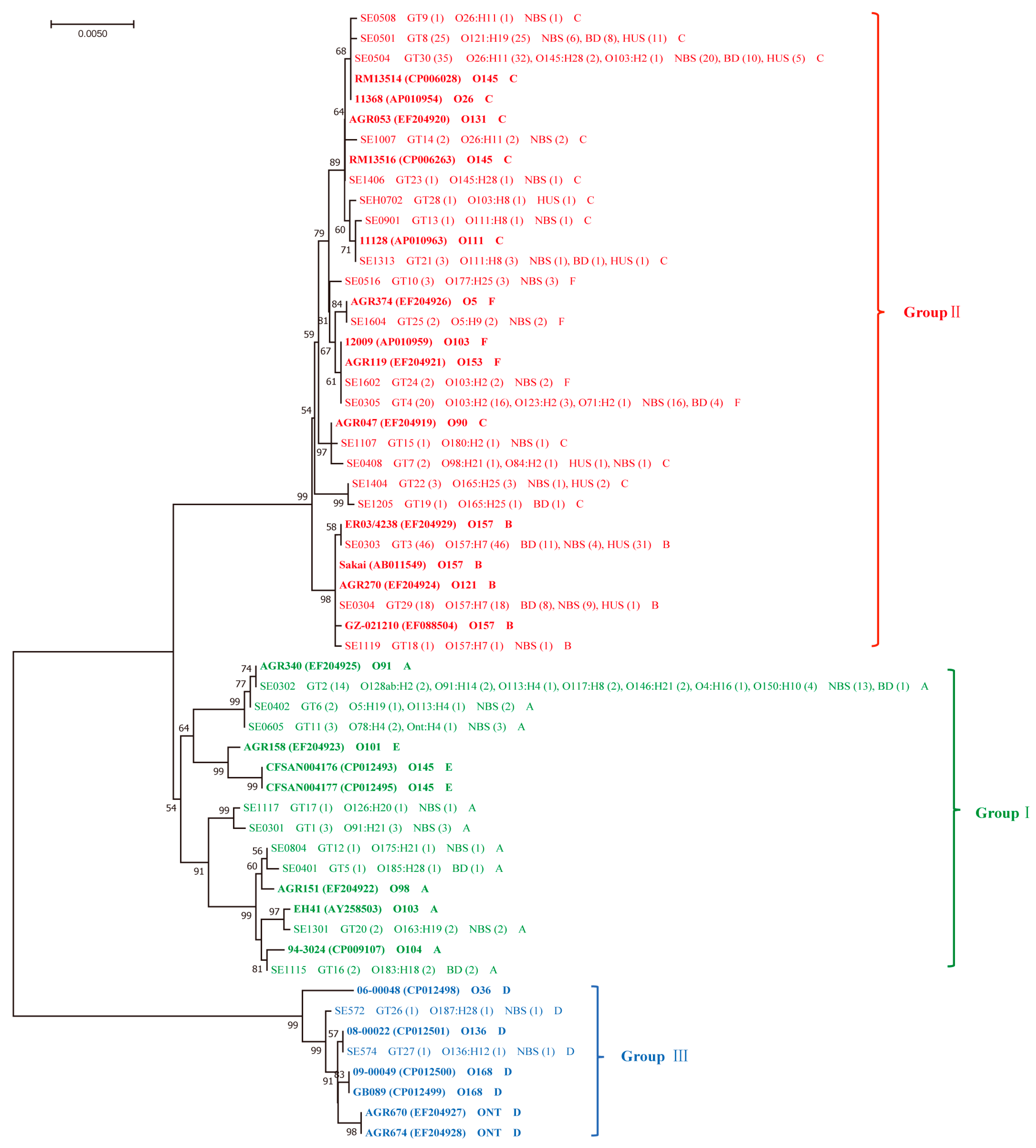
2.2. Diversity of ehxA and Its Correlation to Serotypes and the stx and eae Genes

Thirty unique ehxA sequences (genotypes GT1 to GT30) were identified among the 199 ehxA positive STEC isolates. The nucleotide similarities of ehxA gene sequences in this study ranged from 95.79 to 100%. Five distinct subtypes (A, B, C, D, F) were found, out of which, subtype C (76, 38.19%) was the most predominant subtype, followed by ehxA subtype B (65, 32.66%) and ehxA subtype A (29, 14.57%). In addition, subtype C showed greater genetic diversity than other subtypes. All isolates carrying subtypes B, C, or F were eae positive, with the exception of two ehxA subtype C isolates (Table 2). Interestingly, all ehxA subtype A and D isolates were eae negative and subtype B was only found in O157:H7 isolates (Table 2). Other ehxA subtypes were represented within different serotypes: ehxA subtype C was linked to O121:H19 and O26:H11 strains (Table S2) and subtype F mainly belonged to O103:H2 isolates (Table 2). The presence of ehxA + eae (Table 3) and ehxA + eae + stx2 (Table S3) was statistically associated with O157:H7 isolates. The presence of stx1 and its subtype stx1a was statistically associated with ehxA subtype F, while the presence of stx2 and its subtype stx2a + stx2c was linked to ehxA subtype B (Table 4).
A neighbor-joining tree was generated using 30 unique ehxA sequences from this study and 26 reference ehxA sequences that were reported previously. Three phylogenetic groups were identified, namely, group Ⅰ (ehxA subtype A), where all isolates were eae negative; group Ⅱ (ehxA subtype B, C, F), where all isolates were eae positive; group Ⅲ (ehxA subtype D) containing only two eae-negative isolates (Figure 1).

2.3. ehxA Subtypes and Phylogenic Groups in Correlation with Clinical Variables and the Presence of eae

ehxA subtype B was overrepresented in BD- and HUS-associated isolates. Accordingly, ehxA group Ⅱ was statistically associated with BD and HUS. ehxA subtype A and ehxA group Ⅰ were statistically associated with NBS and individuals ≥10 years of age; ehxA subtype F and ehxA group Ⅱ were significantly linked to individuals <10 years of age; however, these differences had no statistical significance after Benjiamini–Hochberg corrections (Table 5). ehxA subtype B and ehxA group Ⅱ were statistically associated with O157:H7 strains (p < 0.001) (Table 5). No association was observed between the ehxA subtype/phylogenetic group and the duration of bacterial shedding (data not shown). The presence of ehxA + eae (Table 3) and ehxA + eae + stx2 (Table S3) was statistically associated with HUS. In addition, the presence of ehxA + eae was overrepresented in isolates from individuals <10 years of age (Table 3).

3. Discussion

Shiga toxin and intimin have been widely investigated as vital virulence factors of STEC [21]. In addition, enterohemolysin (ehxA) has emerged as a possible marker for the identification of specific STEC strains, such as O26, O157, O145, and O103, which are highly related to severe clinical symptoms, including BD and HUS [13,17,22,23]. The presence of ehxA was shown to be a useful epidemiological marker for the presence of Stx [12,14]. However, the role of ehxA in STEC pathogenicity and the association between ehxA and other key STEC virulence factors, such as stx and eae, have not been fully elucidated. Here, we systematically investigated the prevalence of ehxA in human clinical STEC isolates in Sweden and analyzed the association between ehxA and clinical symptoms, as well as the bacterial features. We found that ehxA was present in 83.26% of all clinical STEC isolates in this study. The majority of the eae-positive isolates also carried the ehxA gene, while only 50% of the eae-negative isolates carried ehxA. This was in line with a previous study, where ehxA was divided into two major phylogenetic clusters based on the presence or absence of eae [24]. ehxA was also shown to have a strong link with eae-positive atypical EPEC strains that were isolated from cattle and sheep [17]. Notably, the presence of ehxA was statistically associated with O157:H7, stx2a + stx2c, and BD, suggesting that ehxA could be included as a virulence marker in clinical diagnostics to predict highly pathogenic STEC strains.
The phylogeny analysis showed that ehxA-positive STEC isolates were divided into three groups, as described in a previous study [20]. ehxA subtypes B, C, and F were assigned to group Ⅱ, among which, 98.8% carried the eae gene. ehxA subtypes A and D were assigned to groups Ⅰ and Ⅲ, respectively, which were all eae negative. This was in concordance with other studies [12,17], indicating that ehxA subtypes B, C, and F were closely associated with eae-positive strains. ehxA subtype C was the most predominant among the five subtypes, in accordance with a previous study demonstrating that clinical isolates mainly carried ehxA subtype C [12]. Importantly, we found that all O157:H7 isolates carried ehxA subtype B, which was consistent with a previous study [12]. This may also explain the associations we observed between ehxA subtype B and stx2, BD, and HUS, since O157:H7 strains often carry stx2 and are highly associated with BD and HUS [19]. In addition, these results suggest that ehxA subtype B could indicate a higher virulence than other subtypes. Correspondingly, ehxA group Ⅱ, which included ehxA subtype B, was found to be statistically associated with O157:H7, BD, and HUS. ehxA group Ⅰ and subtype A was linked to NBS and individuals ≥10 years of age, while ehxA subtype F was associated with individuals <10 years of age, although these differences did not reach statistical significance after Benjiamini–Hochberg corrections. These data indicated that isolates belonging to ehxA group Ⅱ were highly pathogenic; ehxA phylogenic grouping could thus be used in the risk assessment of STEC infection.
Serotype O157:H7, stx2 subtype stx2a, and virulence genes eae and ehxA were often found to be more common in HUS patients [15,25,26]. The combination of stx2 and eae could increase the risk of developing severe clinical outcomes [27,28]. Here, we found that the presence of ehxA + eae and ehxA + eae + stx2 were statistically associated with HUS and O157:H7, indicating that the coexistence of more than one virulence factor could be associated with more severe clinical outcomes in STEC infections. In addition, the coexistence of ehxA and eae was linked to isolates from individuals <10 years of age.
There were some limitations in our study. First, we did not test the enterohemolytic phenotypes of the ehxA-positive clinical STEC isolates in this study. Previous studies showed that some ehxA-positive serotypes, for instance, O157:H [29] and O111:H [30], showed no enterohemolytic phenotype on washed sheep blood agar. Additional work is required to examine the enterohemolytic phenotype and its associations with the presence of the ehxA gene. Second, enterohemolysin can increase the level of the proinflammatory cytokine interleukin-1β in vitro studies [31]; additional studies are warranted to identify the role of enterohemolysin in STEC pathogenesis.
In conclusion, here we describe the prevalence and genetic diversity of ehxA gene in clinical STEC isolates collected in Sweden. Our results show that ehxA was prevalent in most of the clinical STEC isolates with high genetic diversity. We found that ehxA was often presented in eae-positive O157:H7 isolates and isolates from BD patients. Furthermore, ehxA subtype B and phylogenetic group Ⅱ were associated with severe clinical outcomes. Our study suggests that the coexistence of ehxA, eae, and stx2 could be used as a risk predictor for severe clinical symptoms in STEC infections.

4. Materials and Methods

4.1. Ethics Statement

The study was approved by the regional ethics committees in Gothenburg (2015/335-15) and Stockholm (2020-02338), Sweden.

4.2. Collection of STEC Isolates

All STEC isolates used in this study were described previously [20]. In total, 239 STEC isolates that were collected from STEC cases from 1994 to 2018 in Sweden were analyzed in this study (Table S4). Bacterial genomic DNA was extracted and sequenced, as previously described [32]. The clinical picture was classified into HUS, BD, and individuals with non-bloody stool (NBS) [33]. Patients were divided into two age groups: <10 years and ≥10 years.

4.3. ehxA Subtyping and Polymorphism Analysis

The complete sequences of the ehxA gene were extracted from the genomic assemblies according to the genome annotation. The unique ehxA sequences were aligned with reference nucleotide sequences of the previously described ehxA subtypes (Table S5). The sequences were aligned and the genetic distances between the ehxA subtypes were calculated using the maximum composite likelihood method with MEGA 7.0 software (Center for Evolutionary Medicine and Informatics, Tempe, AZ, USA), and a neighbor-joining tree was generated with 1000 bootstrap resamples. The ehxA genotypes based on ehxA sequence polymorphism was used to determine the diversity within each ehxA subtype.

4.4. Statistical Analyses

The associations between the ehxA prevalence or subtypes and bacterial features or clinical outcomes were analyzed using Fisher’s exact test. Statistica12 (StatSoft, Inc. Tibco, San Francisco, CA, USA) was used to determine the statistical significance, where p-value < 0.05 was considered statistically significant. Multiple testing corrections were done using the Benjamini–Hochberg method when needed.

Supplementary Materials

The following are available online at https://0-www-mdpi-com.brum.beds.ac.uk/2072-6651/13/1/71/s1, Table S1: Association between the presence of ehxA and HUS, age of patients, and the duration of bacterial shedding, Table S2: Association between ehxA subtype C and serotypes, Table S3: Association between the presence of ehxA + eae + stx2 and clinical symptoms or serotypes (.doc), Table S4: Metadata of 239 clinical STEC isolates, Table S5: 26 reference sequences of six described ehxA subtypes.

Author Contributions

Y.H. analysed the data and wrote the manuscript; J.Z. carried out sequence data analysis; C.J., M.C., S.H., and A.F. collected the clinical data and contributed to data analysis; Y.X. assisted in exhA gene subtyping and contributed to data interpretation; C.W. contributed to data interpretation; A.M., and X.B. designed the study, supervised the data analysis and writing. All authors have read and agreed to the published version of the manuscript.

Funding

This work was supported by the Scandinavian Society for Antimicrobial Chemotherapy Foundation under grant SLS884041, National Natural Science Foundation of China under grant 81701977, the Natural Science Foundation of Guangdong Province under grant no. 2018B030311063, and the Guangdong Basic and Applied Basic Research Foundation under grant no. 2019A1515111004. No funders had any role in the study design, data collection and interpretation, or the decision to submit the work for publication.

Institutional Review Board Statement

The study was conducted according to the guidelines of the Declaration of Helsinki, and approved by the Ethics Committee of Gothenburg (2015/335-15) and Stockholm (2020-02338), Sweden.

Informed Consent Statement

Patients consent was not required as the clinical data of STEC patients were collected through routine praxis used for the STEC diagnostics and surveillance performed in Sweden in line with local regimens.

Data Availability Statement

The sequences of all strains included in this study are openly available in GenBank with accession numbers and metadata shown in Table S1.

Conflicts of Interest

The authors declare no conflict of interest.

References

  1. Kaper, J.B.; Karmali, M.A. The continuing evolution of a bacterial pathogen. Proc. Natl. Acad. Sci. USA 2008, 105, 4535–4536. [Google Scholar] [CrossRef] [Green Version]
  2. Majowicz, S.; Scallan, E.; Jones-Bitton, A.; Sargeant, J.M.; Stapleton, J.; Angulo, F.J.; Yeung, D.H.; Kirk, M.D. Global Incidence of Human Shiga Toxin–Producing Escherichia coli Infections and Deaths: A Systematic Review and Knowledge Synthesis. Foodborne Pathog. Dis. 2014, 11, 447–455. [Google Scholar] [CrossRef] [Green Version]
  3. Lynn, R.M.; O’Brien, S.J.; Taylor, C.M.; Adak, G.K.; Chart, H.; Cheasty, T.; Coia, J.E.; Gillespie, I.A.; Locking, M.E.; Reilly, W.J.; et al. Childhood Hemolytic Uremic Syndrome, United Kingdom and Ireland. Emerg. Infect. Dis. 2005, 11, 590–596. [Google Scholar] [CrossRef] [PubMed]
  4. Tarr, P.I.; Gordon, C.A.; Chandler, W.L. Shiga toxin-producing Escherichia coli and haemolytic uraemic syndrome. Lancet 2005, 365, 1073–1086. [Google Scholar] [CrossRef]
  5. Scallan, E.; Griffin, P.M.; Angulo, F.J.; Tauxe, R.V.; Hoekstra, R.M. Foodborne Illness Acquired in the United States—Unspecified Agents. Emerg. Infect. Dis. 2011, 17, 16–22. [Google Scholar] [CrossRef]
  6. Conrad, C.C.; Stanford, K.; McAllister, T.A.; Thomas, J.; Reuter, T. Further development of sample preparation and detection methods for O157 and the top 6 non-O157 STEC serogroups in cattle feces. J. Microbiol. Methods 2014, 105, 22–30. [Google Scholar] [CrossRef]
  7. Etcheverria, A.I.; Padola, N.L. Shiga toxin-producing Escherichia coli: Factors involved in virulence and cattle colonization. Virulence 2013, 4, 366–372. [Google Scholar] [CrossRef] [PubMed] [Green Version]
  8. Herold, S.; Karch, H.; Schmidt, H. Shiga toxin-encoding bacteriophages—Genomes in motion. Int. J. Med. Microbiol. 2004, 294, 115–121. [Google Scholar] [CrossRef] [PubMed]
  9. Baranzoni, G.M.; Fratamico, P.; Gangiredla, J.; Patel, I.; Bagi, L.K.; Delannoy, S.; Fach, P.; Boccia, F.; Anastasio, A.; Pepe, T. Characterization of Shiga Toxin Subtypes and Virulence Genes in Porcine Shiga Toxin-Producing Escherichia coli. Front. Microbiol. 2016, 7, 574. [Google Scholar] [CrossRef] [PubMed] [Green Version]
  10. Girard, F.; Batisson, I.; Frankel, G.M.; Harel, J.; Fairbrother, J.M. Interaction of Enteropathogenic and Shiga Toxin-Producing Escherichia coli and Porcine Intestinal Mucosa: Role of Intimin and Tir in Adherence. Infect. Immun. 2005, 73, 6005–6016. [Google Scholar] [CrossRef] [PubMed] [Green Version]
  11. Frankel, G.; Phillips, A.D.; Trabulsi, L.R.; Knutton, S.; Dougan, G.; Matthews, S. Intimin and the host cell—Is it bound to end in Tir(s)? Trends Microbiol. 2001, 9, 214–218. [Google Scholar] [CrossRef]
  12. Lorenz, S.C.; Son, I.; Maounounen-Laasri, A.; Lin, A.; Fischer, M.; Kase, J.A. Prevalence of Hemolysin Genes and Comparison of ehxA Subtype Patterns in Shiga Toxin-Producing Escherichia coli (STEC) and Non-STEC Strains from Clinical, Food, and Animal Sources. Appl. Environ. Microbiol. 2013, 20, 6301–6311. [Google Scholar] [CrossRef] [PubMed] [Green Version]
  13. Beutin, L.; Montenegro, M.A.; Orskov, I.; Orskov, F.; Prada, J.; Zimmermann, S.; Stephan, R. Close association of verotoxin (Shiga-like toxin) production with enterohemolysin production in strains of Escherichia coli. J. Clin. Microbiol. 1989, 11, 2559–2564. [Google Scholar] [CrossRef] [PubMed] [Green Version]
  14. Beutin, L.; Aleksic’, S.; Zimmermann, S.; Gleier, K. Virulence factors and phenotypical traits of verotoxigenic strains of Escherichia coli isolated from human patients in Germany. Med. Microbiol. Immunol. 1994, 183, 13–21. [Google Scholar] [CrossRef]
  15. Ethelberg, S.; Olsen, K.E.P.; Scheutz, F.; Jensen, C.; Schiellerup, P.; Engberg, J.; Petersen, A.M.; Olesen, B.; Gerner-Smidt, P.; Mølbak, K. Virulence Factors for Hemolytic Uremic Syndrome, Denmark1. Emerg. Infect. Dis. 2004, 10, 842–847. [Google Scholar] [CrossRef]
  16. Schwidder, M.; Heinisch, L.; Schmidt, H. Genetics, Toxicity, and Distribution of Enterohemorrhagic Escherichia coli Hemolysin. Toxins 2019, 11, 502. [Google Scholar] [CrossRef] [Green Version]
  17. Cookson, A.L.; Bennett, J.; Thomson-Carter, F.; Attwood, G.T. Molecular Subtyping and Genetic Analysis of the Enterohemolysin Gene (ehxA) from Shiga Toxin-Producing Escherichia coli and Atypical Enteropathogenic E. coli. Appl. Environ. Microbiol. 2007, 73, 6360–6369. [Google Scholar] [CrossRef] [Green Version]
  18. Patel, I.R.; Gangiredla, J.; Lacher, D.W.; Mammel, M.K.; Bagi, L.; Baranzoni, G.M.; Fratamico, P.M.; Roberts, E.L.; Debroy, C.; Lindsey, R.L.; et al. Interlaboratory Evaluation of the U.S. Food and Drug Administration Escherichia coli Identification Microarray for Profiling Shiga Toxin–Producing Escherichia coli. J. Food Prot. 2018, 81, 1275–1282. [Google Scholar] [CrossRef]
  19. Bai, X.; Mernelius, S.; Jernberg, C.; Einemo, I.-M.; Monecke, S.; Ehricht, R.; Löfgren, S.; Matussek, A. Shiga Toxin-Producing Escherichia coli Infection in Jönköping County, Sweden: Occurrence and Molecular Characteristics in Correlation With Clinical Symptoms and Duration of stx Shedding. Front. Cell. Infect. Microbiol. 2018, 8, 125. [Google Scholar] [CrossRef]
  20. Fu, S.; Bai, X.; Fan, R.; Sun, H.; Xu, Y.; Xiong, Y. Genetic diversity of the enterohaemolysin gene (ehxA) in non-O157 Shiga toxin-producing Escherichia coli strains in China. Sci. Rep. 2018, 8, 1–8. [Google Scholar] [CrossRef]
  21. Law, D. Virulence factors of Escherichia coli O157 and other Shiga toxin-producing E. coli. J. Appl. Microbiol. 2000, 88, 729–745. [Google Scholar] [CrossRef]
  22. Cookson, A.L.; Croucher, D.; Pope, C.; Bennett, J.; Thomson-Carter, F.; Attwood, G.T. Isolation, Characterization, and Epidemiological Assessment of Shiga Toxin-Producing Escherichia coli O84 Isolates from New Zealand. J. Clin. Microbiol. 2006, 44, 1863–1866. [Google Scholar] [CrossRef] [Green Version]
  23. Zhang, W.L.; Bielaszewska, M.; Liesegang, A.; Tschäpe, H.; Schmidt, H.; Bitzan, M.; Karch, H. Molecular characteristics and epidemiological significance of Shiga tox-in-producing Escherichia coli O26 strains. J. Clin. Microbiol. 2000, 6, 2134–2140. [Google Scholar] [CrossRef]
  24. Newton, H.J. Shiga Toxin–producing Escherichia coli Strains Negative for Locus of Enterocyte Effacement. Emerg. Infect. Dis. 2009, 15, 372–380. [Google Scholar] [CrossRef] [PubMed]
  25. Byrne, L.; Vanstone, G.L.; Perry, N.T.; Launders, N.; Adak, G.K.; Godbole, G.; Grant, K.A.; Smith, R.; Jenkins, C. Epidemiology and microbiology of Shiga toxin-producing Escherichia coli other than serogroup O157 in England, 2009–2013. J. Med. Microbiol. 2014, 63, 1181–1188. [Google Scholar] [CrossRef] [PubMed] [Green Version]
  26. Haugum, K.; Brandal, L.T.; Lindstedt, B.-A.; Wester, A.L.; Bergh, K.; Afset, J.E. PCR-Based Detection and Molecular Characterization of Shiga Toxin-Producing Escherichia coli Strains in a Routine Microbiology Laboratory over 16 years. J. Clin. Microbiol. 2014, 52, 3156–3163. [Google Scholar] [CrossRef] [PubMed] [Green Version]
  27. Boerlin, P.; McEwen, S.A.; Boerlin-Petzold, F.; Wilson, J.B.; Johnson, R.P.; Gyles, C.L. Associations between virulence factors of Shiga toxin-producing Escherichia coli and disease in humans. J. Clin. Microbiol. 1999, 3, 497–503. [Google Scholar] [CrossRef] [Green Version]
  28. Werber, D.; Fruth, A.; Buchholz, U.; Prager, R.; Kramer, M.H.; Ammon, A.; Tschäpe, H. Strong Association Between Shiga Toxin-Producing Escherichia coli O157 and Virulence Genes stx2 and eae as Possible Explanation for Predominance of Serogroup O157 in Patients with Haemolytic Uraemic Syndrome. Eur. J. Clin. Microbiol. Infect. Dis. 2003, 22, 726–730. [Google Scholar] [CrossRef]
  29. Eklund, M.; Bielaszewska, M.; Nakari, U.-M.; Karch, H.; Siitonen, A. Molecular and phenotypic profiling of sorbitol-fermenting Escherichia coli O157:H− human isolates from Finland. Clin. Microbiol. Infect. 2006, 12, 634–641. [Google Scholar] [CrossRef] [PubMed] [Green Version]
  30. Schmidt, H.; Karch, H. Enterohemolytic phenotypes and genotypes of shiga toxin-producing Escherichia coli O111 strains from patients with diarrhea and hemolytic-uremic syndrome. J. Clin. Microbiol. 1996, 10, 2364–2367. [Google Scholar] [CrossRef] [PubMed] [Green Version]
  31. Taneike, I.; Zhang, H.-M.; Wakisaka-Saito, N.; Yamamoto, T. Enterohemolysin operon of Shiga toxin-producing Escherichia coli: A virulence function of inflammatory cytokine production from human monocytes. FEBS Lett. 2002, 524, 219–224. [Google Scholar] [CrossRef] [Green Version]
  32. Hua, Y.; Bai, X.; Zhang, J.; Jernberg, C.; Chromek, M.; Hansson, S.; Frykman, A.; Yang, X.; Xiong, Y.; Wan, C.; et al. Molecular characteristics of eae-positive clinical Shiga toxin-producing Escherichia coli in Sweden. Emerg. Microbes Infect. 2020, 9, 2562–2570. [Google Scholar] [CrossRef]
  33. Matussek, A.; Jernberg, C.; Einemo, I.-M.; Monecke, S.; Ehricht, R.; Engelmann, I.; Löfgren, S.; Mernelius, S. Genetic makeup of Shiga toxin-producing Escherichia coli in relation to clinical symptoms and duration of shedding: A microarray analysis of isolates from Swedish children. Eur. J. Clin. Microbiol. Infect. Dis. 2017, 36, 1433–1441. [Google Scholar] [CrossRef]
Figure 1. Phylogenetic relationships of the 30 unique ehxA sequences identified in this study and 26 reference sequences of six ehxA subtypes that were downloaded from GenBank based on the neighbor-joining method. The 26 reference sequences of six ehxA subtypes (A to F) are indicated in bold, the strain name of each reference sequence is shown, followed by the accession number in parentheses, the serotype, and the ehxA subtype. For the 30 unique ehxA sequences in this study, the representative isolate of each genotype is shown, followed by the corresponding ehxA genotype (number of isolates), serotypes (number of isolates), symptoms (number of isolates), and ehxA subtype. The phylogenetic groups of ehxA sequences are labeled in different colors. Bootstrap values above 50% are shown at the branch points. The scale bar indicates the genetic distance.
Toxins 13 00071 g001
Table 1. Prevalence of the ehxA gene in 239 STEC isolates from Shiga-toxin-producing E. coli (STEC)-positive individuals a.
ehxANo. (%)p-ValueNo. (%)p-ValueNo. (%)p-ValueNo. (%)p-Value
BD (51)NBS (128)O157:H7 (65)Non-O157 (174)stx2a + stx2c (48)Non-stx2a + stx2c (191)eae + (173)eae − (66)
Positive47 (92.16)99 (77.34)0.021 *65 (100.00)134 (77.01)<0.001 *48 (100.00)151 (79.06)<0.001 *166 (95.95)33 (50.00)<0.001 *
Negative4 (7.84)29 (22.66)0 (0.00)40 (22.99)0(0.00)40 (20.94)7(4.05)33 (50.00)
HUS: hemolytic uremic syndrome; BD: bloody diarrhea; NBS: non-bloody stool. a The associations were analyzed between the presence of ehxA and clinical symptoms (HUS and non-HUS; BD and NBS), age groups (<10 years of age; ≥10 years of age), duration of bacterial shedding (long: >24 days; short: ≤24 days), serotypes (O157 and Non-O157), stx subtypes, the presence of eae; only differences showing statistical significance were shown. * Statistically significant difference.
Table 2. Characteristics of ehxA-positive STEC isolates.
ehxA SubtypeNo. of IsolatesGenotype (No.)Group (No.)eae (No.)Serotype (No.)stx Subtype (No.)Symptoms (No.)Age Group (No.)Duration of Bacterial Shedding (No.)
PositiveNegative
A29GT1 (3), GT2 (14), GT5 (1), GT6 (2), GT11 (3), GT12 (1), GT16 (2), GT17 (1), GT20 (2)Ⅰ (29)029O91:H21 (3), O113:H4 (2), O146:H21 (2), O150:H10 (4), O128ab:H2 (2), O4:H16 (1), O117:H8 (2), O91:H14 (2), O117:H8 (2), O185:H28 (1), O5:H19 (1), Ont:H4 (1), O78:H4 (2), O175:H21 (1), O183:H18 (2), O126:H20 (1), O163:H19 (2)stx2d (5), stx2b (6), stx1c (6), stx2a (1), stx1c + stx2b (5), stx1a + stx2b (2), stx1a + stx2a (4), stx1a + stx2d (2)NBS (25), BD (4)<10 years (11), ≥10 years (18)Short (7),long (9),NA (13)
B65GT3 (46), GT18 (1), GT29 (18)Ⅱ (65)650O157:H7 (65)stx2a + stx2c (45), stx2c (9), stx2a (2), stx1a + stx2c (9)HUS (32), BD (19), NBS (14)<10 years (18), ≥10 years (16), NA (31)Short (17), long (8), NA (40)
C76GT7 (2), GT8 (25), GT9 (1), GT13 (1), GT14 (2), GT15 (1), GT19 (1), GT21 (3), GT22 (3), GT23 (1), GT28 (1), GT30 (35)Ⅱ (76)742O84:H2 (1), O98:H21 (1), O121:H19 (25), O26:H11 (35), O111:H8 (4), O180:H2 (1), O165:H25 (4), O145:H28 (3), O103:H8 (1), O103:H2 (1)stx1a (36), stx2a (33), stx1a + stx2a (4), stx2a + stx2c (3)HUS (21), BD (20), NBS (35)<10 years (36), ≥10 years (21), NA (19)Short (21), long (19), NA (36)
D2GT26 (1), GT27 (1)Ⅲ (2)02O187:H28 (1), O136:H12 (1)stx2g (1), stx2a (1)NBS (2)<10 years (1), ≥10 years (1)Short (2)
F27GT4 (20), GT10 (3), GT24 (2), GT25 (2)Ⅱ (27)270O103:H2 (18), O123:H2 (3), O71:H2 (1), O177:H25 (3), O5:H9 (2)stx1a (24), stx2c (24)NBS (23), BD (4)<10 years (21), ≥10 years (6)Short (12), long (13), NA (2)
Table 3. Association between the presence of ehxA + eae and clinical symptoms, age groups, and serotypes.
ehxA + eaeNo. (%)p-ValueNo. (%)p-ValueNo. (%)p-Value
Non-HUS (146)HUS (53)<10 Years of Age (87)≥10 Years of Age (62)O157:H7 (65)Non-O157 (134)
+114 (78.08)52 (98.11)<0.001 *75 (86.21)42 (67.74)<0.001 *65 (100.00)101 (75.37)<0.001 *
32(21.92)1 (1.89)12 (13.79)20 (32.26)0 (0.00)33 (24.63)
* Statistically significant difference.
Table 4. Association between stx subtypes and ehxA subtypes a.
stx SubtypeehxA Subtype (No. Isolates)p-ValueBH-Corrected p-Value
stx1F (27)Non-F (172)<0.001 *<0.001 *
+24 (88.89)42 (24.12)
3 (11.11)130 (75.58)
stx1aF (27)Non-F (172)<0.001 *<0.001 *
+24 (88.89)36 (20.93)
3 (11.11)136 (79.07)
stx2B (65)Non-B (134)<0.001 *<0.001 *
+56 (86.15)51 (38.06)
9 (13.85)83 (61.94)
stx2a + stx2cB (65)Non-B (134)<0.001 *<0.001 *
+45 (69.23)3 (2.24)
20 (30.77)131 (97.76)
a The association was analyzed between the stx subtypes and ehxA subtypes; only differences showing statistical significance were shown. The stx subtypes and ehxA subtypes (number of isolates) were indicated in bold. * Statistically significant difference. BH: Benjamini–Hochberg.
Table 5. Association between the ehxA subtypes/groups and symptoms, age group, serotypes, and the presence of eae.
ehxA SubtypeNo. IsolatesSymptoms Age Group Serotypes eae
NBS (99)BD (47)p-ValueBH-Corrected p-ValueNon-HUS (146)HUS (53)p-ValueBH-Corrected p-Value<10 Years (87)≥10 Years (62)p-ValueBH-Corrected p-ValueO157:H7 (65)non-O157 (134)p-ValueBH-Corrected p-ValuePositive (166)Negative (33)p-ValueBH-Corrected p-Value
PosPrevalencePosPrevalencePosPrevalencePosPrevalencePosPrevalencePosPrevalencePosPrevalencePosPrevalencePosPrevalencePosPrevalence
A292525.25%48.51%0.018 *0.5352919.86%00.00%<0.001 *0.013 *1112.64%1829.03%0.013 *0.38300.00%2921.64%<0.001 *0.002 *00.00%2987.88%<0.001 *<0.001 *
B331414.14%1940.43%<0.01 *0.012 *3322.60%3260.38%<0.001 *<0.001 *1820.69%1625.81%0.4631.00065100.00%00.00%<0.001 *<0.001 *6539.16%00.00%<0.001 *<0.001 *
C553535.35%2042.55%0.4021.0005537.67%2139.62%0.8021.0003641.38%2133.87%0.3531.00000.00%7656.72%<0.001 *<0.001 *7444.58%26.06%<0.001 *0.001 *
D222.02%00.00%0.3271.00021.37%00.00%0.3921.000150.00%150.00%0.8091.00000.00%21.49%0.321.00000.00%26.06%0.027 *0.810
F272323.23%48.51%0.0320.9692718.49%00.00%<0.001 *0.023 *2124.14%69.68%0.024 *0.71700.00%2720.15%<0.001 *0.003 *2716.27%00.00%0.013 *0.381
ehxA Group
Group Ⅰ292525.25%48.51%0.018 *0.3212919.86%00.00%<0.001 *0.008 *1112.64%1829.03%0.013 *0.23000.00%2921.64%<0.001 *0.001 *00.00%2987.88%<0.001 *<0.001 *
Group Ⅱ1157272.73%4391.49%<0.01 *0.17311578.77%53100.00%<0.001 *0.005 *7586.21%4369.35%0.012 *0.22565100.00%10376.87%<0.001 *<0.001 *166100.00%26.06%<0.001 *<0.001 *
Group Ⅲ222.02%00.00%0.3271.00021.37%00.00%0.3921.000150.00%150.00%0.8091.00000.00%21.49%0.3221.00000.00%26.06%0.027 *0.486
HUS: hemolytic uremic syndrome; BD: bloody diarrhea; NBS: non-bloody stool. Age groups (<10 years of age; ≥10 years of age). Pos: number of positive isolates. * Statistically significant difference.
Publisher’s Note: MDPI stays neutral with regard to jurisdictional claims in published maps and institutional affiliations.

