The Role of Ultrasound for the Personalized Botulinum Toxin Treatment of Cervical Dystonia

 , , ,
, , ,
Abstract
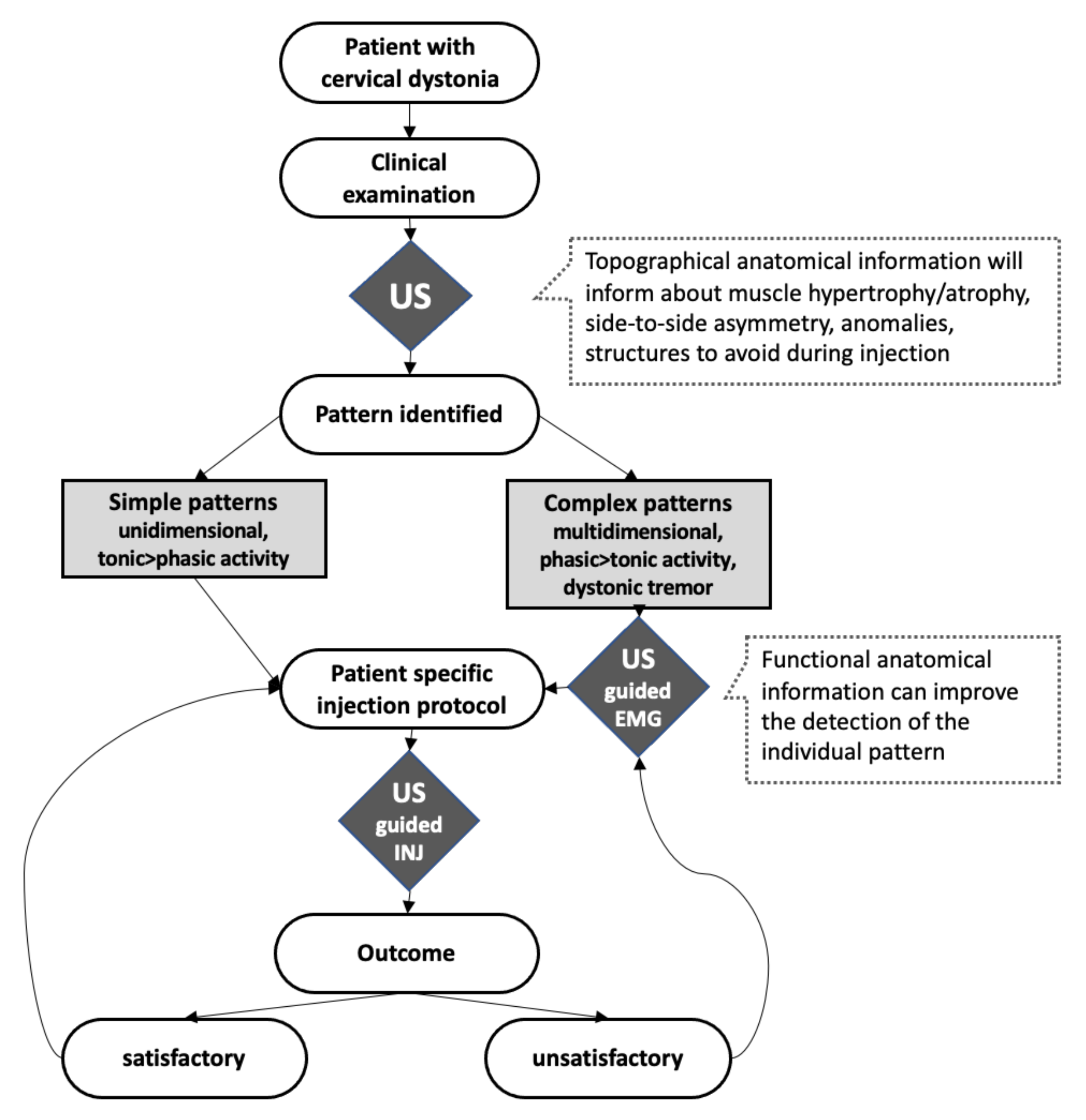
:1. Introduction
2. Technical Background
3. Ultrasound Improves Anatomical Knowledge
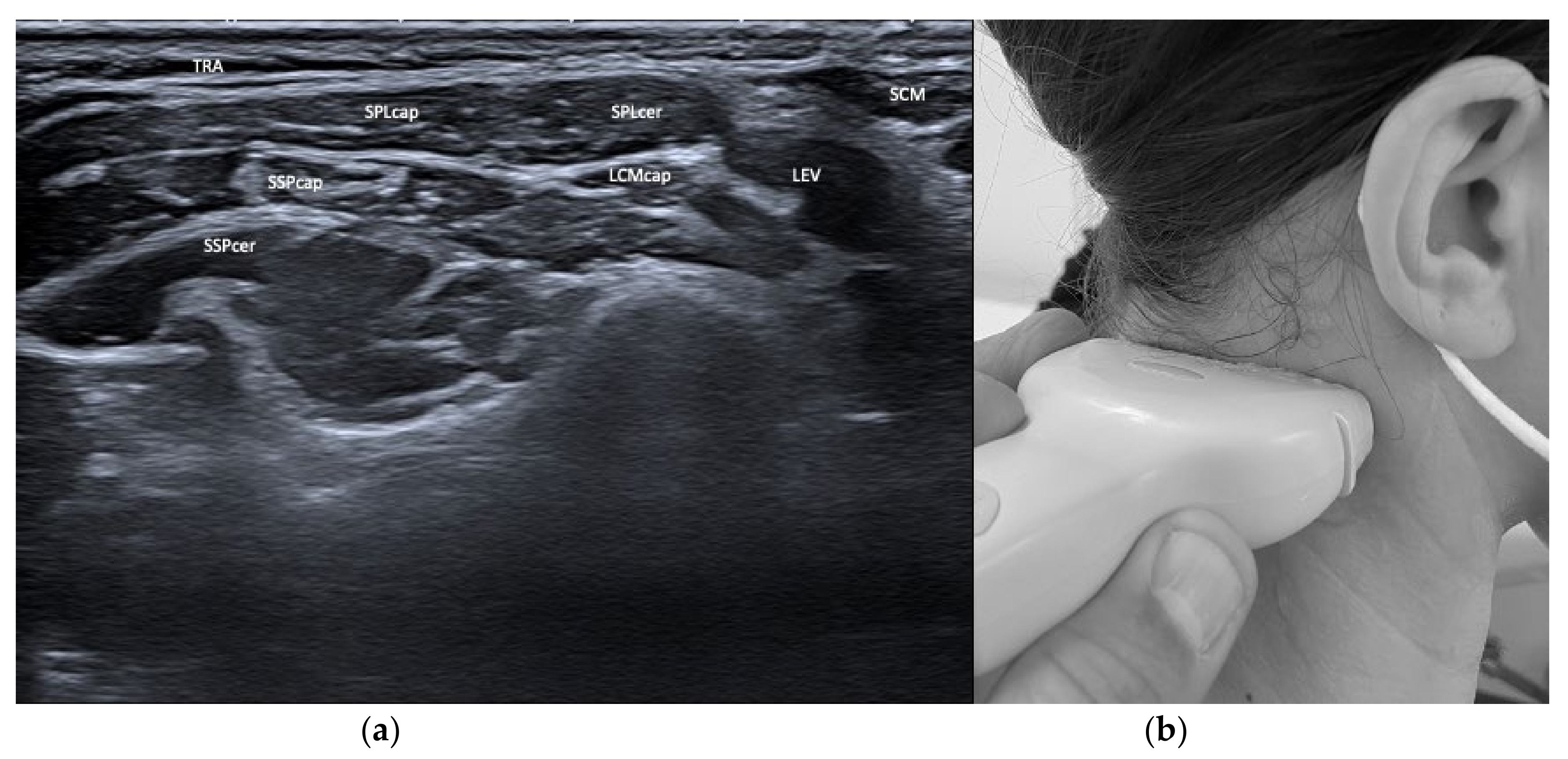
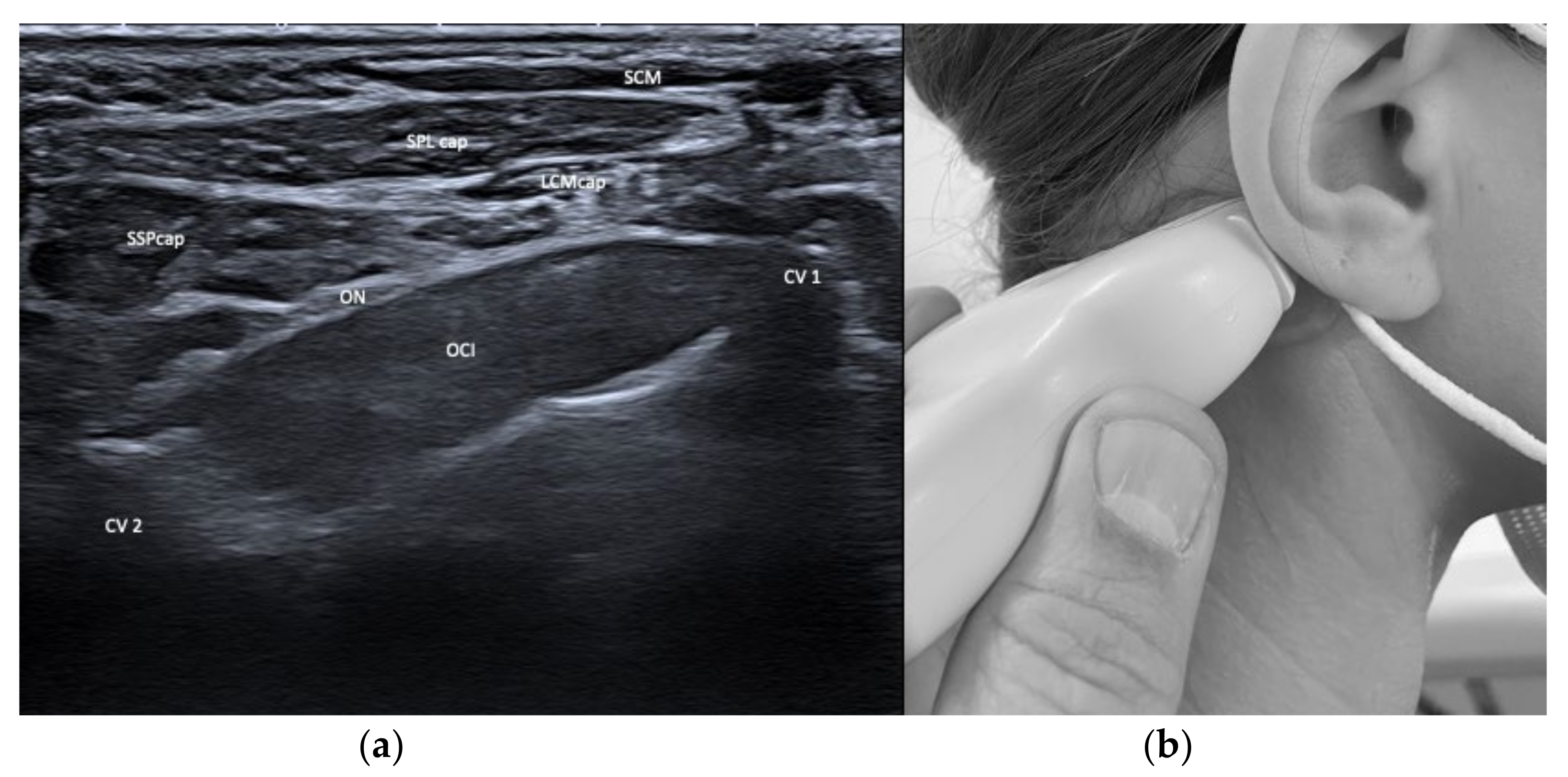
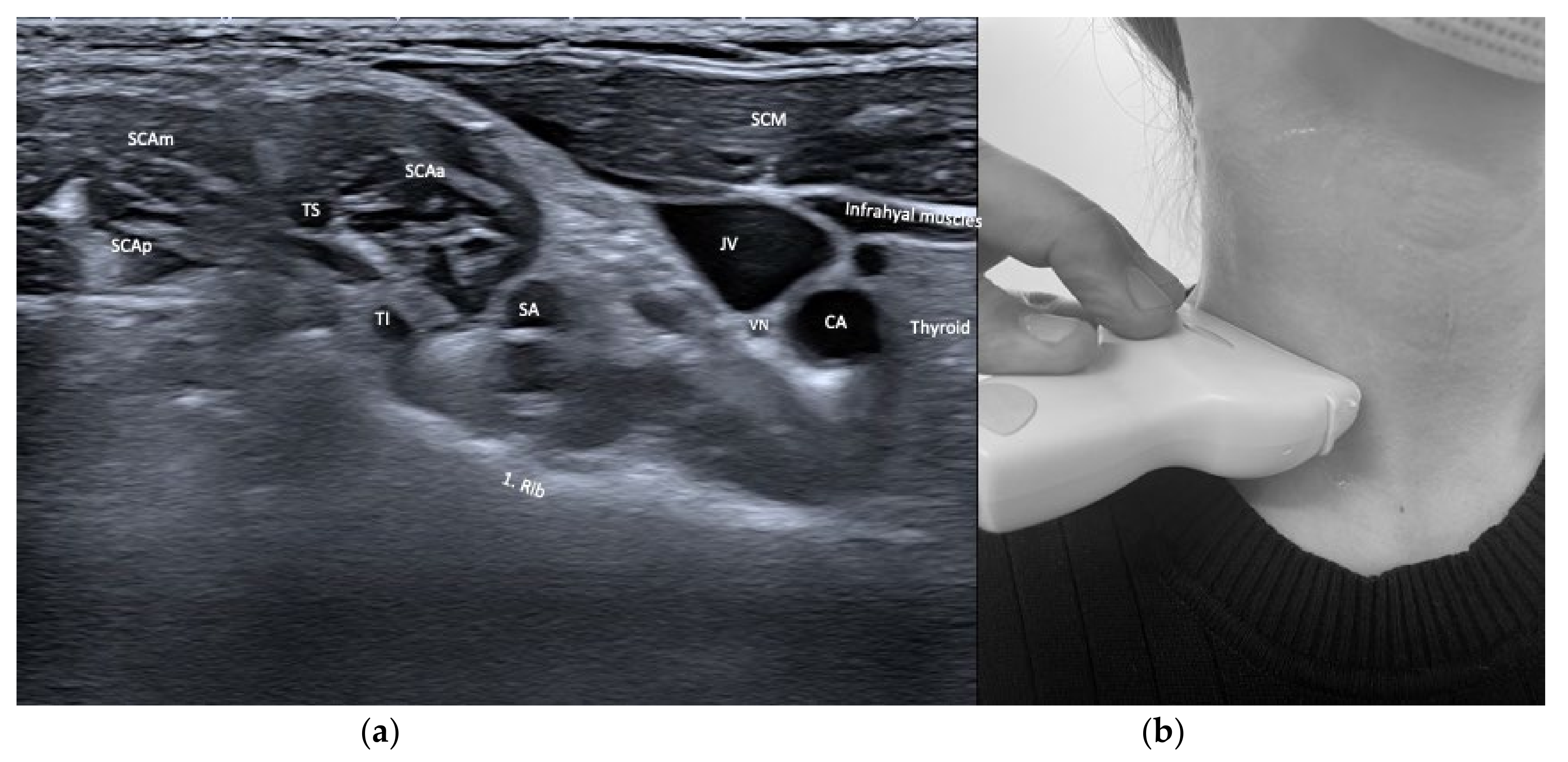
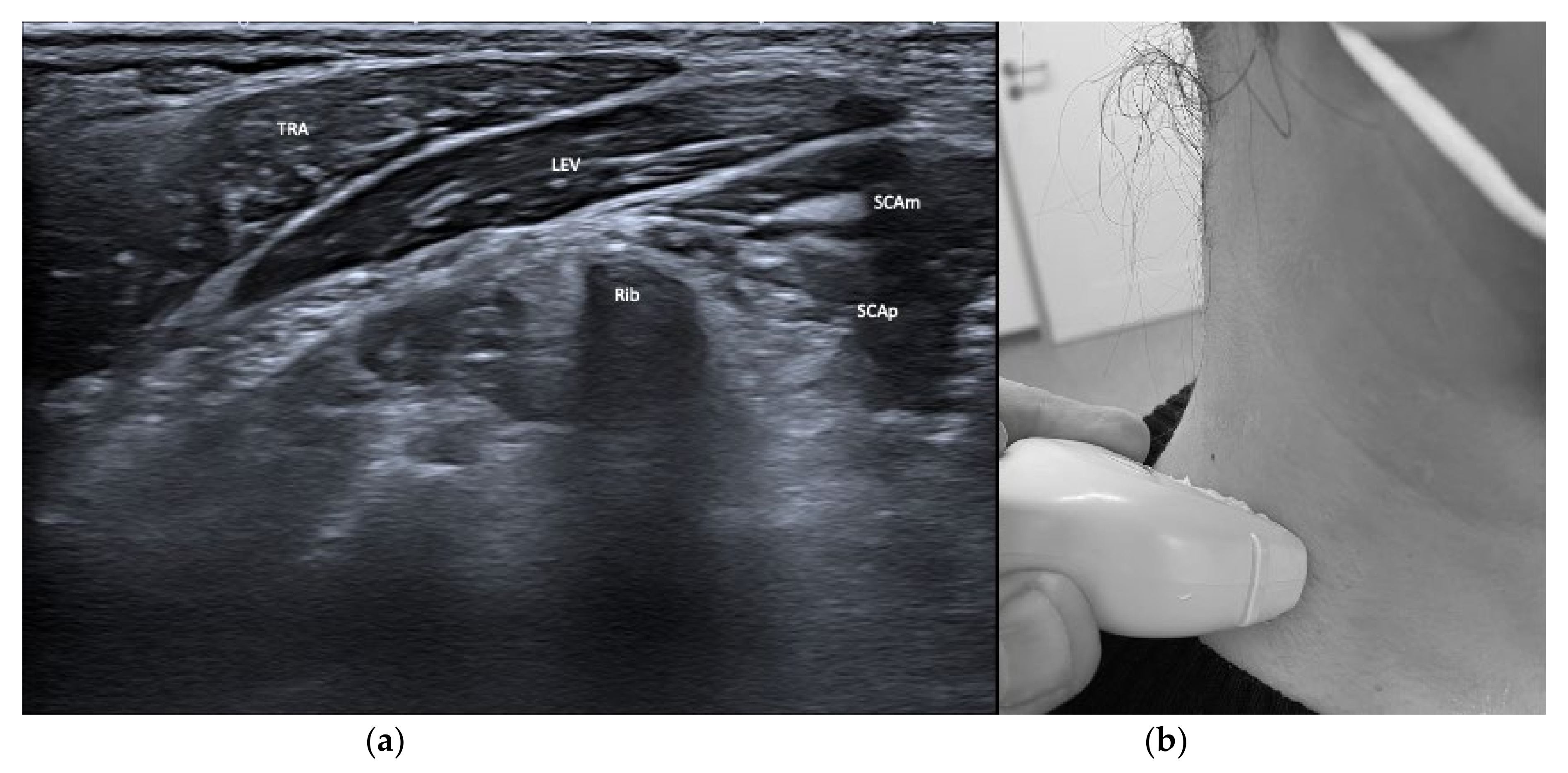
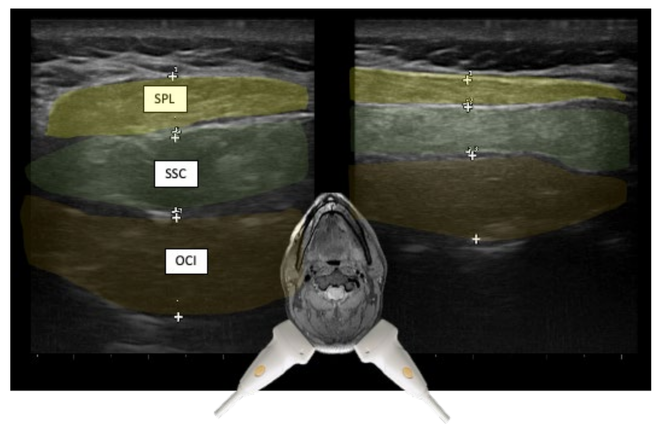
3.1. Layers and Compartments
3.2. Orientation of Layers—Reciprocal Function of Neighboring Structures
3.3. Biomechanical Basic Assumptions: Cross-Section and Lever Arm
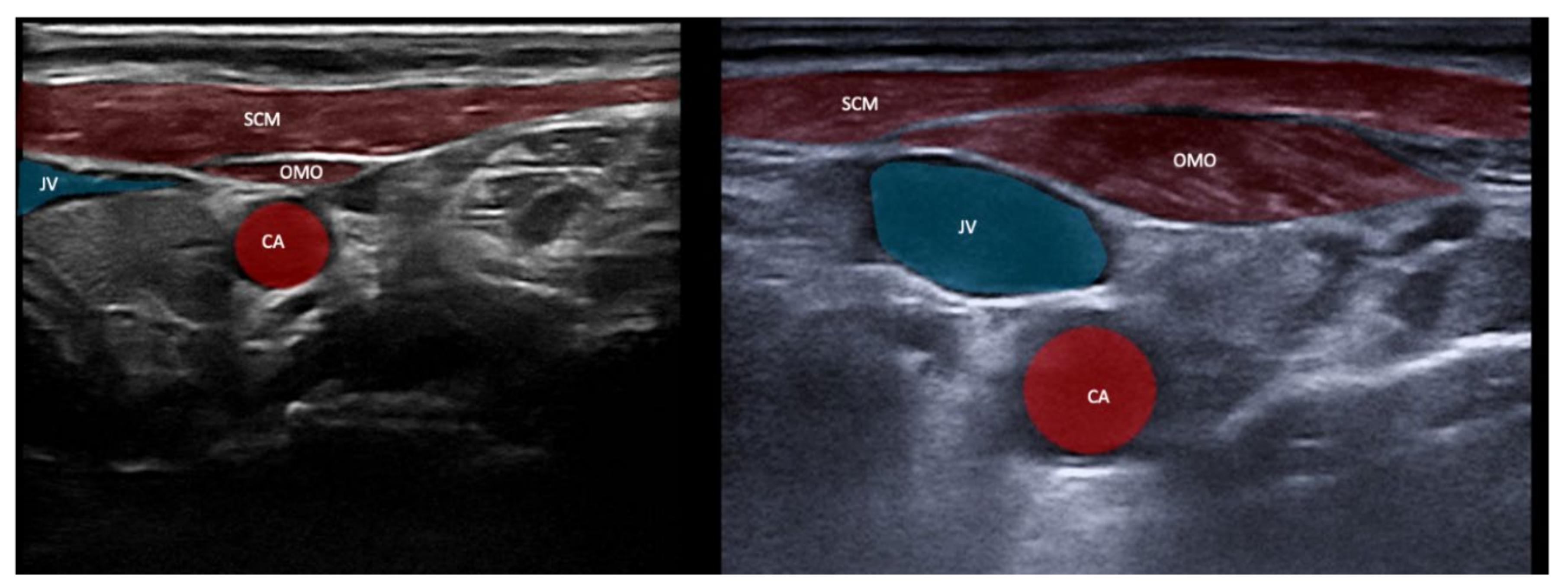
3.4. Safety Issues and Imaging of Relevant Neighboring Structures
3.5. Learning Anatomy Anew—From Clinical Assumptions to Visual Feedback
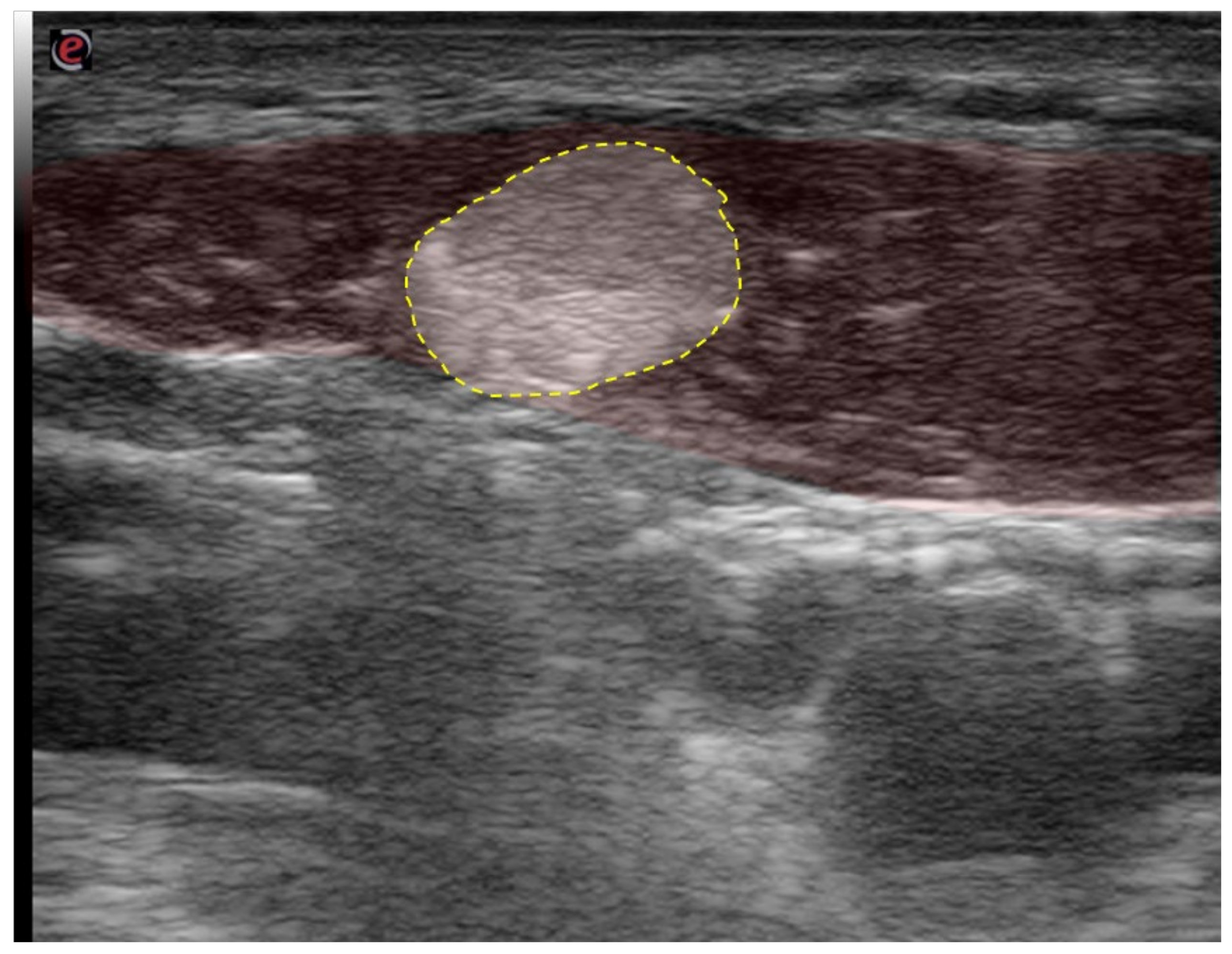
3.6. Injecting BoNT/A Using US Guidance
3.7. The Relevant Muscles for CD
4. Combining US with the EMG
5. Evidence for the Use of US for Botulinum Toxin Treatment for CD
6. Clinical Pearls
6.1. “Depth Matters”—Do Not Underestimate the 3rd Dimension
6.2. “Expect the Unexpected”—Variant Anatomy
6.3. “Watch Out for Tremor”
6.4. “It May Not be Dystonia”
7. Conclusions
- US offers the advantage of unambiguously identifying the targets for BoNT/A treatment of CD.
- US allows the anatomically precise injection even into deep-seated muscles.
- US increases the reproducibility of therapy, and thus, the efficacy and safety of long-term BoNT/A treatment of people with CD.
- Treating with US improves the anatomical knowledge—topographically and functionally—of the practitioner, leading to more individual injection protocols.
- Combining US with EMG guidance opens the path to the analysis of complex clinical patterns of CD that often are unresponsive to standard protocols.
Supplementary Materials
Author Contributions
Funding
Institutional Review Board Statement
Informed Consent Statement
Data Availability Statement
Conflicts of Interest
References
- Simpson, D.M.; Hallett, M.; Ashman, E.J.; Comella, C.L.; Green, M.W.; Gronseth, G.S.; Armstrong, M.J.; Gloss, D.; Potrebic, S.; Jankovic, J.; et al. Practice Guideline Update Summary: Botulinum Neurotoxin for the Treatment of Blepharospasm, Cervical Dystonia, Adult Spasticity, and Headache: Report of the Guideline Development Subcommittee of the American Academy of Neurology. Neurology 2016, 86, 1818–1826. [Google Scholar] [CrossRef] [PubMed] [Green Version]
- Truong, D.; Duane, D.D.; Jankovic, J.; Singer, C.; Seeberger, L.C.; Comella, C.L.; Lew, M.F.; Rodnitzky, R.L.; Danisi, F.O.; Sutton, J.P.; et al. Efficacy and Safety of Botulinum Type A Toxin (Dysport) in Cervical Dystonia: Results of the First US Randomized, Double-Blind, Placebo-Controlled Study. Mov. Disord. 2005, 20, 783–791. [Google Scholar] [CrossRef] [PubMed]
- Jankovic, J.; Schwartz, K.; Donovan, D.T. Botulinum Toxin Treatment of Cranial-Cervical Dystonia, Spasmodic Dysphonia, Other Focal Dystonias and Hemifacial Spasm. J. Neurol. Neurosurg. Psychiatry 1990, 53, 633–639. [Google Scholar] [CrossRef] [PubMed] [Green Version]
- Benecke, R.; Jost, W.H.; Kanovsky, P.; Ruzicka, E.; Comes, G.; Grafe, S. A New Botulinum Toxin Type A Free of Complexing Proteins for Treatment of Cervical Dystonia. Neurology 2005, 64, 1949–1951. [Google Scholar] [CrossRef]
- Tsui, J.K.; Eisen, A.; Stoessl, A.J.; Calne, S.; Calne, D.B. Double-Blind Study of Botulinum Toxin in Spasmodic Torticollis. Lancet 1986, 2, 245–247. [Google Scholar] [CrossRef]
- Finsterer, J.; Maeztu, C.; Revuelta, G.J.; Reichel, G.; Truong, D. Collum-Caput (COL-CAP) Concept for Conceptual Anterocollis, Anterocaput, and Forward Sagittal Shift. J. Neurol. Sci. 2015, 355, 37–43. [Google Scholar] [CrossRef]
- Reichel, G.; Stenner, A.; Jahn, A. The phenomenology of cervical dystonia. Fortschr. Neurol. Psychiatr. 2009, 77, 272–277. [Google Scholar] [CrossRef]
- Lee, I.H.; Yoon, Y.C.; Sung, D.H.; Kwon, J.W.; Jung, J.Y. Initial Experience with Imaging-Guided Intramuscular Botulinum Toxin Injection in Patients with Idiopathic Cervical Dystonia. Am. J. Roentgenol. 2009, 192, 996–1001. [Google Scholar] [CrossRef]
- Berweck, S.; Feldkamp, A.; Francke, A.; Nehles, J.; Schwerin, A.; Heinen, F. Sonography-Guided Injection of Botulinum Toxin A in Children with Cerebral Palsy. Neuropediatrics 2002, 33, 221–223. [Google Scholar] [CrossRef]
- Berweck, S.; Schroeder, A.S.; Fietzek, U.M.; Heinen, F. Sonography-Guided Injection of Botulinum Toxin in Children with Cerebral Palsy. Lancet 2004, 363, 249–250. [Google Scholar] [CrossRef]
- Kirchmair, L.; Entner, T.; Wissel, J.; Moriggl, B.; Kapral, S.; Mitterschiffthaler, G. A Study of the Paravertebral Anatomy for Ultrasound-Guided Posterior Lumbar Plexus Block. Anesth. Analg. 2001, 93, 477–481, 4th contents page. [Google Scholar] [CrossRef]
- Heinen, F.; Desloovere, K.; Schroeder, A.S.; Berweck, S.; Borggraefe, I.; van Campenhout, A.; Andersen, G.L.; Aydin, R.; Becher, J.G.; Bernert, G.; et al. The Updated European Consensus 2009 on the Use of Botulinum Toxin for Children with Cerebral Palsy. Eur. J. Paediatr. Neurol. 2010, 14, 45–66. [Google Scholar] [CrossRef]
- Gray, A.T. Ultrasound-Guided Regional Anesthesia: Current State of the Art. Anesthesiology 2006, 104, 368–373. [Google Scholar] [CrossRef] [Green Version]
- Schramm, A.; Bäumer, T.; Fietzek, U.; Heitmann, S.; Walter, U.; Jost, W.H. Relevance of Sonography for Botulinum Toxin Treatment of Cervical Dystonia: An Expert Statement. J. Neural Transm. 2015, 122, 1457–1463. [Google Scholar] [CrossRef] [Green Version]
- Walter, U.; Dressler, D. Ultrasound-Guided Botulinum Toxin Injections in Neurology: Technique, Indications and Future Perspectives. Expert Rev. Neurother. 2014, 14, 923–936. [Google Scholar] [CrossRef]
- Pillen, S.; van Alfen, N. Skeletal Muscle Ultrasound. Neurol. Res. 2011, 33, 1016–1024. [Google Scholar] [CrossRef] [Green Version]
- Kreisler, A.; Gerrebout, C.; Defebvre, L.; Demondion, X. Accuracy of Non-Guided versus Ultrasound-Guided Injections in Cervical Muscles: A Cadaver Study. J. Neurol. 2021, 268, 1894–1902. [Google Scholar] [CrossRef]
- Kim, B.S.; Kim, D.S.; Kang, S.; Kim, J.Y.; Kang, B.; Rhyu, I.J.; Yoon, J.S. Ultrasound-Guided Injection of the Sternocleidomastoid Muscle: A Cadaveric Study with Implications for Chemodenervation. PM&R 2020. [Google Scholar] [CrossRef]
- Ko, Y.D.; Yun, S.I.; Ryoo, D.; Chung, M.E.; Park, J. Accuracy of Ultrasound-Guided and Non-Guided Botulinum Toxin Injection Into Neck Muscles Involved in Cervical Dystonia: A Cadaveric Study. Ann. Rehabil. Med. 2020, 44, 370–377. [Google Scholar] [CrossRef]
- Hong, J.S.; Sathe, G.G.; Niyonkuru, C.; Munin, M.C. Elimination of Dysphagia Using Ultrasound Guidance for Botulinum Toxin Injections in Cervical Dystonia. Muscle Nerve 2012, 46, 535–539. [Google Scholar] [CrossRef]
- Kutschenko, A.; Klietz, M.; Paracka, L.; Kollewe, K.; Schulte-Sutum, A.; Janssen, T.; Schrader, C.; Wegner, F.; Dressler, D. Dysphagia in Cervical Dystonia Patients Receiving Optimised Botulinum Toxin Therapy: A Single-Center Retrospective Cohort Study. J. Neural Transm. 2020, 127, 1161–1165. [Google Scholar] [CrossRef]
- Hefter, H.; Kupsch, A.; Müngersdorf, M.; Paus, S.; Stenner, A.; Jost, W. Dysport Cervical Dystonia Study Group A Botulinum Toxin A Treatment Algorithm for de Novo Management of Torticollis and Laterocollis. BMJ Open 2011, 1, e000196. [Google Scholar] [CrossRef] [Green Version]
- Flowers, J.M.; Hicklin, L.A.; Marion, M.-H. Anterior and Posterior Sagittal Shift in Cervical Dystonia: A Clinical and Electromyographic Study, Including a New EMG Approach of the Longus Colli Muscle. Mov. Disord. Off. J. Mov. Disord. Soc. 2011, 26, 2409–2414. [Google Scholar] [CrossRef]
- Loram, I.; Siddique, A.; Sanchez, M.B.; Harding, P.; Silverdale, M.; Kobylecki, C.; Cunningham, R. Objective Analysis of Neck Muscle Boundaries for Cervical Dystonia Using Ultrasound Imaging and Deep Learning. IEEE J. Biomed. Health Inform. 2020, 24, 1016–1027. [Google Scholar] [CrossRef]
- Song, Y.; Zhang, T.-J.; Li, Y.; Gao, Y. Application of Real-Time Shear Wave Elastography in the Assessment of Torsional Cervical Dystonia. Quant. Imaging Med. Surg. 2019, 9, 662–670. [Google Scholar] [CrossRef]
- Walter, U.; Dudesek, A.; Fietzek, U.M. A Simplified Ultrasonography-Guided Approach for Neurotoxin Injection into the Obliquus Capitis Inferior Muscle in Spasmodic Torticollis. J. Neural Transm. 2018, 125, 1037–1042. [Google Scholar] [CrossRef]
- Lagnau, P.; Lo, A.; Sandarage, R.; Alter, K.; Picelli, A.; Wissel, J.; Verduzco-Gutierrez, M.; Suputtitada, A.; Munin, M.C.; Carda, S.; et al. Ergonomic Recommendations in Ultrasound-Guided Botulinum Neurotoxin Chemodenervation for Spasticity: An International Expert Group Opinion. Toxins 2021, 13, 249. [Google Scholar] [CrossRef] [PubMed]
- Elwischger, K.; Kasprian, G.; Weber, M.; Meyerspeer, M.; Linder, C.; Auff, E.; Prayer, D.; Sycha, T.; Kranz, G. Intramuscular Distribution of Botulinum Toxin—Visualized by MRI. J. Neurol. Sci. 2014, 344, 76–79. [Google Scholar] [CrossRef]
- Gracies, J.-M.; Lugassy, M.; Weisz, D.J.; Vecchio, M.; Flanagan, S.; Simpson, D.M. Botulinum Toxin Dilution and Endplate Targeting in Spasticity: A Double-Blind Controlled Study. Arch. Phys. Med. Rehabil. 2009, 90, 9–16.e2. [Google Scholar] [CrossRef] [PubMed]
- Delnooz, C.C.S.; Veugen, L.C.; Pasman, J.W.; Lapatki, B.G.; van Dijk, J.P.; van de Warrenburg, B.P.C. The Clinical Utility of Botulinum Toxin Injections Targeted at the Motor Endplate Zone in Cervical Dystonia. Eur. J. Neurol. 2014, 21, 1486-e98. [Google Scholar] [CrossRef] [PubMed]
- Comella, C.L.; Buchman, A.S.; Tanner, C.M.; Brown, T.N.; Goetz, C.G. Botulinum Toxin Injection for Spasmodic Torticollis: Increased Magnitude of Benefit with Electromyographic Assistance. Neurology 1992, 42, 878–882. [Google Scholar] [CrossRef]
- Werdelin, L.; Dalager, T.; Fuglsang-Frederiksen, A.; Regeur, L.; Karlsborg, M.; Korbo, L.; Munck, O.; Winge, K. The Utility of EMG Interference Pattern Analysis in Botulinum Toxin Treatment of Torticollis: A Randomised, Controlled and Blinded Study. Clin. Neurophysiol. Off. J. Int. Fed. Clin. Neurophysiol. 2011, 122, 2305–2309. [Google Scholar] [CrossRef]
- Nijmeijer, S.W.R.; de Bruijn, E.; Forbes, P.A.; Kamphuis, D.J.; Happee, R.; Koelman, J.H.T.M.; Tijssen, M.A.J. EMG Coherence and Spectral Analysis in Cervical Dystonia: Discriminative Tools to Identify Dystonic Muscles? J. Neurol. Sci. 2014, 347, 167–173. [Google Scholar] [CrossRef]
- Nijmeijer, S.W.R.; de Bruijn, E.; Verhagen, R.; Forbes, P.A.; Kamphuis, D.J.; Happee, R.; Tijssen, M.A.J.; Koelman, J.H.T.M. Spectral EMG Changes in Cervical Dystonia Patients and the Influence of Botulinum Toxin Treatment. Toxins 2017, 9, 256. [Google Scholar] [CrossRef] [Green Version]
- Schramm, A.; Huber, D.; Möbius, C.; Münchau, A.; Kohl, Z.; Bäumer, T. Involvement of Obliquus Capitis Inferior Muscle in Dystonic Head Tremor. Parkinsonism Relat. Disord. 2017, 44, 119–123. [Google Scholar] [CrossRef]
- Shaikh, A.G.; Beylergil, S.B.; Scorr, L.; Kilic-Berkmen, G.; Freeman, A.; Klein, C.; Junker, J.; Loens, S.; Brüggemann, N.; Münchau, A.; et al. Dystonia and Tremor: A Cross-Sectional Study of the Dystonia Coalition Cohort. Neurology 2021, 96, e563–e574. [Google Scholar] [CrossRef]
- Fujimoto, H.; Mezaki, T.; Yokoe, M.; Mochizuki, H. Sonographic Guidance Provides a Low-Risk Approach to the Longus Colli Muscle. Mov. Disord. Off. J. Mov. Disord. Soc. 2012, 27, 928–929. [Google Scholar] [CrossRef]
- Huang, L.; Chen, H.-X.; Ding, X.-D.; Xiao, H.-Q.; Wang, W.; Wang, H. Efficacy Analysis of Ultrasound-Guided Local Injection of Botulinum Toxin Type A Treatment with Orthopedic Joint Brace in Patients with Cervical Dystonia. Eur. Rev. Med. Pharmacol. Sci. 2015, 19, 1989–1993. [Google Scholar]
- Allison, S.K.; Odderson, I.R. Ultrasound and Electromyography Guidance for Injection of the Longus Colli With Botulinum Toxin for the Treatment of Cervical Dystonia. Ultrasound Q. 2016, 32, 302–306. [Google Scholar] [CrossRef]
- Tyślerowicz, M.; Jost, W.H. Injection into the Longus Colli Muscle via the Thyroid Gland. Tremor Hyperkinetic Mov. 2019, 9. [Google Scholar] [CrossRef]
- Tatu, L.; Jost, W.H. Anatomy and Cervical Dystonia: “Dysfunction Follows Form”. J. Neural Transm. 2017, 124, 237–243. [Google Scholar] [CrossRef]


| Muscle (Abbreviation) | Predominant Function from Neutral Position | Relevance of US | Neighboring Structures | Comment | |
|---|---|---|---|---|---|
| For Localization | To Avoid Side Effects | ||||
| Infra-/suprahyoid | anteflexion of head and neck | +++ | +++ | Thyroid gland | Dysphagia |
| Sternocleido-mastoid (SCM) | Contraversion of head and neck Ipsitilt of head and neck Anteflexion of head and neck with bilateral activation | + | ++ | Infrahyoid/supra-hyoid muscles, omohyoid, carotid artery, jugular vein | Dysphagia (particularly if injected bilaterally) |
| Longus capitis (LNGcap) | Anteflexion of head and neck | +++ | +++ | Carotid artery, jugular vein, vagus nerve, phrenic nerve | Authors recommend a transoral injection |
| Longus colli (LNGco) | Anteflexion of neck | +++ | +++ | Carotid artery, jugular vein, vagus nerve, phrenic nerve | Authors recommend a transoral injection |
| Scalenus anterior (SCAa) | Anteflexion of neck | +++ | +++ | Thyroid, carotid artery, brachial plexus, phrenic nerve, lung | |
| Scalenus medius/posterior (SCAmp) | Ipsitilt of neck | ++ | ++ | Brachial plexus, lung | |
| Semispinalis capitis (SSPcap) | Extension of head | + | + | SPLcap, OCI | Strongest extensor muscle of head and neck |
| Semispinalis cervicis (SSPcer) | Extension of neck | + | + | SPLcer, TRA | |
| Splenius capitis (SPLcap) | Ipsiversion of head | ++ | ++ | major occipital nerve, SPLcap/cer, LSMcap, OCI | Prominent reduction in bulk from repeated injections possible |
| Ipsitilt of head | |||||
| Splenius cervicis (SPLcer) | Ipsiversion of neck | +++ | ++ | LEV, TRA, Longissimus cervicis | Relevant for full turn of the neck |
| Ipsitilt of the neck | |||||
| Longissimus capitis (LCM) | Ipsiversion of head and neck | +++ | ++ | SPLcap, SSPcap | Obligatory USG |
| Trapezius (TRA) | Extension of neck | + | + | LEV, Supraspinatus | |
| Contraversion of neck | |||||
| Levator scapulae (LEV) | Lift of scapula | + | + | SPLcer, TRA | |
| Ipsitilt of head and neck | Role for antecollis variants is discussed variably | ||||
| Obliquus capitis inferior (OCI) | Ipsiversion of head | +++ | +++ | SSPcap, RCM, vertebral artery, greater occipital nerve | Adjacent muscles are all extensors of the head |
| Rectus capitis major (RCM) | Extension of head | +++ | +++ | SSPcap, OCI | |
| Author | Year | N | Type of Study | Method | Main Result |
|---|---|---|---|---|---|
| Lee et al. [8] | 2008 | 6 | Retrospective chart review | US, CT, EMG, and SPECT | OCI is a relevant muscle in CD. |
| Hong et al. [20] | 2012 | 5 | Retrospective chart review | US and EMG | Dysphagia was eliminated using combined US and EMG guided injection. |
| Fujimoto et al. [37] | 2012 | 1 | Case report | US | For guiding injection into LNGcol US is recommended rather than EMG. |
| Huang et al. [38] | 2015 | 105 | Prospective, randomized, controlled | US combined with joint brace | Significant improvement in dystonia at 1, 3, and 6 months of treatment compared to non-guided injection |
| Allison et al. [39] | 2016 | 1 | Case report | US and EMG | Injection of LongCol was efficient in the improvement of antecollis. |
| Schramm et al. [35] | 2017 | 35 | Prospective, non-controlled | US and EMG | Muscle activity of OCI is present in tremulous CD and the injection of OCI using US is recommended. |
| Walter et al. [26] | 2018 | 5 | Retrospective chart review | US | Add-on US guided injections into the OCI led to better outcomes, especially of the tremulous component. |
| Tyslerowicz et al. [40] | 2019 | 1 | Case report | US and EMG | Injection of LongCol is recommended using EMG and US. |
| Kutschenko et al. [21] | 2020 | 117 | Retrospective chart review | US | US guided application failed to prevent dysphagia (retrospective study) |
Publisher’s Note: MDPI stays neutral with regard to jurisdictional claims in published maps and institutional affiliations. |
© 2021 by the authors. Licensee MDPI, Basel, Switzerland. This article is an open access article distributed under the terms and conditions of the Creative Commons Attribution (CC BY) license (https://creativecommons.org/licenses/by/4.0/).
Share and Cite
Fietzek, U.M.; Nene, D.; Schramm, A.; Appel-Cresswell, S.; Košutzká, Z.; Walter, U.; Wissel, J.; Berweck, S.; Chouinard, S.; Bäumer, T. The Role of Ultrasound for the Personalized Botulinum Toxin Treatment of Cervical Dystonia. Toxins 2021, 13, 365. https://0-doi-org.brum.beds.ac.uk/10.3390/toxins13050365
Fietzek UM, Nene D, Schramm A, Appel-Cresswell S, Košutzká Z, Walter U, Wissel J, Berweck S, Chouinard S, Bäumer T. The Role of Ultrasound for the Personalized Botulinum Toxin Treatment of Cervical Dystonia. Toxins. 2021; 13(5):365. https://0-doi-org.brum.beds.ac.uk/10.3390/toxins13050365
Chicago/Turabian StyleFietzek, Urban M., Devavrat Nene, Axel Schramm, Silke Appel-Cresswell, Zuzana Košutzká, Uwe Walter, Jörg Wissel, Steffen Berweck, Sylvain Chouinard, and Tobias Bäumer. 2021. "The Role of Ultrasound for the Personalized Botulinum Toxin Treatment of Cervical Dystonia" Toxins 13, no. 5: 365. https://0-doi-org.brum.beds.ac.uk/10.3390/toxins13050365