Article

Enzymatic Degradation of Multiple Major Mycotoxins by Dye-Decolorizing Peroxidase from Bacillus subtilis

State Key Laboratory of Animal Nutrition, Institute of Animal Sciences, Chinese Academy of Agricultural Sciences, Beijing 100193, China
*
Authors to whom correspondence should be addressed.
Submission received: 20 May 2021 / Revised: 9 June 2021 / Accepted: 16 June 2021 / Published: 19 June 2021
(This article belongs to the Section Mycotoxins)

Abstract

:
The co-occurrence of multiple mycotoxins, including aflatoxin B1 (AFB1), zearalenone (ZEN) and deoxynivalenol (DON), widely exists in cereal-based animal feed and food. At present, most reported mycotoxins degrading enzymes target only a certain type of mycotoxins. Therefore, it is of great significance for mining enzymes involved in the simultaneous degradation of different types of mycotoxins. In this study, a dye-decolorizing peroxidase-encoding gene BsDyP from Bacillus subtilis SCK6 was cloned and expressed in Escherichia coli BL21/pG-Tf2. The purified recombinant BsDyP was capable of oxidizing various substrates, including lignin phenolic model compounds 2,6-dimethylphenol and guaiacol, the substrate 2,2′-azino-bis (3-ethylbenzothiazoline-6-sulfonic acid), anthraquinone dye reactive blue 19 and azo dye reactive black 5, as well as Mn2+. In addition, BsDyP could efficiently degrade different types of mycotoxins, including AFB1, ZEN and DON, in presence of Mn2+. More important, the toxicities of their corresponding enzymatic degradation products AFB1-diol, 15-OH-ZEN and C15H18O8 were significantly lower than AFB1, ZEN and DON. In summary, these results proved that BsDyP was a promising candidate for the simultaneous degradation of multiple mycotoxins in animal feed and food.
Key Contribution: BsDyP could degrade multiple major mycotoxins including AFB1, ZEN and DON through the catalytic oxidation of Mn2+. The predicted biological toxicity of each corresponding degradation product was significantly removed.

1. Introduction

Mycotoxins are a diverse group of toxic secondary metabolites produced by certain filamentous fungi, and they display carcinogenicity, teratogenicity, immune toxicity, neurotoxicity, hepatotoxicity, nephrotoxicity, reproductive and developmental toxicity towards humans and animals [1]. Until now, more than 400 mycotoxins have been identified and reported; however, only a few mycotoxins dominate in contaminated food and animal feed. These are aflatoxins B1 (AFB1), zearalenone (ZEN), deoxynivalenol (DON), ochratoxin A, fumonisin B1, citrinin and patulin [2,3]. Meanwhile, the co-occurrence of abovementioned major mycotoxins is frequently observed in cereal-based animal feed and food [4,5], which might lead to synergistic and additive toxicological effects in humans and animals due to different mechanisms of action of mycotoxins at the cellular level [6].
The control of mycotoxins in food and feed depends on two main approaches: the prevention of mycotoxin production and detoxification of mycotoxins [7,8]. Most agronomic, physical and chemical strategies at pre-harvest as well as at post-harvest have been shown to be rather ineffective to control mycotoxin contamination [8]. With the increasing demand for effective approaches to reducing mycotoxin contamination, researchers have been paying more attention to biological strategies. The detoxification of mycotoxins through biotransformation by microorganisms or their enzymes has gradually become one of the most promising strategies because of its advantages in efficiency, specificity and environmental friendliness [7,9]. As for biotransformation of multiple mycotoxins, several microorganisms such as Bacillus subtilis, Rhodococcus percolatus and Pseudomonas putida have been reported to be capable of degrading AFB1 and ZEN [7,10,11,12]. In contrast, only a few enzymes have been identified, purified and characterized for multiple mycotoxin biotransformation.
Considering that part of mycotoxins is structurally similar to a lignin monomer or its derivatives, ligninolytic enzymes might be able to degrade different types of mycotoxins. In our previous study, we found that fungal manganese peroxidases could degrade four major mycotoxins, namely AFB1, ZEN, DON and fumonisin B1, in the presence of a dicarboxylic acid malonate [13]. Moreover, bacterial and fungal laccases could simultaneously degrade AFB1 and ZEN, using the laccase/mediator systems [14]. In this study, another ligninolytic enzyme dye-decolorizing peroxidase (DyP) from B. subtilis SCK6 was expressed in Escherichia coli, using the cold shock-inducible expression system coupled with co-expression of chaperones. Then the substrate specificity of purified recombinant BsDyP was characterized. Moreover, the degrading capacity of three major mycotoxins AFB1, ZEN and DON by BsDyP was evaluated, and corresponding enzymatic degradation products were identified by mass spectrometry analysis.

2. Results and Discussion

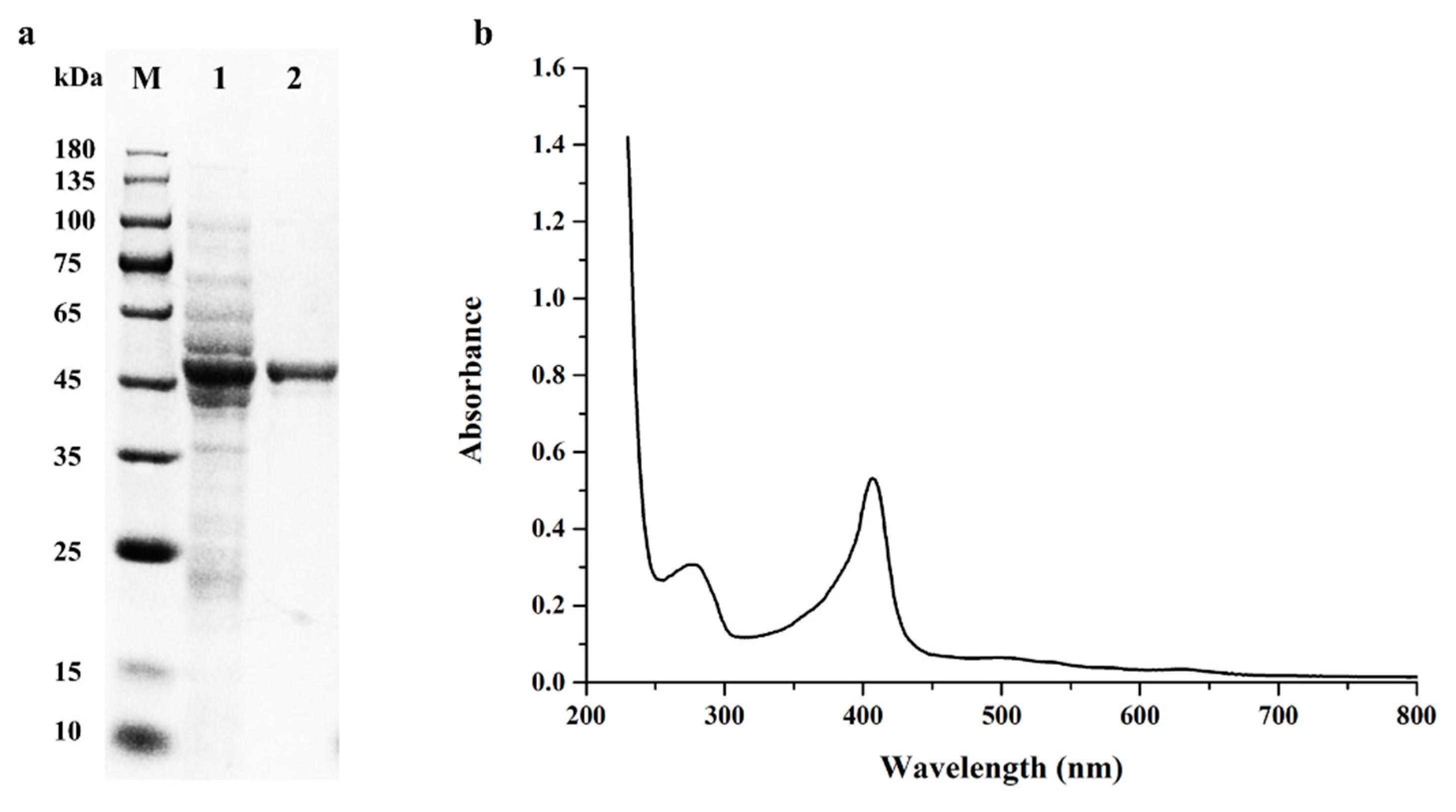
2.1. Gene Cloning, Expression and Purification of BsDyP from B. subtilis SCK6

It has been reported that B. subtilis could be able to efficiently degrade different types of mycotoxins, including AFB1 and ZEN [15,16]. However, very few mycotoxins-degrading enzymes are identified and characterized except for CotA laccase [14]. In the present study, genomic analysis of B. subtilis SCK6 revealed that ligninolytic enzymes were composed of DyP and laccase based on the PeroxiBase and CAZy databases annotation. One DyP gene, 1251 bp, was identified in the genome of B. subtilis SCK6, which encoded 416 amino acids with an estimated molecular weight of 45.7 kDa and an isoelectric point value of 8.71. The cloned BsDyP from B. subtilis SCK6 shared 93% nucleic acid sequence identity and 96% amino acid identity with BsDyP from B. subtilis KCTC2023 [17].
E. coli, one of the most popular hosts for recombinant protein overexpression, was used as the expressing host to heterologously express BsDyP. Given that the ligninolytic enzyme DyP expressed in E. coli accumulated in inclusion bodies which required another refolding process [18,19,20], the cold shock-inducible expression system coupled with co-expression of chaperones was applied to obtain the active DyP. With the help of molecular chaperones groES, groEL and tig, BsDyP was successfully expressed in E. coli BL21 as a soluble form. A significant activity of DyP was detected in the sonicated cell supernatant, indicating the proper formation of disulfide bonds and heme incorporation into recombinant BsDyP. After purification by immobilized metal affinity chromatography, BsDyP showed a single band on SDS-PAGE gel, corresponding to the calculated molecular masses (Figure 1a).

2.2. Biochemical Characterization of the Purified Recombinant BsDyP

Like other ligninolytic peroxidases, including manganese peroxidase, versatile peroxidase and lignin peroxidase, DyP also showed the typical features of heme-containing enzymes with a Soret peak at 404–408 nm [21]. As shown in Figure 1b, BsDyP had an obvious absorbance peak at 407 nm, indicating that the heme group was indeed incorporated into the purified recombinant BsDyP. The Rz (A407/A280) ratio of the purified BsDyP was 1.7.
It had been reported that DyP could act on a broad range of substrates, including lignin-derived phenolic and non-phenolic compounds, synthetic high redox potential anthraquinone and azo dyes [19,22]. In order to explore its substrate specificity, BsDyP was tested on a series of lignin-derived compounds, synthetic dyes and Mn2+. The purified recombinant BsDyP could oxidize the model substrate ABTS, phenolic compounds DMP and GUA, anthraquinone dye RB19, azo dye RB5 and Mn2+ except for the non-phenolic compound VA (Figure 2). Notably, BsDyP had the ability to oxidize Mn2+ to Mn3+, which belongs to the catalytic properties of manganese peroxidase and versatile peroxidase [23,24]. The results suggested that BsDyP might also contain the manganese binding sites.
In addition, the optimum pHs of the purified recombinant BsDyP oxidizing various substrates, including ABTS, DMP, GUA, RB19 and Mn2+, were all 4.0, except that a higher optimum pH of BsDyP against RB5 was 6.0. Meanwhile, BsDyP was completely inactive when the pH was below 3.0, which was different from fungal DyP with a wider acidic pH range [22,25]. The specific activity of BsDyP for ABTS, DMP, GUA, RB19 and Mn2+ at optimum pH were 7.25, 1.79, 0.03, 0.67 and 0.46 U/mg, respectively. Like other dye-decolorizing peroxidases, DyPs from Pseudomonas putida, Thermobifida fusca and Thermomonospora curvata showed higher specific activities towards ABTS and RB19, exhibiting two orders of magnitude higher than that of GUA [17,26,27]. However, the reason for difference in specific activities towards different substrates had not been well elucidated yet.

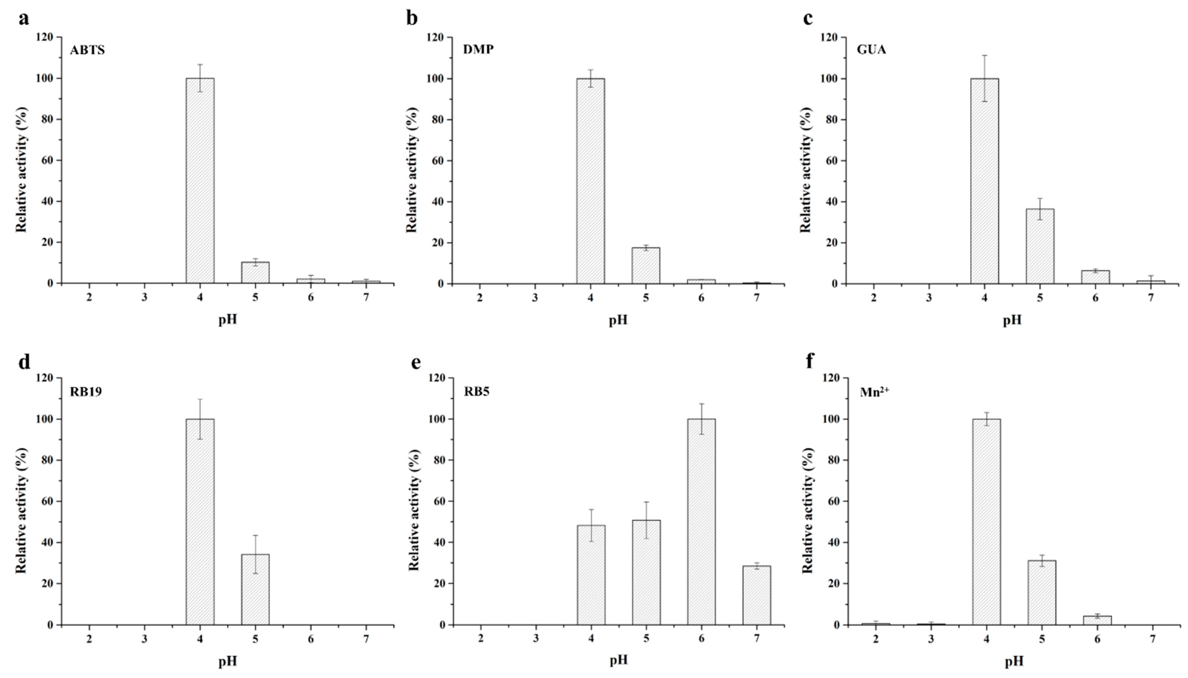
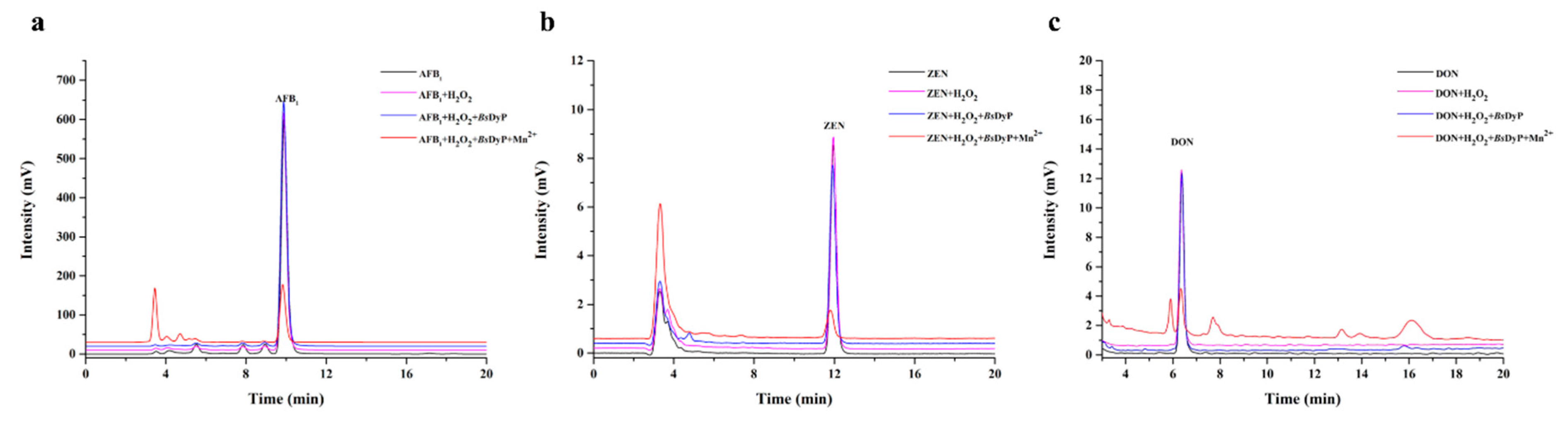
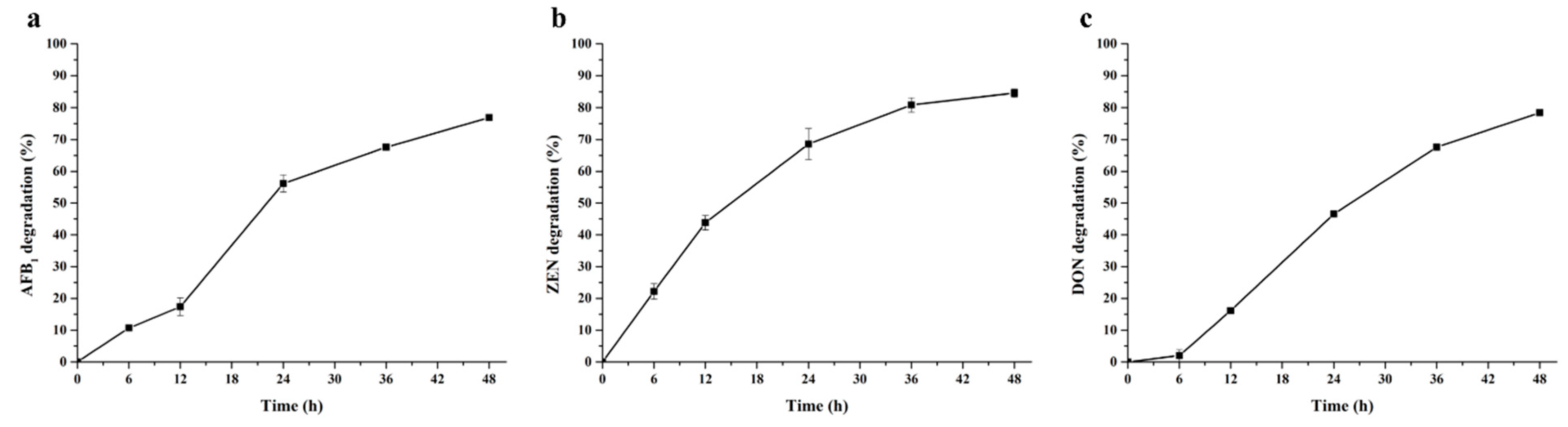
2.3. Degradation of Different Types of Mycotoxins by BsDyP

Although DyP was reported to be involved in lignin degradation, industrial dyes decolorization [21,28], the great application value of DyP in xenobiotic bioremediation gradually attracted more and more attentions in recent years [29,30]. In this study, the different types of mycotoxins including AFB1, ZEN and DON were used to evaluate the mycotoxin degrading capability of BsDyP from B. subtilis SCK6. As shown in Figure 3, BsDyP could efficiently degrade multiple mycotoxins in presence of Mn2+. AFB1, ZEN and DON were degraded up to 76.93%, 84.65% and 78.42% by the purified BsDyP within 48 h, respectively. Unlike AFB1 and DON, ZEN also could be slightly degraded 11.78% by BsDyP in absence of Mn2+, indicating that there might be two different mechanisms of ZEN degradation by BsDyP. On one hand, it was similar to the detoxification mechanism of manganese peroxidases that oxidized Mn3+ and malonate acid mediated degradation of mycotoxins through the formation of radicals [13]. On the other hand, ZEN might be directly degraded by binding to the catalytic pocket of BsDyP.
The time courses of AFB1, ZEN and DON degradation by BsDyP in presence of Mn2+ were shown in Figure 4. The degrading percentages of AFB1 and ZEN were 10.68% and 11.78% at 6 h, and then gradually ascended to 17.37% and 43.84% at 12 h. More than 50% AFB1 and ZEN degradation occurred after a 24 h incubation. In contrast, the initial degradation of DON was slower, with degradation percentages of 1.99% at 6 h, but DON content was significantly decreased at 12 h, and then 46.56% DON was degraded over 24 h of incubation.

2.4. Identification of AFB1, ZEN and DON Degradation Products

In order to further prove the effective detoxification of multiple mycotoxins by BsDyP, it is necessary to identify the corresponding degradation products of mycotoxins and analyze their biological toxicities. Based on the clear relationship between chemical structure and biological activities of AFB1, ZEN and DON [31,32], the toxicology of degradation products could judge from chemical structure changes of different mycotoxins.
The main degradation product of AFB1 was identified by LC-MS/MS. The parent ion appeared at m/z 347.07 [M + H]+ and produced daughter ions of 329.06 [M − 18 + H]+, 301.07 [M − 46 + H]+ and 273.08 [M − 74 + H]+ (Figure 5a). These daughter ions were in accordance with the MS/MS fragments of AFB1-diol [33], suggesting that the C8–9 double bond of terminal furan rings in AFB1 was oxidized by BsDyP and two hydroxyl groups were added to the molecular generate AFB1-diol. Remarkably, AFB1-diol was less toxic than AFB1 due to the removal of double bond in the terminal furan ring, which was considered as the basis for toxic and carcinogenic activity [32,34]. Similarly, the manganese peroxidase from the white rot fungus Phanerochaete sordida YK-624 could convert AFB1 to AFB1-diol and effectively remove the mutagenic activity of AFB1 [35]. Manganese peroxidases from the white rot fungus Irpex lacteus CD2 were able to oxidize AFB1 to AFB1-8,9-epoxide as the intermediate product [13]. Thus, the epoxidation and hydrolysis of AFB1 to produce AFB1-diol might be the common characteristic of AFB1 degradation by Mn3+ derived radicals.
The mass spectrum for the corresponding degradation product of ZEN is shown in Figure 5b. Daughter ions were obtained by fragmenting the parent ion with m/z 335.30 [M + H]+, including 317.15 [M − 18 + H]+ and 205.05 [M − 130 + H]+. These daughter ions were in agreement with the MS/MS fragments of 15-OH-ZEN [33], indicating that ZEN was hydroxylated at the C15 position of aromatic moiety by BsDyP and 15-OH-ZEN was the major degradation product. The result revealed that there were significant differences between DyP and the well-known ZEN degrading enzyme lactonohydrolase ZHD101. Unlike the lactonohydrolase ZHD101, BsDyP hydroxylated the aromatic ring rather than cleaved the ester bond of the macrolactone ring in ZEN [36]. Meanwhile, it had been reported that the hydroxylation of aromatic moiety in ZEN exhibited a significantly decreased estrogenicity and the estrogenicity of 15-OH-ZEN was remarkably decreased by 98% compared with ZEN [37]. These indicated that it might be a novel strategy feasible for biodetoxification of ZEN by BsDyP from B. subtilis SCK6.
Although new product peaks of DON degradation by BsDyP appeared in HPLC analysis, only one of the main degradation products was identified by LC–MS/MS, exhibiting a parent ion peak at m/z 327.11 [M + H]+. The corresponding daughter ions were m/z 309.09 [M − 18 + H]+, 257.10 [M − 70 + H]+ and 221.09 [M − 106 + H]+, which were consistent with the MS/MS fragments of C15H18O8 [31]. Although the enzymatic degradation mechanism of DON by BsDyP was unclear, the possible structure of the degradation product C15H18O8 was shown in Figure 5c. According to structure-activity relationship studies, the toxicity of C15H18O8 might be significantly reduced due to the breakage of C9–10 double bond and the loss of C3-hydroxyl group [38]. To our knowledge, this is the first report about biotransformation of multiple mycotoxins, including AFB1, ZEN and DON by DyP. Moreover, the biological toxicities of each corresponding enzymatic degradation product, namely AFB1-diol, 15-OH-ZEN and C15H18O8, were significantly lower than AFB1, ZEN and DON. Though the other degradation products and their biological toxicity remain to be unveiled, these results might be of great value for practical application of BsDyP in the biodetoxification of multiple mycotoxins in animal feed and food.

3. Conclusions

In this study, a dye-decolorizing peroxidase gene was cloned from Bacillus subtilis SCK6, and then successfully expressed in E. coli BL21 as a soluble form, using the cold shock-inducible expression system coupled with co-expression of chaperones. The recombinant BsDyP could oxidize various lignin-derived phenolic compounds and high redox potential synthetic dyes, as well as Mn2+. Moreover, BsDyP could efficiently degrade different types of mycotoxins, including AFB1, ZEN and DON, in the presence of Mn2+. The major degradation products of multiple mycotoxins were AFB1-diol, 15-OH-ZEN and C15H18O8, respectively. The predicted biological toxicity of each corresponding degradation product was significantly removed. Moreover, further studies are needed to confirm the detoxification effects for the pretreatment of contaminated food and animal feed. Nonetheless, the BsDyP exhibited a great application potential for the enzymatic degradation of different types of mycotoxins in animal feed and food.

4. Material and Methods

4.1. Strain and Substrates

B. subtilis SCK6 was isolated and preserved in Institute of Animal Sciences, Chinese Academy of Agricultural Sciences, Beijing, China. Substrates 2,2′-azino-bis (3-ethylbenzothiazoline-6-sulfonic acid) (ABTS), 2,6-dimethylphenol (DMP), guaiacol (GUA), veratryl alcohol (VA), reactive blue 19 (RB19), reactive black 5 (RB5) and mycotoxins, including AFB1, ZEN and DON, were purchased from Sigma-Aldrich (St. Louis, MO, USA). Hemin was purchased from TCI (Tokyo, Japan).

4.2. Cloning and Expression of BsDyP

Based on the 5′ and 3′-end sequences of the BsDyP structural gene, the BsDyP was amplified with gene-specific primers (BsDyP-NdeI-F: 5′ ATCATCATATCGAAGGTAGGCATATGAGCGATGAACAGAAAAAGC 3′; BsDyP-XbaI-R: 5′ TTTTAAGCAGAGATTACCTATCTAGATGATTCCAGCAAACGCTG 3′). The PCR product was assembled into the linearized vector pCold I and then transformed into commercial E. coli BL21/pG-Tf2 chaperone competent cells (Takara, Japan), which harbored a plasmid pG-Tf2 encoding three molecular chaperones: groES, groEL and tig. The cells harboring pCold I-BsDyP were in pre-cultured in LB medium supplemented with 100 μg/mL ampicillin and 20 μg/mL chloramphenicol at 37 °C overnight and used as the inocula of 300 mL LB medium containing 5 ng/mL tetracyclin, 100 μg/mL ampicillin and 20 μg/mL chloramphenicol. Tetracyclin was used as an inducer for the expression of molecular chaperones. The cultures were grown at 37 °C for 4 h, followed by the addition of 0.5 mM isopropy-β-d-thiogalactoside (IPTG) and 20 μM hemin for 12 h induction at 16 °C.

4.3. Purification and Characterization of BsDyP

After induction, the cells were harvested by centrifugation and resuspended in 30 mL binding buffer (20 mM sodium phosphate, 500 mM NaCl, pH 7.4). The cytosol containing the recombinant BsDyP was released by sonication at 130 W for 30 min. BsDyP was then purified using immobilized metal affinity chromatography with the washing buffer (20 mM sodium phosphate, 500 mM NaCl, 40 mM imidazole, pH 7.4) and elution buffer (20 mM sodium phosphate, 500 mM NaCl, 200 mM imidazole, pH 7.4). The purified recombinant BsDyP was verified by SDS-PAGE, using 10% polyacrylamide gel. The protein content was determined by the Bradford method, using bovine serum albumin as the standard.
The DyP activity was measured by monitoring the oxidation of ABTS (ε420 = 36,000 M−1·cm−1) at 420 nm in the kinetic model by recording absorbance at 30 s intervals for 5 min, in a buffer containing 50 mM malonate, 1 mM ABTS and 0.1 mM H2O2 (pH 5.0 and 25 °C). One unit of enzyme activity was defined as the amount of enzyme that oxidizes 1 μmol of ABTS per min at 25 °C. The purified recombinant BsDyP was first subjected to UV–visible spectroscopic analysis in the range of 230 to 800 nm in the 20 mM pH 5.0 malonate buffer. Then the substrate specificity of BsDyP was studied for the oxidation of different substrates, including ABTS, DMP (ε470 = 12,100 M−1·cm−1), GUA (ε465 = 49,600 M−1·cm−1), VA (ε310 = 9300 M−1·cm−1), RB19 (ε595 = 10,000 M−1·cm−1), RB5 and Mn2+270 = 11,590 M−1·cm−1), in 50 mM sodium malonate buffers with pH values ranging from 2.0 to 7.0 at 25 °C. The maximum activity of BsDyP was considered to be 100%.

4.4. Degradation of Different Types of Mycotoxins

BsDyP (1.25 U/mL) was incubated with different types of mycotoxins (AFB1 and ZEN 1 µg/mL; DON 10 µg/mL) in 50 mM pH 4.0 malonate buffer supplemented with 0.1 mM H2O2 in the presence or absence of 1 mM MnSO4. The reaction was carried out at 30 °C for 48 h. Periodically, samples were taken out and equal volume methanol were added to stop the reaction.
HPLC analysis of AFB1, ZEN and DON degradation were performed by using a SHIMADZU 20A series instrument (Kyoto, Japan) equipped with a UV/Vis detector and RF-20A fluorescence detector with a Waters XBridge C18 column (5 µm, 4.6 mm × 150 mm). The elution condition for AFB1 was set as follows: 45% methanol at a flow rate of 0.8 mL/min; AFB1 was monitored at 360 nm excitation wavelength and 440 nm emission wavelength. The elution condition for ZEN was set as follows: 45% acetonitrile (ACN) at a flow rate of 0.8 mL/min; ZEN was monitored at 274 nm excitation wavelength and 440 nm emission wavelength. The elution condition for DON was set as follows: 10% ACN at a flow rate of 0.8 mL/min; DON were monitored at 218 nm.
AFB1, ZEN and DON degradation products were further analyzed by using LC–MS/MS, which was carried out by coupling a SHIMADZU Nexera UHPLC system (Kyoto, Japan) to an AB-SCIEX 5600+ Triple TOF mass spectrometer. For LC analysis, the elution program was as follows: 0–70% ACN, 7 min; 70–100% ACN, 5 min; and 100% ACN, 1 min. For MS analysis, the parameters were set as ion source gases GS1 and GS2 and curtain gas, at 55, 55 and 25 psi, respectively; temperature was 500 °C; and ion spray voltage floating was at 5500 V.

Author Contributions

X.Q.: Conceptualization, Methodology, Investigation, Data Curation, Writing—Original Draft, Visualization. X.S.: Conceptualization, Investigation, Methodology. T.T.: Formal analysis, Investigation, Funding acquisition. J.Z.: Investigation, Data Curation, Visualization. X.W.: Validation, Formal analysis. Y.W. (Yaru Wang): Resources, Data Curation. Y.W. (Yuan Wang): Data Curation, Visualization. Y.B.: Resources. B.Y.: Supervision, Funding acquisition. H.L.: Conceptualization, Project administration, Writing—Review & Editing, Funding acquisition. H.H.: Resources, Investigation, Project administration. All authors have read and agreed to the published version of the manuscript

Funding

This research was supported by the National Key Research and Development Program of China (2021YFC2100200), the State Key Laboratory of Animal Nutrition Project (2004DA125184G2101), and the China Agriculture Research System of MOF and MARA (CARS-41).

Institutional Review Board Statement

Not applicable.

Informed Consent Statement

Not applicable.

Data Availability Statement

The data presented in this study are available on request from the corresponding author.

Conflicts of Interest

The authors declare that they have no competing interests.

References

  1. Pleadin, J.; Frece, J.; Markov, K. Chapter Eight—Mycotoxins in Food and Feed. In Advances in Food and Nutrition Research; Toldrá, F., Ed.; Academic Press: Cambridge, MA, USA, 2019; Volume 89, pp. 297–345. [Google Scholar]
  2. Alshannaq, A.; Yu, J.-H. Occurrence, toxicity, and analysis of major mycotoxins in food. Int. J. Environ. Res. Public Health 2017, 14, 632. [Google Scholar] [CrossRef] [Green Version]
  3. Gruber-Dorninger, C.; Jenkins, T.; Schatzmayr, G. Global mycotoxin occurrence in feed: A ten-year survey. Toxins 2019, 11, 375. [Google Scholar] [CrossRef] [Green Version]
  4. Martins, C.; Assunção, R.; Cunha, S.C.; Fernandes, J.O.; Jager, A.; Petta, T.; Oliveira, C.A.; Alvito, P. Assessment of multiple mycotoxins in breakfast cereals available in the Portuguese market. Food Chem. 2018, 239, 132–140. [Google Scholar] [CrossRef]
  5. Palumbo, R.; Crisci, A.; Venâncio, A.; Cortiñas Abrahantes, J.; Dorne, J.-L.; Battilani, P.; Toscano, P. Occurrence and co-occurrence of mycotoxins in cereal-based feed and food. Microorganisms 2020, 8, 74. [Google Scholar] [CrossRef] [PubMed] [Green Version]
  6. Sobral, M.M.C.; Faria, M.A.; Cunha, S.C.; Ferreira, I.M.P.L.V.O. Toxicological interactions between mycotoxins from ubiquitous fungi: Impact on hepatic and intestinal human epithelial cells. Chemosphere 2018, 202, 538–548. [Google Scholar] [CrossRef] [PubMed]
  7. Ben Taheur, F.; Kouidhi, B.; Al Qurashi, Y.M.A.; Ben Salah-Abbès, J.; Chaieb, K. Review: Biotechnology of mycotoxins detoxification using microorganisms and enzymes. Toxicon 2019, 160, 12–22. [Google Scholar] [CrossRef]
  8. Conte, G.; Fontanelli, M.; Galli, F.; Cotrozzi, L.; Pagni, L.; Pellegrini, E. Mycotoxins in feed and food and the role of ozone in their detoxification and degradation: An update. Toxins 2020, 12, 486. [Google Scholar] [CrossRef] [PubMed]
  9. Li, P.; Su, R.; Yin, R.; Lai, D.; Wang, M.; Liu, Y.; Zhou, L. Detoxification of mycotoxins through biotransformation. Toxins 2020, 12, 121. [Google Scholar] [CrossRef] [PubMed] [Green Version]
  10. Risa, A.; Krifaton, C.; Kukolya, J.; Kriszt, B.; Cserháti, M.; Táncsics, A. Aflatoxin B1 and zearalenone-detoxifying profile of Rhodococcus type strains. Curr. Microbiol. 2018, 75, 907–917. [Google Scholar] [CrossRef] [PubMed]
  11. Altalhi, A.D.; El-Deeb, B. Localization of zearalenone detoxification gene(s) in pZEA-1 plasmid of Pseudomonas putida ZEA-1 and expressed in Escherichia coli. J. Hazard. Mater. 2009, 161, 1166–1172. [Google Scholar] [CrossRef] [PubMed]
  12. Singh, J.; Mehta, A. Protein-mediated degradation of aflatoxin B1 by Pseudomonas putida. Braz. J. Microbiol. 2019, 50, 1031–1039. [Google Scholar] [CrossRef] [PubMed]
  13. Wang, X.; Qin, X.; Hao, Z.; Luo, H.; Yao, B.; Su, X. Degradation of four major mycotoxins by eight manganese peroxidases in presence of a dicarboxylic acid. Toxins 2019, 11, 566. [Google Scholar] [CrossRef] [Green Version]
  14. Wang, X.; Bai, Y.; Huang, H.; Tu, T.; Wang, Y.; Wang, Y.; Luo, H.; Yao, B.; Su, X. Degradation of aflatoxin B1 and zearalenone by bacterial and fungal laccases in presence of structurally defined chemicals and complex natural mediators. Toxins 2019, 11, 609. [Google Scholar] [CrossRef] [PubMed] [Green Version]
  15. Gao, X.; Ma, Q.; Zhao, L.; Lei, Y.; Shan, Y.; Ji, C. Isolation of Bacillus subtilis: Screening for aflatoxins B1, M1, and G1 detoxification. Eur. Food Res. Technol. 2011, 232, 957. [Google Scholar] [CrossRef]
  16. Cho, K.J.; Kang, J.S.; Cho, W.T.; Lee, C.H.; Ha, J.K.; Song, K.B. In vitro degradation of zearalenone by Bacillus subtilis. Biotechnol. Lett. 2010, 32, 1921–1924. [Google Scholar] [CrossRef] [PubMed]
  17. Min, K.; Gong, G.; Woo, H.M.; Kim, Y.; Um, Y. A dye-decolorizing peroxidase from Bacillus subtilis exhibiting substrate-dependent optimum temperature for dyes and β-ether lignin dimer. Sci. Rep. 2015, 5, 8245. [Google Scholar] [CrossRef] [PubMed]
  18. Alfi, A.; Zhu, B.; Damnjanović, J.; Kojima, T.; Iwasaki, Y.; Nakano, H. Production of active manganese peroxidase in Escherichia coli by co-expression of chaperones and in vitro maturation by ATP-dependent chaperone release. J. Biosci. Bioeng. 2019, 128, 290–295. [Google Scholar] [CrossRef] [PubMed]
  19. de Eugenio, L.I.; Peces-Pérez, R.; Linde, D.; Prieto, A.; Barriuso, J.; Ruiz-Dueñas, F.J.; Martínez, M.J. Characterization of a dye-decolorizing peroxidase from Irpex lacteus expressed in Escherichia coli: An enzyme with wide substrate specificity able to transform lignosulfonates. J. Fungi 2021, 7, 325. [Google Scholar] [CrossRef] [PubMed]
  20. Linde, D.; Coscolín, C.; Liers, C.; Hofrichter, M.; Martínez, A.T.; Ruiz-Dueñas, F.J. Heterologous expression and physicochemical characterization of a fungal dye-decolorizing peroxidase from Auricularia auricula-judae. Protein Expr. Purif. 2014, 103, 28–37. [Google Scholar] [CrossRef]
  21. Catucci, G.; Valetti, F.; Sadeghi, S.J.; Gilardi, G. Biochemical features of dye-decolorizing peroxidases: Current impact on lignin degradation. Biotechnol. Appl. Biochem. 2020, 67, 751–759. [Google Scholar] [CrossRef]
  22. Qin, X.; Luo, H.; Zhang, X.; Yao, B.; Ma, F.; Su, X. Dye-decolorizing peroxidases in Irpex lacteus combining the catalytic properties of heme peroxidases and laccase play important roles in ligninolytic system. Biotechnol. Biofuels 2018, 11, 302. [Google Scholar] [CrossRef] [PubMed]
  23. Janusz, G.; Kucharzyk, K.H.; Pawlik, A.; Staszczak, M.; Paszczynski, A.J. Fungal laccase, manganese peroxidase and lignin peroxidase: Gene expression and regulation. Enzym. Microb. Technol. 2013, 52, 1–12. [Google Scholar] [CrossRef] [PubMed]
  24. Knop, D.; Levinson, D.; Makovitzki, A.; Agami, A.; Lerer, E.; Mimran, A.; Yarden, O.; Hadar, Y. Limits of versatility of versatile peroxidase. Appl. Environ. Microbiol. 2016, 82, 4070–4080. [Google Scholar] [CrossRef] [PubMed] [Green Version]
  25. Fernández-Fueyo, E.; Linde, D.; Almendral, D.; López-Lucendo, M.F.; Ruiz-Dueñas, F.J.; Martínez, A.T. Description of the first fungal dye-decolorizing peroxidase oxidizing manganese(II). Appl. Microbiol. Biotechnol. 2015, 99, 8927–8942. [Google Scholar] [CrossRef] [PubMed] [Green Version]
  26. Santos, A.; Mendes, S.; Brissos, V.; Martins, L.O. New dye-decolorizing peroxidases from Bacillus subtilis and Pseudomonas putida MET94: Towards biotechnological applications. Appl. Microbiol. Biotechnol. 2014, 98, 2053–2065. [Google Scholar] [CrossRef]
  27. Chen, C.; Shrestha, R.; Jia, K.; Gao, P.F.; Geisbrecht, B.V.; Bossmann, S.H.; Shi, J.; Li, P. Characterization of dye-decolorizing peroxidase (DyP) from Thermomonospora curvata reveals unique catalytic properties of A-type DyPs. J. Biol. Chem. 2015, 290, 23447–23463. [Google Scholar] [CrossRef] [Green Version]
  28. Linde, D.; Ruiz-Dueñas, F.J.; Fernández-Fueyo, E.; Guallar, V.; Hammel, K.E.; Pogni, R.; Martínez, A.T. Basidiomycete DyPs: Genomic diversity, structural–functional aspects, reaction mechanism and environmental significance. Arch. Biochem. Biophys. 2015, 574, 66–74. [Google Scholar] [CrossRef] [PubMed] [Green Version]
  29. Loi, M.; Renaud, J.B.; Rosini, E.; Pollegioni, L.; Vignali, E.; Haidukowski, M.; Sumarah, M.W.; Logrieco, A.F.; Mulè, G. Enzymatic transformation of aflatoxin B1 by Rh_DypB peroxidase and characterization of the reaction products. Chemosphere 2020, 250, 126296. [Google Scholar] [CrossRef]
  30. Wang, X.; Yao, B.; Su, X. Linking enzymatic oxidative degradation of lignin to organics detoxification. Int. J. Mol. Sci. 2018, 19, 3373. [Google Scholar] [CrossRef] [PubMed] [Green Version]
  31. Li, M.; Guan, E.; Bian, K. Structure elucidation and toxicity analysis of the degradation products of deoxynivalenol by gaseous ozone. Toxins 2019, 11, 474. [Google Scholar] [CrossRef] [PubMed] [Green Version]
  32. Li, J.; Huang, J.; Jin, Y.; Wu, C.; Shen, D.; Zhang, S.; Zhou, R. Aflatoxin B1 degradation by salt tolerant Tetragenococcus halophilus CGMCC 3792. Food Chem. Toxicol. 2018, 121, 430–436. [Google Scholar] [CrossRef] [PubMed]
  33. Slobodchikova, I.; Sivakumar, R.; Rahman, M.S.; Vuckovic, D. Characterization of phase I and glucuronide phase II metabolites of 17 mycotoxins using liquid chromatography-high-resolution mass spectrometry. Toxins 2019, 11, 433. [Google Scholar] [CrossRef] [PubMed] [Green Version]
  34. Iram, W.; Anjum, T.; Iqbal, M.; Ghaffar, A.; Abbas, M. Structural elucidation and toxicity assessment of degraded products of aflatoxin B1 and B2 by aqueous extracts of Trachyspermum ammi. Front. Microbiol. 2016, 7, 346. [Google Scholar] [CrossRef] [Green Version]
  35. Wang, J.; Ogata, M.; Hirai, H.; Kawagishi, H. Detoxification of aflatoxin B1 by manganese peroxidase from the white-rot fungus Phanerochaete sordida YK-624. FEMS Microbiol. Lett. 2011, 314, 164–169. [Google Scholar] [CrossRef]
  36. Takahashi-Ando, N.; Kimura, M.; Kakeya, H.; Osada, H.; Yamaguchi, I. A novel lactonohydrolase responsible for the detoxification of zearalenone: Enzyme purification and gene cloning. Biochem. J. 2002, 365, 1–6. [Google Scholar] [CrossRef] [Green Version]
  37. Drzymala, S.S.; Binder, J.; Brodehl, A.; Penkert, M.; Rosowski, M.; Garbe, L.-A.; Koch, M. Estrogenicity of novel phase I and phase II metabolites of zearalenone and cis-zearalenone. Toxicon 2015, 105, 10–12. [Google Scholar] [CrossRef]
  38. He, J.; Zhou, T.; Young, J.C.; Boland, G.J.; Scott, P.M. Chemical and biological transformations for detoxification of trichothecene mycotoxins in human and animal food chains: A review. Trends Food Sci. Technol. 2010, 21, 67–76. [Google Scholar] [CrossRef]
Figure 1. The analysis of purified recombinant BsDyP by SDS-PAGE (a) and UV–visible spectroscopy (b). Lanes: M, the protein molecular mass marker; 1, the whole-cell lysis protein; 2, the purified recombinant BsDyP.
Toxins 13 00429 g001
Figure 2. Optimum pH of the purified recombinant BsDyP oxidizing different substrates: ABTS (a), DMP (b), GUA (c), RB19 (d), RB5 (e) and Mn2+ (f). Each assay was performed with three independent biological replicates.
Toxins 13 00429 g002
Figure 3. Degradation of different mycotoxins including AFB1 (a), ZEN (b) and DON (c) by 1.25 U/mL BsDyP in 50 mM malonate buffer (pH 4.0) at 30 °C for 48 h.
Toxins 13 00429 g003
Figure 4. Time-course analysis of AFB1 (a), ZEN (b) and DON (c) by 1.25 U/mL BsDyP in 50 mM malonate buffer (pH 4.0) supplemented with 1 mM MnSO4 and 0.1 mM H2O2 at 30 °C. Each assay was performed with three independent biological replicates.
Toxins 13 00429 g004
Figure 5. HPLC–MS/MS analysis of AFB1 (a), ZEN (b) and DON (c) degradation products by 1.25 U/mL BsDyP in 50 mM malonate buffer (pH 4.0) supplemented with 1 mM MnSO4 and 0.1 mM H2O2 at 30 °C for 48 h.
Toxins 13 00429 g005
Publisher’s Note: MDPI stays neutral with regard to jurisdictional claims in published maps and institutional affiliations.

