Article

The Dragon’s Paralysing Spell: Evidence of Sodium and Calcium Ion Channel Binding Neurotoxins in Helodermatid and Varanid Lizard Venoms

1
Venom Evolution Lab, School of Biological Sciences, University of Queensland, St. Lucia, QLD 4072, Australia
2
Department of Pharmacology, Biomedicine Discovery Institute, Monash University, Clayton, VIC 3800, Australia
3
Department of Basic and Applied Medical Sciences, Ghent University, 9000 Ghent, Belgium
4
Alphabiotoxine Laboratory sprl, Barberie 15, 7911 Montroeul-au-Bois, Belgium
*
Author to whom correspondence should be addressed.
Submission received: 9 July 2021 / Revised: 26 July 2021 / Accepted: 27 July 2021 / Published: 6 August 2021
(This article belongs to the Special Issue Drivers of Venom Potency across the Animal Kingdom)

Abstract

:
Bites from helodermatid lizards can cause pain, paresthesia, paralysis, and tachycardia, as well as other symptoms consistent with neurotoxicity. Furthermore, in vitro studies have shown that Heloderma horridum venom inhibits ion flux and blocks the electrical stimulation of skeletal muscles. Helodermatids have long been considered the only venomous lizards, but a large body of robust evidence has demonstrated venom to be a basal trait of Anguimorpha. This clade includes varanid lizards, whose bites have been reported to cause anticoagulation, pain, and occasionally paralysis and tachycardia. Despite the evolutionary novelty of these lizard venoms, their neuromuscular targets have yet to be identified, even for the iconic helodermatid lizards. Therefore, to fill this knowledge gap, the venoms of three Heloderma species (H. exasperatum, H. horridum and H. suspectum) and two Varanus species (V. salvadorii and V. varius) were investigated using Gallus gallus chick biventer cervicis nerve–muscle preparations and biolayer interferometry assays for binding to mammalian ion channels. Incubation with Heloderma venoms caused the reduction in nerve-mediated muscle twitches post initial response of avian skeletal muscle tissue preparation assays suggesting voltage-gated sodium (NaV) channel binding. Congruent with the flaccid paralysis inducing blockage of electrical stimulation in the skeletal muscle preparations, the biolayer interferometry tests with Heloderma suspectum venom revealed binding to the S3–S4 loop within voltage-sensing domain IV of the skeletal muscle channel subtype, NaV1.4. Consistent with tachycardia reported in clinical cases, the venom also bound to voltage-sensing domain IV of the cardiac smooth muscle calcium channel, CaV1.2. While Varanus varius venom did not have discernable effects in the avian tissue preparation assay at the concentration tested, in the biointerferometry assay both V. varius and V. salvadorii bound to voltage-sensing domain IV of both NaV1.4 and CaV1.2, similar to H. suspectum venom. The ability of varanid venoms to bind to mammalian ion channels but not to the avian tissue preparation suggests prey-selective actions, as did the differential potency within the Heloderma venoms for avian versus mammalian pathophysiological targets. This study thus presents the detailed characterization of Heloderma venom ion channel neurotoxicity and offers the first evidence of varanid lizard venom neurotoxicity. In addition, the data not only provide information useful to understanding the clinical effects produced by envenomations, but also reveal their utility as physiological probes, and underscore the potential utility of neglected venomous lineages in the drug design and development pipeline.
Key Contribution: This study identifies modes of action for helodermatid lizard neurotoxins. It also offers the first evidence of neurotoxicity in varanid lizards.

1. Introduction

Voltage-gated ion channels allow the movement of ions across cellular membranes, regulating resting and action potentials. Depending on the type of ions transported, they are categorized as voltage-gated sodium (NaV), calcium (CaV), potassium (KV), or chloride channels (CLC) [1]. Due to their common evolutionary origin, NaV, CaV and KV channels share architectural similarities [2]. Their α-subunits are comprised of four identical or homologous domains each, with associated voltage-sensing domains (I-IV). Each domain consists of six transmembrane segments (S1–S6). Segments S5–S6 form the ion-selective pore while S1–S4 form the voltage sensor. In NaV channels, voltage-sensing domains I–III are important in channel opening while voltage-sensing domain IV is involved in gating regulation and the fast termination of ion flux post-activation [3,4,5]. Similarly, in CaV gating, voltage-sensing domains II and III make a major contribution to channel activation, while voltage-sensing domain I are thought to have a minor role in activation [6]. The potential role of voltage-sensing domain IV in CaV fast inactivation is still debated [6,7].
Ion channels are divided into closely related subcategories with specific physiological roles and are often more highly expressed in particular locations within the nervous system. For example, NaV1.4 and NaV1.5 are predominantly expressed in skeletal and cardiac muscle, respectively, whereas NaV1.6 are found in both the CNS and peripheral nervous system (PNS) [8]. Similarly, the L-type CaV channels show tissue-selective expression, with CaV1.1 abundant in skeletal muscle and CaV1.2 in cardiac muscle [1,9]. Both NaV and CaV channels play roles in neurotransmission, endocrine secretion, muscle contraction and sensory perception [10,11,12,13,14]. Therefore, NaV and CaV channels in physiologically relevant locations have been convergently bound by neurotoxins produced by venomous organisms to aid in prey capture and deter potential predators [15].
Ultimately, ion channel-binding toxins can lead to an increase in, or inhibition of, the release of the cholinergic neurotransmitter acetylcholine. Toxins can bind to the pore region or bind to the various voltage-sensing domains, to inhibit ion channel opening, allow opening at more negative voltages, or delay the inactivation of the channel, thus prolonging ion flux [16]. Common targets of ion channel-binding toxins are the extracellular loops between S3 and S4, a region that flexes in response to membrane depolarizations and drives voltage-sensor activation. As the voltage-sensing domains are structurally similar, toxins that interact with this motif often interact with multiple voltage-sensing domains [17]. Toxins that bind to this locus in voltage-sensing domains I–III typically inhibit channel opening, while toxins that exclusively target voltage-sensing domain IV delay fast inactivation, resulting in spastic paralysis, such as that of the elapid snake Calliophis bivirgatus [18]. However, exceptions to this paradigm have been found. A toxin isolated from the tarantula Hysterocrates gigas interacts with voltage-sensing domains III and IV to inhibit CaV channel gating [19]. Similarly, ProTx-II from the tarantula Thrixopela pruriens inhibits NaV channel gating via voltage-sensing domains II and IV binding [20], which may cause flaccid paralysis.
Despite their evolutionary novelty, lizard venoms are a neglected area of research.. From a clinical perspective, members of the Heloderma genus are an under-investigated venomous reptile lineage partly due to envenomations rarely requiring medical assistance. Bites with serious symptoms are infrequent, often following a bite where the lizard has been attached to the victim for a considerable amount of time, maximizing venom delivery [21,22,23]. This is due to their venom apparatus lacking associated muscles the high-pressure venom delivery seen in snakes. Instead, helodermatid lizards possess mandibular venom glands with multiple ducts leading to the gums at the base of grooved teeth [24]. Therefore, clamping and chewing is required to deliver venom [22].
In addition to coagulotoxic, myotoxic and cytotoxic traits, Heloderma bites are often reported to cause pain, paresthesia, tachycardia and paralysis consistent with neurotoxic/cardiotoxic envenomation [23,25,26]. Previous research on Heloderma venom has revealed a myriad of protein families often recruited as neurotoxins [24,27,28,29,30]. However, few toxins have been isolated and characterized. Injecting purified helothermine, a Cysteine-rich secretory protein (CRiSP) family toxin isolated from H. horridum, produces lethargy, rear limb paralysis and death in rodents [31]. Further studies revealed that helothermine selectively binds ryanodine receptors and blocks K+ and Ca2+ ion flux in rat cerebellar cells [32,33,34]. Komori et al. (1988) reported on the inhibitory effect of “lethal toxin-1” isolated from H. horridum on the direct electrical stimulation of mouse hemidiaphragm [35]. This toxin was later revealed to be constructed of β-defensin domain repeats, thus inducing the renaming of the toxin as helofensin [36]. However, the physiological targets of helofensins and helothermine have yet been determined.
Helodermatid lizards were long thought to be the only venomous lizards. However, studies in many “omics” fields have provided evidence for venom in other anguimorphan lizard lineages, including varanid species, such as the legendary Komodo dragon (Varanus komodoensis) [24,37,38,39,40,41]. The majority of varanid lizards are active predators that exhibit extreme differences in size between species: from the 20 cm V. sparnus to the over 2 m V. komodoensis [42,43]. This disparity in size is also accompanied by variations in diet, hunting behavior and habitat [43,44,45]. Differences in selection pressures due to the variety in the habits of these lizards have led to considerable variation in their venom compositions and activities, rivalling that of the diversity seen within genera of venomous snakes, consistent with active evolution under purifying selection pressure [37,38]. Previous studies incubating varanid venoms with human fibrinogen revealed extensive variation in fibrinogenolytic activity, and other effects include the induction of hypotension [37,38,39,40]. Variable venom activity is reflected in the severity of bites in reports, ranging from minor injuries to hospitalizations requiring medical treatment [46,47,48,49].
The toxins likely responsible for coagulotoxic activities in varanid lizards are kallikrein enzymes, which destructively cleave fibrinogen, and phospholipase A2 (PLA2), which block platelet aggregation, with both toxin types being homologous to Heloderma toxins with the same activity, reflective of their shared molecular evolutionary history and arising from the same venom glands [37,38,40,50,51]. However, many toxin families often recruited as neurotoxins in other organisms, such as AVIT, PLA2, cysteine rich secretory proteins (CRiSPs) and kunitz peptides, have been identified in the venom gland transcriptomes or venom proteomes of varanid and helodermatid lizards [15,24,27,28,29,30,37,40]. Indeed, the neurotoxic peptides in the AVIT and cholecystoxin classes were likely responsible for the strong contraction of rat smooth muscle after incubation with V. varius venom [37,38]. In addition, a study noted that injecting birds and rodents with crude V. griseus venom resulted in immediate paralysis of rodents, suggesting the presence of taxon-selective receptor-binding neurotoxins [48,52].
Human envenomations by varanid lizards that present with neurotoxic symptoms are rare, but have been reported. Due to the similarities of their venom apparatus to those of Heloderma, symptoms of envenomation by varanids follow incidents where the lizard has been attached to the victim for considerable time [46,47,48]. Whilst bites from larger species such as V. varius and V. salvadorii have anticoagulant effects, any neurotoxic symptoms caused may be masked by the pain caused by the often severe mechanical damage these species can inflict [49]. However, bites from V. griseus have been reported to produce specific symptoms (muscle weakness, local and systemic pain, respiratory distress and difficulty walking), and tests of the venoms on animal models have confirmed these actions [46,47,48]. The site of action, however, remains enigmatic. Similarly, other victims have observed neurotoxic symptoms post-bites from V. scalaris, comparing the sensation to that of a paper wasp sting [44,49].
Despite the evidence presented for neurotoxins within varanid lizard venoms, few studies have investigated the activity of the venom on assays assessing neurotoxicity. Furthermore, studies have yet identified the neurological targets of Heloderma venom neurotoxins. Therefore, to fill these knowledge gaps, we investigated the neuropathology of helodermatid and varanid lizard venoms. To observe the effects on whole receptors, we used the Gallus gallus chick biventer nerve–muscle preparation assay, an avian skeletal muscle preparation well-validated through numerous studies on snake venoms [18,53,54,55,56]. To determine the binding affinity of the venoms for various ion channels, we used biolayer interferometry to ascertain binding to the voltage-sensing domain IV S3–S4 extracellular loop mimotopes for human sodium and calcium channels, an approach validated using venoms as diverse as snakes and stonefish [57,58,59,60,61,62,63]. The present study represents the most rigorous investigation of the ion channel neurotoxicity of the Heloderma genus, and the first for varanid lizard venoms. Information regarding the fundamental biochemical actions of the venoms may be of use in the evidence-based design of clinical management strategies for envenomed patients. In addition, the results may reveal useful probes of physiological pathways, or reveal novel lead compounds for the drug design and development pipeline.

2. Results

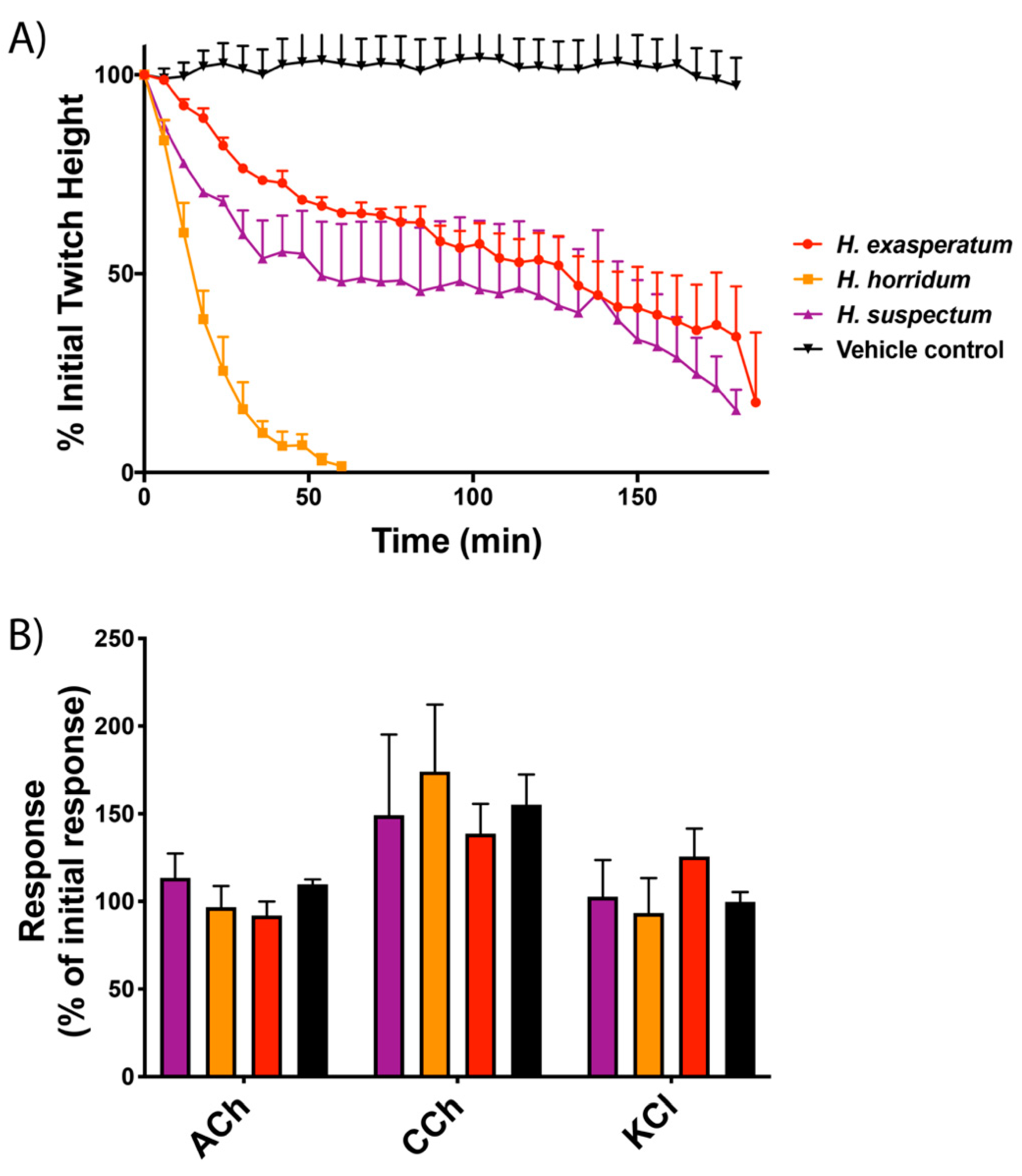
2.1. Gallus gallus Chick Biventer Cervicis Nerve–Muscle Assays

Testing on the avian neuromuscular preparation revealed that the muscle twitch response heights were markedly attenuated when Heloderma venoms were added (Figure 1). The most potent of the three Heloderma venoms was H. horridum, which abolished twitches within 60 min (Figure 1A). The venoms of H. exasperatum and H. suspectum had similar activities, inhibiting twitches by approximately 80% over the time-course of the experiment (Figure 1). There were no significant differences between responses to the agonists acetylcholine (ACh), carbachol (CCh) and potassium chloride (KCl) in the presence or absence of the venoms, with all groups producing responses comparable to the initial contractile response (Figure 1B). Parallel experiments run with V. varius venom did not produce notable results at the concentrations tested on this avian tissue preparation (see Supplementary Data).

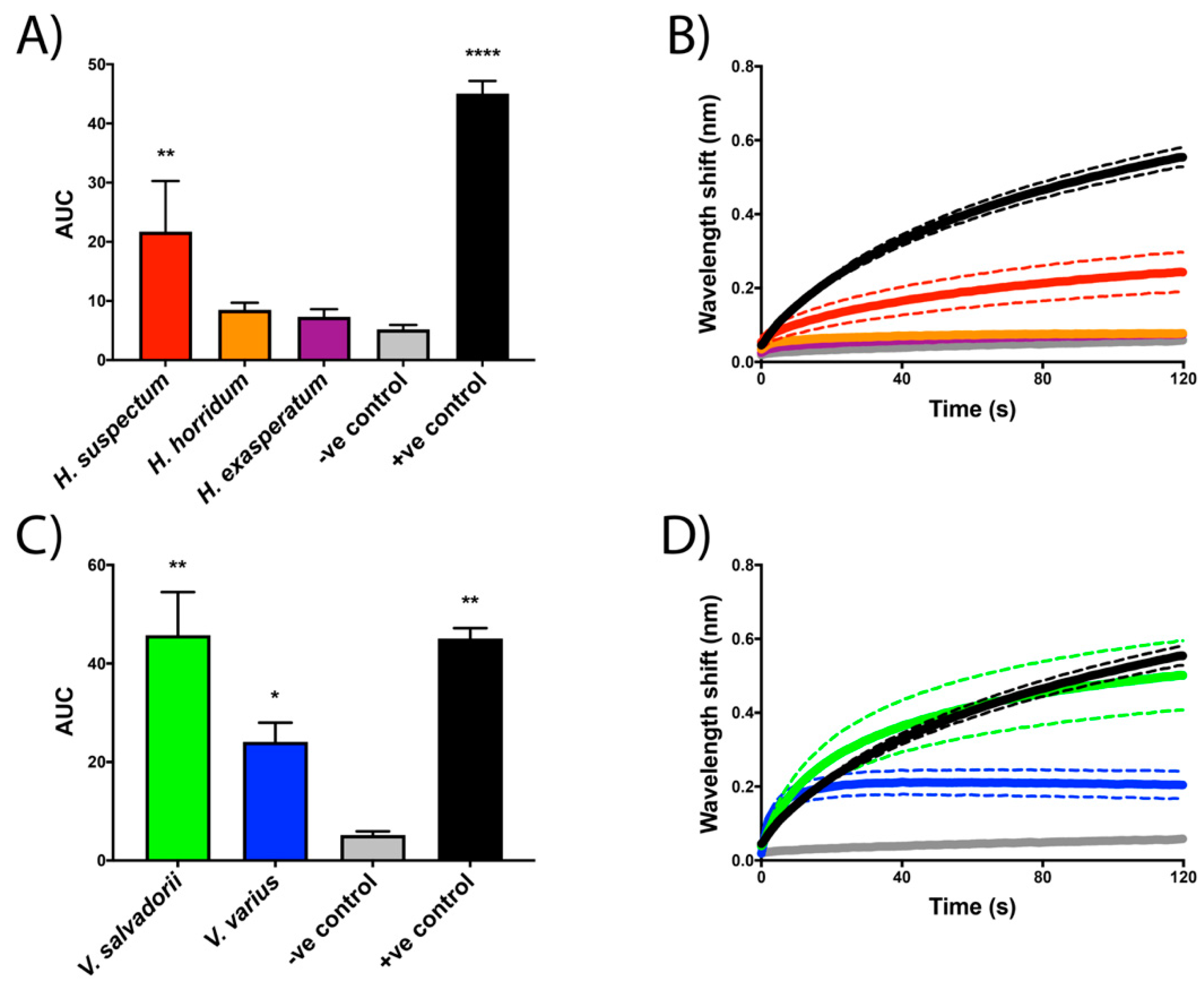
2.2. Biolayer Interferometry Assays

As it is a well-described ion channel-attacking venom, L. quinquestriatus was used as a positive control to validate the assay parameters, displaying strong binding to the S3–S4 extracellular loop mimotope of voltage-sensing domain IV of human NaV1.4 and CaV1.2 (Figure 2A,B and Figure 3A,B, respectively). Wavelength response shifts indicate Heloderma suspectum venom binds strongly to the voltage-sensing domain IV motif of human NaV1.4 (Figure 3A,B) and CaV1.2 (Figure 3A,B). In contrast, the other helodermatid lizard species tested, H. exasperatum and H. horridum, did not display significant binding to these mammalian targets (Figure 2 and Figure 3). However, wavelength response shifts indicate that V. salvadorii venom strongly binds to the voltage-sensing domain IV motif of both NaV1.4 (Figure 2C,D) and CaV1.2 (Figure 3C,D). Although still binding significantly, V. varius venom was less potent than V. salvadorii on both targets, and proportionally weaker on CaV1.2 than NaV1.4 (Figure 2C,D), being equipotent to H. suspectum on CaV1.2 (Figure 3) but slightly less potent than H. suspectum on NaV1.4 (Figure 2).

3. Discussion

Helodermatid lizard venoms have previously been shown to inhibit the electrical stimulation of mouse hemidiaphragm and induce paralysis in mice [31,35], but the site of action was not determined in this prior work. Here, we present similar results displaying a reduction in contraction ability in avian skeletal muscle (Figure 1A). Responses to postsynaptic agonists suggest Heloderma venoms act pre-synaptically, with the absence of myotoxicity (Figure 1B). Congruent with the avian tissue preparation results of presynaptic neurotoxicity, biolayer interferometry assays revealed Heloderma suspectum bound to the S3–S4 loop of voltage-sensing domain IV within NaV1.4, inhibiting channel fast inactivation and altering Na+ influx (Figure 2A,B). While many toxins that bind to voltage-sensing domain IV have a stimulatory effect, such as delaying inactivation [18], binding to produce an inhibitory effect has been documented [20], thus supporting our results. In addition, many toxins found to bind to voltage-sensing domain IV also interact with voltage sensing domains I-III, where binding would inhibit gating [17]. The dichotomy between the avian tissue preparation and the mammalian-target biolayer interferometry assays is suggestive of differential taxon-specific effects between the three Heloderma species tested and thus is a rich area for future research regarding the evolutionary selection pressures leading to such lineage-selective effects.
While V. varius venom did not produce discernable results on the avian-tissue preparation assays at the concentrations tested, both V. varius and V. salvadorii bound mammalian NaV1.4 with V. varius nearly equipotent to H. suspectum while V. salvadorii was almost twice as potent (Figure 2). These results support the clinical symptoms of lethargy and paralysis reported in the bites from helodermatid and, occasionally, varanid lizards [21,46,48]. Further support can be found from previously conducted in vivo studies [31,35,52]. Varanus griseus venom is reported to produce paralyzing effects on mice, consistent with NaV1.4 antagonistic activity, similar to the effects of helothermine [31,52]. The study by Gorelov (1971) also injected sparrows with V. griseus venom, which did not result in the immediate paralysis observed in mice, corroborating the lack of effect of the varanid venoms on the avian tissue preparation assays and the strong binding in mammalian target assays.
The disparity between avian and mammalian data is suggestive of differences in ion channel physiology between organisms. Structural differences between voltage-gated ion channels of avian and mammalian species (as shown in the sequence variation of key domains in uniprot accessions P35499 and P35499) could explain the lack of venom activity displayed by the varanid lizards in avian tissue preparation assays and the strong binding in mammalian biolayer interferometry assays, and the converse where H. exasperatum and H. horrium were much more potent in the avian assay than the mammalian (Figure 1, Figure 2 and Figure 3). As the Heloderma and Varanus species studied here are known to consume both mammals and birds, different toxin classes may be selected for the subjugation of different prey, as has been observed for snake venoms [64]. Such differences observed for snake venoms include extreme variations between prey lineages, suggestive of taxon-specific effects [65,66,67,68,69,70,71]. Other lineage-specific effects include the binding affinity of μ-conotoxins, which are significantly more potent binders of frog TTX-resistant NaV channels as compared to homologous channels in rat and mouse assays and while such variation is not reflective of any sort of biological reality (as cone snails do not feed on frogs or rodents) this specificity of venoms never-the-less underscores the potential use of venoms as probes for differences in physiological pathways of disparate animal lineages [72,73].
Compared to other Heloderma venoms tested, H. horridum venom was more potent on avian tissue preparation assays (Figure 1) and showed a lower binding affinity to the mammalian ion channel domains, possibly reflecting a predatory role of the venom with variations in diet between Heloderma species. Due to the elusive subterranean nature of Heloderma species, studies on diet comparisons are lacking, often limited to seasonal above-ground observations. Thus, the abundant observations of helodermatid lizard diets preying upon defenseless prey, such as eggs and nestlings [25,43,74], are likely due to the opportunistic nature of these observations when the lizards are feeding upon birds and eggs during annual avian breeding periods. Supporting this supposition are records of H. suspectum consuming mammals, which rapidly succumb to the venom’s effects ([75], Fry’s personal observations).
An alternative testable hypothesis is that differences in venom activity between species could be selected for defensive purposes. Heloderma suspectum are the smaller of the species tested and thus are likely more vulnerable to mammalian predators, such as Canis latrans [25]. This is consistent with the more prominent aposematic markings found on H. suspectum, indicating that a bite causes pain. Indeed, severe pain is often recorded in bite reports from this species [76]. Many varanid lizard species are also vulnerable to mammalian and avian predators as juveniles, and for smaller species, as adults; therefore, a painful bite would confer an advantage [43]. It is unclear if the activity presented in this study may contribute to the pain from a bite, as there is extensive cross-reactivity between sodium channel types, and thus paralytic toxins binding to the neuromuscular target NaV1.4 may also bind to other sodium channels involved in pain. However, the interactions of NaV1.4 shown in this study would produce paralysis of skeletal muscles, such as is shown in the tissue preparation, which is consistent with evolutionary selection pressures for a predatory role. Thus, a testable hypothesis for future work is that Heloderma venom contains components for prey subjugation, in addition to serving a defensive role due to the cross-reactivity between sodium channel subtypes.
Commonly reported in severe Heloderma human bite clinical cases are tachycardia, arrythmia, and the potentially fatal cardiac ischemia [22,23,76]. An increased, irregular heart rate has also been observed following varanid lizard bites [46,48]. As CaV1.2 is primarily a cardiac muscle ion channel, binding to this channel could be the mode of action responsible for these symptoms. Harris et al. (2021) reported on the binding of stone fish (Synanceia verrucosa) venom to the S3–S4 extracellular loop of voltage-sensing domain IV of CaV1.2, suggesting this activity could explain the arrythmia and tachycardia that manifest post-stone fish envenomation. In addition, severe scorpion envenomations can cause cardiac ischemia [77]. A direct cardiac effect may act synergistically with the mass release of catecholamines that has been suggested as the cause of similar symptoms seen in Heloderma bite victims [76]. Thus, the role of the CaV1.2 binding effect is another rich area for future research into the predatory or defensive ecology of anguimorphan lizards. In addition to the calcium channel-binding toxins, the effects upon heart rate may also be due to the sodium channel-binding toxins binding to the cardiac-associated NaV1.5 channel [8]. While not investigated in this study, toxins that bind to NaV1.4 have also been reported to bind to other NaV channels, including NaV1.5 [78]. Thus, the ion channel interactions responsible for cardiac dysfunction is another rich area of future research.
Venom binding to both NaV and CaV channels is often due to the possession of a complex cocktails of toxins which individually may have selective binding affinities for either channel family. Conversely, some toxins are promiscuous in being able to bind across not only NaV or CaV channel subtypes, but due to the homologous architecture from a shared molecular evolutionary history, some ion channel toxins are able to act upon both NaV and CaV channels. Examples include hanatoxin, which binds to multiple subtypes of NaV, CaV and KV channels [16]. Similarly, other tarantula venom peptides have been found to be active on both NaV and CaV channels [79]. Thus, the structure-function responsible for such diverse binding by some venom toxins is a rich area for future research.
Based on the results presented in this study, future work should investigate other species of varanid lizards for their potential neurotoxic venom activities. While we were limited by species available, experiments should be performed on species previously shown to produce neurotoxic activities and symptoms, such as V. griseus, V. scalaris, and other closely related species. Binding to the S3–S4 extracellular loop of domains I-III could potentially be investigated using biolayer interferometry mimotope assays similar to those used in this study. Despite evidence of toxins binding to the voltage-sensing domain IV of NaV1.4 and CaV1.2, translating these data into the effects on whole ion channel gating would require future work on additional testing platforms, such as the Kv channel chimeric approach or electrophysiological techniques such as oocyte patch-clamp assays. In addition, future work should include fractionating the venoms to ascertain the toxin types responsible for these intriguing activities.
In summary, we have elucidated the calcium and sodium channel-binding mechanisms of Heloderma venom, and report for the first time evidence of ion channel binding neurotoxins in Varanus venoms. This work provides a foundation for natural history investigations to ascertain the biological roles of lizard venoms, whether as predatory weapons or as part of a defensive arsenal, or a combination thereof. Notably, the neurotoxic effects revealed in this study are indicative of both defensive and predatory roles for the venoms of Heloderma and Varanus species, and further suggest prey-selective effects of both Heloderma and Varanus venoms. The modes of action of these venoms presented in this study could also be important in understanding the clinical effects of envenomations, by revealing the underlying biochemistry of neurotoxic symptoms, thereby providing data which may be useful for the evidence-based design of clinical management strategies. The results further underscore the utility of using venoms as rich sources of novel compounds, useful as probes to ascertain physiological function, and even as lead compounds for drug design and development. Animal toxins have been used as key probes in the discovery of voltage-gated ion channel subtypes, their structural properties, and their functional characteristics. Our results reinforce the use of biolayer interferometry assays as a potential tool to investigate the role of voltage-sensing domains in ion channel gating. Thus, in addition to being selective probes for ion channel functions, these novel compounds may have utility as starting substrates in the drug design and development pipeline. It is only by investigating the full range of venomous animals, such as the neglected venomous lizard lineages, that such resources can be harnessed to their fullest potential.

4. Materials and Methods

4.1. Sample Acquisition

Venoms were collected by encouraging the specimens to chew on soft rubber tubing, with the mandibular secretions collected with pipettes (Varanus) or allowed to flow into a 50 mL tube (Heloderma). Secretions were then centrifuged at 14,000 RCF (4 °C, 10 min.) to remove insoluble material, filtered with a 40 µm syringe filter, flash-frozen in liquid nitrogen, and then freeze-dried. Varanus varius samples (adult male, n = 3, pooled) were collected from captive specimens under University of Queensland animal ethics approval SBS/403/16 (approval date: 1 February 2016). Non-Australian lizard (H. exasperatum, H. horridum, H. suspectum, and V. salvadorii) samples were supplied by licensed biotechnology company Alphabiotoxine Laboratory, Montroel-au-bois, Belgium.

4.2. Gallus Gallus Chick Biventer Cervicis Nerve–Muscle Assays

Neurotoxicity was ascertained using a previously validated organ bath protocol [80]. Gallus gallus chicks aged between 4–10 days were euthanized with CO2 (animal ethics number 22575 was approved by Monash University Ethics Committee on 18 December 2019). Biventer cervicis nerve–muscle samples were removed and mounted under 1 g tension in 5 mL organ baths containing physiological salt solution (NaCl, 118.4 mM; KCl, 4.7 mM; MgSO4, 1.2 mM; KH2PO4, 1.2 mM; CaCl2, 2.5 mM; NaHCO3, 25 mM; and glucose, 11.1 mM). Organ baths were bubbled with carbogen (95% O2; 5% CO2) at a constant temperature of 34 °C. Electrodes were placed around the tendon of the biventer muscle and electrical stimulation at the motor nerve (0.2 ms duration, 0.1 Hz, supramaximal V) using a Grass S88 stimulator (Grass Instruments, Quincy, MA, USA) evoked indirect twitches. Selective stimulation of the nerve was confirmed by the abolition of twitches with d-tubocurarine (10 µM), a nAChR competitive antagonist. Tissues were then washed repeatedly with physiological salt solution to restore twitch responses to nerve stimulation. The stimulation was ceased, and the contractile responses to acetylcholine (ACh, 1 mM for 30 s), carbochol (CCh, 20 µM for 60 s), and potassium chloride (KCl, 40 mM for 30 s) were obtained and recorded. The organ bath was then washed, and electrical stimulation was resumed and maintained for 30 min to allow the preparation to equilibrate. Venom (10 µg/mL) was added to the organ bath, and the twitch height was recorded until the abolition of twitch response or was stopped after a 1 h period. The stimulator was turned off again and the bath was washed. Contractile responses to ACh, CCh and KCl were obtained again to compare with responses prior to venom addition. The twitch responses to electrical stimulation and contractile responses to agonists (ACh, CCh, and KCI) were measured using a Grass FT03 force displacement transducer (Grass Instruments, Quincy, MA, USA) and recorded on a PowerLab system (ADInstruments Pty Ltd., Bella Vista, NSW, Australia). The data were then converted into figures using Prism8.0 software (GraphPad Software Inc., La Jolla, CA, USA).

4.3. Biolayer Interferometry

The voltage-sensing domain IV S3–S4 motifs were ascertained using previously validated protocols [57,81]. The amino acid sequences for human voltage-sensing domain IV S3–S4 motifs are shown in Figure 4 for CaV1.2 (Homo sapiens, uniprot accession code Q13936) and NaV1.4 (Homo sapiens, uniprot accession code P35499). These regions were synthesized by GenicBio Ltd. (Shanghai, China) and joined to two aminohaxanoic acid (Ahx) spacers forming a 30 Å linker, with the end Ahx then bound to biotin, providing clearance between the biotin and mimotope and thereby allowing the mimotope to maintain its natural conformational freedom when binding to the analyte in solution. Dried stocks of mimotopes were solubilized in 100% dimethyl sulfoxide (DMSO) prior to dilution with deionized water in a 1:10 ratio to produce a working stock concentration of 50 µg/mL. This stock was then stored at −80 °C until required for experiments. Binding kinetics were analyzed by biolayer interferometry utilizing the Octet HTX system (ForteBio). All assays were conducted in standard Greiner black 96-microtiter well plates (ref: 655209). Analyte (venom) experimental concentrations of 50 µg/mL (10 µg per well) were produced by diluting stock solutions 1:20 with matrix buffer (1× DPBS with 0.1% BSA and 0.05% Tween-20). Mimotope aliquots were diluted 1:50 with matrix buffer to produce a final concentration of 1 µg/mL (0.2 µg per well). Streptavidin sensors were hydrated in the matrix buffer for 30–60 min, whilst being agitated at 2.0 RPM on a shaker prior to experiments. A standard acidic solution glycine buffer (10 mM glycine (pH 1.5–1.7)) in deionized water was used to regenerate the sensor tips during experimentation while still leaving the mimotope attached to the sensor. Controls consisted of a buffer control (deionized water:glycerol 1:1) and the negative control Naja kaouthia (monocled cobra) venom in the place of the analyte sample in the wells. The positive control for NaV1.4- and CaV1.2-binding was Leiurus quinquestriatus (deathstalker scorpion) venom, which is known to strongly bind to both channels.
Data were processed in accordance with the validation study [59]. The association step data (.csv file) were extracted for each triplicate and imported into Prism 9.0 software (GraphPad Software Inc., La Jolla, CA, USA) where area under the curve (AUC) calculations were made and data graphed. Dunnett’s multiple comparison tests were performed to determine statistical significances between treatment groups and negative controls.

Supplementary Materials

The following are available online at https://0-www-mdpi-com.brum.beds.ac.uk/article/10.3390/toxins13080549/s1, S1: Raw Data.

Author Contributions

Conceptualization, B.G.F. and F.B.; methodology, F.B. and W.C.H.; validation, R.J.H., C.N.Z., T.H. and W.C.H.; formal analysis, J.S.D.; writing—original draft preparation, J.S.D.; writing—review and editing, R.J.H., C.N.Z., T.H., W.C.H., F.B., R.F., A.V. and B.G.F. Sample provision, B.G.F., R.F. and A.V. All authors have read and agreed to the published version of the manuscript.

Funding

This research was funded by Australian Research Council Discovery Project DP210102406 and Australian Research Council Linkage Infrastructure & Equipment Fund LE200100140 to BGF. T.H. and W.C.H. were supported by an Australian National Health and Medical Research Council (NHMRC) Centres for Research Excellence Grant (ID: 1110343).

Institutional Review Board Statement

Varanus varius samples (adult male, n = 3, pooled) were collected from captive specimens under University of Queensland animal ethics approval SBS/403/16 (approval date: 1 February 2016). Non-Australian lizard (H. exasperatum, H. horridum, H. suspectum, and V. salvadorii) samples were supplied by licensed biotechnology company Alphabiotoxine Laboratory, Montroel-au-bois, Belgium.

Informed Consent Statement

Not applicable.

Data Availability Statement

The data presented in this study are available in supplementary material.

Conflicts of Interest

The authors declare no conflict of interest.

References

  1. Ertel, E.A.; Campbell, K.P.; Harpold, M.M.; Hofmann, F.; Mori, Y.; Perez-reyes, E.; Schwartz, A.; Snutch, T.P.; Tanabe, T.; Birnbaumer, L.; et al. Nomenclature of Voltage-Gated Calcium Channles. Neuron 2000, 25, 533–535. [Google Scholar] [CrossRef] [Green Version]
  2. Anderson, P.A.V.; Greenberg, R.M. Phylogeny of Ion Channels: Clues to Structure and Function. Comp. Biochem. Physiol. Part B 2001, 129, 17–28. [Google Scholar] [CrossRef]
  3. Ahern, C.A.; Payandeh, J.; Bosmans, F.; Chanda, B. The Hitchhiker’s Guide to the Voltage-Gated Sodium Channel Galaxy. J. Gen. Physiol. 2016, 147, 1–24. [Google Scholar] [CrossRef] [Green Version]
  4. Chanda, B.; Bezanilla, F. Tracking Voltage-Dependent Conformational Changes in Skeletal Muscle Sodium Channel during Activation. J. Gen. Physiol. 2002, 120, 629–645. [Google Scholar] [CrossRef] [PubMed]
  5. Goldschen-ohm, M.P.; Capes, D.L.; Oelstrom, K.M.; Chanda, B. Multiple Pore Conformations Driven by Asynchronous Movements of Voltage Sensors in a Eukaryotic Sodium Channel. Nat. Commun. 2013, 4, 1350. [Google Scholar] [CrossRef] [PubMed] [Green Version]
  6. Pantazis, A.; Savalli, N.; Sigg, D.; Neely, A.; Olcese, R.; Pantazisa, A.; Savalli, N.; Siggbf, D.; Neelycf, A.; Olcesea, R. Functional Heterogeneity of the Four Voltage Sensors of a Human L-type Calcium Channel. Proc. Natl. Acad. Sci. USA 2021, 111, 18381–18386. [Google Scholar] [CrossRef] [PubMed] [Green Version]
  7. Beyl, S.; Depil, K.; Hohaus, A.; Stary-weinzinger, A.; Linder, T.; Timin, E.; Hering, S. Neutralisation of a Single Voltage Sensor Affects Gating Determinants in All Four Pore-Forming S6 Segments of CaV1.2: A Cooperative Gating Model. Pflügers Arch.-Eur. J. Physiol. 2012, 464, 391–401. [Google Scholar] [CrossRef] [PubMed] [Green Version]
  8. Catterall, W.A.; Goldin, A.L.; Waxman, S.G. International Union of Pharmacology. XLVII. Nomenclature and Structure-Function Relationships of Voltage-Gated Sodium Channels. Pharmacol. Rev. 2005, 57, 397–409. [Google Scholar] [CrossRef]
  9. Dolphin, A.C. Calcium Channel Diversity: Multiple Roles of Calcium Channel Subunits. Curr. Opin. Neurobiol. 2009, 19, 237–244. [Google Scholar] [CrossRef]
  10. Catterall, W.A. Voltage-Gated Calcium Channels. Cold Spring Harb. Perspect. Biol. 2011, 3, 1–23. [Google Scholar] [CrossRef]
  11. Catterall, W.A. Voltage-Gated Sodium Channels at 60: Structure, Function and Pathophysiology. J. Physiol. 2012, 590, 2577–2589. [Google Scholar] [CrossRef]
  12. George, A.L.J. Inherited Disorders of Voltage-Gated Sodium Channels. J. Clin. Investig. 2005, 115, 1990–1999. [Google Scholar] [CrossRef] [PubMed]
  13. Cannon, S.C. Pathomechanisms in Channelopathies of Skeletal Muscle and Brain. Annu. Rev. Neurosci. 2006, 29, 387–417. [Google Scholar] [CrossRef] [PubMed]
  14. Waxman, S.G.; Merkies, I.S.J.; Gerrits, M.M.; Dib-hajj, S.D.; Lauria, G.; Cox, J.J.; Wood, J.N.; Woods, C.G.; Drenth, J.P.H.; Faber, C.G. Sodium Channel Genes in Pain-Related Disorders: Phenotype–Genotype Associations and Recommendations for Clinical Use. Lancet Neurol. 2014, 13, 1152–1160. [Google Scholar] [CrossRef]
  15. Fry, B.G.; Roelants, K.; Champagne, D.E.; Scheib, H.; Tyndall, J.D.A.; King, G.F.; Nevalainen, T.J.; Norman, J.A.; Lewis, R.J.; Norton, R.S.; et al. The Toxicogenomic Multiverse: Convergent Recruitment of Proteins Into Animal Venoms. Annu. Rev. Genomics Hum. Genet. 2009, 10, 483–511. [Google Scholar] [CrossRef] [PubMed] [Green Version]
  16. Bosmans, F.; Swartz, K.J. Targeting Voltage Sensors in Sodium Channels with Spider Toxins. Trends Pharmacol. Sci. 2010, 31, 175–182. [Google Scholar] [CrossRef] [PubMed] [Green Version]
  17. Bosmans, F.; Martin-Eauclaire, M.F.; Swartz, K.J. Deconstructing Voltage Sensor Function and Pharmacology in Sodium Channels. Nature 2008, 456, 202–208. [Google Scholar] [CrossRef] [Green Version]
  18. Yang, D.C.; Deuis, J.R.; Dashevsky, D.; Dobson, J.; Jackson, T.N.W.; Brust, A.; Xie, B.; Koludarov, I.; Debono, J.; Hendrikx, I.; et al. The Snake with the Scorpion’s Sting: Novel Three-Finger Toxin Sodium Channel Activators from the Venom of the Long-Glanded Blue Coral Snake (Calliophis bivirgatus). Toxins 2016, 8, 303. [Google Scholar] [CrossRef]
  19. Bourinet, E.; Stotz, S.C.; Spaetgens, R.L.; Dayanithi, G.; Lemos, J.; Nargeot, J.; Zamponi, G.W. Interaction of SNX482 with Domains III and IV Inhibits Activation Gating of α1E (CaV2. 3) calcium channels. Biophys. J. 2001, 81, 79–88. [Google Scholar] [CrossRef] [Green Version]
  20. Gilchrist, J.; Bosmans, F. Using Voltage-Sensor Toxins and their Molecular Targets to Investigate NaV1.8 Gating. J. Physiol. 2018, 10, 1863–1872. [Google Scholar] [CrossRef] [Green Version]
  21. Hooker, K.R.; Caravati, M.E. Gila Monster Envenomation. Ann. Emerg. Med. 1994, 24, 731–735. [Google Scholar] [CrossRef]
  22. French, R.N.E.; Ash, J.; Brooks, D.E.; French, R.N.E.; Ash, J.; Brooks, D.E. Gila Monster Bite. Clin. Toxicol. 2012, 50, 151–152. [Google Scholar] [CrossRef] [PubMed]
  23. Amri, K.; Chippaux, J. Report of a Severe Heloderma suspectum Envenomation. Clin. Toxicol. 2020, 59, 343–346. [Google Scholar] [CrossRef]
  24. Fry, B.G.; Winter, K.; Norman, J.A.; Roelants, K.; Nabuurs, R.J.A.; van Osch, M.J.P.; Teeuwisse, W.M.; van der Weerd, L.; Mcnaughtan, J.E.; Kwok, H.F.; et al. Functional and Structural Diversification of the Anguimorpha Lizard Venom System. Mol. Cell. Proteom. 2010, 9, 2369–2390. [Google Scholar] [CrossRef] [Green Version]
  25. Russell, F.E.; Bogert, C.M. Gila Monster: Its Biology, Venom and Bite-A Review. Toxicon 1981, 19, 341–359. [Google Scholar] [CrossRef]
  26. French, R.; Brooks, D.; Ruha, A.; Shirazi, F.; Boesen, K.; Walter, F.; French, R.; Brooks, D.; Ruha, A.; Shirazi, F.; et al. Gila Monster (Heloderma suspectum) Envenomation: Descriptive Analysis of Calls to United States Poison Centers with Focus on Arizona Cases. Clin. Toxicol. 2015, 53, 60–70. [Google Scholar] [CrossRef] [PubMed]
  27. Lino-López, G.J.; Valdez-Velázquez, L.L.; Corzo, G.; Romero-Gutiérrez, M.T.; Jiménez-Vargas, J.M.; Rodríguez-Vázquez, A.; Vazquez-Vuelvas, O.F.; Gonzalez-Carrillo, G. Venom Gland Transcriptome from Heloderma horridum horridum by High-Throughput Sequencing. Toxicon 2020, 180, 62–78. [Google Scholar] [CrossRef]
  28. Koludarov, I.; Jackson, T.N.W.; Sunagar, K.; Nouwens, A.; Hendrikx, I.; Fry, B.G. Fossilized Venom: The Unusually Conserved Venom Profiles of Heloderma Species (Beaded Lizards and Gila Monsters). Toxins 2014, 6, 3582–3595. [Google Scholar] [CrossRef] [PubMed] [Green Version]
  29. Kwok, H.F.; Ivanyi, C.; Morris, A.; Shaw, C. Proteomic and Genomic Studies on Lizard Venoms in the Last Decade. Proteom. Insights 2010, 3, 25–31. [Google Scholar] [CrossRef]
  30. Sanggaard, K.W.; Dyrlund, T.F.; Thomsen, L.R.; Nielsen, T.A.; Brøndum, L.; Wang, T.; Thøgersen, I.B.; Enghild, J.J. Characterization of the Gila Monster (Heloderma suspectum suspectum) Venom Proteome. J. Proteom. 2015, 117, 1–11. [Google Scholar] [CrossRef] [PubMed] [Green Version]
  31. Mochca-Morales, J.; Martin, B.M.; Possani, L.D. Isolation and Characterization of Helothermine, a Novel Txin from Heloderma horridum horridum (Mexican Beaded Lizard) Lenom. Toxicon 1990, 28, 299–309. [Google Scholar] [CrossRef]
  32. Morrissette, J.; Krätzschmar, J.; Haendler, B.; El-Hayek, R.; Mochca-Morales, J.; Martin, B.M.; Patel, J.R.; Moss, R.L.; Schleuning, W.D.; Coronado, R.; et al. Primary Structure and Properties of Helothermine, a Peptide Toxin that Blocks Ryanodine Receptors. Biophys. J. 1995, 68, 2280–2288. [Google Scholar] [CrossRef] [Green Version]
  33. Nobile, M.; Magnelli, V.; Lagostena, L.; Mochca-Morales, J.; Possani, L.D.; Prestipino, G. The Toxin Helothermine Affects Potassium Currents in Newborn Rat Cerebellar Granule Cells. J. Membr. Biol. 1994, 139, 49–55. [Google Scholar] [CrossRef]
  34. Nobile, M.; Noceti, F.; Prestipino, G.; Possani, L.D. Helothermine, a Lizard Venom Toxin, Inhibits Calcium Current in Cerebellar Granules. Exp. Brain Res. 1996, 110, 15–20. [Google Scholar] [CrossRef]
  35. Komori, Y.; Nikai, T.; Sugihara, H. Purification and Characterization of a Lethal Toxin from the Venom of Helodermahorridumhorridum. Biochem. Biophys. Res. Commun. 1988, 154, 613–619. [Google Scholar] [CrossRef]
  36. Fry, B.G.; Roelants, K.; Winter, K.; Hodgson, W.C.; Griesman, L.; Kwok, H.F.; Scanlon, D.; Karas, J.; Shaw, C.; Wong, L.; et al. Novel Venom Proteins Produced by Differential Domain-Expression Strategies in Beaded Lizards and Gila Monsters (Genus Heloderma). Mol. Biol. Evol. 2010, 27, 395–407. [Google Scholar] [CrossRef] [PubMed] [Green Version]
  37. Koludarov, I.; Jackson, T.N.W.; op den Brouw, B.; Dobson, J.; Dashevsky, D.; Arbuckle, K.; Clemente, C.J.; Stockdale, E.J.; Cochran, C.; Debono, J.; et al. Enter the Dragon: The Dynamic and Multifunctional Evolution of Anguimorpha Lizard Venoms. Toxins 2017, 9, 242. [Google Scholar] [CrossRef] [Green Version]
  38. Dobson, J.S.; Zdenek, C.N.; Hay, C.; Violette, A.; Fourmy, R.; Cochran, C.; Fry, B.G. Varanid Lizard Venoms Disrupt the Clotting Ability of Human Fibrinogen through Destructive Cleavage. Toxins 2019, 11, 255. [Google Scholar] [CrossRef] [Green Version]
  39. Fry, B.G.; Wroe, S.; Teeuwisse, W.; Van Osch, M.J.P.; Moreno, K.; Ingle, J.; Mchenry, C.; Ferrara, T.; Clausen, P.; Scheib, H.; et al. A Central Role for Venom in Predation by Varanus komodoensis (Komodo Dragon) and the Extinct Giant Varanus (Megalania) priscus. Proc. Natl. Acad. Sci. USA 2009, 106, 8969–8974. [Google Scholar] [CrossRef] [Green Version]
  40. Fry, B.G.; Vidal, N.; Norman, J.A.; Vonk, F.J.; Scheib, H.; Ramjan, S.F.R.; Kuruppu, S.; Fung, K.; Hedges, S.B.; Richardson, M.K.; et al. Early Evolution of the Venom System in Lizards and Snakes. Nature 2006, 439, 584–588. [Google Scholar] [CrossRef] [PubMed]
  41. Koludarov, I.; Sunagar, K.; Undheim, E.A.B.; Jackson, T.N.W.; Ruder, T.; Whitehead, D.; Saucedo, A.C.; Mora, G.R.; Alagon, A.C.; King, G.; et al. Structural and Molecular Diversification of the Anguimorpha Lizard Mandibular Venom Gland System in the Arboreal Species Abronia graminea. J. Mol. Evol. 2012, 75, 168–183. [Google Scholar] [CrossRef] [PubMed]
  42. Wilson, S.; Swan, G. A Complete Guide to Reptiles of Australia, 5th ed.; Reed New Holland Publishers: Chatswood, Australia, 2017; ISBN 9781925546026. [Google Scholar]
  43. Pianka, E.R.; King, D.; King, R. Varanoid Lizards of the World; Pianka, E.R., King, D.R., King, R.A., Eds.; Indiana University Press: Indianapolis, IN, USA, 2004. [Google Scholar]
  44. Pianka, E.R.; Sweet, S.S. Field Observations by Two American Varanophiles. In Proceedings of the 2015 Interdisciplinary World Conference on Monitor Lizards; Cota, M., Ed.; Institute for Research and Development, Suan Sunandha Rajabhat University: Bangkok, Thailand, 2016; pp. 1–68. [Google Scholar]
  45. Arbuckle, K. Ecological Function of Venom in Varanus, with a Compilation of Dietary Records from the Literature. Biawak 2009, 3, 46–56. [Google Scholar]
  46. Sopiev, O.; Makeev, V.M.; Kudryavcev, S.V.; Makarov, A.N. The Case of Intoxication with the Bite of the Gray Monitor Lizard Varanus. Izvestia Academii nauk Turkmenskoi SSR. Seriya Biol. Nauk 1987, 598, 615. (In Rissian) [Google Scholar]
  47. Ballard, V.; Antonio, F.B. Varanus griseus (Desert Monitor) Toxicity. Herpetol. Rev. 2001, 32, 261. [Google Scholar]
  48. Zima, Y.A. On the Toxicity of the Bite of the Caspian Desert Monitor Lizard (Varanus griseus caspius). Biawak 2019, 13, 115–118. [Google Scholar]
  49. Sweet, S.S. Chasing Flamingos: Toxicofera and the Misinterpretation of Venom in Varanid Lizards. In Proceedings of the 2015 Interdisciplinary World Conference on Monitor Lizards; Cota, M., Ed.; Institute for Research and Development, Suan Sunandha Rajabhat University: Bangkok, Thailand, 2016; pp. 123–149. [Google Scholar]
  50. Huang, T.-F.; Chiang, H.-S. Effect on Human Platelet Aggregation of Phospholipase A2 Purified from Heloderma horridum (Beaded Lizard) Venom. Biochim. Biophys. Acta Lipids Lipid Metab. 1994, 1211, 61–68. [Google Scholar] [CrossRef]
  51. Utaisincharoenso, P.; Mackessyn, S.P.; Miller, R.A.; Tusii, A.T. Complete Primary Structure and Biochemical Properties of Gilatoxin, a Serine Protease with Kallikrein-like and Angiotensin-Degrading Activities. J. Biol. Chem. 1993, 268, 21975–21983. [Google Scholar] [CrossRef]
  52. Gorelov, Y.K. On the Toxicity of Saliva Varanus griseus. Izvestia Academii nauk Turkmenskoi SSR. Seriya Biol. Nauk 1971, 6, 75–76. (In Russian) [Google Scholar]
  53. Yang, D.C.; Dobson, J.; Cochran, C.; Dashevsky, D.; Arbuckle, K.; Benard, M.; Boyer, L.; Alagón, A.; Hendrikx, I.; Hodgson, W.C.; et al. The Bold and the Beautiful: A Neurotoxicity Comparison of New World Coral Snakes in the Micruroides and Micrurus Genera and Relative Neutralization by Antivenom. Neurotox. Res. 2017, 32, 487–495. [Google Scholar] [CrossRef]
  54. Fry, B.G.; Lumsden, N.G.; Wuster, W.; Wickramaratna, J.C.; Hodgson, W.C.; Kini, R.M. Isolation of a Neurotoxin (Alpha-Colubritoxin) from a Nonvenomous Colubrid: Evidence for Early Origin of Venom in Snakes. J. Mol. Evol. 2003, 57, 446–452. [Google Scholar] [CrossRef]
  55. Op den Brouw, B.; Coimbra, F.C.P.; Bourke, L.A.; Huynh, T.M.; Vlecken, D.H.W.; Ghezellou, P.; Visser, J.C.; Dobson, J.S.; Fernandez-Rojo, M.A.; Ikonomopoulou, M.P.; et al. Extensive Variation in the Activities of Pseudocerastes and Eristicophis Viper Venoms Suggests Divergent Envenoming Strategies Are Used for Prey Capture. Toxins 2021, 13, 112. [Google Scholar] [CrossRef]
  56. Lumsden, N.G.; Fry, B.G.; Manjunatha Kini, R.; Hodgson, W.C. In Vitro Neuromuscular Activity of ‘Colubrid’ Venoms: Clinical and Evolutionary Implications. Toxicon 2004, 43, 819–827. [Google Scholar] [CrossRef]
  57. Harris, R.J.; Youngman, N.J.; Chan, W.; Bosmans, F.; Cheney, K.L.; Fry, B.G. Getting Stoned: Characterisation of the Coagulotoxic and Neurotoxic Effects of Reef Stonefish (Synanceia verrucosa) Venom. Toxicol. Lett. 2021, 346, 16–22. [Google Scholar] [CrossRef] [PubMed]
  58. Jones, L.; Harris, R.J.; Fry, B.G. Not Goanna Get Me: Mutations in the Savannah Monitor Lizard (Varanus exanthematicus) Nicotinic Acetylcholine Receptor Confer Reduced Susceptibility to Sympatric Cobra Venoms. Neurotox. Res. 2021, 39, 1116–1122. [Google Scholar] [CrossRef] [PubMed]
  59. Zdenek, N.C.; Harris, J.R.; Kuruppu, S.; Youngman, J.N.; Dobson, S.J.; Debono, J.; Khan, M.; Smith, I.; Yarski, M.; Harrich, D.; et al. A Taxon-Specific and High-Throughput Method for Measuring Ligand Binding to Nicotinic Acetylcholine Receptors. Toxins 2019, 11, 600. [Google Scholar] [CrossRef] [Green Version]
  60. Harris, R.J.; Youngman, N.J.; Zdenek, C.N.; Huynh, T.M.; Nouwens, A.; Hodgson, W.C.; Harrich, D.; Dunstan, N.; Portes-junior, J.A.; Fry, B.G. Assessing the Binding of Venoms from Aquatic Elapids to the Nicotinic Acetylcholine Receptor Orthosteric Site of Di ff erent Prey Models. Int. J. Mol. Sci. 2020, 21, 7377. [Google Scholar] [CrossRef] [PubMed]
  61. Harris, R.J.; Fry, B.G. Electrostatic Resistance to Alpha-Neurotoxins Conferred by Charge Reversal Mutations in Nicotinic Acetylcholine Receptors. Proc. R. Soc. B 2021, 288, 7–9. [Google Scholar] [CrossRef]
  62. Harris, R.J.; Zdenek, C.N.; Debono, J.; Harrich, D.; Fry, B.G. Evolutionary Interpretations of Nicotinic Acetylcholine Receptor Targeting Venom Effects by a Clade of Asian Viperidae Snakes. Neurotox. Res. 2020, 38, 312–318. [Google Scholar] [CrossRef]
  63. Youngman, N.J.; Harris, R.J.; Huynh, T.M.; Coster, K.; Sundman, E.; Braun, R.; Naude, A.; Hodgson, W.C.; Fry, B.G. Widespread and Differential Neurotoxicity in Venoms from the Bitis Genus of Viperid Snakes. Neurotox. Res. 2021, 39, 697–704. [Google Scholar] [CrossRef]
  64. Modahl, C.; Mrinalini; Frietze, S.; Mackessy, S.P. Adaptive Evolution of Distinct Prey-Specific Toxin Genes in Rear-Fanged Snake Venom. Proc. R. Soc. B Biol. Sci. 2018, 285, 20181003. [Google Scholar] [CrossRef]
  65. Harris, R.J.; Zdenek, C.N.; Harrich, D.; Frank, N.; Fry, B.G. An Appetite for Destruction: Detecting Prey-Selective Binding of α-Neurotoxins in the Venom of Afro-Asian Elapids. Toxins 2020, 12, 205. [Google Scholar] [CrossRef] [Green Version]
  66. Heyborne, W.H.; Mackessy, S.P. Identification and Characterization of a Taxon-Specific Three- Finger Toxin from the Venom of the Green Vinesnake (Oxybelis fulgidus; family Colubridae). Biochimie 2013, 95, 1923–1932. [Google Scholar] [CrossRef]
  67. Pawlak, J.; Mackessy, S.P.; Fry, B.G.; Bhatia, M.; Mourier, G.; Fruchart-gaillard, C.; Servent, D.; Stura, E.; Ménez, A.; Kini, R.M. Denmotoxin, a Three-finger Toxin from the Colubrid Snake Boiga dendrophila (Mangrove Catsnake) with Bird-specific Activity. J. Biol. Chem. 2006, 281, 29030–29041. [Google Scholar] [CrossRef] [Green Version]
  68. Youngman, N.J.; Chowdhury, A.; Zdenek, C.N.; Coster, K.; Sundman, E.; Braun, R.; Fry, B.G. Utilising Venom Activity to infer Dietary Composition of the Kenyan Horned Viper (Bitis worthingtoni). Comp. Biochem. Physiol. Part C 2021, 240, 108921. [Google Scholar] [CrossRef]
  69. Youngman, N.J.; Zdenek, C.N.; Dobson, J.S.; Bittenbinder, M.A.; Gillett, A.; Hamilton, B.; Dunstan, N.; Allen, L.; Veary, A.; Veary, E.; et al. Mud in the Blood: Novel Potent Anticoagulant Coagulotoxicity in the Venoms of the Australian Elapid Snake Genus Denisonia (Mud Adders) and Relative Antivenom Efficacy. Toxicol. Lett. 2019, 302, 1–6. [Google Scholar] [CrossRef] [Green Version]
  70. Cipriani, V.; Debono, J.; Goldenberg, J.; Jackson, T.N.W.; Arbuckle, K.; Dobson, J.; Koludarov, I.; Li, B.; Hay, C.; Dunstan, N.; et al. Correlation Between Ontogenetic Dietary Shifts and Venom Variation in Australian Brown Snakes (Pseudonaja). Comp. Biochem. Physiol. Part C Toxicol. Pharmacol. 2017, 197, 53–60. [Google Scholar] [CrossRef] [Green Version]
  71. Daltry, J.C.; Wüster, W.; Thorpe, R.S. Diet and Snake Venom Evolution. Nature 1996, 379, 537–540. [Google Scholar] [CrossRef] [PubMed]
  72. Zhang, M.; Fiedler, B.; Green, B.R.; Catlin, P.; Watkins, M.; Garrett, J.E.; Smith, B.J.; Yoshikami, D.; Olivera, B.M.; Bulaj, G. Structural and Functional Diversities among µ-Conotoxins Targeting TTX-Resistant Sodium Channels. Biochem. 2019, 13, 115–118. [Google Scholar] [CrossRef] [PubMed]
  73. Wilson, M.J.; Yoshikami, D.; Azam, L.; Gajewiak, J.; Olivera, B.M.; Bulaj, G. μ -Conotoxins that Differentially Block Sodium Channels NaV1.1 through 1.8 Identify those Responsible for Action Potentials in Sciatic Nerve. Proc. Natl. Acad. Sci. USA 2011, 108, 10302–10307. [Google Scholar] [CrossRef] [PubMed] [Green Version]
  74. Beck, D.D.; Lowe, C.H. Ecology of the Beaded Lizard, Heloderma horridum, in a Tropical Dry Forest in Jalisco, México. J. Herpetol. 1991, 25, 395–406. [Google Scholar] [CrossRef]
  75. Hensley, M. Mammal Diet of Heloderma. Herpetologica 1949, 5, 5–6. [Google Scholar]
  76. Chippaux, J.-P.; Amri, K. Severe Heloderma spp. Envenomation: A Review of the Literature. Clin. Toxicol. 2021, 59, 179–184. [Google Scholar] [CrossRef] [PubMed]
  77. Abroug, F.; Souheil, E.; Ouanes, I.; Dachraoui, F.; Fekih-Hassen, M.; Besbes, L.O. Scorpion-Related Cardiomyopathy: Clinical Characteristics, Pathophysiology, and Treatment. Clin. Toxicol. 2015, 53, 511–518. [Google Scholar] [CrossRef]
  78. Gilchrist, J.; Olivera, B.M.; Bosmans, F. Animal Toxins Influence Voltage-Gated Sodium Channel Function. In Voltage Gated Sodium Channels; Ruben, P.C., Ed.; Springer: Berlin/Heidelberg, Germany, 2014; pp. 203–229. ISBN 978-3-642-41588-3. [Google Scholar]
  79. Cardoso, F.C.; Castro, J.; Grundy, L.; Schober, G.; Garcia-caraballo, S.; Zhao, T.; Herzig, V.; King, G.F.; Brierley, S.M.; Lewis, R.J. A Spider-Venom Peptide with Multitarget Activity on Sodium and Calcium Channels Alleviates Chronic Visceral Pain in a Model of Irritable Bowel Syndrome. Pain 2021, 162, 569–581. [Google Scholar] [CrossRef]
  80. Dobson, J.; Yang, D.C.; op den Brouw, B.; Cochran, C.; Huynh, T.; Kurrupu, S.; Sánchez, E.E.; Massey, D.J.; Baumann, K.; Jackson, T.N.W.; et al. Rattling the Border Wall: Pathophysiological Implications of Functional and Proteomic Venom Variation between Mexican and US Subspecies of the Desert Rattlesnake Crotalus scutulatus. Comp. Biochem. Physiol. Part C Toxicol. Pharmacol. 2018, 205, 62–69. [Google Scholar] [CrossRef] [PubMed] [Green Version]
  81. Martin-Eauclaire, M.F.; Ferracci, G.; Bosmans, F.; Bougis, P.E. A Surface Plasmon Resonance Approach to Monitor Toxin Interactions with an Isolated Voltage-Gated Sodium Channel Paddle Motif. J. Gen. Physiol. 2015, 145, 155–162. [Google Scholar] [CrossRef] [PubMed] [Green Version]
Figure 1. Gallus gallus chick biventer cervicis nerve–muscle preparation results after incubation with Heloderma venoms; (A) inhibition of indirect muscle twitches by Heloderma exasperatum (red), H. horridum (orange) and H. suspectum (purple) venoms relative to the initial twitches recorded prior to venom addition. Vehicle control shown in black; (B) effect of venoms on the contractile responses of the agonists acetylcholine (ACh), carbachol (CCh) and potassium chloride (KCl) relative to the initial response. All experiments were performed in triplicate.
Toxins 13 00549 g001
Figure 2. Binding affinity of Heloderma (A,B) and Varanus (C,D) crude venoms to the S3–S4 extracellular loop mimotope of domain IV of the human sodium channel NaV1.4. Figures (A,C) depict the area under the curve (AUC) of the curves displayed in figures (B,D). The Y axis of figure (B) shows the wavelength (nm) from the association (Ka binding) step. Asterisk symbols (*) above the bars (A,C) denote the level of statistical significance relative to the negative control. The negative control (-ve) was Naja kaouthia crude venom, while the positive control was Leiurus quinquestriatus crude venom. Dotted lines surrounding curves represent the error bars (B,D) based on SEM values from experiments performed in triplicate.
Toxins 13 00549 g002
Figure 3. Binding affinity of Heloderma (A,B) and Varanus (C,D) crude venoms to the S3–S4 extracellular loop mimotope of domain IV of the human calcium channel CaV1.2. Figures (A,C) depict the area under the curve (AUC) of the curves displayed in (B) and (D). The Y axis of (B) shows the wavelength (nm) from the association (Ka binding) step. Asterisk symbols (*) above the bars (B,D) denote the level of statistical significance relative to the negative control. The negative control (-ve) was Naja kaouthia crude venom while the positive control was Leiurus quinquestriatus crude venom. Dotted lines surrounding curves (C,D) represent the error bars based on SEM values from experiments performed in triplicate.
Toxins 13 00549 g003
Figure 4. Graphic depicting the basic structure of a voltage-gated ion channel α-subunit displaying the position of the voltage-sensing domains I–IV in the phospholipid bilayer. Blue cylinders represent transmembrane helices while black lines represent connective loops. Highlighted in red is the S3–S4 extracellular loop of voltage-sensing domain IV used in the biolayer interferometry assays. As previously validated [57,80], biosensor mimotopes were designed from sequences for this region, as follows: for CaV1.2 (Homo sapiens), AEHTQSSPSMNAEENSRISITFFRLFRVMRLVK, and for NaV1.4 (Homo sapiens), SIVGLALSDLIQKYFVSPTLFRVIRLARIGRVLR.
Toxins 13 00549 g004
Publisher’s Note: MDPI stays neutral with regard to jurisdictional claims in published maps and institutional affiliations.

