1. Introduction
Cholera toxin (CT) is a major virulence factor produced by
Vibrio cholerae, the causative agent of human cholera [
1]. The action of CT on intestinal epithelial cells results in net fluid secretion and the massive watery diarrhea characteristic of cholera. CT is the prototype AB5 toxin, consisting of a single enzymatic A subunit non-covalently linked to a homopentameric B subunit [
2]. The A and B polypeptides are made as pre-proteins, secreted into the periplasm, processed to mature forms, and assembled into catalytically inactive holotoxin that is exported across the outer membrane. CT binds to lipid raft-associated ganglioside GM1 receptors on enterocytes in the small intestine, is internalized by endocytosis, and traffics via the retrograde pathway through the Golgi to the endoplasmic reticulum (ER) [
3]. CT is susceptible to proteolytic nicking of its A subunit within a disulfide-linked loop between the CTA1 and CTA2 domains, and subsequent reduction of that disulfide bond generates non-covalently-linked CTA1 and CTA2 fragments. Nicking can be accomplished either by
Vibrio-produced proteases or proteases of enterocytes to which CT is exposed during uptake and internalization. Reduction occurs in the ER of intoxicated enterocytes, after which chaperone-facilitated dissociation from holotoxin initiates retrotranslocation of reduced CTA1 into the cytosol. Although the reduced CTA1 fragment has a basal level of catalytic activity, it is allosterically activated in the cytosol by binding to the GTP-bound forms of cellular co-factors called ADP-ribosylation factors (ARFs) [
4,
5]. Allosterically-activated CTA1 ADP-ribosylates and activates the alpha subunit of the stimulatory heterotrimeric G-protein (Gsα) resulting sequentially in constitutive activation of adenylate cyclase, increased production of cAMP, activation of protein kinase A, phosphorylation and activation of the cystic fibrosis transmembrane conductance regulator chloride channel, and enhanced secretion of Cl
− ions into the intestinal lumen [
6]. These events, plus, cAMP-dependent inhibition of Na
+-uptake [
7], cause fluid loss into the intestine that presents clinically as watery diarrhea.
CTA1 undergoes several conformational changes during its transition from the catalytically latent CTA1 domain in holotoxin to the nicked, reduced and ARF-bound active CTA1 fragment that is presumed to ADP ribosylate Gsα. The structure of a nicked but not reduced form of the related heat-labile enterotoxin LT-I showed no major conformational differences from un-nicked toxin [
8]. The latent catalytic site within the CTA1 domain of holotoxin is occluded by an “active site” loop (consisting of residues 47–56) that is held in place by interactions with an “activation loop” (consisting of residues 25–36) that also interacts with CTA2 [
9]. The structure of an enzymatically inactive R7K variant of LT provided early evidence that the corresponding activation loop of LT was disordered. Analysis of that structure generated a model whereby nicking and reduction of the holotoxin were proposed to initiate activation of the corresponding A1 domain by a three-step process, starting at the site of nicking and reduction, propagating to the active site, and involving: (1) increased flexibility of the long alpha helical segment of the A2 domain leading to disruption of its interaction with the “activation loop”; (2) increased flexibility of the “activation loop” leading to disruption of its interaction with the “active site” loop; and (3) increased flexibility of the “active site” loop enabling it be displaced and permit entry of the substrates NAD and the target arginine residue of Gsα into the active site [
10]. Additional support for this model was provided by crystal structures of a CT variant with a Y30S substitution in the “activation” loop that permitted the holotoxin to exhibit intrinsic enzymatic activity without any requirement for proteolysis or reduction [
9]. The “activation” loop in each of several crystal forms of this Y30S holotoxin variant was disordered, and the “active site” loop displayed varying degrees of order.
CTA1 has low intrinsic enzymatic activity
in vitro, but interaction of CTA1 with any of several eukaryotic ARFs results in allosteric activation of its catalytic activity. A bacterial two-hybrid analysis of CTA1 and ARF6 showed that they form a tight interaction complex and identified residues in CTA1 that are required for binding to ARF6, thereby identifying a potential interaction interface [
11]. Subsequent studies led to production in
E. coli and purification of a complex containing a catalytically inactive CTA1 variant (with E110D and E112D substitutions) and ARF6-GTP, and comparisons of crystal structures of this complex (with or without bound NAD) [
12] showed that residues 25–33 within the “activation loop” (residues 25–40) of CTA1 rearrange from an ordered coil in the catalytically latent holotoxin to a short amphipathic helix in the CTA1:ARF6-GTP complex (see
Figure 1, Results and Discussion). Additional conformational changes that occur in CTA1 when it binds to ARF6-GTP include re-arranging residues 48–52 within the “active site” loop (residues 47–56) to form a knob near the active site and positioning the ADP ribosylating turn-turn (ARTT) motif (residues 104–110) near the active site. The ARTT motif is conserved among several ADP-ribosylating toxins (including pertussis toxin, CT, LT, diphtheria toxin, and
Pseudomonas aeruginosa exotoxin A), and it participates in target protein recognition in some of them [
13,
14]. Taken together, these findings allowed us to predict several surface-exposed residues near the active site of CTA1 that might participate in binding and recognition of Gsα as an ADP ribosylation target. Nevertheless, Gsα alone is a poor substrate for CT, and under certain conditions CT can also ADP-ribosylate other G proteins including Giα [
15,
16,
17]. Current biochemical and cell biological evidence suggests that the preferred
in vivo substrate for CT may be a short-lived heterotrimeric Gαβγ complex bound to an activated G protein coupled-receptor (GPCR) and with Gsα in a nucleotide-free state—
i.e., after release of bound GDP but before Gβγ dissociates from Gsα and before Gsα binds GTP [
18].
In vitro, CT can also ADP-ribosylate several small guanidino-group-containing artificial substrates that include agmatine [
19] and diethylamino-(benzylidineamino)-guanidine (DEABAG) [
20]. Structural and mutational studies identified several critically important residues of CTA1 or the related LTA1 that are required for enzymatic activity and NAD-substrate/ARF-co-factor binding [
11,
21,
22,
23], but the molecular interactions of CTA1 with Gsα are not well characterized. We hypothesized that ADP-ribosylation of a small artificial substrate like DEABAG would depend primarily on its interactions with ligands within the CTA1 active site, whereas ADP-ribosylation of the much larger protein substrate Gsα would likely require additional interactions with CTA1 ligands outside the active site. In this study, we introduced alanine substitutions into CT holotoxin at selected surface-exposed positions near the CTA1 active site, screened the purified, nicked and reduced holotoxin variants
in vitro to identify ones that retained full or nearly-full ability to ADP ribosylate DEABAG but exhibited decreased ability to ADP ribosylate Gsα, and thereby identified specific amino acid residues in CTA1 that likely participate in recognition and binding interactions with Gsα.
2. Results and Discussion
Figure 1 compares the conformation of a catalytically inactive CTA1
E110D,E112D variant in complex with ARF6-GTP and NAD with the proenzyme form of wild-type CTA1 in native (un-nicked and un-reduced) CT. Based on the novel conformation of the allosterically-activated CTA1
E110D,E112D fragment in complex with ARF6-GTP and NAD, we selected several surface-exposed residues near the active-site cleft to investigate as possible contributors to the interaction interface between CTA1 and Gsα. The selected residues (see
Figure 1) are as follows: R25 at the
N-terminal end of the activation loop (residues 25–40); T50 and F52 (within the knob formed upon activation) plus H55 in the active-site loop (residues 47–56); R67 (not in a defined motif); L71, T75, I76, and S78 in a loop (residues 71–78) that may be comparable to the PN loop in the Ia fragment of the ADP-ribosylating iota toxin from Clostridium perfringens [
24,
25] (although lacking any amino-acid homology); and H107, D109, and E110 in the ARTT motif. It is noteworthy that the positions of most of these selected residues differ significantly between the proenzyme and allosterically-activated conformations of CTA1. Mutations encoding single or multiple alanine substitutions for these selected residues were introduced by site-directed mutagenesis into the
ctxA1-coding region of the inducible holotoxin-encoding plasmid clone pARCT5. Introduced mutations were confirmed by restriction digests of PCR amplicons of the
ctxA1 coding region to detect the novel restriction site(s) introduced with the alanine codon(s), followed by DNA sequencing to confirm that no unintended mutations were introduced in the cloned genes.
Figure 1.
Conformational changes in cholera toxin A1 subunit CTA1 associated with binding to ADP-ribosylation factor 6 (ARF6)-GTP and NAD and predictions of candidate residues in CTA1 that may participate in Gsα binding. Space-filling projection of CTA1
wt from the crystal structures of latent holotoxin (1XTC, left) or CTA1
E110D,E112D with NAD
+ and ARF6 (2A5F, right). Coloring and feature annotation is based on
Figure 4B in reference [
12]. ARF6 (not shown) binds to an interface on the rear of this projection. Residues 25–40 (activation loop) are yellow, residues 47–56 (active site loop, including knob in ARF6-bound form) are orange, residues 104–110 (ARTT motif) are brown, and active site residues 110 (stick) and 112 (space-fill) are blue. NAD is shown as sticks and is red. Each residue substituted by an alanine in this study is numbered, shown in stick format, colored cyan or as described above, and identified by the standard IUPAC one-letter code and position.
Wild type (wt) and variant forms of CT holotoxin (listed below in the legend for
Figure 2) were produced in
E. coli, isolated from cellular extracts by Talon affinity chromatography, and further purified by ion-exchange chromatography. Holotoxins produced in
E. coli have an intact CTA polypeptide that must be cleaved within the disulfide-linked loop joining the CTA1 and CTA2 domains and also reduced to produce the catalytically active CTA1 fragment. One sensitive measure of correct folding and assembly of variant toxins is their resistance to limited trypsin digestion. The CTA subunit of native toxin is nicked to give stable CTA1 and CTA2 fragments, while variants that are not folded correctly have A subunits that are rapidly and progressively degraded in the presence of limiting amounts of trypsin [
22]. Therefore, we assessed the effects of limited trypsin digestion on the wt and variant forms of CT constructed for this study to see if any of the introduced alanine substitutions altered toxin stability. Each CT variant behaved comparably to wt CT and was nicked by trypsin to produce a stable A1 fragment, showing that these alanine substitutions did not measurably affect toxin assembly or resistance to limited trypsin digestion (
Figure 2).
Figure 2.
Purification and trypsin resistance of wt CT and CT variants with alanine substitutions in CTA1.SDS-PAGE analysis (15% gels) of wt and variant holotoxins. All samples were boiled with 5% β-ME prior to loading and electrophoresis at 200 V, 45 mins, followed by colloidal staining with Coomassie Blue [
26]. Top Left panel: St, protein standards; T, partially purified wt CT after Talon purification; IE, purified wt CT after ion-exchange chromatography. Lanes T and IE each contain 2.5 µg of un-nicked toxin. Top Right panel: analysis of 2.5 µg samples of trypsin-treated wt and single- or double-alanine-substitution CT variants. St, protein standards; A, wt CT; B, CT-H55A; C, CT-R67A; D, CT-L71A; E, CT-S78A; F, CT-D109A; G, CT-H55A/L71A; H, CT-H55A/S78A. Bottom panel, analysis of 2.5 µg samples of trypsin-treated double- or triple-alanine-substitution CT variants. I, CT-H55A/D109A; St, protein standards; J, CT-L71A/D109A; K, CT-S78A/D109A; L, CT-L71A/S78A; M, CT-R67A/S78A; N, CT-R67A/D109A; O, CT-H55A/L71A/S78A; P, CT-H55A/L71A/D109A; Q, CT-H55A/R67A; R, CT-H55A/R67A/L71A; S, CT-R67A/L71A; St, protein standards. Molecular masses: protein standards (top to bottom), 97, 66, 45, 31, 21.5 and 14 kDa; un-nicked CTA, 28 kDa (filled triangle); CTA1, 21.5 kDa (open triangle); CTB, 11.5 kDa (circle); CTA2, 6.5 kDa (does not stain). The figure is a composite of multiple gels that differ only in the degree of destaining.
![Toxins 07 00919 g002]()
To analyze the biochemical and biological activities of these wt and variant CT holotoxins, we tested them in three assays: (1) ADP ribosyltransferase activity
in vitro with the native substrate Gsαβγ; (2) ADP ribosyltransferase activity
in vitro with the artificial substrate DEABAG; and (3) toxicity for cultured Y1 adrenal cells (exhibited by increased intracellular cAMP). ADP-ribosylation of Gsα by CT was facilitated by the addition of Gβγ (
Figure 3A), and Gβγ was included in all subsequent assays for ADP ribosylation of Gsα. The apparent
Km for Gsα during ADP-ribosylation by wt CT was 1.95 ± 0.23 µM (
Figure 3B). This
Km for Gsα is close to the calculated (low µM) concentration of Gsα in rat myocytes (based on approximately 5 × 10
7 molecules of both short and long Gsα isoforms per cell [
27] and an average cell volume of 34 pL [
28]). We then determined the activity of each of our CT variants for ADP-ribosylation of Gsα under identical conditions, and representative results are shown in
Figure 3C.
Figure 3.
In vitro ADP-ribosylation of Gsα by nicked and reduced wt and variant forms of CT in the presence of ARF6-GTP and Gβγ. Top left panel (A), Gβγ is required for efficient ADP-ribosylation of Gsα by wt CT. Top right panel (B), determination of apparent Km for ADP-ribosylation of Gsα by wt CT. Lower left panel (C), reproducibility of Gsα ADP-ribosylation assay. Independent assays (upper and lower sections) were performed one week apart with (L to R) wt CT, CT-H55A, CT-L71A, CT-S78A, and CT-D109A. Lower right panel, ADP-ribosylation of Gsα by representative CT variants: (L to R) wt CT, CT-S78A, CT-D109A, empty lane, CT-H55A/L71A, CT-H55A/S78A, CT-H55A/D109A, CT-L71A/D109A, and CT-S78A/D109A.
Quantitative ADP ribosylation assays catalyzed by wt CT and each of the CT variants were performed separately with Gsα and DEABAG as acceptor molecules, and for each set of assays the activity obtained with wt CT was set to 1.0 and the activity of each CT variant was normalized to the activity of wt CT (
Figure 4). The CT-T75A variant did not differ significantly in activity from wt CT and was not considered further. The CT-I76A, CT-H107A and CT-E110A variants exhibited greatly decreased ADP-ribosyltransferase activity against both DEABAG and Gsα.
Figure 4.
Relative activity of CTA variants in ADP-ribosylation assays with diethylamino-(benzylidineamino)-guanidine (DEABAG) or Gsα.
Figure 4.
Relative activity of CTA variants in ADP-ribosylation assays with diethylamino-(benzylidineamino)-guanidine (DEABAG) or Gsα.
Because the alanine substitutions in these three CT variants nearly abolished catalytic activity, we could not evaluate whether or not the I76, H107 and E110 residues contributed to recognition of Gsα by CTA1. The CT-R25A, CT-F52A, and CT-T50A variants exhibited partial activity against DEABAG but much less activity against Gsα, suggesting that the wt residues at these positions contribute both to intrinsic catalytic activity of CTA1 and to recognition of Gsα by CTA1. Finally, the CT-H55A, CT-R67A, CT-L71A, CT-S78A, and CT-D109A variants exhibited full activity against DEABAG but substantially decreased activity against Gsα, suggesting that the alanine substitutions at each of these positions decreased the ability of CTA1 to recognize Gsα without affecting the intrinsic catalytic activity of CTA1.
We also generated CT variants with two or three alanine substitutions distributed among positions 55, 67, 71, 78, or 109 in CTA1 to assess their cumulative effects on holotoxin assembly, susceptibility to degradation by trypsin, and catalytic activity against DEABAG and Gsα (
Figure 5).
Figure 5.
Effects of single-, double-, and triple-alanine-substitutions on the ratios of enzymatic activity for Gsα and DEABAG, normalized to the activities of wt CT.
Figure 5.
Effects of single-, double-, and triple-alanine-substitutions on the ratios of enzymatic activity for Gsα and DEABAG, normalized to the activities of wt CT.
We confirmed that these multiple substitutions did not affect toxin assembly or susceptibility to degradation by trypsin (see
Figure 2, lanes G-I, L-N, and Q for representative data on double mutants; and lanes O-P and R-S for representative data on triple mutants). All of these doubly- and triply-alanine-substituted CT variants retained full or partial catalytic activity with DEABAG (
Figure 5), and only the CT-H55A/D109A, R67A/D109A, and CT-H55A/L71A/D109A variants exhibited less than 75% of wt CT catalytic activity with DEABAG.
Most of the CT variants with multiple alanine substitutions retained ADP-ribosyltransferase for Gsα that was only slightly less than the activity of the least active of the corresponding single-alanine-substitution variants. In contrast, the CT-R67A/L71A double-substitution variant was slightly more active with DEABAG and slightly less active with Gsα than the CT-R67A and CT-L71A single-substitution variants, and it was substantially more active with DEABAG and less active with Gsα than wild-type CT. Furthermore, the CT-L71A/S78A double substitution variant had significantly greater activity with Gsα than the CT-L71A single substitution variant. We speculate that the diethylamino-benzylidine moiety of DEABAG interacts with CTA1 and that the R67A, L71A, or R67A plus L71A substitutions may increase CTA1 activity with DEABAG by modifying that interaction. It is also striking, that the CT variants with multiple alanine substitutions in CTA1 did not show dramatically greater losses of enzymatic activity with Gsα than the corresponding variants with single alanine substitutions.
We determined apparent
Km’s for DEABAG and for Gsα in assays with wt CT and with each of the five CT variants that exhibited preferential loss of catalytic activity with Gsα and retention of full catalytic activity with DEABAG (
Table 1). Each of these 5 CT variants had an apparent
Km for DEABAG that was comparable or slightly lower than that of wt CT. In contrast, the CT-R67A and CT-L71A variants had apparent
Km’s for Gsα that were somewhat higher than that of wt CT. Furthermore, wt CT and the CT-H55A, CT-R67A and CT-L71A variants had comparable
Km’s for NAD (in the presence of 2 mM DEABAG) that were also close to the reported intracellular concentration for NAD (0.37 mM) in mouse erythrocytes [
29].
Table 1.
Km determinations for CTA1 variants with Gsα, DEABAG or NAD.
Table 1.
Km determinations for CTA1 variants with Gsα, DEABAG or NAD.
| CT Variant | Gsα (µM) | DEABAG (mM) | NAD (mM) |
|---|
| native | 1.66 ± 0.88 * | 2.9 ± 1.3 | 0.354 |
| H55A | 1.8 ± 0.4 | 1.8 ± 0.3 | 0.334 |
| R67A | 4.5 ± 1.2 | 2.4 ± 0.5 | 0.382 |
| L71A | 2.5 ± 0.5 | 1.7 ± 0.4 | 0.299 |
| S78A | 1.5 ± 0.3 | 1.5 ± 0.2 | ND |
| D109A | 1.0 ± 0.1 | 3.2 ± 0.5 | ND |
| H55A + L71A | 8.6 ± 4.0 | ND | ND |
| H55A + S78A | 8.6 ± 4.3 | ND | ND |
| R67A + L71A | 5.0 ± 1.0 | ND | ND |
| H55A + R67A + L71A | 3.3 ± 0.6 | ND | ND |
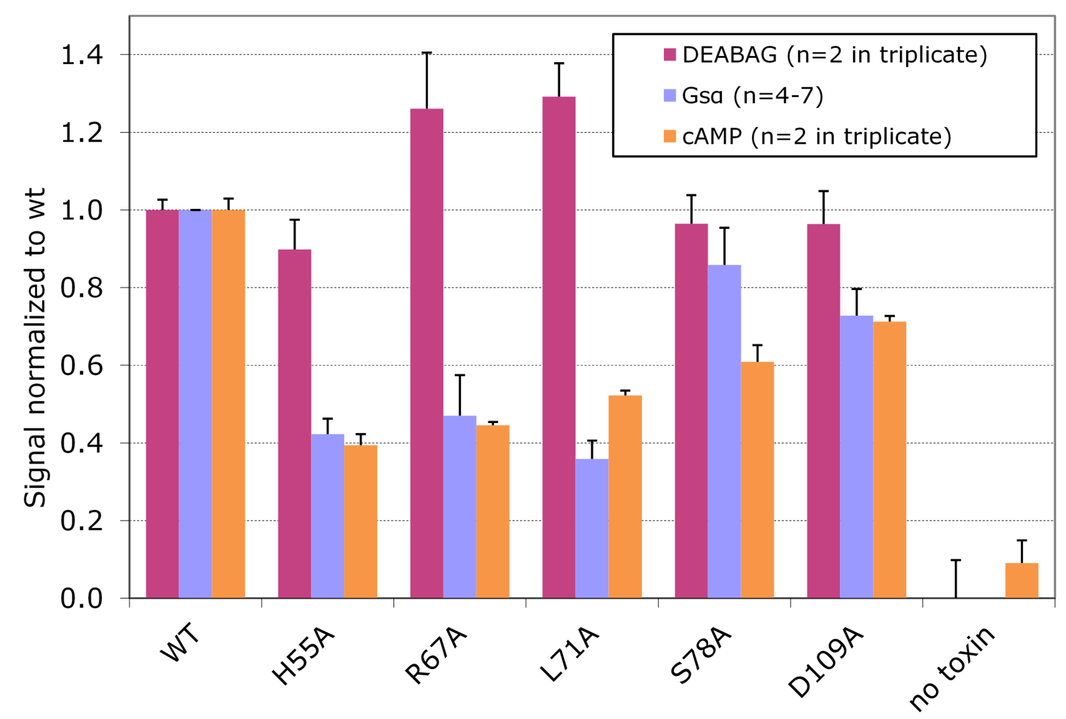
The effects of wt CT on cultured Y1 adrenal cells are mediated by increases in the intracellular concentration of cAMP produced as a consequence of intoxication. We performed quantitative assays for intracellular cAMP in Y1 adrenal cells after exposing them to wt CT or to each of the five CT variants associated with full ADP ribosylating activity against DEABAG and selective loss of ADP ribosylating activity against Gsα.
Figure 6 compares the values for the
in vitro ADP-ribosylation activity with DEABAG, the in vitro ADP-ribosylation activity with Gsα, and the intracellular cAMP concentration for each of the CT variants, all normalized to values set to 1.0 for the corresponding results obtained with wt CT. These results show that the
in vivo cAMP concentrations in Y1 adrenal treated with each of these five CT variants correlate well with the ADP ribosyltransferase activities of the variants against Gsα, but not against DEABAG, and support the conclusion that the residues replaced by alanine substitutions in these variants are directly involved in interactions of CTA1 with Gsα.
Figure 6.
Normalized in vitro enzymatic activities with DEABAG or Gsα and normalized intracellular cAMP concentrations in intoxicated Y1 cells for selected CT variants vs. wt CT.
Figure 6.
Normalized in vitro enzymatic activities with DEABAG or Gsα and normalized intracellular cAMP concentrations in intoxicated Y1 cells for selected CT variants vs. wt CT.
Toxin assays were done as described in experimental section, using equal amounts of wt or variant activated holotoxins (2 µg for in vitro enzymatic assays and 1 ng for in vivo cAMP assay). Calculated activities were then normalized to wt levels (wt CT = 1.0) and plotted for each CT variant, showing SEM for replicates.
In this study, we identified five amino acids (H55, R67, L71, S78, and D109) located near but not in the active site of CTA1 that are likely involved in interactions with the Gsα substrate, based on the demonstration that single alanine substitutions at each of these positions decreased the ADP ribosylating activity of the corresponding CT variants for Gsα but not for the small artificial substrate DEABAG. We also identified three amino acids (R25, T50, and F52) that likely participate in interactions with Gsα substrate but also affect intrinsic catalytic activity, based on the demonstration that single alanine substitutions at each of these positions decreased the ADP ribosylating activity against both substrates, but caused substantially greater loss of activity for Gsα than for DEABAG. Because double and triple alanine substitutions did not cause substantially greater loss of ADP ribosylating activity with Gsα than single alanine substitutions, it seems likely that other residues in CTA1, yet to be identified, also make important contributions to binding interactions between CTA1 and Gsα. If the binding interface between CTA1 and Gsα is large and complex, the consequences of any single alanine substitution for a residue in CTA1 that contributes to that interface may be small. It would be interesting in future studies to determine if more dramatic changes in substrate recognition specificity of CTA1 could be accomplished by substituting residues at the positions identified in the current study that would introduce larger side groups with various properties into the interface (instead of the methyl group provided by an alanine substitution), thereby creating potential steric hindrance as well as altering the local environment within the interface with respect to its hydrophobicity, hydrophilicity or charge. There is only one available structure for an arginine-specific ADP-ribosylating toxin in complex with its substrate, that of the
Clostridium perfringens iota toxin Ia fragment with actin [
24,
25]. The Ia-actin interface involves 32 residues from each protein partner and covers 829–895 Å
2, or about 5% of the surface area of each protein. Furthermore, the interface involves five loops on the Ia fragment, only one of which has amino-acid homology in CTA1 (the ARTT loop), which include residues targeted in this study. Because actin and Gsα are not homologous, we cannot yet compare the relative contributions of amino acids at specific positions (other than the acceptor arginine residue) to the interactions of these substrates with their cognate ADP-ribosylating toxins.
We have shown here, and others have shown [
30,
31,
32], that both
in vitro and
in vivo Gsα alone is a poor substrate for ADP-ribosylation, even in the presence of GTP-bound ARFs. Unlike the case for pertussis toxin, where peptides encompassing the last 10–20 amino acids of PT-sensitive alpha subunits (surrounding the target cysteine in Giα) are equally as effective substrates for PT-catalyzed ADP-ribosylation as is native Giα [
33], CTA1 likely requires a very particular ternary complex that includes Gsα to enable ADP-ribosylation. Recent crystal structures of G protein complexes have given insight into why this is so. Early work with Gt (transducin) [
18] and Gsα [
34] produced data that were consistent with the natural substrate for cholera toxin being the activated GPCR-bound, nucleotide-free form of Gsα. Gsα in the GDP bound (inactive) form has a high affinity for Gβγ, and they form a stable heterotrimeric complex.
In vivo, this complex is tightly bound to membrane-anchored quiescent GPCRs. Upon stimulation by receipt of an activation signal, the activated receptor induces a conformational change that results in opening of the interdomain interface in the G protein heterotrimer with release of GDP, generating a nucleotide-free Gα(0) as part of the ternary complex of activated GPCR-G protein heterotrimer [
35]. The structure of this activated complex has recently been determined [
36,
37] and reveals a remarkable conformational change in the nucelotide-free Gsα upon release of GDP, compared to its structure in the GPCR-coupled GDP-bound heterotrimer [
38] or Giα in the isolated heterotrimer [
39]. This complex is short-lived, and the Gsα(0) rapidly binds GTP. Gsα(GTP) has dramatically reduced affinity for Gβγ, and it quickly dissociates from the ternary complex and proceeds to activate downstream effectors, one of which in the case of Gsα is adenylate cyclase.
Our
in vitro system for ADP-ribosylation of Gsα does not include a membrane-anchored GPCR or the upstream components required for its activation. Interestingly, it has been shown that ARF1 can also interact with Gsα and that the amino-terminus of ARF1 is required for CT-catalyzed ADP-ribosylation of Gsα [
40], but not the small artificial substrate agmatine [
40]. The presence of Gβγ and the other components in our system must be sufficient (but not necessarily optimal) to generate a form of Gsα that is ADP-ribosylated by CTA1. The availability of these new structures of the activated complexes should facilitate future studies on the interactions of CTA1 and Gsα.


