Article

Occurrence of Penicillium brocae and Penicillium citreonigrum, which Produce a Mutagenic Metabolite and a Mycotoxin Citreoviridin, Respectively, in Selected Commercially Available Rice Grains in Thailand

by
Nozomi Shiratori
1,†,
Naoki Kobayashi
1,2,†,
Phitsanu Tulayakul
3,
Yoshitsugu Sugiura
1,
Masahiko Takino
4,
Osamu Endo
2 and
Yoshiko Sugita-Konishi
1,2,*
1
Graduate School of Life and Environmental Sciences, Azabu University, 1-17-71, Fuchinobe Chuo-ku, Sagamihara 252-5201, Japan
2
Faculty of Life and Environmental Sciences, Azabu University, 1-17-71, Fuchinobe Chuo-ku, Sagamihara 252-5201, Japan
3
Department of Veterinary Public Health, Faculty of Veterinary Medicine, Kasetsart University, Kamphaeng Saen campus, Nakhon Pathom 73140, Thailand
4
Agilent Technologies, Japan, Ltd., 9-1 Takakura-cho, Hachioji, Tokyo 192-8510, Japan
*
Author to whom correspondence should be addressed.
These authors contributed equally to this work.
Submission received: 17 February 2017 / Revised: 4 June 2017 / Accepted: 12 June 2017 / Published: 15 June 2017
(This article belongs to the Section Mycotoxins)

Abstract

:
Commercially available rice grains in Thailand were examined to isolate the monoverticillate Penicillium species responsible for toxic yellowed rice. Penicillium species were obtained from seven out of 10 rice samples tested. Among them, one Penicillium citreonigrum isolate and six Penicillium brocae isolates were morphologically identified. The P. citreonigrum isolate produced the mycotoxin citreoviridin on a yeast extract sucrose broth medium. Mycotoxin surveys showed that citreoviridin was not detected in any samples, but one out of 10 rice samples tested was positive for aflatoxin B1 at a level of 5.9 μg/kg. An Ames test revealed that methanol extracts from rice grains inoculated with selected P. brocae isolates were positive for strains TA100 and YG7108 of Salmonella typhimurium, suggesting the presence of base-pair substitution and DNA alkylation mutagens. Our data obtained here demonstrated that aflatoxin B1 and toxic P. citreonigrum were present on domestic rice grains in Thailand, although limited samples were tested. Penicillium brocae, which may produce mutagenic metabolites, was isolated for the first time from the surface of Thai rice grains.

1. Introduction

Fungal contamination in food and foodstuffs causes a number of problems in commercially available products, such as food spoilage and food poisoning. In terms of fungi, mycotoxin contamination is of great concern to human health around the world. In Thailand, the major mycotoxins associated with cancer risk found in market commodities are aflatoxin (AF) B1 and other aflatoxins [1]. AFB1 is a strong class I carcinogen according to the International Agency for Research on Cancer, and several studies have reported rice contaminated with AFB1 and other aflatoxins among Thai commodities [2,3,4]. In the postharvest condition, harvested rice grains may be damaged in several ways, such as fungal invasion, mycotoxin contamination, physicochemical changes, and insect activity. Particularly, fungal invasion of stored agricultural products leads to a loss of quality in the storage environment [5]. However, there is currently insufficient information on stored rice as an agricultural commodity in Thailand.
A staple cereal plant, rice (Oryza sativa L.) is widely cultivated in Asia, North and South America, the European Union, and Middle Eastern and African countries. Rice provides 20% of the world’s dietary energy supply, while wheat and maize supply 19% and 5%, respectively [6]. The nutritive contents of rice vary depending on the type of rice, but rice is known to be a good source of thiamine, riboflavin and niacin, along with some kinds of amino acids. Thus, the contamination of rice grains with mycotoxins is a serious problem for human health [7]. In Asian countries, the contamination of rice with Aspergillus and Fusarium toxins such as aflatoxins, trichothecenes, and fumonisins, has been of great concern [8,9], but little attention has been paid to Penicillium toxin due to its low presence in rice.
Before and after World War II, the toxic rice problems in Japan occurred after rice was imported, and three historical cases have been documented [10,11]. It was called “yellow rice” and was caused by contamination of rice grains with toxigenic Penicillium species. The first case was the isolation of Penicillium citreonigrum (formerly, P. citreo-viride). Fungus-damaged rice was imported from Taiwan in 1937, and found to be contaminated with the toxic metabolite citreoviridin (CTV), which has been suggested to be responsible for beriberi disease. The second case was an occurrence of Penicillium islandicum (current taxon, Talaromyces islandicus) in a batch of not yellowed but yellowish-brown rice. This rice was imported from Egypt in 1948. Mycotoxin studies have revealed that P. islandicum produces hepatotoxic mycotoxins, such as luteoskyrin, and cyclochlorotine. The third case was caused by the citrinin-producer Penicillium citrinum. The rice was imported from Thailand in 1951 [12]. Citrinin is nephrotoxic, but has low acute toxicity. These toxicological characteristics, including the induction of cancer and chemical structures, have been reported by Japanese mycotoxin researchers [10,12].
Recently, an outbreak of beriberi disease occurred in the state of Maranhao, Brazil, and 1207 cases were reported, including 40 deaths. In rice samples from the outbreak region, the neurotoxic mycotoxin CTV was detected at ranges from 0.9 to 31.1 μg/kg. CTV is a known toxic metabolite of P. citreonigrum that adversely affects the cardiovascular system, and may be involved in acute cardiac beriberi [13]. However, the relationship between CTV and the disease is still unclear.
In the present study, we examined rice commodities in Thai markets for surveillance of mycoflora. In addition to the alcohol treatment, the rice grains were tested without any treatment in order to assess the stored rice environment in Thailand. Particularly, the presence of the toxic yellowed rice fungus P. citreonigrum and related species were surveyed. These monoverticillate species are characterized by slow growth and yellow pigment production on the surface or reverse of the colonies on Czapek yeast agar (CYA) medium. The isolates obtained were tested for CTV production. As an indicator of human toxicity, their metabolites were assayed for mutagenic activities using an Ames test. Contamination of Thai rice samples with mycotoxins was also analyzed.

2. Results

2.1. Mycoflora on Rice Grains

Based on macroscopic observation, the developed fungi were classified simply as genera of fungi. In the alcohol-treated group, the developed fungi were Aspergillus, Eurotium (teleomorph of xerophilic Aspergillus), Mucor and Rhizopus shown as Zygomycetes in Figure 1, and other fungi. Similar findings were noted in the untreated group, except for Penicillium also being observed. As shown in Figure 1, the frequency of the genera differed depending on the rice type and region of Thailand. However, almost the same major fungal genera developed between the groups, except for the genus Penicillium. Isolates of Penicillium species were only found on untreated rice grains. In addition, in the sample from the Surin region, monoverticillate and yellow pigment-producing Penicillium isolates were observed. These isolates were subjected to identification at the species level.

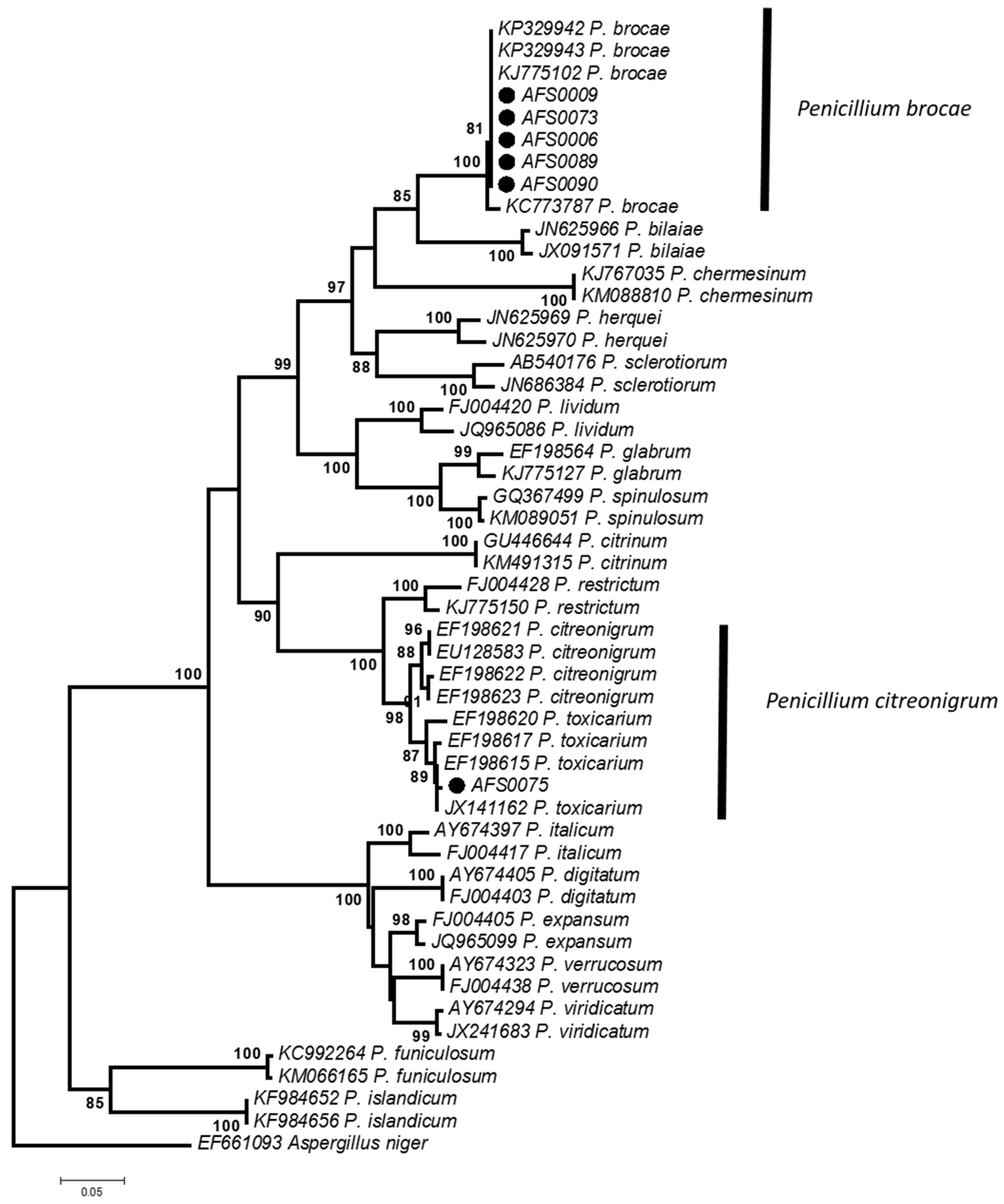
2.2. Morphological and Phylogenetic Identification

Monoverticillate and yellow pigment-producing Penicillium isolates were cultured on CYA and malt extract agar (MEA) media at 25 °C for seven days. Based on the morphological characteristics of Penicillium isolates, one isolate was identified as P. citreonigrum, but the taxonomic positions of two similar Penicillium isolates could not be determined by morphological observation alone. Therefore, these isolates were subjected to a molecular phylogeny analysis.
Figure 2 shows the NJ-tree inferred from the β-tubulin gene (β-tub) of selected Penicillium species. Each reference species formed a monophyletic clade with high bootstrap support. The P. citreonigrum isolate identified morphologically belonged to Penicillium toxicarium, which is included in the clade of P. citreonigrum. Recently, P. toxicarium was moved to Penicillium citreosulfuratum by molecular phylogeny alone [14], and the isolate was suggested to identify as P. citreosulfuratum. However, due to finely roughened conidia, especially with roughness arranged in undulation, the isolate differed from the IMI 92228 strain, which is an ex-type of P. citreosulfuratum [14,15]. Thus, the isolate was identified as P. citreonigrum based on the criterion of Penicillium taxonomy by Pitt [15]. Two unknown Penicillium isolates were identified as Penicillium brocae based on a phylogenetic analysis (Figure 2). The morphological characteristics of these isolates were then compared with the description of P. brocae reported by Peterson et al. [16].
As shown in Figure 3, P. brocae isolates grew slowly when cultured on CYA at 25 °C for seven days. Colonies were 23–25 mm in diameter, grayish blue-green, velvety, and centrally produced moderate to abundant yellow exudate. The reverse color was yellowish brown or brownish purple, producing soluble naphthalene yellow pigment at the edge. The isolates did not grow at 37 °C. Conidiophores were short and smooth-walled, and vesiculate, monoverticillate penicillus was observed. Phialides were ampulliform, 8–9 × 2–3 μm. Conidia were spherical, 2.5–3.5 µm in diameter, with slightly roughened walls, forming short chains. These characteristics of the observed P. brocae isolates were almost the same as the original description of the species.
The sequence data of P. citreonigrum AFS-0075, and P. brocae AFS-0006 and AFS-0073 strains were deposited in the DNA Data Bank of Japan (DDBJ) under the accession numbers LC216411, LC216406, and LC216408, respectively.
As relatively few Penicillium species were observed, an additional trial was conducted in the same sample but using different rice grains. The trial showed that no P. citreonigrum isolate and four P. brocae isolates were obtained. Among them, three P. brocae isolates were subjected to the molecular phylogeny analysis. The identification of the isolates was confirmed using the β-tub region (Figure 2). The sequence data of P. brocae AFS-0009, AFS-0089, and AFS-0090 strains were deposited in the DDBJ under the accession numbers LC216407, LC216409, and LC216410, respectively. The strains obtained were not used for further experiments.

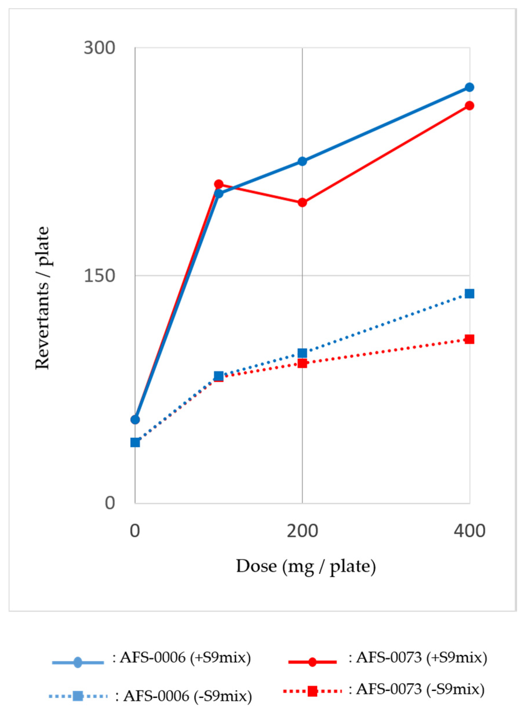
2.3. Mutagenicity of P. brocae Metabolites

The AFS-0006 and AFS-0073 strains of P. brocae were cultured on 20 g of rice grains at 25 °C for 14 days. Early in the culture, the rice was stained yellow, and then covered with abundant green conidia. The cultures were extracted with 100 mL of methanol by sonication for 20 min. The methanol extracts were evaporated to dryness under a nitrogen gas flow and resolved with 5 mL of dimethylsulfoxide.
The assay dose was calculated from the extract amounts per 4 g of rice grains and prepared with serial dilutions. Assays were performed with and without S9 mix using the tester strains of Salmonella typhimurium and Escherichia coli. Results showed that the methanol extracts of both strains were positive against the TA100 and YG7108 strains of S. typhimurium, as base pair substitution mutations occurred primarily at one of the G-C pairs. The mutagenic activity of YG7108 was higher than that of TA100. Figure 4 shows the dose-response curve of mutagenicity of the methanol extracts from the P. brocae AFS-0006 and AFS-0073 strains for S. typhimurium YG7108, both with and without rat liver S9 mix. The mutagenicity increased dose dependently, especially when treated with the S9 mix. These data indicated the presence of alkylating agents in the P. brocae metabolites.

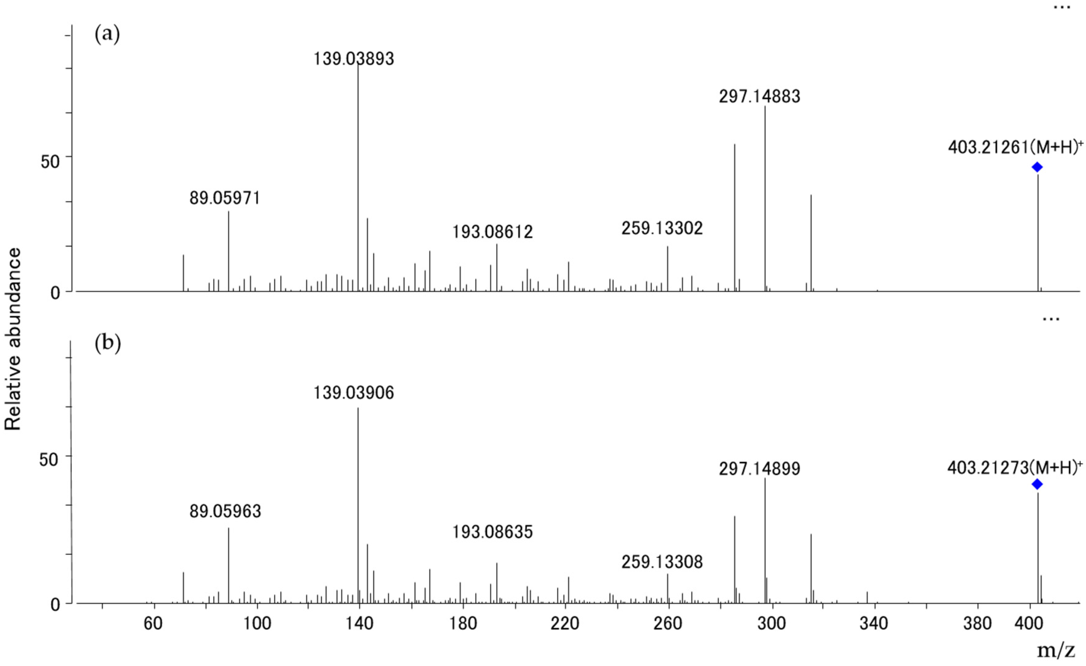
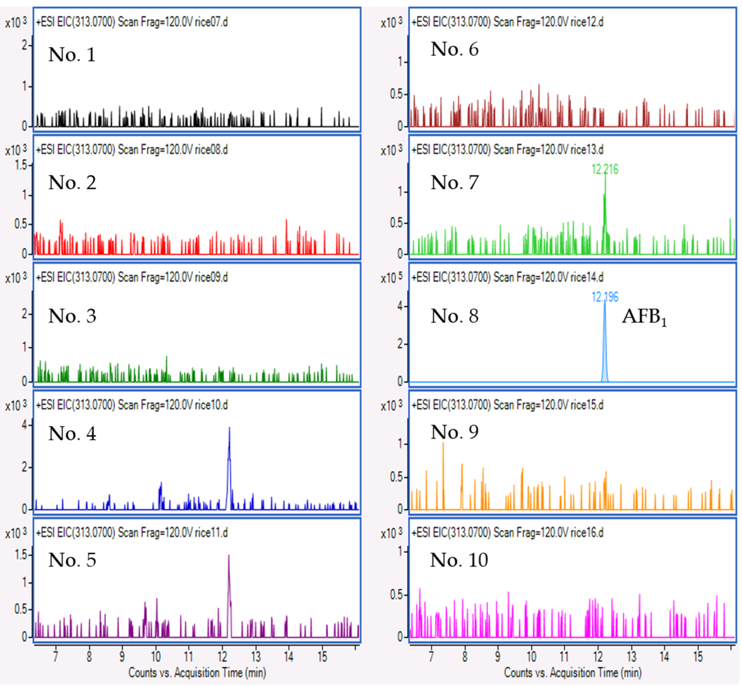
2.4. Mycotoxin Analysis

For the P. citreonigrum isolate obtained, an LC-QTOF analysis revealed that the isolate produced high amounts of CTV, when cultured on a yeast extract sucrose (YES) liquid cultural medium. Figure 5 and Figure 6 show the total ion chromatogram of the fungal extract, the accurate mass spectra of the main peak of the fungal extract, and citreoviridin A standard solution, respectively. The relative mass error of the protonated molecular ion at main peak was 2.71 ppm.
Multiple mycotoxins survey showed that CTV was negative for any samples of Thai rice grains, although CTV-producing fungus was found. Among the 10 rice samples tested, AFB1 was detected at a concentration of 5.9 µg/kg in sample No. 8 obtained from the Pathum Thani region. Figure 7 shows extracted ion chromatograms of the rice samples for AFB1 detection. The other mycotoxins examined were not detected in any samples (data not shown).

3. Discussion

An incident involving rice with yellow coloration occurred in 1937 in Japan and was found to have been caused by P. citreonigrum [12]. The fungus produced a toxic metabolite, CTV, which is responsible for acute cardiac beriberi [17]. As beriberi disease did not occur in Japan after World War II, this yellowed rice toxic syndrome was largely forgotten. However, in 2006 to 2008, an outbreak occurred in Brazil due to the consumption of CTV-contaminated rice [18]. This incident reminded us of past issues associated with yellowed rice in Japan.
In this study, we obtained the target fungus P. citreonigrum from the surface of rice grains in Thai commodities. In addition, we also isolated P. brocae from the same samples, which was unexpected, as P. brocae is regarded as an endophytic fungus. These two Penicillium species showed slow growth on culture media.
In general, fungal isolation in rice grains is conducted with surface disinfection using chlorine, as several kinds of air-borne fungi may be present on the surface of rice grains. These contaminants can interfere with the surveillance of mycoflora on rice grains. However, we compared two methods in the present study, surface disinfection and direct plating without disinfection in order to examine the surface mycobiota on rice grains. We found toxigenic P. citreonigrum and endophytic P. brocae, along with other Penicillium species, only in the untreated group (Figure 1). As these two Penicillium species are not common air-borne fungi, our untreated approach seems to have been useful for the isolation of uncommon Penicillium species, possibly due to their susceptibility to the disinfecting agents compared with the common field or storage fungi of Aspergillus, Bipolaris, Colletotrichum, Curvularia, Mucor, Phoma, and Rhizopus [19]. This may explain why previous studies on mycoflora in rice failed to find any P. citreonigrum or P. brocae, except in the case of the P. citreonigrum responsible for food poisoning in Brazil [18].
An isolate of P. citreonigrum identified morphologically was obtained from a rice sample in the Surin region, which is in the northeast of Thailand, close to Cambodia. It produced CTV on YES medium. Kim et al. [20] reported that P. toxicarium was an endophytic fungus, because it was isolated from healthy needles of Pinus rigida, along with Penicillium fellutanum. P. citreonigrum and P. toxicarium are very close in the phylogenetic tree (Figure 2), but the taxon remains unclear. P. brocae was isolated firstly from the coffee berry borer Hypothenemus hampei [16], but it is known to be an endophytic fungus, generally derived from the coffee plant [21], or the Zyzzya sp. sponge [22], or the marine mangrove plant Avicennia marina [23]. Two research reports have shown that insects carried experimentally toxigenic Aspergillus and Penicillium species on stored maize kernels [24] and wheat grains [25]. Therefore, although P. citreonigrum and P. brocae were both isolated from rice grains in this study, they may have been introduced to the rice by insects, such as grain weevils, which tend to favor rice. Further research will be needed to confirm our speculation.
In the mutagenicity test, two P. brocae strains were tested to determine whether or not they could produce mutagens. The methanol extracts obtained from moldy rice grains were positive for base-pair substitution mutation and DNA alkylation using S. typhimurium strains TA100 and YG7108, respectively. Interestingly, these strains suggested the mutation of GC sites. As the mutagenic activity in the YG7108 strain was high, the data revealed that the metabolites of P. brocae AFS-0006 and AFS-0073 strains induced the alkylation of the GC sites (Figure 4). Further studies are underway to clarify the mutagenic agents produced by P. brocae.
Our fungal survey showed that toxigenic monoverticillate Penicillium species were present in commercially available Thai rice grains. However, CTV produced by P. citreonigrum was negative in any rice samples tested. AFB1 alone was detected in the No. 8 rice sample, although toxigenic Aspergillus species were not found in the sample (Figure 1). The culture plate of the No. 8 sample was fully covered with Mucor or Rhizopus. Anti-fungal chemicals, dichloran and rose bengal were inactive in depressing the growth of these fungi in the sample. Aflatoxin contamination in Thai commodities has been reported by several studies [2,8]. In this study, we also detected AFB1 in a sample of Luem phua rice in the Pathum Thani region, close to Bangkok. Although our survey was admittedly limited, the level of AFB1 contamination was higher than that in previous reports. Levels of contamination with AFB1 may depend on rice types.

4. Conclusions

Commercially available Thai rice samples were contaminated with several fungal species. Most of the species observed were not toxic, but a few toxigenic fungi were present, such as P. citreonigrum and P. brocae. Of note, these toxigenic Penicillium species possessed good ability to produce CTV or a mutagen. Thus, these species could be a potential threat to human health if environmental conditions are suitable for their growth.

5. Materials and Methods

5.1. Sample and Fungal Strain

Ten samples of rice were purchased from nine markets in Thailand. The types of rice and market regions were as follows: Jasmine rice (No. 1) in Bangkok, Dong Ruk rice (No. 2) in Surin, Pratum rice (No. 3) in Ayuddhaya, Riceberry gaba (No. 4) in Nakorn Pathom, Jasmine rice (No. 5) and brown jasmine rice (No. 10) in Chiang Rai, Red jasmine rice (No. 6) in Chaiyaphum, Organic brown rice (No. 7) in Chaing Mai, Luem phua rice (No. 8) in Pathum Thani, and Riceberry (No. 9) in Petchabune. These rice samples were chosen randomly, and packed into sealed plastic bags. The bags were kept closed until use. The samples sizes of the rice grains were ca. 1 kg each.
As a reference strain, the P. citreo-viride IMI 92228 strain was obtained from the culture collection of CABI in the United Kingdom.

5.2. Fungal Isolation and Morphological Identification

One hundred rice grains from all 10 samples were used, and treated in two different manners: 50 grains were exposed to 70% ethanol solution for 30 s and then washed twice with sterile distilled water, and the other 50 grains were not treated in any manner. Following this treatment, five rice grains per plate were placed onto dichloran rose bengal chloramphenicol agar (DRBC) or dichloran 18% glycerol agar (DG18) medium and cultured at 25 °C for 5 days. After incubation, the genera of the developed fungi were simply classified by a macroscopic observation [26]. Among them, Penicillium species, showing monoverticillate and yellow pigmentation, were transferred to CYA and MEA media (BD, Sparks, MD, USA) and cultured at 25 °C for 7 days. According to the taxonomic criterion of the Penicillium species [26], the isolates were morphologically identified based on colony textures and microscopic observation.

5.3. Molecular Phylogenetic Analysis

A molecular phylogenetic analysis was carried out as follows. Fungi were pre-cultured on potato dextrose (PD) broth medium (Difco Laboratories, Franklin Lakes, NJ, USA) at 25 °C for 3–4 days. The genomic DNA was extracted from fungal mycelia by the sodium dodecyl sulfate method [27]. The β-tub regions were chosen for the phylogenic analysis. PCR amplification was done using Bt2a and Bt2b primers [28]. Sequencing was performed using a BigDye Terminator v3.1 Cycle Sequencing Kit and ABI3130 DNA Analyzer (Applied Biosystems, Foster City, CA, USA) according to the manufacturer’s instructions. Forty-five sequences of β-tub regions of the various Penicillium species and Aspergillus niger were downloaded from GenBank and used as reference data for the phylogenetic analysis. The species were selected with regard to various Penicillium clades reported by Visagie et al. [29] and A. niger was used as an outer species. The sequences were automatically aligned and the alignments were manually adjusted using MEGA6 [30]. A phylogenetic tree was constructed with the neighbor joining method by the maximum composite likelihood model using MEGA6.

5.4. Mutagenicity Assay

The mutagenicity assay was performed using S. typhimurium strains TA98, TA100, TA1537, and YG7108, and E. coli strain WP2/uvrA/pKM101 with and without rat liver S9 mix. These strains were chosen for detecting frameshift mutations (TA98 and TA1537) and base-pair substitution mutations (TA100, YG7108 and WP2), and YG7108 was particularly sensitive for DNA alkylation. The assay was tested for methanol extracts obtained from moldy rice grains, which were individually inoculated with tester isolates and cultured at 25 °C for 14 days. In accordance with the official guide of the mutagenicity test using microorganisms [31], which was revised by the Ministry of Health, Labor and Welfare of Japan in 1991, along with the method of Yamada et al. [32] for the S. typhimurium YG7108 strain, the assay was performed at five doses with two plates per dose for each sample. In addition, mutagenic standard chemicals were used as positive references against tester strains. The experiments were repeated twice, and the mutagenic activity was calculated from the slope of the linear portion of the dose-response curve using the statistical model of least squares linear regression.

5.5. Mycotoxin Analysis

For the CTV analysis, the tester isolate was cultured on YES liquid medium at 25 °C for 10 days. The CTV extract was then obtained in accordance with the method described by da Rocha et al. [13]. In target MS/MS acquisition for CTV, a data rate of three mass spectra per sec in MS and MS/MS mode was used. Target compounds were detected and reported from accurate-mass data using Agilent Mass Hunter Qualitative and The Agilent Mycotoxins and Related Metabolites Personal Compound Database and Library (PCDL) software programs. The PCDL database contains 455 target compound names, molecular formulae, CAS numbers, structure and 297 MSMS spectra. The Qualitative Analysis software program tentatively identified target compounds using a “Find by Formula” algorithm with the following criteria: relative mass error, 5 ppm; isotope abundance and spacing score, greater than 90 out of 100. The reagent of citreoviridin A standard used was obtained from Fermentek Ltd. (Jerusalem, Israel).
Fifty grams of rice grains from each sample were powdered using a blender machine, and a 7.5 g portion of the powder sample was weighed and transferred to a 50-mL centrifuge tube. Mycotoxins were extracted using the QuEChERS extraction kits (Agilent Technologies, Inc., Tokyo, Japan) with slightly modified citrate buffer. For clean-up, a dispersive solid-phase extraction (Agilent Technologies, Inc.) was used. In accordance with the manufacturer’s protocol, the purified extracts obtained were analyzed by liquid chromatography coupled with quadrupole time-of-flight mass spectrometry (LC-QTOFMS) equipped with a dual-spray Jet Stream electrospray ionization source. The LC system was an Agilent 1290 Infinity II LC system (Agilent Technologies, Inc.) consisting of binary pumps, degasser, column compartment and autosampler with a reverse-phase column of ZORBAX Eclipse plus C18 (100 × 2.1 mm, 1.8 μm) and operated with a gradient mobile phase containing 0.1% formic acid + 10 mM ammonium acetate (A) and acetonitrile, or methanol (B). The initial combination was 90% A and 10% B, changing linearly to 0% A and 100% B over 30 min. The flow rate was 0.2 mL/min.
The quadrupole time-of-flight mass spectrometry system was the Agilent 6550 iFunnel Quadrupole Time-of-Flight LC/MS (Agilent Technologies, Inc.) operated in positive ion mode with a mass ranging from 100 to 1000 Da and a data rate of 1.5 mass spectra per sec. The instrument acquired data using the following parameters: drying gas temperature, 200 °C; drying gas flow, 20 L/min; nebulizer, 50 psi; sheath gas temperature, 400 °C; sheath gas flow, 12 L/min; VCap. 4000 V; nozzle, 0 V; fragmentor, 360 V; skimmer, 65 V; and octapole RF peak, 750. A constant flow of Agilent TOF reference solution containing purine (m/z = 121.05087) and HP-921 (m/z = 922.00979) through the reference nebulizer allowed the system to continuously correct for any mass drift.
Twenty-six mycotoxins; AFB1, AFB2, AFG1, AFG2, AFM1, citrinin, CTV, cyclochlorotine, cyclopiazonic acid, deoxynivalenol (DON), DON-glucoside, 3-acetylDON, 15-acetylDON, diacetoxyscirpenol, fumonisin B1, fumonisin B2, luteoskyrin, nivalenol (NIV), NIV-glucoside, 4-acetylNIV, ochratoxin, patulin, sterigmatocystin, T-2 toxin, HT-2 toxin, and zearalenone, were examined by LC-QTOFMS.

Acknowledgments

This work was supported by the Health and Labour Sciences Research Grants (Research on Food Safety) from the Ministry of Health, Labour and Welfare of Japan.

Author Contributions

Y.S.-K., Y.S. and N.K. conceived and designed the experiments; P.T. performed the sample preparation; N.S. and Y.S. performed the microbiological experiments and the metabolites extraction; N.S. and M.T. performed the chemical analysis; N.K. performed the molecular analysis; O.E. contributed reagents, materials and analysis for Ames assay; Y.S. wrote the paper. Y.S.-K. and N.S. reviewed the paper. Y.S.-K. was responsible for submission.

Conflicts of Interest

The authors declare no conflicts of interest.

References

  1. Anukarahanonta, T.; Chudhabuddhi, C.; Temcharoen, P.; Sukroongreung, S. Cancer Risk from aflatoxins in Thailand. In Developments in Food Science 7 Toxigenic Fungi—Their Toxins and Health Hazard; Kurata, H., Ueno, Y., Eds.; Kodansha Ltd.: Tokyo, Japan, 1984; pp. 229–347. [Google Scholar]
  2. Tansakul, N.; Limsuwan, S.; Böhm, J.; Hollmann, M.; Razzazi-Fazeli, E. Aflatoxins in selected Thai commodities. Food Addit. Contam. Part B Surveill. 2013, 6, 254–259. [Google Scholar] [CrossRef] [PubMed]
  3. Bansal, J.; Pantazopoulos, P.; Tam, J.; Cavlovic, P.; Kwong, K.; Turcotte, A.M.; Lau, B.P.; Scott, P.M. Surveys of rice sold in Canada for aflatoxins, ochratoxin A and fumonisins. Food Addit. Contam. Part A Chem. Anal. Control Expo. Risk Assess. 2011, 28, 767–774. [Google Scholar] [CrossRef] [PubMed]
  4. Suttajit, M. Current status of aflatoxin and its control in Thailand. J. Toxicol. Sci. 1998, 23, 173–176. [Google Scholar] [CrossRef] [PubMed]
  5. Abramson, D. Mycotoxin formation and environmental factors. In Mycotoxins in Agriculture and Food Safety; Sinha, K.K., Bhatnagar, D., Eds.; Marcel Dekker, Inc.: New York, NK, USA, 1998; pp. 255–277. [Google Scholar]
  6. Food and Agriculture Organization of the United Nations. Rice and Human Nutrition. International Year of Rice 2004, Rice Is Life. Available online: http://www.fao.org/rice2004/en/f-sheet/factsheet3.pdf (accessed on 15 May 2017).
  7. Rohman, A.; Helmiyati, S.; Hapsari, M.; Setyaningrum, D.L. Rice in health and nutrition. Int. Food Res. J. 2014, 21, 13–24. [Google Scholar]
  8. Kushiro, M.; Zheng, Y.; Noriduki, H.; Sugita-Konishi, Y. Limited Surveillance of Mycoflora and Mycotoxins in Thai Rice Retailed in Japan. Asia-Pac. J. Food Saf. Secur. 2015, 1, 25–37. [Google Scholar]
  9. Tanaka, K.; Sago, Y.; Zheng, Y.; Nakagawa, H.; Kushiro, M. Mycotoxins in rice. Int. J. Food Microbiol. 2007, 119, 59–66. [Google Scholar] [CrossRef] [PubMed]
  10. Udagawa, S.; Tatsuno, T. Safety of rice grains and mycotoxins—A historical review of yellow rice mycotoxicoses. Yakushigaku Zasshi 2004, 39, 321–342. [Google Scholar] [PubMed]
  11. Kushiro, M. Historical review of researches on yellow rice and mycotoxigenic fungi adherent to rice in Japan. JSM Mycotoxins 2015, 65, 19–23. [Google Scholar] [CrossRef]
  12. Tsunoda, H. Micro-organisms which deteriorate stored cereals and grains. In Proceedings of the First U.S.-Japan Conference on Toxic Micro-organisms Mycotoxins Botulism, Honolulu, Hawaii, 10 October 1968; Herzberg, M., Ed.; The UJNR Joint Panels on Toxic Micro-organisms and the U.S. Department of the Interior: Washington, DC, USA, 1970; pp. 143–162. [Google Scholar]
  13. Da Rocha, M.W.; Resck, I.S.; Caldas, E.D. Purification and full characterisation of citreoviridin produced by Penicillium citreonigrum in yeast extract sucrose (YES) medium. Food Addit. Contam. Part A Chem. Anal. Control Expo. Risk Assess. 2015, 32, 584–595. [Google Scholar] [CrossRef] [PubMed]
  14. Visagie, C.M.; Seifert, K.A.; Houbraken, J.; Samson, R.A.; Jacobs, K. A phylogenetic revision of Penicillium sect. Exilicaulis, including nine new species from fynbos in South Africa. IMA Fungus 2016, 7, 75–117. [Google Scholar] [PubMed]
  15. Pitt, J.I. Chapter 8 Subgenus Aspergilloides Dierckx. In The Genus Penicillium and Its Teleomorphic States Eupenicillium and Talaromyces; Academic Press: New York, NY, USA, 1979; pp. 164–232. [Google Scholar]
  16. Peterson, S.W.; Pérez, J.; Vega, F.E.; Infante, F. Penicillium brocae, a new species associated with the coffee berry borer in Chiapas, Mexico. Mycologia 2003, 95, 141–147. [Google Scholar] [CrossRef] [PubMed]
  17. Uraguchi, K. Mycotoxic origin of cardiac beriberi. J. Stored Prod. Res. 1969, 5, 227–236. [Google Scholar] [CrossRef]
  18. Rosa, C.A.; Keller, K.M.; Oliveira, A.A.; Almeida, T.X.; Keller, A.M.; Marassi, A.C.; Kruger, C.D.; Deveza, M.V.; Monteiro, B.S.; Nunes, L.M.; et al. Production of citreoviridin by Penicillium citreonigrum strains associated with rice consumption and beriberi cases in the Maranhão State, Brazil. Food Addit. Contam. Part A Chem. Anal. Control Expo. Risk Assess. 2010, 27, 241–248. [Google Scholar] [CrossRef] [PubMed]
  19. Pitt, J.I.; Hocking, A.D.; Bhudhasamai, K.; Miscamble, B.F.; Wheeler, K.A.; Tanboon-Ek, P. The normal mycoflora of commodities from Thailand. 2. Beans, rice, small grains and other commodities. Int. J. Food Microbiol. 1994, 23, 35–43. [Google Scholar] [CrossRef]
  20. Kim, C.S.; Park, M.S.; Yu, S.H. Two Species of Endophytic Penicillium from Pinus rigida in Korea. Mycobiology 2008, 36, 222–227. [Google Scholar] [CrossRef] [PubMed]
  21. Peterson, S.W.; Vega, F.E.; Posada, F.; Nagai, C. Penicillium coffeae, a new endophytic species isolated from a coffee plant and its phylogenetic relationship to P. fellutanum, P. thiersii and P. brocae based on parsimony analysis of multilocus DNA sequences. Mycologia 2005, 97, 659–666. [Google Scholar] [CrossRef] [PubMed]
  22. Bugni, T.S.; Bernan, V.S.; Greenstein, M.; Janso, J.E.; Maiese, W.M.; Mayne, C.L.; Ireland, C.M. Brocaenols A–C: Novel polyketides from a marine-derived Penicillium brocae. J. Org. Chem. 2003, 68, 2014–2017. [Google Scholar] [CrossRef] [PubMed]
  23. Meng, L.H.; Li, X.M.; Lv, C.T.; Huang, C.G.; Wang, B.G. Brocazines A−F, Cytotoxic Bisthiodiketopiperazine Derivatives from Penicillium brocae MA-231, an Endophytic Fungus Derived from the Marine Mangrove Plant Avicennia marina. J. Nat. Prod. 2014, 77, 1921–1927. [Google Scholar] [CrossRef] [PubMed]
  24. Dharmaputra, O.S.; Halid, H.; Sunjaya; Khim, K.S. The effect of Sitophilus zeamais on fungal infection, aflatoxin production, moisture content and damage to kernels of stored maize. In Proceedings of the 6th International Working Conference on Stored-Product Protection, Canberra, Australia, 17–23 April 1994; Highley, E., Wright, E.J., Banks, H.J., Champ, B.R., Eds.; CAB International: Wallingford, UK, 1994; Volume 2, pp. 981–984. Available online: http://spiru.cgahr.ksu.edu/proj/iwcspp/pdf2/6/981.pdf (accessed on 16 March 2016).
  25. Sinha, A.K.; Sinha, K.K. Incidence of mycotoxigenic fungi on stored grain insects, Sitophilus oryzae and Tribolium casteneum. Proc. Indian Natl. Sci. Acad. 1991, B57, 77–80. [Google Scholar]
  26. Pitt, J.I.; Hocking, A.D. Fungi and Food Spoilage, 3rd ed.; Springer: New York, NY, USA, 2009. [Google Scholar]
  27. Watanabe, M.; Lee, K.; Goto, K.; Kumagai, S.; Sugita-Konishi, Y.; Hara-Kudo, Y. Rapid and effective DNA extraction method with bead grinding for a large amount of fungal DNA. J. Food Prot. 2010, 73, 1077–1084. [Google Scholar] [CrossRef] [PubMed]
  28. Glass, N.L.; Donaldson, G.C. Development of primer sets designed for use with the PCR to amplify conserved genes from filamentous ascomycetes. Appl. Environ. Microbiol. 1995, 61, 1323–1330. [Google Scholar] [PubMed]
  29. Visagie, C.M.; Hirooka, Y.; Tanney, J.B.; Whitfield, E.; Mwangen, K.; Meijer, M.; Amend, A.S.; Seifert, K.A.; Samson, R.A. Aspergillus, Penicillium and Talaromyces isolated from house dust samples collected around the world. Stud. Mycol. 2014, 78, 63–139. [Google Scholar] [CrossRef] [PubMed]
  30. Tamura, K.; Stecher, G.; Peterson, D.; Filipski, A.; Kumar, S. MEGA6: Molecular Evolutionary Genetics Analysis version 6.0. Mol. Biol. Evol. 2013, 30, 2725–2729. [Google Scholar] [CrossRef] [PubMed]
  31. Labor Standard Bureau, Ministry of Labor. Guidebook for Mutagenicity Test Using Microorganisms; Japan Industrial Safety and Health Association: Tokyo, Japan, 1986; pp. 33–81.
  32. Yamada, M.; Sedgwick, B.; Sofuni, T.; Nohni, T. Construction and characterization of mutants of Salmonella typhimurium deficient in DNA repair of O6-methylguanine. J. Bacteriol. 1995, 177, 1511–1519. [Google Scholar] [CrossRef] [PubMed]
Figure 1. Percentage of fungal genera isolated from 10 samples of Thai rice, which were treated with 70% ethanol exposure (a) and without surface disinfection (b), and cultured on DG18 (left) and DRBC (right) media.
Toxins 09 00194 g001
Figure 2. Phylogenetic tree based on the β-tubulin gene. The tree was generated by the neighbor joining method using 1000 bootstrap replicates. Only the bootstrap values above 70% are indicated near the branches. The tree was rooted with the strain of Aspergillus niger. The strain numbers marked with black circles are the species obtained in this study. Others were downloaded from GenBank.
Toxins 09 00194 g002
Figure 3. Penicillium brocae. (a) Colonies grown on CYA medium at 25 °C for 7 days. (b) Conidiophores and conidia in a chain. Bar, 10 μm.
Toxins 09 00194 g003
Figure 4. Mutagenicity of the methanol extract from Penicillium brocae for Salmonella typhimurium YG7108 with (●) and without (■) rat liver S9 mix.
Toxins 09 00194 g004
Figure 5. Total ion chromatograms of the Penicillium citreonigrum extract (upper) and citreoviridin A standard (lower) by LC/MS/MS.
Toxins 09 00194 g005
Figure 6. Accurate product ion spectra of peak 1 in Figure 5 and citreoviridin A standard. (a) Peak 1, (b) Citreoviridin A standard.
Toxins 09 00194 g006
Figure 7. Extracted ion chromatograms of 10 rice samples for AFB1 detection.
Toxins 09 00194 g007

