
Towards a New μ→eγ Search with the MEG II Experiment: From Design to Commissioning

 ,
,  , , , , and
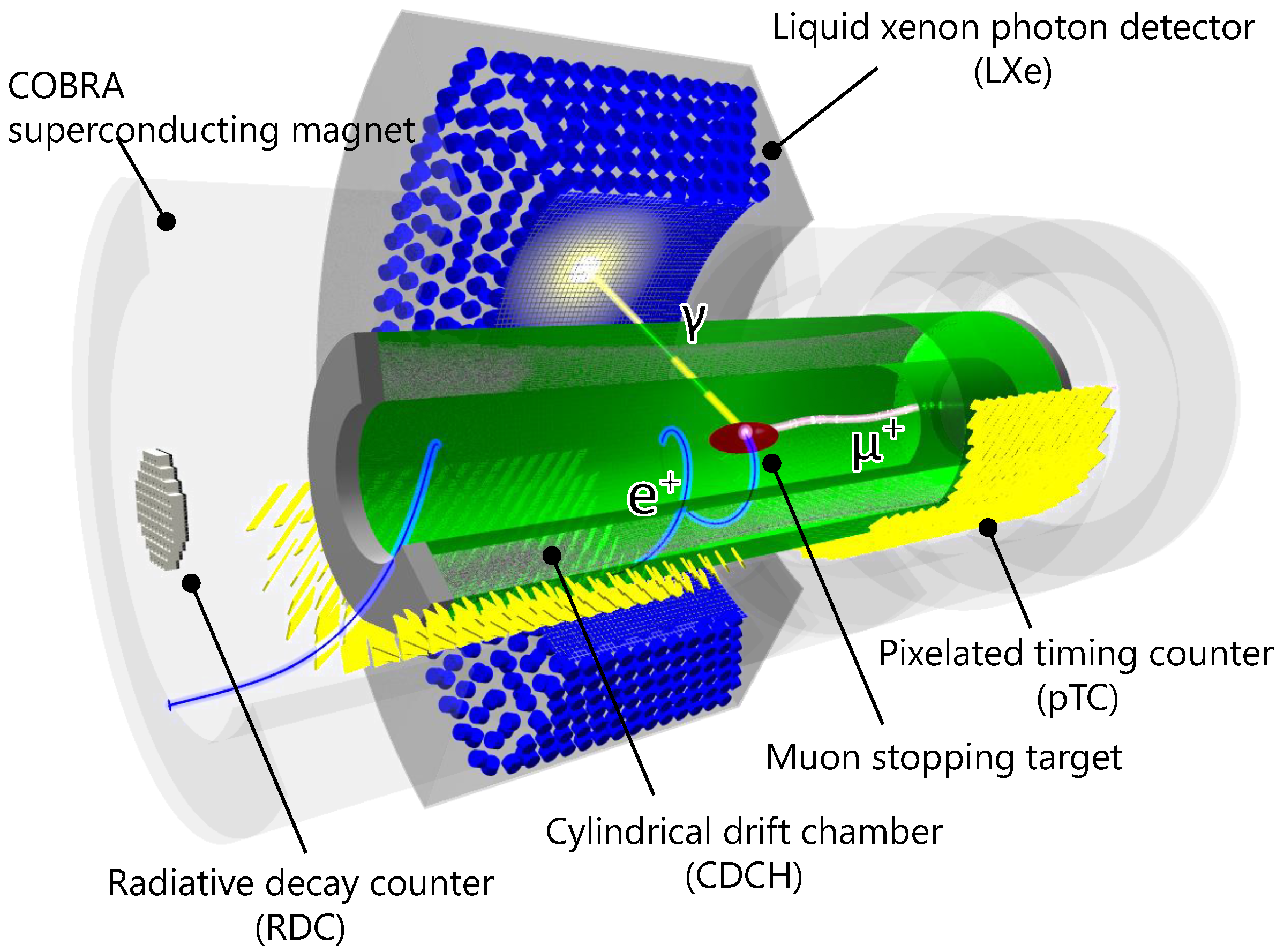
Abstract
:1. Introduction
2. Materials and Methods
2.1. Beamline and Target
2.2. Positron Spectrometer
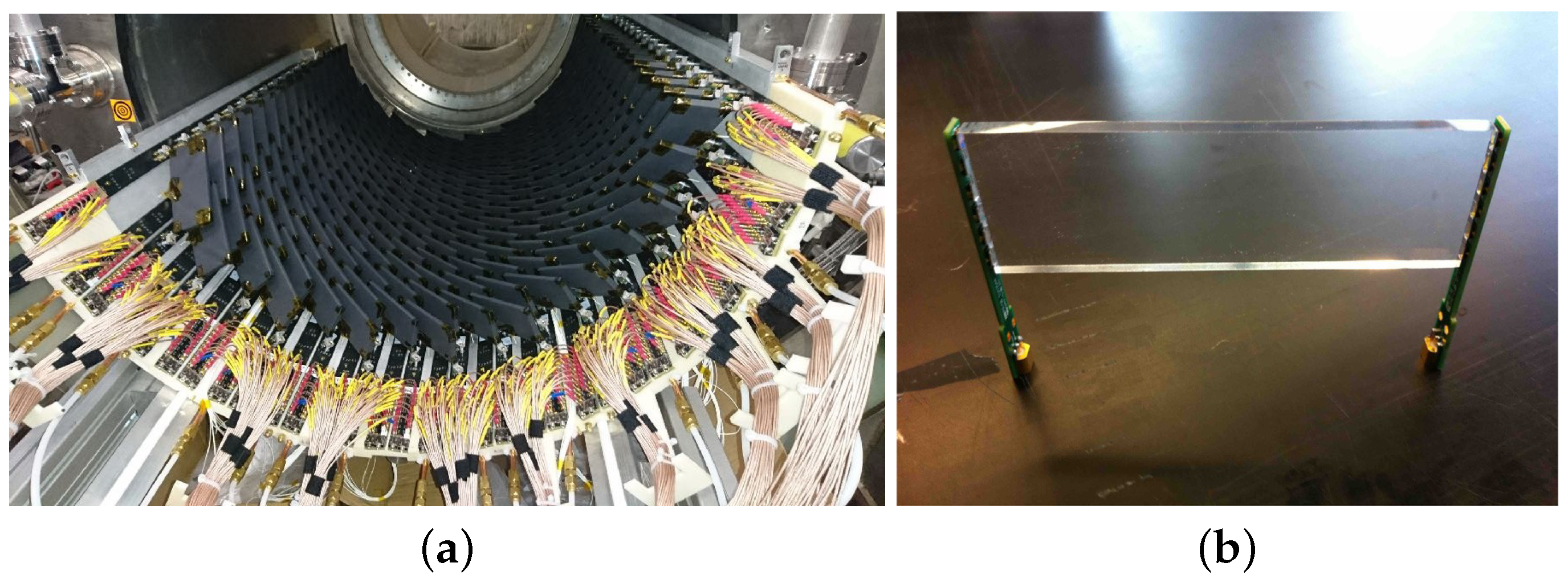
2.2.1. Pixelated Timing Counter-pTC
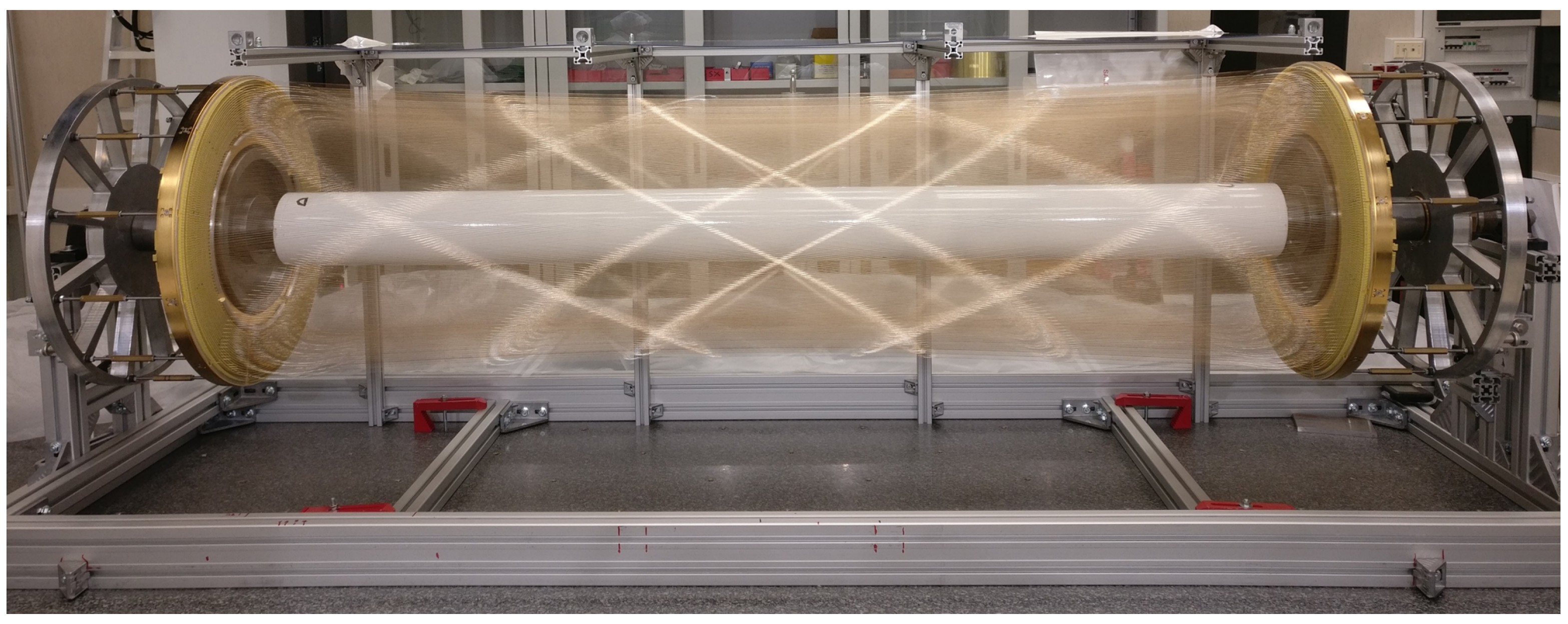
2.2.2. Cylindrical Drift CHamber-CDCH
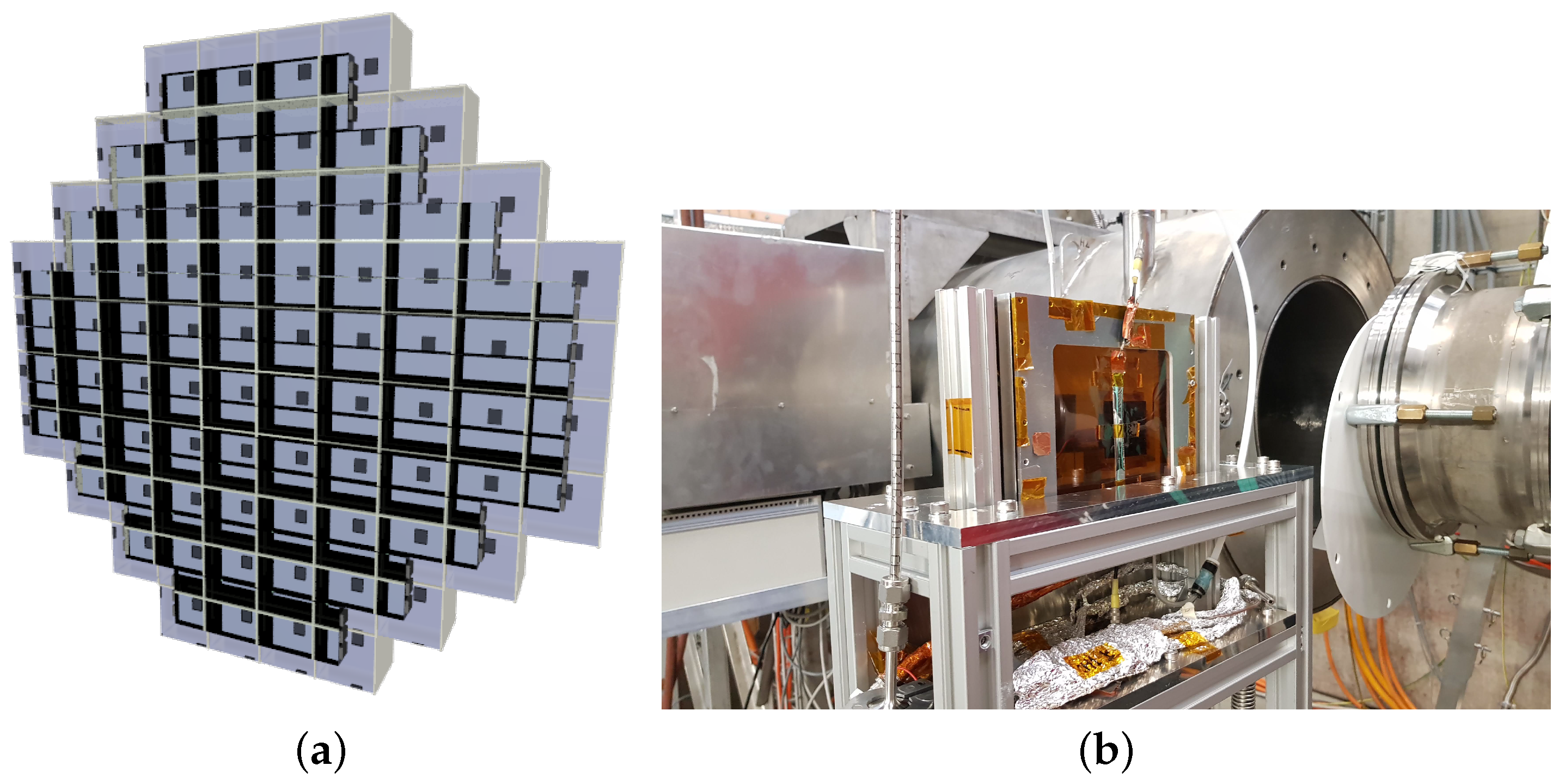
2.3. Liquid Xenon Gamma Detector-LXe
2.4. Radiative Decay Counter-RDC
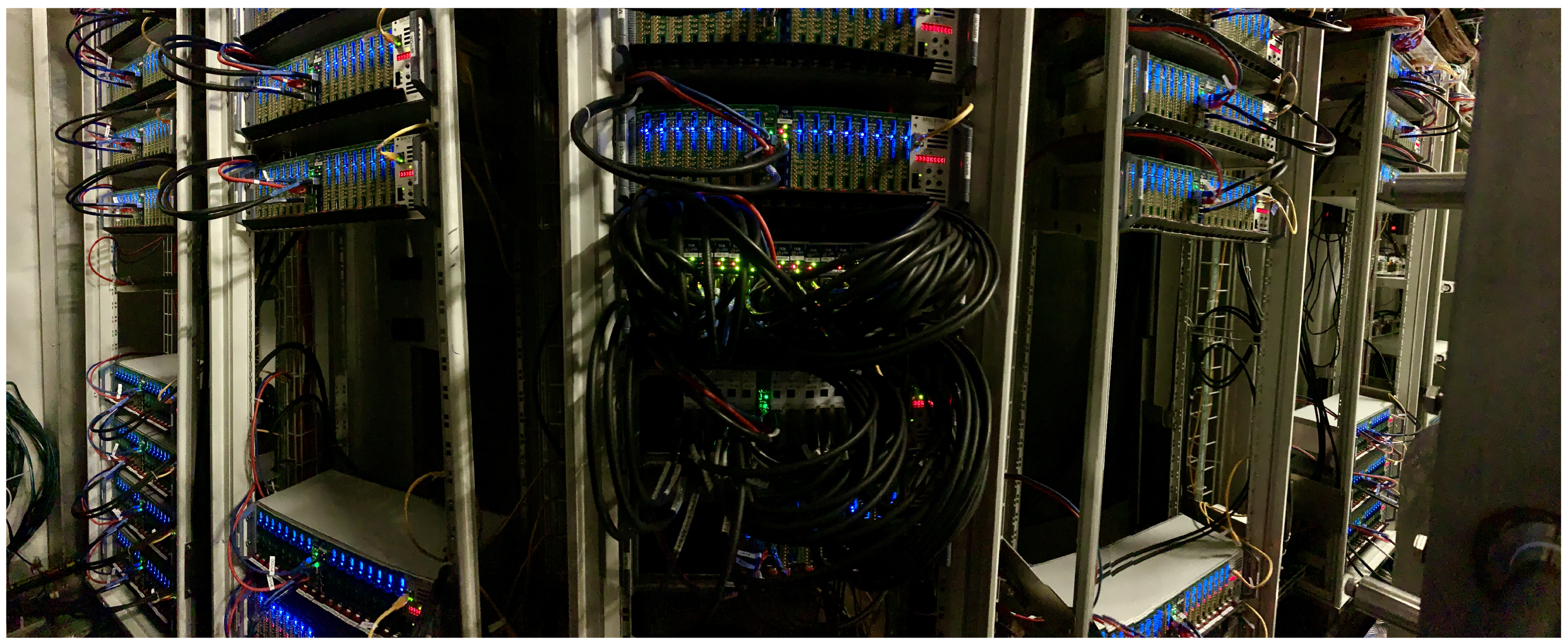
2.5. Trigger and Data Acquisition-TDAQ
3. Results
3.1. Pre-Engineering Runs: Results
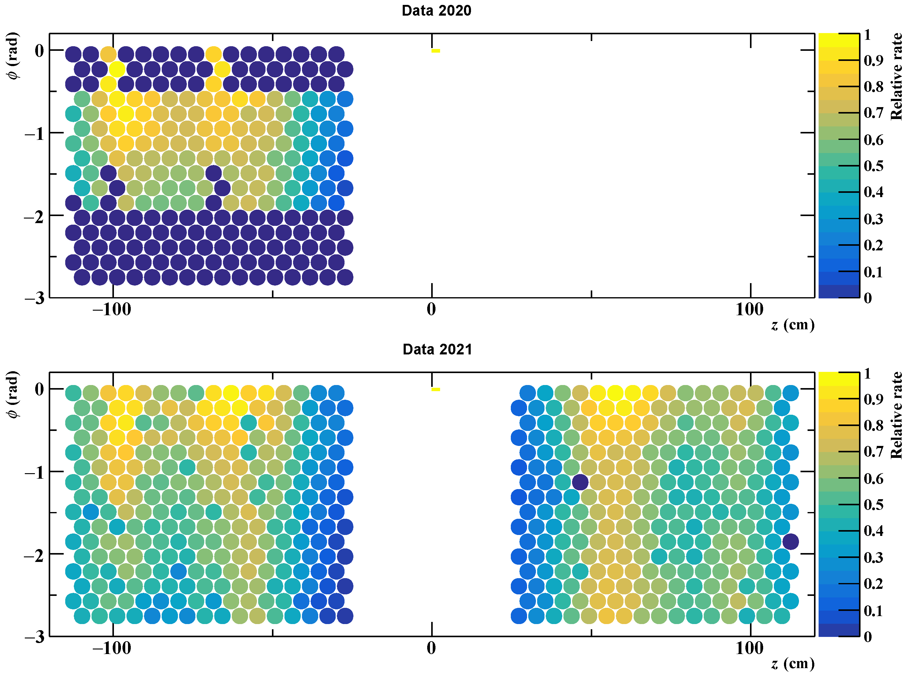
3.1.1. pTC
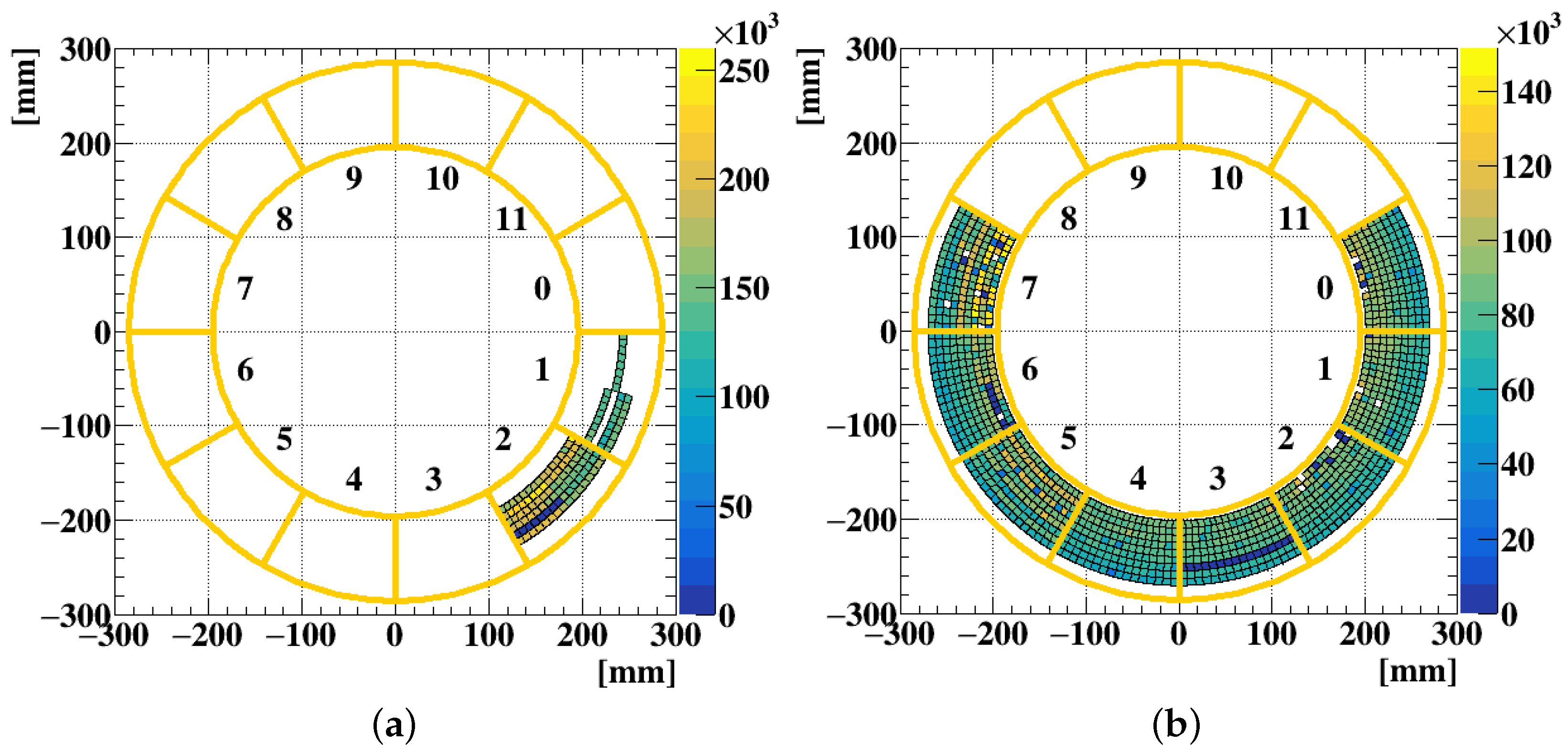
3.1.2. CDCH
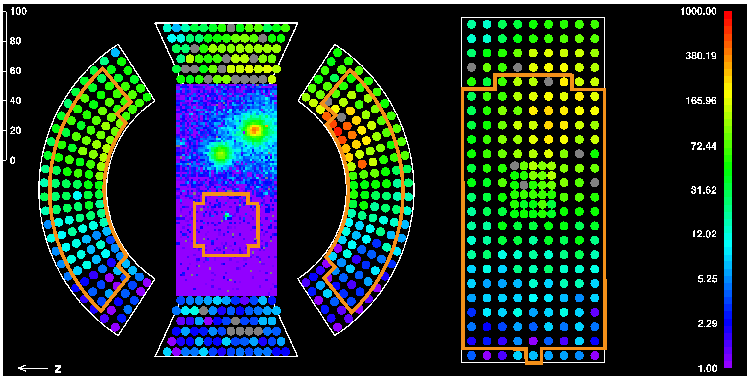
3.1.3. LXe
3.2. Engineering Run 2021: Highlights
X(17) Boson Measurement
4. Conclusions
Author Contributions
Funding
Acknowledgments
Conflicts of Interest
References
- Calibbi, L.; Signorelli, G. Charged Lepton Flavor Violation: An experimental and theoretical introduction. Riv. Nuovo Cimento 2018, 41, 71–174. [Google Scholar] [CrossRef]
- Hincks, E.P.; Pontecorvo, B. On the absence of photons among the decay products of the 2.2 microsecond meson. Can. J. Res. 1950, 28a, 29–43. [Google Scholar] [CrossRef]
- Fukuda, Y.; Hayakawa, T.; Ichihara, E.; Inoue, K.; Ishihara, K.; Ishino, H.; Itow, Y.; Kajita, T.; Kameda, J.; Kasuga, S.; et al. Evidence for Oscillation of Atmospheric Neutrinos. Phys. Rev. Lett. 1998, 81, 1562. [Google Scholar] [CrossRef] [Green Version]
- Kuno, Y.; Okada, Y. Muon decay and physics beyond the standard model. Rev. Mod. Phys. 2001, 73, 151–202. [Google Scholar] [CrossRef] [Green Version]
- Hernández-Tomé, G.; Castro, G.L.; Roig, P. Flavor violating leptonic decays of τ and μ leptons in the Standard Model with massive neutrinos. Eur. Phys. J. C 2019, 79, 84. [Google Scholar] [CrossRef]
- Calibbi, L.; López-Ibáñez, M.L.; Melis, A.; Vives, O. Implications of the Muon g-2 result on the flavour structure of the lepton mass matrix. arXiv 2021, arXiv:2104.03296. [Google Scholar] [CrossRef]
- Abi, B.; Albahri, T.; Al-Kilani, S.; Allspach, D.; Alonzi, L.P.; Anastasi, A.; Anisenkov, A.; Azfar, F.; Badgley, K.; Baeßler, S.; et al. Measurement of the Positive Muon Anomalous Magnetic Moment to 0.46 ppm. Phys. Rev. Lett. 2021, 126, 141801. [Google Scholar] [CrossRef] [PubMed]
- Baldini, A.M.; Bao, Y.; Baracchini, E.; Bemporad, C.; Berg, F.; Biasotti, M.; Boca, G.; Cascella, M.; Cattaneo, P.W.; Cavoto, G.; et al. Search for the lepton flavour violating decay μ+→e+γ with the full dataset of the MEG experiment. Eur. Phys. J. C 2016, 76, 434. [Google Scholar] [CrossRef] [Green Version]
- Davidson, S. Completeness and complementarity for μ→eγ, μ→ee and μA→eA. J. High Energy Phys. 2021, 2021, 1–36. [Google Scholar]
- Arndt, K.; Augustin, H.; Baesso, P.; Berger, N.; Berg, F.; Betancourt, C.; Bortoletto, D.; Bravar, A.; Briggl, K.; vom Bruch, D.; et al. Technical design of the phase I Mu3e experiment. Nucl. Instrum. Methods Phys. Res. Sect. A Accel. Spectrometers Detect. Assoc. Equip. 2021, 1014, 165679. [Google Scholar] [CrossRef]
- Bartoszek, L.; Barnes, E.; Miller, J.P.; Mott, J.; Palladino, A.; Quirk, J.; Roberts, B.L.; Crnkovic, J.; Polychronakos, V.; Tishchenko, V.; et al. Mu2e Technical Design Report. arXiv 2014, arXiv:1501.05241. [Google Scholar]
- Abramishvili, R.; Adamov, G.; Akhmetshin, R.R.; Allin, A.; Angélique, J.C.; Anishchik, V.; Aoki, M.; Aznabayev, D.; Bagaturia, I.; Ban, G.; et al. COMET Phase-I Technical Design Report. Prog. Theor. Exp. Phys. 2020, 2020, 033C01. [Google Scholar] [CrossRef] [Green Version]
- Nguyen, T.M. [DeeMe Collaboration]. Search for μ→e conversion with DeeMe experiment at J-PARC MLF. PoS FPCP 2015, 248, 060. [Google Scholar]
- Adam, J.; Bai, X.; Baldini, A.M.; Baracchini, E.; Bemporad, C.; Boca, G.; Cattaneo, P.W.; Cavoto, G.; Cei, F.; Cerri, C.; et al. The MEG detector for μ+→e+γ decay search. Eur. Phys. J. C 2013, 73, 2365. [Google Scholar] [CrossRef] [Green Version]
- Baldini, A.M.; Bao, Y.; Baracchini, E.; Bemporad, C.; Berg, F.; Biasotti, M.; Boca, G.; Cattaneo, P.W.; Cavoto, G.; Cei, F.; et al. Measurement of the radiative decay of polarized muons in the MEG experiment. Eur. Phys. J. C 2016, 76, 108. [Google Scholar] [CrossRef] [Green Version]
- Baldini, A.M.; Baracchini, E.; Bemporad, C.; Berg, F.; Biasotti, M.; Boca, G.; Cattaneo, P.W.; Cavoto, G.; Cei, F.; Chiappini, M.; et al. The design of the MEG II experiment. Eur. Phys. J. C 2018, 78, 380. [Google Scholar] [CrossRef]
- PSI. The Swiss Research Infrastructure for Particle Physics CHRISP. Available online: https://www.psi.ch/en/media/chrisp-overview (accessed on 25 November 2021).
- Baldini, A.M.; Bao, Y.; Baracchini, E.; Bemporad, C.; Berg, F.; Biasotti, M.; Boca, G.; Cattaneo, P.W.; Cavoto, G.; Cei, F.; et al. Muon polarization in the MEG experiment: Predictions and measurements. Eur. Phys. J. C 2016, 76, 223. [Google Scholar] [CrossRef] [Green Version]
- Papa, A.; Barchetti, F.; Gray, F.; Ripiccini, E.; Rutar, G. A multi-purposed detector with silicon photomultiplier readout of scintillating fibers. Nucl. Instr. Meth. A 2015, 787, 130–133. [Google Scholar] [CrossRef]
- Papa, A.; Rutar, G.; Barchetti, F.; Hildebrandt, M.; Kettle, P.R. A fast and quasi non-invasive muon beam monitor working at the intensity frontier. Nucl. Instr. Meth. A 2019, 936, 634–635. [Google Scholar] [CrossRef]
- Cavoto, G.; Chiarello, G.; Hildebrandt, M.; Hofer, A.; Ieki, K.; Meucci, M.; Milana, S.; Pettinacci, V.; Renga, F.; Voena, C. A photogrammetric method for target monitoring inside the MEG II detector. Rev. Sci. Instrum. 2021, 92, 043707. [Google Scholar] [CrossRef] [PubMed]
- Palo, D.; Hildebrandt, M.; Hofer, A.; Kyle, W.; Lad, D.; Libeiro, T.; Molzon, W. Precise Photographic Monitoring of MEG II Thin-film Muon Stopping Target Position and Shape. Nucl. Instrum. Methods A 2019, 944, 162511. [Google Scholar] [CrossRef] [Green Version]
- Ootani, W.; Odashima, W.; Kimura, S.; Kobayashi, T.; Makida, Y.; Mitsuhashi, T.; Mizumaki, S.; Ruber, R.; Yamamoto, A. Development of a thin-wall superconducting magnet for the positron spectrometer in the MEG experiment. IEEE Trans. Appl. Supercond. 2004, 14, 568–571. [Google Scholar] [CrossRef] [Green Version]
- Nishimura, M.; Berg, F.; Biasotti, M.; Boca, G.; Cattaneo, P.W.; De Gerone, M.; De Bari, A.; Francesconi, M.; Galli, L.; Gatti, F.; et al. Full system of positron timing counter in MEG II having time resolution below 40 ps with fast plastic scintillator readout by SiPMs. Nucl. Instrum. Methods A 2020, 958, 162785. [Google Scholar] [CrossRef]
- Saint-Gobain Ceramics & Plastics, Inc. BC-418, BC-420, BC-422 Premium Plastic Scintillators. Available online: http://www.crystals.saint-gobain.com/sites/imdf.crystals.com/files/documents/sgc-bc418-420-422-data-sheet_69699.pdf (accessed on 25 November 2021).
- 3M. ESR Reflector Datasheet. Available online: https://www.3m.com/3M/en_US/p/d/eebgdar000006/ (accessed on 25 November 2021).
- Boca, G.; Cattaneo, P.W.; De Gerone, M.; Francesconi, M.; Galli, L.; Gatti, F.; Koga, J.; Nakao, M.; Nishimura, M.; Ootani, W.; et al. The laser-based time calibration system for the MEG II pixelated Timing Counter. Nucl. Instrum. Methods A 2019, 947, 162672. [Google Scholar] [CrossRef] [Green Version]
- Boca, G.; Cattaneo, P.W.; De Gerone, M.; Gatti, F.; Nakao, M.; Nishimura, M.; Ootani, W.; Rossella, M.; Uchiyama, Y.; Usami, M.; et al. Timing resolution of a plastic scintillator counter read out by radiation damaged SiPMs connected in series. Nucl. Instrum. Methods A 2021, 999, 165173. [Google Scholar] [CrossRef]
- Chiappini, M.; Baldini, A.M.; Cavoto, G.; Cei, F.; Chiarello, G.; Francesconi, M.; Galli, L.; Grancagnolo, F.; Grassi, M.; Hildebrandt, M.; et al. The new drift chamber of the MEG II experiment. Nucl. Instrum. Methods Phys. Res. Sect. A Accel. Spectrometers Detect. Assoc. Equip. 2019, 936, 501–502. [Google Scholar] [CrossRef]
- Baldini, A.M.; Cavoto, G.; Cei, F.; Chiappini, M.; Chiarello, G.; Corvaglia, A.; Francesconi, M.; Galli, L.; Grancagnolo, F.; Grassi, M.; et al. The ultra light Drift Chamber of the MEG II experiment. Nucl. Instrum. Methods Phys. Res. Sect. A Accel. Spectrometers Detect. Assoc. Equip. 2020, 958, 162152. [Google Scholar] [CrossRef]
- Chiarello, G.; Chiri, C.; Corvaglia, A.; Grancagnolo, F.; Miccoli, A.; Panareo, M.; Pinto, C.; Spedicato, M.; Tassielli, G.F. The construction technique of the high granularity and high transparency drift chamber of MEG II. J. Instrum. 2017, 12, C07022. [Google Scholar] [CrossRef]
- Baldini, A.M.; Baracchini, E.; Cavoto, G.; Cei, F.; Chiappini, M.; Chiarello, G.; Chiri, C.; Francesconi, M.; Galli, L.; Grancagnolo, F.; et al. Gas Distribution and Monitoring for the Drift Chamber of the MEG-II Experiment. J. Instrum. 2018, 13, P06018. [Google Scholar] [CrossRef] [Green Version]
- Baldini, A.M.; Baracchini, E.; Cavoto, G.; Cascella, M.; Cei, F.; Chiappini, M.; Chiarello, G.; Chiri, C.; Dussoni, S.; Galli, L.; et al. Single-hit resolution measurement with MEG II drift chamber prototypes. J. Instrum. 2016, 11, P07011. [Google Scholar] [CrossRef]
- Venturini, M.; Baldini, A.M.; Baracchini, E.; Cei, F.; Dussoni, S.; Galli, L.; Grassi, M.; Nicolò, D.; Signorelli, G.; Tenchini, F.; et al. Ageing tests for the MEG II drift chamber. Nucl. Instrum. Methods Phys. Res. Sect. A Accel. Spectrometers Detect. Assoc. Equip. 2016, 824, 592–594. [Google Scholar] [CrossRef]
- Chiarello, G.; Chiri, C.; Corvaglia, A.; Grancagnolo, F.; Panareo, M.; Pepino, A.; Pinto, C.; Tassielli, G. A high performance Front End Electronics for drift chamber readout in MEG experiment upgrade. Nucl. Instrum. Methods A 2016, 824, 336–339. [Google Scholar] [CrossRef]
- Baldini, A.M.; Cavoto, G.; Cei, F.; Chiappini, M.; Chiarello, G.; Chiri, C.; Cocciolo, G.; Corvaglia, A.; Cuna, F.; Francesconi, M.; et al. Detailed analysis of chemical corrosion of ultra-thin wires used in drift chamber detectors. arXiv 2021, arXiv:2108.13948. [Google Scholar]
- Ieki, K.; Iwamoto, T.; Kaneko, D.; Kobayashi, S.; Matsuzawa, N.; Mori, T.; Ogawa, S.; Onda, R.; Ootani, W.; Sawada, R.; et al. Large-area MPPC with enhanced VUV sensitivity for liquid xenon scintillation detector. Nucl. Instrum. Methods A 2019, 925, 148–155. [Google Scholar] [CrossRef] [Green Version]
- Baldini, A.M.; Bemporad, C.; Cei, F.; Dussoni, S.; Gatti, F.; Grassi, M.; Haruyama, T.; Hisamatsu, Y.; Iwamoto, T.; Mihara, S.; et al. A radioactive point-source lattice for calibrating and monitoring the liquid xenon calorimeter of the MEG experiment. Nucl. Instrum. Methods A 2006, 565, 589–598. [Google Scholar] [CrossRef]
- Adam, J.; Bai, X.; Baldini, A.; Baracchini, E.; Bemporad, C.; Boca, G.; Cattaneo, P.W.; Cavoto, G.; Cei, F.; Cerri, C.; et al. Calibration and monitoring of the MEG experiment by a proton beam from a Cockcroft–Walton accelerator. Nucl. Instrum. Methods A 2011, 641, 19–32. [Google Scholar] [CrossRef]
- Oya, A.; Ieki, K.; Ochi, A.; Onda, R.; Ootani, W.; Yamamoto, K. Development of high-rate capable and ultra-low mass Resistive Plate Chamber with Diamond-Like Carbon. arXiv 2021, arXiv:2109.13525. [Google Scholar]
- Ritt, S.; Dinapoli, R.; Hartmann, U. Application of the DRS chip for fast waveform digitizing. Nucl. Instrum. Methods A 2010, 623, 486–488. [Google Scholar] [CrossRef]
- Galli, L.; Baldini, A.M.; Cei, F.; Chiappini, M.; Francesconi, M.; Grassi, M.; Hartmann, U.; Meucci, M.; Morsani, F.; Nicolò, D.; et al. WaveDAQ: An highly integrated trigger and data acquisition system. Nucl. Instrum. Methods A 2019, 936, 399–400. [Google Scholar] [CrossRef]
- Ritt, S. MIDAS: Midas.psi.ch. Available online: http://midas.psi.ch (accessed on 25 November 2021).
- Galli, L.; Baldini, A.M.; Cattaneo, P.W.; Cei, F.; De Gerone, M.; Dussoni, S.; Gatti, F.; Grassi, M.; Morsani, F.; Nicolò, D.; et al. Operation and performance of the trigger system of the MEG experiment. J. Instrum. 2014, 9, P04022. [Google Scholar] [CrossRef]
- Francesconi, M.; Baldini, A.M.; Cei, F.; Chiappini, M.; Galli, L.; Grassi, M.; Hartmann, U.; Morsani, F.; Nicolò, D.; Papa, A.; et al. Low latency serial communication for MEG II trigger system. Nucl. Instrum. Methods Phys. Res. Sect. A Accel. Spectrometers Detect. Assoc. Equip. 2019, 936, 331–332. [Google Scholar] [CrossRef]
- Nicolò, D.; Baldini, A.M.; Bemporad, C.; Cei, F.; Chiappini, M.; Francesconi, M.; Galli, L.; Grassi, M.; Iwamoto, T.; Morsani, F.; et al. Real-time particle identification in liquid xenon. IEEE Trans. Nucl. Sci. 2021, 68, 2630–2636. [Google Scholar] [CrossRef]
- Baldini, A.M.; Bao, Y.; Baracchini, E.; Bemporad, C.; Berg, F.; Biasotti, M.; Boca, G.; Cascella, M.; Cattaneo, P.W.; Cavoto, G.; et al. The Search for μ→ eγ with 10-14 Sensitivity: The Upgrade of the MEG Experiment. Symmetry 2021, 13, 1591. [Google Scholar] [CrossRef]
- Chiappini, M.; Baldini, A.M.; Cavoto, G.; Cei, F.; Chiarello, G.; Corvaglia, A.; Francesconi, M.; Galli, L.; Grancagnolo, F.; Grassi, M.; et al. Commissioning of the MEG II tracker system. J. Instrum. 2020, 15, C06056. [Google Scholar] [CrossRef]
- Cascella, M.; Grancagnolo, F.; Tassielli, G. Cluster Counting/Timing Techniques for Drift Chambers. Nucl. Phys. B (Proc. Suppl.) 2014, 248–250, 127–130. [Google Scholar] [CrossRef]
- Li, A.N.F. CCD Image Sensors in Deep-Ultraviolet; Springer: Berlin/Heidelberg, Germany, 2005. [Google Scholar] [CrossRef]
- Kobayashi, S.; Francesconi, M.; Galli, L.; Ieki, K.; Iwamoto, T.; Libeiro, T.; Matsuzawa, N.; Molzon, W.; Mori, T.; Nakao, M.; et al. Precise measurement of 3D-position of SiPMs in the liquid xenon gamma-ray detector for the MEGII experiment. Nucl. Instrum. Methods Phys. Res. Sect. A Accel. Spectrometers Detect. Assoc. Equip. 2019, 936, 189–191. [Google Scholar] [CrossRef]
- Krasznahorkay, A.J.; Csatlós, M.; Csige, L.; Gácsi, Z.; Gulyás, J.; Hunyadi, M.; Kuti, I.; Nyakó, B.M.; Stuhl, L.; Timár, J.; et al. Observation of anomalous internal pair creation in 8Be: A possible indication of a light, neutral boson. Phys. Rev. Lett. 2016, 116, 042501. [Google Scholar] [CrossRef] [Green Version]
- Krasznahorkay, A.J.; Csatlós, M.; Csige, L.; Gulyás, J.; Koszta, M.; Szihalmi, B.; Timár, J.; Firak, D.S.; Nagy, Á.; Sas, N.J.; et al. A new anomaly observed in 4He supporting the existence of the hypothetical X17 particle. J. Phys. Conf. Ser. 2020, 1643, 012001. [Google Scholar] [CrossRef]
- Feng, J.L.; Fornal, B.; Galon, I.; Gardner, S.; Smolinsky, J.; Tait, T.M.P.; Tanedo, P. Protophobic Fifth-Force Interpretation of the Observed Anomaly in 8Be Nuclear Transitions. Phys. Rev. Lett. 2016, 117, 071803. [Google Scholar] [CrossRef] [Green Version]
- Aleksejevs, A.; Barkanova, S.; Kolomensky, Y.G.; Sheff, B. A Standard Model explanation for the “ATOMKI anomaly”. arXiv 2021, arXiv:2102.01127. [Google Scholar]


Publisher’s Note: MDPI stays neutral with regard to jurisdictional claims in published maps and institutional affiliations. |
© 2021 by the authors. Licensee MDPI, Basel, Switzerland. This article is an open access article distributed under the terms and conditions of the Creative Commons Attribution (CC BY) license (https://creativecommons.org/licenses/by/4.0/).
Share and Cite
Chiappini, M.; Francesconi, M.; Kobayashi, S.; Meucci, M.; Onda, R.; Schwendimann, P.; on behalf of the MEG II Collaboration. Towards a New μ→eγ Search with the MEG II Experiment: From Design to Commissioning. Universe 2021, 7, 466. https://0-doi-org.brum.beds.ac.uk/10.3390/universe7120466
Chiappini M, Francesconi M, Kobayashi S, Meucci M, Onda R, Schwendimann P, on behalf of the MEG II Collaboration. Towards a New μ→eγ Search with the MEG II Experiment: From Design to Commissioning. Universe. 2021; 7(12):466. https://0-doi-org.brum.beds.ac.uk/10.3390/universe7120466
Chicago/Turabian StyleChiappini, Marco, Marco Francesconi, Satoru Kobayashi, Manuel Meucci, Rina Onda, Patrick Schwendimann, and on behalf of the MEG II Collaboration. 2021. "Towards a New μ→eγ Search with the MEG II Experiment: From Design to Commissioning" Universe 7, no. 12: 466. https://0-doi-org.brum.beds.ac.uk/10.3390/universe7120466