Article

Genotypic Characterization of Epstein Barr Virus in Blood of Patients with Suspected Nasopharyngeal Carcinoma in Ghana

1
West African Center for Cell Biology of Infectious Pathogens (WACCBIP), Department of Biochemistry, Cell and Molecular Biology, University of Ghana, Legon, Accra 00233, Ghana
2
Ear, Nose and Throat Unit, Korle-Bu Teaching Hospital, Accra 00233, Ghana
3
School of Life Sciences, University of Sussex, Brighton BN1 9QG, UK
*
Author to whom correspondence should be addressed.
Submission received: 1 May 2020 / Revised: 5 June 2020 / Accepted: 8 June 2020 / Published: 16 July 2020

Abstract

:
Nasopharyngeal cancer (NPC) is associated with Epstein Barr virus (EBV) infection. However different viral strains have been implicated in NPC worldwide. This study aimed to detect and characterize EBV in patients diagnosed with NPC in Ghana. A total of 55 patients diagnosed with NPC by CT scan and endoscopy were age-matched with 53 controls without a known oncological disease. Venous blood was collected from the study participants and DNA extracted from the blood samples. Detection of EBV and genotyping were done by amplifying Epstein Barr nuclear antigen 1 (EBNA-1) and Epstein Barr nuclear antigen 2 (EBNA-2), respectively, using specific primers. Viral load in patients and controls was determined using real-time polymerase chain reaction. EBV positivity in controls (92%) was significantly greater than that of NPC patients (67%) (χ2 = 19.17, p < 0.0001), and viral infection was independent of gender (χ2 = 1.770, p = 0.1834). The predominant EBV genotypes in patients and controls were genotype 2 (52%) and genotype 1 (62%), respectively. Median EBV load was significantly higher in NPC patients than the control group (p < 0.01). In summary, prevalence of EBV genotype 2 infection was higher in NPC patients than the control group. Assessment of EBV load may be used as a biomarker for the diagnosis of NPC.

1. Introduction

Nasopharyngeal cancer (NPC) is a malignant tumor of epithelial squamous cell which arises from the lateral wall of the nasopharynx, notably, the fossa of Rosenmüller located superior and posterior to the torus tubarius [1,2]. NPC has an incidence of 1 person per 100,000 in non-endemic regions, with an incidence of 25 to 30 persons per 100,000 in endemic regions like China and Southeast Asia [3]. In Africa, specifically Northern Africa, the yearly incidence of NPC varies between 5 to 7 persons per 100,000 [4]. NPC is one of the leading cancers in Ghana, with a prevalence of 29% among the head and neck cancers [5], and about 1.2% of all cancers [6]. There are ethnic and geographic variations in the occurrence of NPC, which indicates a multifactorial etiology involving EBV infection, genetic susceptibility, ethnicity, environmental factors and food consumption [7,8].
The most common etiological factor associated with NPC development is Epstein Barr virus (EBV) infection. EBV is a ubiquitous oncogenic virus belonging to the family Herpesviridae [9], which is further classified into subfamilies: alpha-, beta-, and gammaherpes virus. EBV is a classical member of the subfamily Gammaherpesvirinae, and among nine viruses of the subfamily that have been identified to solely infect humans [10,11]. Two genotypes of the virus, namely genotypes 1 and 2, exist and exhibit variation in geographical distribution. Although EBV genotype 1 is globally distributed, it is predominantly found in American, Chinese, European and South-East Asian (SEA) populations, whereas genotype 2 is predominantly found in Africa [12]. The two genotypes also vary in biological properties; EBV genotype 1 is more efficient in immortalizing B cells while genotype 2 has a higher lytic ability [13,14].
Despite the increased number of NPC cases reported globally, little has been done to characterize EBV and determine the association of EBV genotype with NPCs, especially in Africa. The current case-control study was aimed at determining the prevalence of EBV infection and the viral genotype associated with NPC in Ghanaian patients who are not undergoing cancer therapy.

2. Methods

2.1. Study Site

This study was conducted at the Ear, Nose, and Throat (ENT) Unit of the Korle-Bu Teaching Hospital (KBTH), Accra. The Unit serves as a referral center for nasopharyngeal cancer (NPC) cases in Ghana and the West African region.

2.2. Recruitment of Study Participants

A total of 108 study participants reporting to the ENT Unit of the KBTH from April 2018 to March 2019 were enrolled into the study. Out of the total number of recruited participants, 55 were suspected cases of NPC, which was determined by endoscopy and computed tomography (CT) scan, although biopsies from the participants were yet to undergo histopathological examination. The remaining 53 participants were NPC negative subjects and served as controls. A well-structured questionnaire was administered to acquire demographic data from the study participants after informed consent had been obtained. The study was approved by the Institutional Review Board of the Noguchi Memorial Institute for Medical Research (NMIMR) with reference number 051/16-17.

2.3. Collection of Blood Samples and DNA Extraction

Venous blood (5 mL) was collected from the study participants and transferred into EDTA tubes. Genomic DNA was extracted from the blood samples using Qiagen QIAamp Blood Mini Kit (QIAGEN, Hilden, Germany) by following the manufacturer’s protocol. The eluted DNA was stored at −20 °C until ready to use.

2.4. Positive Controls

Plasmids pJET-EBNA-1 and pJET-EBNA-2 containing EBV DNA were used as positive controls for EBV detection, and EBV genotypes 1 and 2, respectively. Moreover, pJET-Beta plasmid was used as positive control for beta globulin.

2.5. Detection of EBV by Amplification of Epstein Barr Nuclear Antigen-1 (EBNA-1)

The extracted genomic DNA samples were screened for the presence of EBV by polymerase chain reaction (PCR) amplification of EBNA-1 using primers (QP1, QP2) that have previously been reported, and for the human beta globulin gene as internal control (Table 1). Each PCR reaction mixture of 12.5 µL contains the following components with the final concentrations: 1X One TaqR Quick-Load 2X Master Mix with Standard Buffer (New England Bio Labs, Hertfordshire, UK), 0.5 µM each of the forward and reverse primers, and 0.5 µg/µL of genomic DNA. The volume was made up with nuclease-free water. PCR reaction cycling conditions involved initial denaturation at 94 °C for 30 s, followed by 35 cycles of amplification with denaturation at 94 °C for 30 s, annealing at 56 °C for 1 min, extension at 68 °C for 1 min, and a final extension at 68 °C for 5 min. An aliquot of 8 μL of the amplicons were resolved on 2% ethidium bromide-stained agarose gel, and bands were visualized using Amersham gel imager.

2.6. EBV Genotyping by Epstein Barr Nuclear Antigen 2 (EBNA-2) Nested PCR Amplification

The genotypes of EBV in both patients and controls were detected by nested PCR using specific primers as previously reported (Table 1), with slight modifications to cycling conditions. The first round of the PCR was done by amplifying a common region of EBNA-2 using EBNA-2F and EBNA-2I as sense and antisense primers, respectively. Each PCR reaction mixture of 12.5 µL contains the following components with the final concentrations: 1X One TaqR Quick-Load 2X Master Mix with Standard Buffer (New England Bio Labs, UK), 0.5 µM each of the forward and reverse primers, and 0.5 µg/µL of genomic DNA. The cycling conditions for the reaction were as follow: initial denaturation at 94 °C for 2 min, followed by 35 cycles of amplification with denaturation at 94 °C for 60 s, annealing at 52 °C for 90 s, extension at 72 °C for 4 min, and a final extension at 72 °C for 10 min. A second round PCR (nested) was performed using 0.5 μL of the amplicons from the first round as template; all other reaction components were the same as the first-round reaction mixture, except for the primers. A forward primer (EBNA-2C), that is common to both genotypes, and reverse primers EBNA-2G and EBNA-2B, which are specific for the respective genotypes were used for the second-round amplification. The reaction was carried out at initial denaturation of 94 °C for 2 min, followed by 35 cycles of amplification with denaturation at 94 °C for 30 s, annealing at 52 °C for 60 s, extension at 72 °C for 2 min and a final extension at 72 °C for 10 min. Eight microliters of the amplicons were resolved on 2% ethidium bromide-stained agarose gel and visualized using the Amersham gel imager.

2.7. EBV Load Quantification in Whole Blood by EBNA-1 Real-Time PCR

Measurement of EBV load in blood samples was carried out by real-time PCR, QauntStudio5 PCR System (Applied Biosystems, Waltham, MA, USA). The genomic DNA samples were used for the quantification of EBV by amplifying EBNA-1 and beta-globulin gene (internal control). The PCR reaction volume was made up of the following components: 5 μL 1X Sybr Green SuperMix (Quantabio, Beverly, MA, USA), 0.5 µg/µL template DNA, 0.1 μL of 0.1 μM each of primers (QP1, QP2, B-globulin forward and reverse) (Table 1), and 3.8 μL of nuclease free water. The cycling conditions consisted of a hold at 95 °C for 10 min, followed by an initial denaturation step at 95 °C for 10 min, and then 40 cycles of denaturation at 95 °C for 15 s and 60 °C for 1 min re-annealing and extension. All samples were analyzed in duplicates. A standard reference curve was obtained by amplifying a 10-fold serial dilution of EBNA-1 DNA in pJET-EBNA-1 plasmid. EBV load was expressed as log EBV DNA copies/mL.

2.8. Statistical Analysis

Statistical analysis was performed using IBM SPSS statistical software version 20 and GraphPad Prism 8. Categorical variables were compared using Chi-squared test of independence for contingency table and corrected by Yates’ continuity test or Fisher’s Exact test. After confirmation of the normal distribution of EBV load results by Kolmogorov and Smirnov tests, Mann–Whitney rank-sum test was used to compare median viral load between cases and controls for binary categories. A p-value less than 0.05 was considered statistically significant.

3. Results

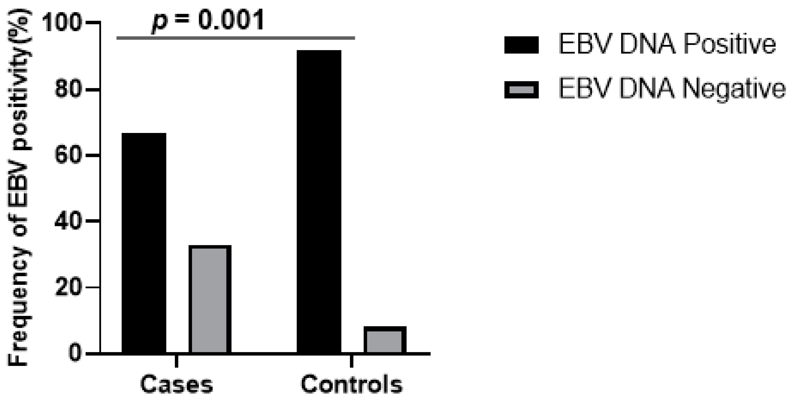
3.1. Demographics of Study Participants and Molecular Detection of EBV in Whole Blood

The median ages of the patients and controls were 40 and 47 years, respectively. The proportion of males suspected with NPC and those in the control group were 47% (26/55) and 30% (16/53), respectively. Furthermore, the proportion of females suspected with NPC and those in the control group were 53% (29/55) and 70% (37/53), respectively. EBV DNA was detected by EBNA-1 amplification (213 bp) (Figure S1) and the number of positives were 67% (37/55) and 92% (48/53) for patients and controls, respectively. The EBV DNA positivity was significantly higher in controls than the patients (χ2 = 19.17, p < 0.0001) (Figure 1). The EBV DNA positivity in females (81.8%) was significantly higher than in males (66.7%). If we assume EBV DNA positivity represents EBV infection history, it means that females were two times more at risk of having EBV infection than males (OR = 2.24, 95% CI = 1.16–4.34, p = 0.0225).

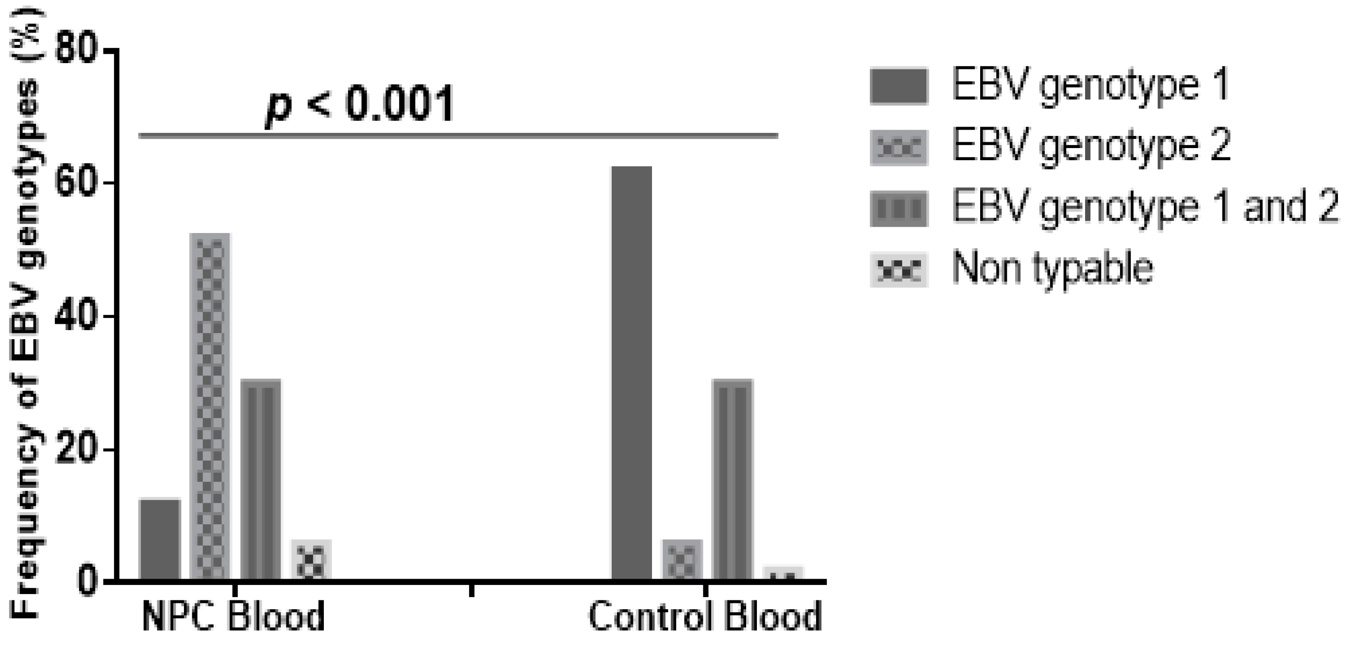
3.2. EBV Genotypes in NPC Patients and Controls of the Study Participants

EBV genotyping using the EBNA-2 gene showed 250 bp and 300 bp fragments which are characteristic of genotypes 1 and 2, respectively (Figure S2). Frequencies of EBV genotype 1 in blood samples of NPC patients and controls were 11.5% (6/52) and 62.3% (33/53), respectively, whilst genotype 2 was 51.9% (27/52) in the NPC patients compared to 5.7% (3/53) in controls (Figure 2). Co-infection with EBV genotypes 1 and 2 in NPC cases was 30.8% (16/52) and in 30.2% (16/53) in the controls. Differences in EBV genotype distribution in both patients and controls were statistically significant (p < 0.001); the EBV genotype 1 was higher in the controls than the patients (χ2 = 51.5, p < 0.001), and the reverse was observed for EBV genotype 2 (χ2 = 49.2, p < 0.001). Individuals infected with EBV genotype 2 were almost 17 times more at risk of developing NPC (OR = 16.97, 95% CI = 6.86–42.32, p < 0.0001) compared to the weakly associated genotype 1 (OR = 0.08, 95% CI = 0.04–0.17).

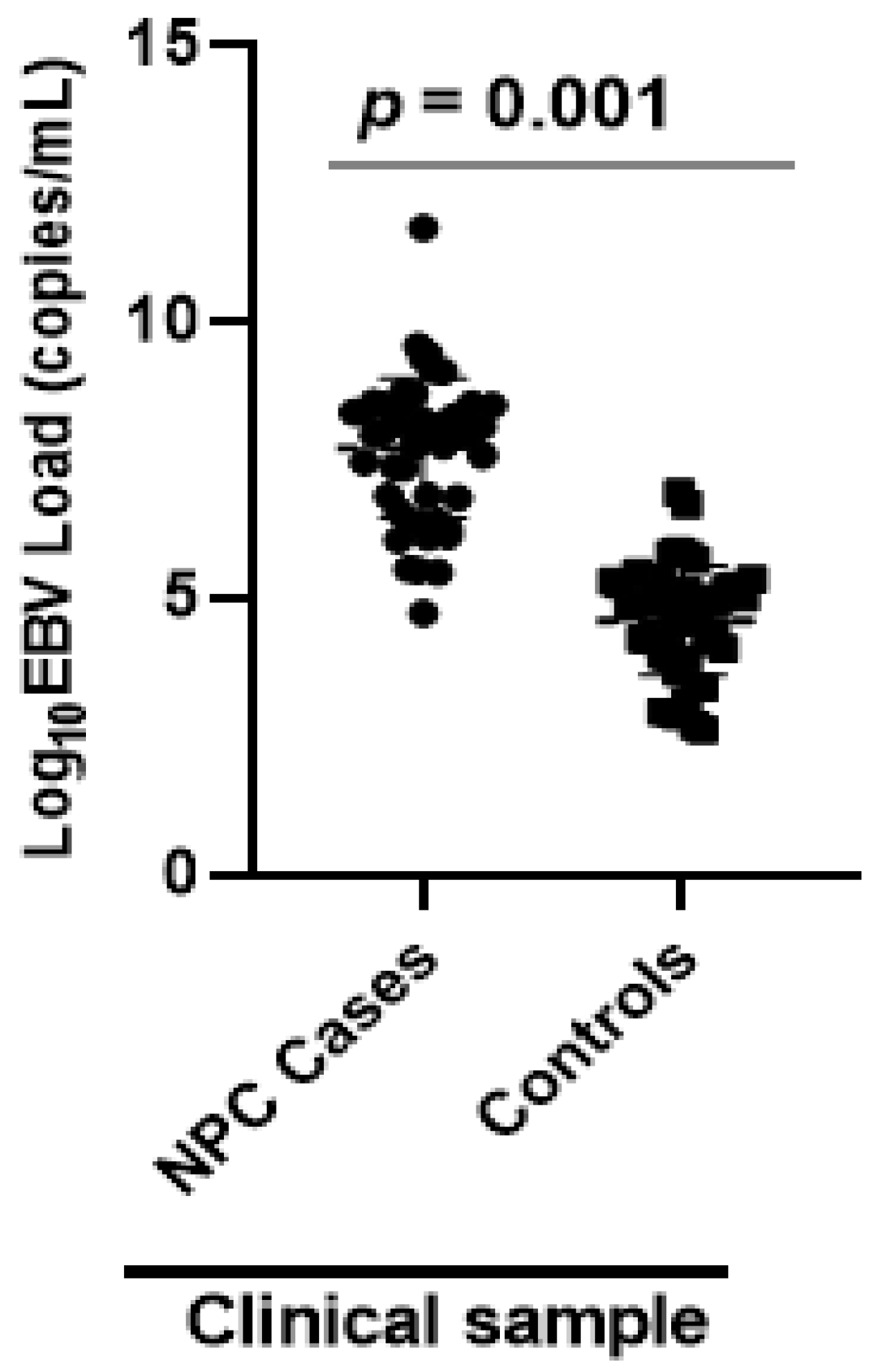
3.3. EBV Load in NPC Patients and Controls

The median Epstein Barr viral load of 9 × 10⁷ copies/mL (53 × 103 to 5 × 10¹¹ copies/mL) in NPC patients was significantly elevated when compared to controls of 6 × 10⁴ copies/ mL (4 × 10²–8 × 10⁶ copies/mL) (p < 0.01) (Figure 3).

4. Discussion

Nasopharyngeal cancer (NPC) is a malignant tumor of epithelial squamous cells which arises from the lateral wall of the nasopharynx, notably, the fossa of Rosenmüller, which is located superior and posterior to the torus tubarius [2]. The etiology of NPC is commonly associated with EBV infection. Although EBV infects more than 90% of the world’s population, most of the infections are asymptomatic [3]. The replication of the virus is held in check by EBV-specific cytotoxic T cells in immunocompetent individuals. However, in instances where immune surveillance by cytotoxic T cells wanes, the viral outgrowth is enabled, and this may result in various EBV-associated cancers [15]. In the host, EBV expresses restricted sets of latent genes that vary from one malignancy to another, as well as in asymptomatic carriers [16].
In the current study, we detected EBV EBNA-1 gene in the whole blood of both patients suspected of NPC and control subjects without a known oncological disease. The virus was present in over 90% of the control group of the study and supported the finding in literature that about 90% of the world’s population are asymptomatic carriers of the virus [17]. A public health factor that may contribute to high prevalence of EBV in healthy individuals is the infection risk of EBV transfer through blood transfusion, and this has been reported in some studies [18,19]. However, the results obtained from the current study, which suggest a high prevalence in asymptomatic carriers, was inconsistent with results reported from similar studies that have been conducted in other parts of the world [3,20]. With regards to females being two times more at risk of having EBV infection compared to males, the authors suspect this observation could be due to hormonal differences. There was, however, no information in the literature to explain the suspicion, and therefore the observation remains a subject for further investigation.
EBV genotype 2 was the predominant genotype in NPC patients whilst the genotype 1 was common in the controls. This observation was made from the genotyping of EBV in the NPC cases and control samples, and consistent with the genotype 2 being the virulent type. To the best of our knowledge, this study is the first and provides a baseline information on the prevalence and co-infection patterns of the genotypes in Ghanaian patients. Our research group is currently investigating the pathogenicity of the two EBV genotypes (type 1 and type 2). The follow-up study will associate virulent factors with the EBV genotypes, and possibly suggest a mechanistic role of the factors in disease etiology, which will ultimately drive the search for therapeutic targets. The classification of EBV into the two main geographically distinct genotypes, 1 and 2, is based on sequence polymorphism in EBNA-2, -3A, -3B and -3C [21,22]. However, since EBNA-2 has the least percentage of sequence homology EBV typing has been done using this protein [17]. EBV genotype 1 has been reported as the predominant genotype globally, and both NPC patients and healthy individuals showed similar viral distribution of the genotype [14,23]. Studies have mapped geographical regions with high prevalence of NPC and EBV genotypes. Moreover, EBV genotypes 1 and 2 co-infected in both NPC patients and healthy participants, which is consistent with reports from other parts of the world [14,24].
The study patients showed significantly high EBV load than the control group. This conclusion was drawn from the quantitative PCR amplification of the EBNA-1 gene, which had high levels of viral DNA compared to the controls. A previous report showed that, NPC patients with significantly higher EBV DNA level developed local recurrence or distant metastasis with high risk of disease-associated mortality within a year of treatment [25]. Quantification of EBV load in NPC patients has proven useful in prognostication, early detection, tumor staging and monitoring of treatment response [26]. Many studies have quantified EBV load in plasma, serum and saliva samples from NPC patients [25,27,28,29], but only a few studies have been done in whole blood specimens [30,31]. The current study therefore presents for the first-time whole blood EBV load in Ghanaian patients suspected of NPC and naïve to chemotherapy. The high viral load observed in whole blood from NPC patients was consistent with findings from other parts of the world [32,33]. EBV establishes latent infection in memory B cells at a low viral level after primary infection, and hence, healthy individuals may carry quantifiable EBV loads in blood circulation [34]. In NPC patients, however, whole blood EBV load is derived from cancerous epithelia tumor cells and circulating B cells. The DNA from the epithelial and B cells may account for the significantly high viral load in patients compared to healthy controls [35].
Despite the above findings, the results from the current study should be explained with the consideration of some limitations. The limitations to this study are the relatively small sample size used, and the unavailability of histopathological results to confirm the NPC status of the patients.
This study identified EBV genotype 2 infection as the virulent genotype in Ghana and a possible risk factor in the development of NPC in Ghanaian patients. EBV genotype 1 was predominant in the control group and consistent with the genotype being relatively less virulent. The detection and quantification of EBV load can be used as a non-invasive biomarker for the diagnosis of NPC.

Supplementary Materials

The following are available online at https://0-www-mdpi-com.brum.beds.ac.uk/1999-4915/12/7/766/s1, Figure S1: Gel electropherogram of EBV EBNA-1 PCR amplification, Figure S2: Gel electropherogram of EBV genotyping.

Author Contributions

Conceptualization, E.A.T., K.S., E.K. and O.Q.; methodology, R.A., M.E.O.O., E.A.T., S.L., K.S., L.A., E.B.-A., K.B., E.K. and O.Q.; software, O.Q.; validation, R.A., M.E.O.O., E.A.T., S.L., E.K. and O.Q.; formal analysis, R.A., M.E.O.O., E.A.T., S.L., E.K. and O.Q.; investigation, R.A., M.E.O.O., E.A.T., S.L., K.S., L.A., E.B.-A., K.B., E.K. and O.Q.; resources, K.B., E.K., E.W. and O.Q.; data curation, R.A., M.E.O.O., E.A.T., S.L. and O.Q.; writing—original draft preparation, R.A., M.E.O.O., S.L., O.Q.; writing—review and editing, R.A., M.E.O.O., E.A.T., S.L., K.S., L.A., E.B.-A., K.B., E.K., E.W. and O.Q.; visualization, R.A. and O.Q.; supervision, E.A.T., K.S., K.B., E.K. and O.Q.; project administration, R.A., M.E.O.O., E.A.T., S.L., K.S., L.A., E.B.-A., K.B., E.K., E.W. and O.Q.; funding acquisition, R.A. and O.Q. All authors have read and agreed to the published version of the manuscript.

Funding

This research was funded by a World Bank grant, ACE02-WACCBIP: Awandare and a Wellcome Trust DELTAS Africa grant, DEL-15-007: Awandare. The DELTAS Africa Initiative is an independent funding scheme of the African Academy of Sciences (AAS) Alliance for Accelerating Excellence in Science in Africa (AESA) and supported by the New Partnership for Africa’s Development Planning and Coordinating Agency (NEPAD Agency) with funding from the Wellcome Trust (107755/Z/15/Z: Awandare) and the UK government.

Acknowledgments

The authors acknowledge the staff of the ENT Unit of the Korle Bu Teaching Hospital and the members of the Virology Laboratory of the West African Centre for Cell Biology of Infectious Pathogens for their technical support for the study. We are grateful to Professor Alison Sinclair (University of Sussex) for advice regarding the PCRs that were used in the study. The authors also express their profound gratitude to all the study participants.

Disclosure

Richmond Ayee was supported by a WACCBIP Masters/PhD fellowship.

Conflicts of Interest

The authors declare no conflict of interest. The funders had no role in the design of the study; in the collection, analyses, or interpretation of data; in the writing of the manuscript, or in the decision to publish the results.

References

  1. Borthakur, P.; Kataki, K.; Keppen, C.; Khamo, V.; Medhi, S.; Deka, M. Expression of Epstein Barr Virus encoded EBNA1 and LMP1 oncoproteins in nasopharyngeal carcinomas from Northeast India. Asian Pac. J. Cancer Prev. 2016, 17, 3411–3416. [Google Scholar]
  2. Carle, L.N.; Ko, C.C.; Castle, J.T. Nasopharyngeal carcinoma. Head Neck Pathol. 2012, 6, 364–368. [Google Scholar] [CrossRef] [PubMed]
  3. Breda, E.; Catarino, R.J.F.; Azevedo, I.; Lobão, M.; Monteiro, E.; Medeiros, R. Epstein-barr virus detection in nasopharyngeal carcinoma-implications in a low-risk area. Braz. J. Otorhinolaryngol. 2010, 76, 310–315. [Google Scholar] [CrossRef] [PubMed] [Green Version]
  4. Hila, L.; Farah, F.; Ayari, H.; Ferjaoui, M.; Dehria, W.; Jilani, S.B. Epidemiological study, immunohistochemistry and in situ hybridization studies of nasopharyngeal carcinomas: A Tunisian report. Pathol. Biol. 2009, 57, 427–429. [Google Scholar] [CrossRef] [PubMed]
  5. Larsen-Reindorf, R.; Owusu-Afriyie, O.; Acheampong, A.O.; Boakye, I.; Awuah, B. A six-year review of head and neck cancers at the Komfo Anokye Teaching Hospital, Kumasi, Ghana. Int. J. Otorhinolaryngol. Head Neck Surg. 2014, 3, 271. [Google Scholar] [CrossRef] [Green Version]
  6. Kitcher, E.; Yarney, J.; Gyasi, R.; Cheyuo, C. Nasopharyngeal cancer: A review of cases at the Korle-Bu Teaching Hospital. Ghana Med. J. 2004, 38, 104–108. [Google Scholar] [CrossRef]
  7. Brennan, B. Nasopharyngeal carcinoma. Orphanet J. Rare Dis. 2006, 1, 23. [Google Scholar] [CrossRef] [PubMed]
  8. Li, Y.-H.; Hu, C.-F.; Shao, Q.; Huang, M.-Y.; Hou, J.-H.; Xie, D.; Zeng, Y.X.; Shao, J.Y. Elevated expressions of survivin and VEGF protein are strong independent predictors of survival in advanced nasopharyngeal carcinoma. J. Transl. Med. 2008, 6, 1. [Google Scholar] [CrossRef] [PubMed]
  9. Schäfer, G.; Blumenthal, M.; Katz, A. Interaction of human tumor viruses with host cell surface receptors and cell entry. Viruses 2015, 7, 2592–2617. [Google Scholar] [CrossRef] [PubMed]
  10. Longnecker, R.M.; Kieff, E.; Cohen, J.I. Epstein-Barr Virus. Fields Virology, 6th ed.; Wolters Kluwer Health Adis (ESP): Bridgewater, NJ, USA, 2013. [Google Scholar]
  11. Sathiyamoorthy, K.; Hu, Y.X.; Möhl, B.S.; Chen, J.; Longnecker, R.; Jardetzky, T.S. Structural basis for Epstein–Barr virus host cell tropism mediated by gp42 and gHgL entry glycoproteins. Nat. Commun. 2016, 7, 13557. [Google Scholar] [CrossRef] [PubMed] [Green Version]
  12. Peh, S.-C.; Kim, L.-H.; Poppema, S. Frequent presence of subtype a virus in Epstein-Barr virus-associated malignancies. Pathology 2002, 34, 446–450. [Google Scholar] [CrossRef] [PubMed]
  13. Griffiths, P. Cytomegalovirus. In Principles and Practice of Clinical Virology; Zuckerman, A.J., Banatvala, J.E., Pattison, J.R., Griffiths, P.D., Schoub, B.D., Eds.; John Wiley and Sons, Ltd.: West Suusex, UK, 2004. [Google Scholar]
  14. Klemenc, P.; Marin, J.; Šoba, E.; Gale, N.; Koren, S.; Strojan, P. Distribution of Epstein–Barr virus genotypes in throat washings, sera, peripheral blood lymphocytes and in EBV positive tumor biopsies from Slovenian patients with nasopharyngeal carcinoma. J. Med. Virol. 2006, 78, 1083–1090. [Google Scholar] [CrossRef] [PubMed]
  15. Hatton, O.L.; Harris-Arnold, A.; Schaffert, S.; Krams, S.M.; Martinez, O.M. The interplay between Epstein–Barr virus and B lymphocytes: Implications for infection, immunity, and disease. Immunol. Res. 2014, 58, 268–276. [Google Scholar] [CrossRef] [PubMed] [Green Version]
  16. Salahuddin, S.; Khan, J.; Azhar, J.; Whitehurst, C.B.; Qadri, I.; Shackelford, J.; Pagano, J.S.; Muhammad, D.; Richards, K.L. Prevalence of Epstein–Barr Virus Genotypes in Pakistani Lymphoma Patients. Asian Pac. J. Cancer Prev. 2018, 19, 3153. [Google Scholar] [CrossRef] [PubMed]
  17. Smatti, M.K.; Al-Sadeq, D.W.; Ali, N.H.; Pintus, G.; Abou-Saleh, H.; Nasrallah, G.K. Epstein–Barr virus epidemiology, serology, and genetic variability of LMP-1 oncogene among healthy population: An update. Front. Oncol. 2018, 8, 211. [Google Scholar] [CrossRef] [PubMed] [Green Version]
  18. Trottier, H.; Alfieri, C.; Robitaille, N.; Duval, M.; Buteau, C.; Tucci, M.; Lacroix, J. Transfusion-related Epstein-Barr virus (EBV) infection among stem cell transplant recipients: A retrospective cohort study in children. Am. Soc. Hematol. 2010, 116, 3340. [Google Scholar] [CrossRef]
  19. San-Juan, R.; Manuel, O.; Hirsch, H.H.; Fernandez-Ruiz, M.; Lopez-Medrano, F.; Comoli, P.; Caillard, S.; Grossi, P.; Aguado, J.M. Current preventive strategies and management of Epstein-Barr virus-related post-transplant lymphoproliferative disease in solid organ transplantation in Europe. Results of the ESGICH Questionnaire-based Cross-sectional Survey. Clin. Microbiol. Infect. 2015, 21, 604.e1–604.e9. [Google Scholar] [CrossRef] [PubMed] [Green Version]
  20. Edris, A.; Mohamed, M.A.; Mohamed, N.S.; Siddig, E.E. Erratum to: Molecular Detection of Epstein—Barr virus in Nasopharyngeal Carcinoma among Sudanese population. Infect Agents Cancer 2016, 11, 60. [Google Scholar] [CrossRef] [PubMed] [Green Version]
  21. Banko, A.V.; Lazarevic, I.B.; Folic, M.M.; Djukic, V.B.; Cirkovic, A.M.; Karalic, D.Z.; Cupic, M.D.; Jovanovic, T.P. Characterization of the variability of Epstein-Barr virus genes in nasopharyngeal biopsies: Potential predictors for carcinoma progression. PLoS ONE 2016, 11, e0153498. [Google Scholar] [CrossRef] [PubMed]
  22. Ayee, R.; Ofori, M.E.O.; Wright, E.; Quaye, O. Epstein Barr Virus Associated Lymphomas and Epithelia Cancers in Humans. Int. J. Cancer 2020, 11, 1737. [Google Scholar] [CrossRef] [PubMed] [Green Version]
  23. Kwok, H.; Chan, K.W.; Chan, K.H.; Chiang, A.K.S. Distribution, persistence and interchange of Epstein-Barr virus strains among PBMC, plasma and saliva of primary infection subjects. PLoS ONE 2015, 10, e0120710. [Google Scholar] [CrossRef] [PubMed]
  24. Lo, Y.D.; Chan, A.T.; Chan, L.Y.; Leung, S.-F.; Lam, C.-W.; Huang, D.P.; Johnson, P.J. Molecular prognostication of nasopharyngeal carcinoma by quantitative analysis of circulating Epstein-Barr virus DNA. Cancer Res. 2000, 60, 6878–6881. [Google Scholar] [PubMed]
  25. Chen, Y.; Zhao, W.; Lin, L.; Xiao, X.; Zhou, X.; Ming, H.; Huang, T.; Liao, J.; Li, Y.; Zeng, X.; et al. Nasopharyngeal Epstein-Barr virus load: An efficient supplementary method for population-based nasopharyngeal carcinoma screening. PLoS ONE 2015, 10, e0132669. [Google Scholar] [CrossRef] [PubMed]
  26. He, S.S.; Wang, Y.; Bao, Y.; Cai, X.Y.; Yang, X.L.; Chen, D.M.; Chen, Y.; Lu, L.X. Dynamic changes in plasma Epstein–Barr virus DNA load during treatment have prognostic value in nasopharyngeal carcinoma: A retrospective study. Cancer Med. 2018, 7, 1110–1117. [Google Scholar] [CrossRef] [PubMed]
  27. Lin, J.-C.; Wang, W.-Y.; Chen, K.Y.; Wei, Y.-H.; Liang, W.-M.; Jan, J.-S.; Jiang, R.S. Quantification of plasma Epstein–Barr virus DNA in patients with advanced nasopharyngeal carcinoma. N. Engl. J. Med. 2004, 350, 2461–2470. [Google Scholar] [CrossRef] [PubMed]
  28. Lay, M.-L.J.; Lucas, R.M.; Ratnamohan, M.; Taylor, J.; Ponsonby, A.-L.; Dwyer, D.E. Measurement of Epstein-Barr virus DNA load using a novel quantification standard containing two EBV DNA targets and SYBR Green I dye. Virol. J. 2010, 7, 252. [Google Scholar] [CrossRef] [PubMed] [Green Version]
  29. Shan, J.; Pow, E.H.; Tsang, P.C.; Perera, R.A.; Kwong, D.L. Comparison of two laboratory extraction techniques for the detection of E pstein–B arr virus in the saliva of nasopharyngeal carcinoma patients. J. Investig. Clin. Dent. 2014, 5, 104–108. [Google Scholar] [CrossRef]
  30. Stevens, S.J.; Verkuijlen, S.A.; Hariwiyanto, B.; Fachiroh, J.; Paramita, D.K.; Tan, I.B.; Haryana, S.M.; Middeldorp, J.M. Diagnostic value of measuring Epstein-Barr virus (EBV) DNA load and carcinoma-specific viral mRNA in relation to anti-EBV immunoglobulin A (IgA) and IgG antibody levels in blood of nasopharyngeal carcinoma patients from Indonesia. J. Clin. Microbiol. 2005, 43, 3066–3073. [Google Scholar] [CrossRef] [PubMed] [Green Version]
  31. Stevens, S.J.; Pronk, I.; Middeldorp, J.M. Toward standardization of Epstein-Barr virus DNA load monitoring: Unfractionated whole blood as preferred clinical specimen. J. Clin. Microbiol. 2001, 39, 1211–1216. [Google Scholar] [CrossRef] [PubMed]
  32. Lourembam, D.S.; Singh, A.R.; Sharma, T.D.; Singh, T.S.; Singh, T.R.; Singh, L.S. Evaluation of risk factors for nasopharyngeal carcinoma in a high-risk area of India, the Northeastern Region. Asian Pac. J. Cancer Prev. 2015, 16, 4927–4935. [Google Scholar] [CrossRef] [PubMed] [Green Version]
  33. Adham, M.; Greijer, A.E.; Verkuijlen, S.A.; Juwana, H.; Fleig, S.; Rachmadi, L.; Malik, O.; Kurniawan, A.N.; Roezin, A.; Gondhowiardjo, S.; et al. Epstein-Barr virus DNA load in nasopharyngeal brushings and whole blood in nasopharyngeal carcinoma patients before and after treatment. Clin. Cancer Res. 2013, 19, 2175–2186. [Google Scholar] [CrossRef] [PubMed] [Green Version]
  34. Thorley-Lawson, D.A.; Gross, A. Persistence of the Epstein–Barr virus and the origins of associated lymphomas. N. Engl. J. Med. 2004, 350, 1328–1337. [Google Scholar] [CrossRef] [PubMed]
  35. Chan, K.A.; Zhang, J.; Chan, A.T.; Lei, K.I.; Leung, S.-F.; Chan, L.Y.; Chow, K.C.; Lo, Y.D. Molecular characterization of circulating EBV DNA in the plasma of nasopharyngeal carcinoma and lymphoma patients. Cancer Res. 2003, 63, 2028–2032. [Google Scholar] [PubMed]
Figure 1. Graph showing frequency of EBV positivity in NPC cases and controls. Frequency of EBV positivity was significantly higher in control subjects than cases, when comparison was done using Chi-squared test of independence for contingency table (p = 0.001).
Viruses 12 00766 g001
Figure 2. Frequency distribution of EBV genotypes in cases and control samples. Frequency of EBV genotype 1 was significantly higher in control subjects than cases, whereas frequency of EBV genotype 2 was significantly higher in cases than controls, when comparison was done using Chi-squared test of independence for contingency table (p < 0.001).
Viruses 12 00766 g002
Figure 3. Comparison of EBV load (copies/mL) in whole blood samples of NPC patients and control group. Epstein Barr virus (EBV) load quantification in whole blood samples was done by EBNA-1 real-time PCR in nasopharyngeal cancer (NPC) patients and healthy controls. EBV copies are represented on logarithmic scale (Log10). Comparison of the median EBV load between cases (n = 37) and controls (n = 48) was performed using a Mann–Whitney test. EBV load was significantly higher in NPC patients than controls (p = 0.001).
Viruses 12 00766 g003
Table 1. Primers used for EBV detection, genotyping and beta globulin amplification.
Gene Target Primer NamePrimer SequenceAmplicon Size
EBNA-1QP1 (forward)(GCC GGT GTG TTC GTA TAT GG) 213 bp
QP2 (reverse)(CAA AAC CTC AGC AAA TATATG AG)
EBNA-2First round primers 801 bp
EBNA-2F (sense)(TGGAAACCCGTCACTCTC)
EBNA-2I (antisense) (TAATGGCATAGGTGGAATG)
Second round primers 250 bp
300 bp
EBNA-2C (common sense primer)(AGGGATGCCTGGACACAAGA)
EBNA-2G (type-1 antisense) (GCCTCGGTTGTGACAGAG)
EBNA-2B (type-2 antisense)(TTGAAGAGTATGTCCTAAGG)
Beta globulinB-globulin F (ACACAACTGTGTTCACTAGC)119 bp
B-globulin R(CAACTTCATCCACGTTCACC)

