Article

Cellular microRNAs Repress Vesicular Stomatitis Virus but Not Theiler’s Virus Replication

Université Catholique de Louvain, de Duve Institute, VIRO B1.74.07, 74 Avenue Hippocrate, B-1200 Brussels, Belgium
*
Author to whom correspondence should be addressed.
Submission received: 24 November 2015 / Revised: 26 February 2016 / Accepted: 3 March 2016 / Published: 10 March 2016
(This article belongs to the Section Animal Viruses)

Abstract

:
Picornavirus’ genomic RNA is a positive-stranded RNA sequence that also serves as a template for translation and replication. Cellular microRNAs were reported to interfere to different extents with the replication of specific picornaviruses, mostly acting as inhibitors. However, owing to the high error rate of their RNA-dependent RNA-polymerases, picornavirus quasi-species are expected to evolve rapidly in order to lose any detrimental microRNA target sequence. We examined the genome of Theiler’s murine encephalomyelitis virus (TMEV) for the presence of under-represented microRNA target sequences that could have been selected against during virus evolution. However, little evidence for such sequences was found in the genome of TMEV and introduction of the most under-represented microRNA target (miR-770-3p) in TMEV did not significantly affect viral replication in cells expressing this microRNA. To test the global impact of cellular microRNAs on viral replication, we designed a strategy based on short-term Dicer inactivation in mouse embryonic fibroblasts. Short-term Dicer inactivation led to a >10-fold decrease in microRNA abundance and strongly increased replication of Vesicular stomatitis virus (VSV), which was used as a microRNA-sensitive control virus. In contrast, Dicer inactivation did not increase TMEV replication. In conclusion, cellular microRNAs appear to exert little influence on Theiler’s virus fitness.

Graphical Abstract

1. Introduction

RNA interference (RNAi) is a key mechanism of post-transcriptional gene expression regulation in eukaryotic cells [1]. MicroRNAs (miRNAs) are short double stranded RNA molecules, formed from precursor transcripts carrying a hairpin structure. In the nucleus, this hairpin structure is cleaved off the precursor RNA by the Drosha endonuclease. The hairpin structure is then exported to the cytoplasm where it is processed by Dicer to generate mature miRNAs which are RNA duplexes of about 21 bp [2]. miRNAs are incorporated into the RNA-induced silencing complex (RISC) where one strand is degraded and the other strand acts as a guide to trigger translation arrest and/or degradation of target mRNAs. Targets are mostly determined by sequence complementarity of the seed sequence corresponding to the first eight nucleotides of the miRNA. According to the extent of base-pairing, target mRNA will undergo translation arrest or degradation [3]. Typically, miRNA can bind the 3′ UTR of dozens of cellular mRNAs and thereby fine-tune the expression of corresponding proteins. In mammals, miRNAs regulate various physiological processes such as cell proliferation, cell differentiation, cell signaling and apoptosis. Their dysfunction can lead to development abnormalities or to cancer [4].
In plants and insects, small interfering RNAs (siRNAs) play a crucial role in antiviral immunity [5,6]. Double stranded viral RNAs are recognized and processed by a Dicer-like enzyme into virus-derived siRNAs. These siRNAs are then loaded into the RISC complex and bind their viral RNA target with perfect base pairing to induce viral RNA degradation [7]. Plants also express RNA-dependent RNA polymerases that amplify virus-derived siRNAs to establish a systemic RNA silencing defense against viral infection [8]. In return, the majority of plant viruses and some insect viruses express proteins that interfere with the RNA silencing pathway [9,10,11]. Mutations in the RNAi machinery can increase the susceptibility of these organisms to viral infection [12,13]. Thus, in these eukaryotes, RNAi is a key antiviral mechanism.
As plant and insect viruses, some mammalian viruses have been shown to express proteins that inhibit the miRNA synthesis machinery (e.g., Influenza A virus NS1, Ebola virus VP35, Human immunodeficiency virus-1 Tat) [14,15,16,17]. These viral proteins act by binding double-stranded (ds) RNAs and thereby also inhibit other antiviral pathways that depend on dsRNA recognition, notably the interferon (IFN) pathway, which is critical to control viral infections. Thus, the biological significance of the RNAi suppressor activity of these proteins remains unclear.
It is still debated how much miRNAs can directly target viral genomes and have antiviral activity in mammals. In vivo, Dicer-deficient mice display increased susceptibility to Vesicular stomatitis virus (VSV) infection [18] due to impaired expression of miR-93 and miR-24 which directly target viral RNA. Similarly, miR-29 was reported to bind the 3′UTR of HIV mRNA, thereby inhibiting its translation and inducing its relocation into processing bodies (P bodies). These data support a direct role for host miRNAs in antiviral immunity.
In mammals, a link does exist between the miRNA and IFN pathways. Interferon beta (IFN-β) treatment of hepatocytes raises the expression of several cellular miRNAs whose seed sequence is complementary to Hepatitis C virus (HCV) RNA [19]. These miRNAs can directly target viral RNA to attenuate HCV replication. Mahajan et al. [20] suggest that HCV could, however, take advantage of this replication reduction to establish a persistent infection. Recently, it has been described that miR-142-3p inhibits the replication of Eastern equine encephalitis virus in murine macrophages and dendritic cells [21]. By limiting infection of hematopoietic cells, this RNA virus might limit the induction of innate immunity and thereby promote infection.
Infection of Dicer-deficient mouse embryonic fibroblasts (MEFs) with a large variety of viruses (e.g., Influenza A virus, VSV, HIV, Human herpes simplex virus 1) suggested that absence of cellular miRNAs had little if any impact on the replication of these viruses [22]. Bogerd et al. [23] concluded that human viruses evolved to escape targeting by cellular miRNAs. A recent study compared the antiviral function of miRNAs and IFN by using recombinant VSV viruses expressing either a viral suppressor of the RNAi machinery or a viral inhibitor of the IFN pathway. Inhibition of RISC-associated small RNAs resulted in a slight decrease of viral infection, likely due to a reduced repression of interferon-stimulated genes by miRNAs. In contrast, IFN inhibition strongly enhanced viral replication [23].
Theiler’s murine encephalomyelitis virus (TMEV or Theiler’s virus) belongs to the Picornaviridae family that includes, among others, the prototypic poliovirus. Typical picornavirus genomes are about 8000 nucleotide-long positive-stranded RNA molecules encoding a unique polyprotein. In the picornavirus cycle, replication and translation are inter-dependent as the full-length genome is used as a template for both processes. As replication occurs in semi-confined compartments known as replication complexes, it is unclear how much the viral genome would be accessible to miRNAs activity. Several studies have examined the interaction between host miRNAs and picornaviruses. Introduction of artificial miRNA target sequences in the genome of poliovirus or Coxsackie virus A21 led to a dramatic reduction of viral load in vitro and in vivo, in cells that expressed the corresponding miRNA [24,25]. In vivo, however, virus escape mutants rapidly emerged that lost their miRNA target sequence, suggesting that picornavirus genomes can easily adapt to minimize the impact of RNAi. On the other hand, certain picornaviruses like Foot-and-mouth disease virus (FMDV), Encephalomyocarditis virus (EMCV), Enterovirus 71 (EV71) and Coxsackie virus B3 (CVB3), naturally carry cellular miRNA binding sites in their genome [26,27,28,29]. Targeting of these sequences by cellular miRNAs generally decreases viral replication, suggesting that viruses carrying such target sequences evolved to avoid replicating in specific cell types that express high amounts of these miRNAs. In the case of CVB3, however, a miRNA targeting the viral RNA-dependent RNA polymerase coding sequence was reported to enhance virus biogenesis [30].
The aim of this study was to assess the global influence of miRNAs on Theiler’s virus replication. We first analyzed the frequency of miRNA sequences in the virus genome to test if selection against specific miRNA targets occurred during evolution. Next, we developed a transient Dicer inhibition system to compare viral replication levels in Dicer-positive and Dicer-negative cells.

2. Materials and Methods

2.1. Cell Culture

L929 cells (ATCC) were maintained in Dulbecco modified Eagle medium (DMEM) (Lonza Vervier, Belgium, ref 12-604F) containing 4.5 gr/L glucose, supplemented with 10% foetal calf serum (Sigma-Aldrich, Diegem, Belgium) and 100 U/mL of penicillin-streptomycin (Lonza, Vervier, Belgium).
Mouse embryonic fibroblasts (MEFs) were isolated from a Dicerflox/null embryo derived from a Dicerflox/flox female mouse that had been crossed with a Dicerwt/null male (kind gift of Frédéric Lemaigre, University of Louvain, de Duve Institute, Brussels, Belgium) [31]. MEFs were isolated by standard procedures, as described previously [32] and grown in DMEM containing ultraglutamine and 4.5 gr/L glucose, supplemented with 10% of foetal calf serum and 50 units/mL of penicillin/streptomycin. Genomic DNA was then extracted from MEFs to genotype the Dicer gene by PCR. Briefly, cells were centrifuged for 5 min at 4 °C and then washed twice with PBS. After resuspension in 500 μL PBS, MEFs were warmed at 99 °C during 20 min followed by 10 min centrifugation. Supernatant was used for PCR amplification. The following primers were used for PCR screening: 23F-460R to amplify the floxed Dicer allele and 458F-460R to amplify the excised or the null Dicer allele (23F ATTGTTACCAGCGCTTAGAATTCC; 458F TCGGAATAGGAACTTCGTTTAAAC; 460R GTACGTCTACAATTGTCTATG). After screening, one Dicerflox/null MEF population was immortalized by stable transfection of a pBABE-neo vector expressing the Simian virus 40 large T antigen.

2.2. Viruses

The VSV derivative expressing the green fluorescent protein (GFP) was a gift from Martin Schwemmle (University of Freiburg, Freiburg, Germany).
KJ6 and KJ7 are derivatives of the persistent DA1 strain of TMEV [33]. Both viruses carry capsid mutations that enhance L929 cell infection [34]. KJ7 additionally possesses the GFP coding sequence inserted between codons 5 and 67 of the L-coding region [32].
A KJ6 derivative (ADC43) was constructed in which 3 miR-770-3p target sequences were introduced by site-directed mutagenesis. The target site sequences thus introduced in the viral genome were those predicted for one of the randomly generated virus sequence (see below) that contained 3 miR-770-3p target sites (5′-CCGGCAGUGUCGUCCACG-3′, 5′-UUGCGCCGAACAGUCGCCCAUG-3′, 5′-CCGUGUCACGGCACGCUUACG-3′).
Viruses were propagated and titrated on BHK-21 cells [35]. Recombinant viruses used in this study are listed in Table 1.

2.3. miRNA Target Sequence Predictions

We compared the wild type (WT) sequence of the viral RNA with corresponding randomly generated sequences to identify target sites of miRNAs that were “under-represented” in the WT viral genome. To do this, we used the original amino acid sequence of the TMEV polyprotein that we back-translated to generate 100 different nucleotide sequences that encode the same polyprotein sequence. To avoid codon usage bias, we excluded rare codons from the computational generation of viral nucleotides sequences. Then, we used the RNA22 server to identify potential binding sites of murine miRNAs in the WT viral RNA and in the hundred back-translated nucleotide sequences [36].

2.4. Construction of a CRE Expressing Lentiviral Vector and Cell Transduction

Lentiviral vectors were derived from pCCLsin. PPT. hPGK. GFP.pre (kindly provided by Luigi Naldini, Ospedale San Raffaele—Milano, Italy) [37]. pTM945 was generated by inserting, in the backbone of this vector, a cytomegalovirus promoter, a multicloning site, an internal ribosome entry site (IRES) from TMEV and the mCherry coding sequence. The ORF of bacteriophage P1 Cre recombinase was subcloned in this plasmid using BsiWI and XbaI restriction sites (pADC82). Lentivirus particles were generated by transient transfection of 293T cells grown in 75-cm2 Petri dishes as previously described [38]. Cells were transduced with lentiviral particles 4 or 8 days before infection. Lentiviral vectors used in this study are presented in Table 1.

2.5. Generation of L929 Cells Overexpressing miR-770-3p and Construction of a miR-142-3p Expressing Vector

A lentiviral vector was derived from pLKO.1, which drives the expression of a sequence corresponding to miR770-3p from a RNA polymerase III promoter [39]. To this end, complementary forward and reverse primers containing the short hairpin RNA (shRNA) sequence of miR-770-3p were annealed and cloned in pLKO.1, using AgeI/EcoRI restriction sites. Lentiviral particles were generated, as described previously [38] and used to transduce L929 cells. RT-qPCR was used to assess the expression of miR-770-3p in transduced and mock-transduced L929 cells, using Taqman® MiRNA Reverse Transcription Kit and Taqman® Gene Expression Master Mix (Applied Biosystems-Thermofisher, Erembodegem, Belgium). Expression of miR-770-3p was not detected in L929 cells and was upregulated > 271 ± 68 times in the cell populations transduced with ADC48 as compared to background.
miR-142-3p was overexpressed in cells by transient transfection of pADC38, a pGIPZ (GE Dharmacon-Thermofisher, Erembodegem, Belgium) derivative that drives the expression of artificial miRNAs based on the backbone of miR30, from a RNA polymerase II promoter. To construct this plasmid, a 430 bp genomic sequence encompassing the miR-142-3p short hairpin sequence was amplified by PCR from C57BL/6 mouse genomic DNA and subcloned in pGIPZ using XhoI/BamHI restriction sites.

2.6. Reporter Vectors and Luciferase Assays

As reporter vector, we used a lentiviral vector (pADC1) that expresses the Renilla luciferase gene derived from pGL4.78 (Promega, Leiden, The Netherlands). In the 3′ UTR region of the Renilla luciferase gene, we subcloned a BsiWI/XbaI fragment carrying 4 tandem target sequences for miR-770-3p and miR-142-3p, yielding plasmids pADC30 and pADC39, respectively. Control plasmids pADC62 and pADC64 were obtained similarly by cloning 4 tandem miR-770-3p or mirR-142-3p target sequences that carry point mutations preventing their recognition by their cognate miRNA (Table 2).
As a control of transfection efficiency, we used a pcDNA3 derivative, pADC13 carrying the Photinus pyralis (firefly) luciferase gene from pGL2 (Promega). Transfection was performed with TransIT-LT1 reagent (Mirus Bio LLC, Madison, WI, USA) according to the manufacturer’s protocol. Luciferase assays were carried out with the Dual-Luciferase Reporter Assay System (Promega, Leiden, The Netherlands) and results are reported as relative luciferase units (Luciferase of the reporter/Luciferase of the control vector). Reporter vectors used in this study are listed in Table 1. Corresponding nucleotides sequences of miRNA binding sites are given in Table 2.

2.7. RNA Extraction and Quantitative RT-PCR

Total RNA preparations, reverse transcription and quantitative PCR (RT-qPCR) were performed as previously described [40]. Primers used for amplification are presented in Table 3. qPCR standards consisted of 10-fold dilutions of quantified plasmid DNA carrying the sequence to be amplified. Plasmids pTM793 (Actb), pCS40 (Oasl2), pMIP01 (Usp18), pSPA31 (Stat1) and pSPA33 (Pkr) correspond to pCR4-Topo (Invitrogen, Merelbeke, Belgium) derivatives carrying the corresponding amplicons. Plasmid pTM410 carries nucleotides 1–1730 of TMEV (DA1 strain), plasmid pMD2-VSV-G carries the sequence encoding VSV glycoprotein and plasmid pCDNA3-IFN-β carries the mouse gene encoding IFN-β. Reference plasmids are given in Table 3.
Quantitative RT-PCR for miR-let-7b was performed as previously described [41,42]. Forward (ACACTCCAGCTGGGTGAGGTAGTAGGTTGT) and reverse primers (CTCAACTGGTGTCGTGGAGTCGGCAATTCAGTTGAGAACCACAC) were kindly provided by Frédéric Lemaigre (University of Louvain, de Duve Institute, Brussels, Belgium).
Expression of miR-770-3p was quantified by RT-qPCR in different tissues of C57BL/6 mice, using 10 ng of total RNA prepared previously [32]. RT was performed using TaqMan® MiRNA Reverse Transcription Kit (Applied Biosystems-Thermofisher, Erembodegem, Belgium) according to the manufacturer’s protocol. 1 μL of cDNA was used to perform qPCR using the TaqMan® Gene Expression Master Mix (Applied Biosystems-Thermofisher, Erembodegem, Belgium) in an ABI 7300 thermal cycler, according to the manufacturer’s protocol. PCR programs were 95 °C for 10 min, 40 cycles at 95 °C for 10 s and 60 °C for 1 min.

2.8. Flow Cytometry

Cells were trypsinized and resuspended in phosphate-buffered saline containing 5% of filtered fetal calf serum and 0.5% of paraformaldehyde. Data acquisition was performed on a LSRFortessa cell analyzer (BD biosciences, Erembodegem, Belgium) using the FACSDiva software. Data were analyzed using FlowJo 9.6.4. In cells transduced with mCherry-expressing vectors, viral replication (GFP fluorescence) was measured in the mCherry-positive-gated population. The rate of infection was defined as the percentage of GFP-positive cells. Viral replication was estimated by the median of GFP fluorescence intensity in the GFP-positive population. Infection efficiency was calculated by combining the rate of infection and viral replication (percentage of GFP-positive cells multiplied by median of GFP fluorescence of these cells in the mCherry-positive population).

2.9. Statistical Analysis

Statistical significance was calculated for at least 3 replicates, using the Student t test. (*, p < 0.005; **, p < 0.01; ***, p < 0.001).

3. Results

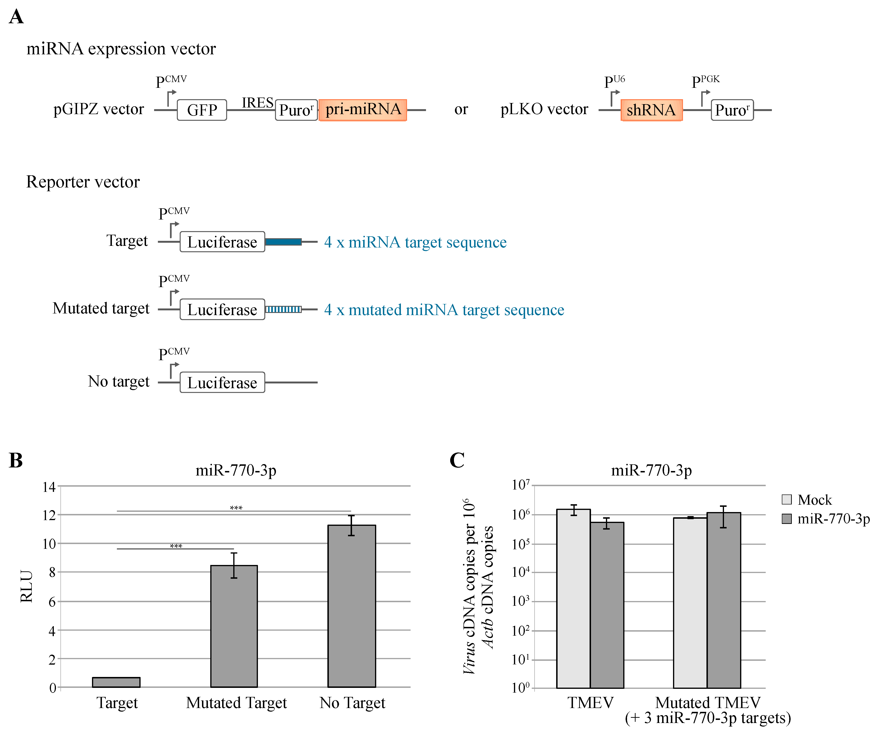
3.1. Under-Represented miRNA Target Sequences in TMEV Genome

It was proposed that viral genomes might have evolved to suppress miRNA target sequences in order to escape miRNA-mediated replication inhibition [43]. We analyzed whether TMEV evolved to suppress the presence of specific miRNA target sequences in its genome. Therefore, we compared the frequency of predicted miRNA target sequences that are present in the coding sequence of the DA1 strain of TMEV and in randomly generated nucleotide sequences that encode an identical polyprotein sequence.
The miR-770-3p target sequence was the most under-represented in TMEV genome (no occurrence in TMEV and a mean of 1.39 occurrences in the 100 randomly generated sequences) (Figure S1A). As this miRNA is mainly expressed in the brain and spinal cord (Figure S1B), we hypothesized that the virus could have evolved to suppress these target sequences, thus avoiding the restriction imposed by miR-770-3p on viral replication in the central nervous system.
To investigate this hypothesis, we generated L929 cells that over-express miR-770-3p and constructed a TMEV mutant virus, ADC43, where 3 predicted miR-770-3p target sites were introduced by site-directed mutagenesis. Then we compared the replication of the WT and mutant viruses in L929 cells that overexpressed or not the miRNA construct (Figure 1).
shRNA expressed in these cells efficiently decreased Renilla luciferase expression from transfected reporter plasmids carrying the corresponding miRNA target sequences in the 3′UTR of the luciferase gene (Figure 1B).
Replication of WT and mutant viruses were assessed, by RT-qPCR, 8 h after infection. As shown in Figure 1C, the presence of miR-770-3p target sequences in the viral genome did not restrict viral replication in cells expressing this miRNA (Figure 1C).

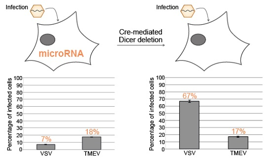
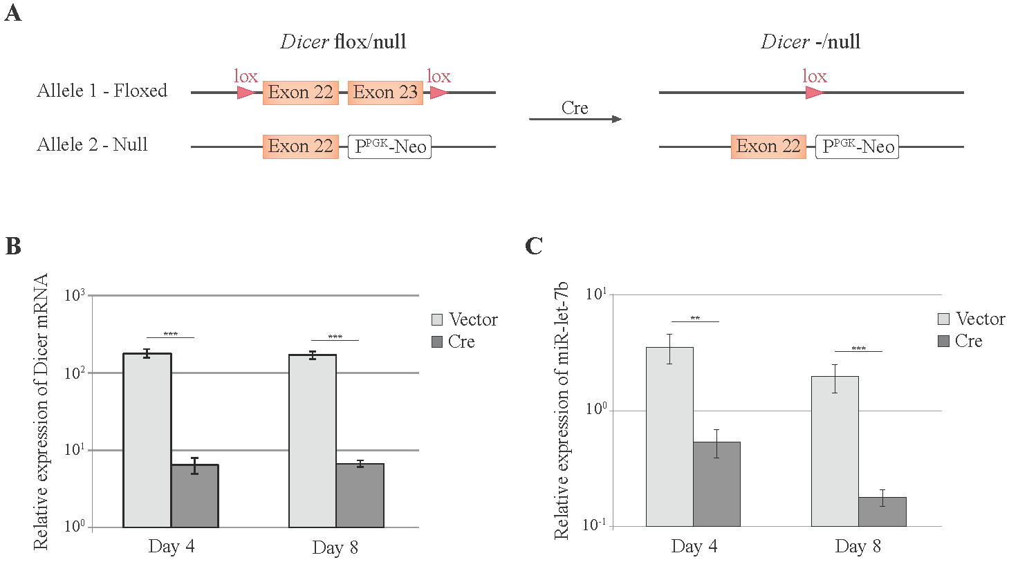
3.2. Cre-Mediated Dicer Inactivation Reduces Expression and Activity of Cellular miRNAs

In order to investigate the global impact of cellular miRNAs on viral replication, we attempted to derive cells that would differ only by the expression of Dicer. To do so, we immortalized and cloned Dicerflox/null MEFs, which carry one Dicer allele with lox sequences framing exons 22 and 23, and one Dicer “null” allele (i.e., with a deletion of the RNaseIII domain) [31] (Figure 2A). All attempts to derive a Dicer/null cell line from these Dicerflox/null cells, by transiently expressing the Cre recombinase from either adenovirus or plasmid vectors, led to gradual growth arrest of the cells (not shown). These cells were thus not suitable to compare viral replication in Dicer-positive and Dicer-negative cells. We therefore designed a short-term Dicer inactivation strategy based on Cre-expressing lentiviral vectors.
Dicerflox/null MEFs were transduced with a lentiviral vector (ADC82) co-expressing mCherry and Cre, in order to inactivate the floxed Dicer allele (Figure 2A). Dicer expression and miRNA abundance were analyzed 4 and 8 days after transduction of the Cre vector. WT Dicer expression was reduced by 30-fold at both time points (Figure 2B). Abundance of the cellular miRNA miR-let-7b decreased by 7-fold at day 4 and by 11-fold at day 8 post-transduction (Figure 2C).
We further tested the influence of Dicer inactivation on a reporter system where miR-142-3p expressed from a pGIPZ derivative inhibits the expression of a reporter luciferase construct carrying miR-142-3p target sequences. In cells transduced with the control lentiviral vector that does not express Cre, miR-142-3p triggered an important decrease of Renilla luciferase activity (Figure 3A,B). In cells transduced with the Cre-expressing vector, the decrease of luciferase activity was reduced, as expected from Dicer inactivation (Figure 3A,B).
Thus, Cre expression in Dicerflox/null MEFs induces a significant decrease of miRNAs expression and of miRNAs function that is detectable four days and more pronounced eight days after transduction.

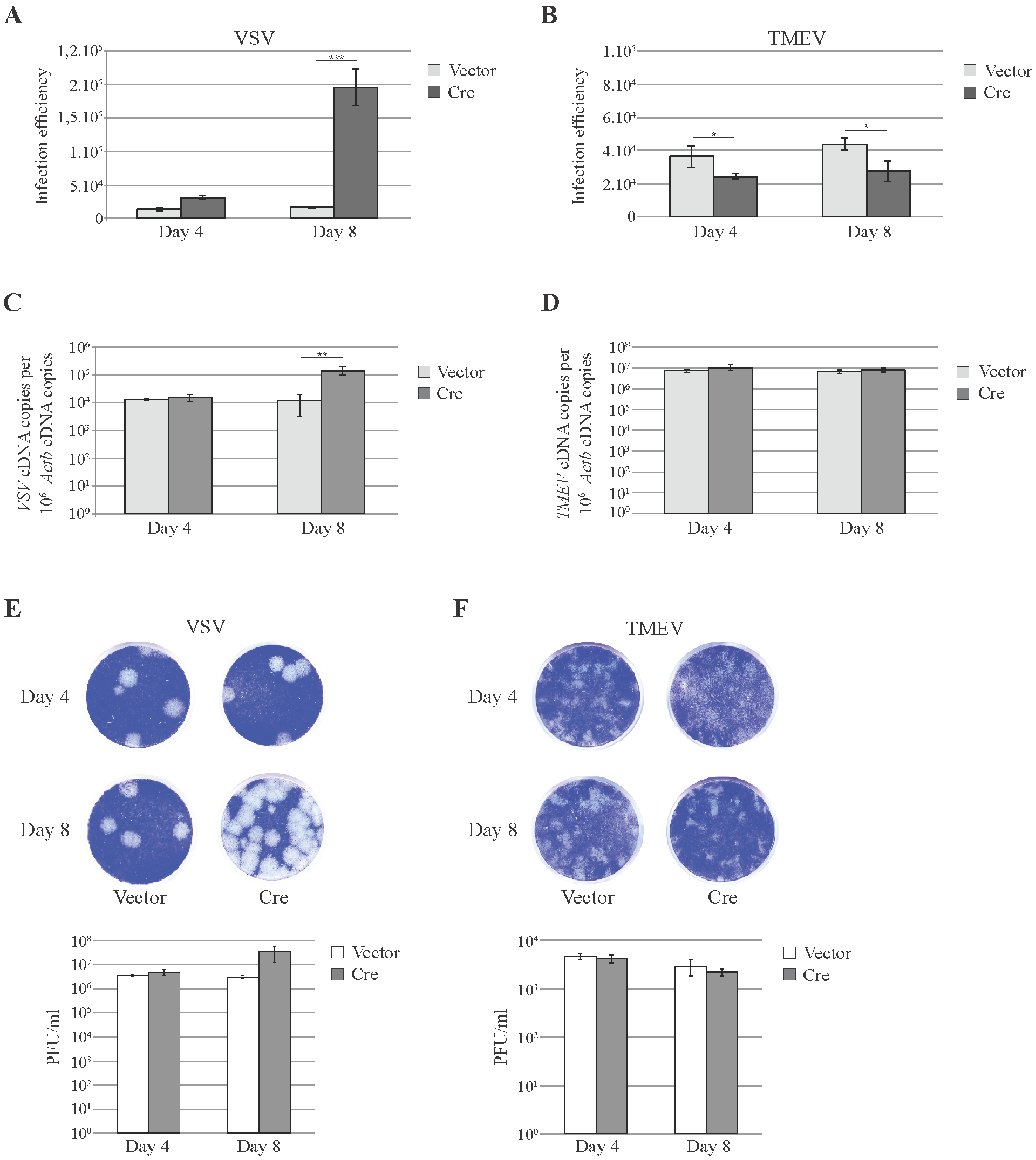
3.3. Knockdown of Cellular miRNAs Increases VSV but Not TMEV Replication

Next, we analyzed the influence of Dicer inactivation on TMEV replication. VSV was used as a control since replication of this virus was reported to be susceptible to host miRNA activity. Thus, Dicerflox/null MEFs were transduced with the lentiviral vectors co-expressing Cre and mCherry (ADC82) or expressing mCherry alone (TM945). Four and eight days after transduction, cells were infected with VSV or TMEV derivatives expressing GFP and viral replication was monitored by flow cytometry. This strategy allows the specific analysis of virus replication (GFP fluorescence) in Cre-expressing (i.e., Dicer-/null, mCherry fluorescence) cells. As shown in Figure 4A, expression of Cre in Dicerflox/null MEFs led to a significant increase of VSV infection efficiency at 8 days post transduction (Figure 4A). In contrast, Cre triggered a slight decrease of TMEV infection efficiency (Figure 4B). Analysis of viral replication by RT-qPCR and plaque assay confirmed that the inactivation of the floxed Dicer allele by CRE increased VSV but not TMEV replication (Figure 4C–F).

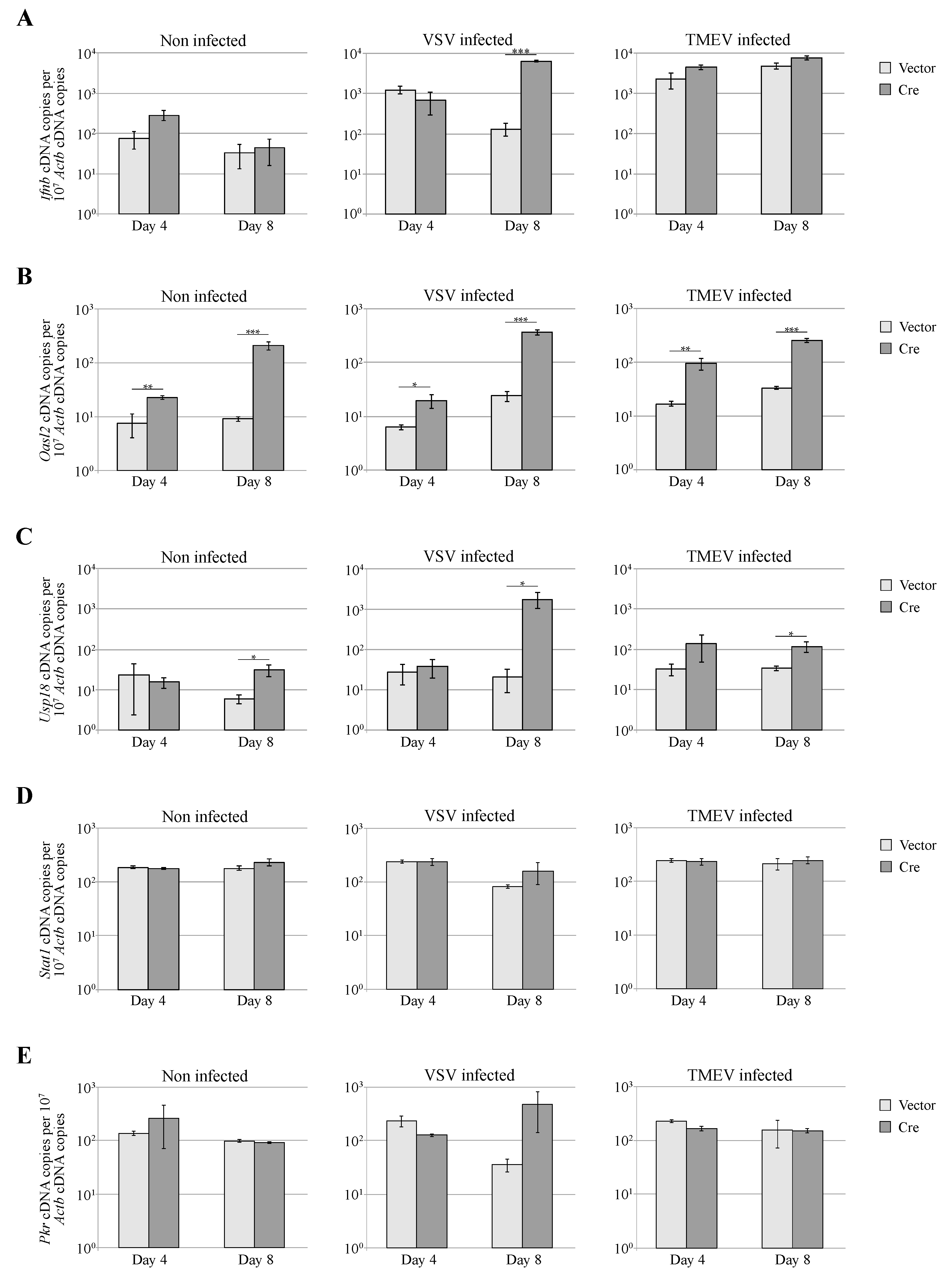
3.4. Dicer Inactivation Increases Oasl2 Expression

To verify that the increase of VSV replication was not due to an impairment of the interferon response in Dicer-deficient cells, we used RT-qPCR to measure the expression of Ifnb, the gene coding interferon β and of 4 interferon-stimulated genes (ISG)(Figure 5). In non-infected cells (left graphs), Dicer inactivation did not influence Ifnb, Stat1 or Pkr expression but clearly induced Oasl2 expression. This suggests that basal Oasl2 levels are maintained low by miRNAs, as reported by Seo et al. [44] who suggested that miRNAs limit ISG expression to avoid their detrimental effects on cell proliferation. As expected, infection of MEFs with either VSV or TMEV increased the expression of Ifnb (Figure 5A) and of Oasl2, Usp18 and Pkr (Figure 5B,C,E). Upregulation of Ifnb and of ISG expression correlated with the extent of virus replication, in agreement with the fact that viral RNA is the main trigger of IFN-β expression. ISG expression was thus higher in Cre-expressing MEFs infected with VSV (Figure 5A). This shows that Dicer inactivation did not increase VSV replication through inhibition of the IFN response.

4. Discussion

In this work, we wanted to evaluate the global influence of miRNAs on TMEV replication. We first analyzed whether miRNAs shaped the viral genome sequence by selection against miRNA target sequences. The most under-represented miRNA target sequence in the TMEV genome (miR-770-3p) was absent from the viral genome and occurred at a mean frequency of 1.39 in randomly generated sequences coding the same protein, the probability to find 0 miR-770-3p target sequence in random sequences being 0.24. In addition, overexpression of a shRNA corresponding to miR-770-3p in L929 cells did not significantly affect replication of TMEV mutants where predicted miR-770-3p target sequences were introduced. The latter results must, however, be taken with caution because this shRNA was expressed from a RNA polymerase III promoter, which reportedly generates RNA that might be off one or few nucleotides [45]. The integrity of the 3′ end of the shRNA was attested by the RT-PCR procedure but alterations of the 5′ end could not be excluded. Taken together, these results fail to suggest a strong selection pressure exerted against the presence of miRNA target sequences in the genome of TMEV.
We next engineered a Dicer inactivation system that enables the comparison of virus replication in otherwise identical Dicer-positive and Dicer-negative cells. Complete inactivation of Dicer consistently led to cell growth inhibition. Therefore, short-term Dicer inactivation was used to analyze the impact of Dicer inactivation on the replication of TMEV and VSV, before the occurrence of cell growth arrest.
Eight days after Cre-mediated deletion of Dicer, the amount and activity of cellular miRNAs were significantly decreased. Replication of VSV was strongly enhanced while replication of TMEV was not affected. Enhancement of VSV replication in Dicer-negative cells was not a consequence of miRNA-mediated inhibition of the interferon antiviral response since the loss of Dicer did not prevent the expression of IFN-stimulated genes. Inhibition of VSV replication was thus likely a direct effect of cellular miRNAs, in line with a previous report showing that VSV can be targeted by miR93 and miR24 [18]. In contrast, TMEV replication was not increased after Dicer inactivation. In contrast, a very small but reproducible decrease of TMEV replication was observed. This increased TMEV replication may result from the upregulation of some ISGs, as observed for Oasl2, that may contribute to restrict viral replication.
Altogether, our results suggest that TMEV exhibits little susceptibility to the action of cellular miRNAs. A recent study showed that the IFN system was much more active than RNAi to restrict the replication of VSV [23]. In the case of TMEV, the balance between RNAi and IFN antiviral effects tips even more toward IFN, even if TMEV evolved to produce proteins that act to antagonize both IFN production and IFN effector activity [46,47]

Supplementary Materials

Figure S1 is available online at www.mdpi.com/1999-4915/8/3/75/s1, Figure S1: Distribution of the number of predicted miR-770-3p targets in viral genomes and expression of miR-770-3p and miR-709 in mouse tissues.

Acknowledgments

We are grateful to Jean-Pierre Szikora (Computer manager, de Duve Institute, University of Louvain, Brussels, Belgium) for help in designing a back-translation routine, to Pierre-Paul Prévot (LPAD unit, de Duve Inst., Univ. of Louvain) for providing a Dicer mutant mouse, to Sabrina El Bachiri (Statistic platform, SMCS, University of Louvain) for assistance in statistics, and to Nicolas Dauguet (GECE unit, de Duve Institute, University of Louvain, Brussels, Belgium), for expert assistance in flow cytometry. ADC was the recipient of a FRIA fellowship from the Belgian Fund for Scientific research and was further supported by a grant of the Health sector of the University of Louvain. Work was supported by the Belgian fund for Scientific Research (conv. 3.4519.11).

Author Contributions

Aurélie De Cock and Thomas Michiels conceived and analyzed the experiments; Aurélie De Cock performed the experiments; Aurélie De Cock and Thomas Michiels wrote the text.

Conflicts of Interest

The authors declare no conflict of interest.

References

  1. Nilsen, T.W. Mechanisms of microRNA-mediated gene regulation in animal cells. Trends Genet. 2007, 23, 243–249. [Google Scholar] [CrossRef] [PubMed]
  2. Zeng, Y. Principles of micro-RNA production and maturation. Oncogene 2006, 25, 6156–6162. [Google Scholar] [CrossRef] [PubMed]
  3. Valencia-Sanchez, M.A.; Liu, J.; Hannon, G.J.; Parker, R. Control of translation and mRNA degradation by miRNAs and siRNAs. Genes Dev. 2006, 20, 515–524. [Google Scholar] [CrossRef] [PubMed]
  4. Kloosterman, W.P.; Plasterk, R.H. The diverse functions of microRNAs in animal development and disease. Dev. Cell 2006, 11, 441–450. [Google Scholar] [CrossRef] [PubMed]
  5. Ding, S.W.; Voinnet, O. Antiviral immunity directed by small RNAs. Cell 2007, 130, 413–426. [Google Scholar] [CrossRef] [PubMed]
  6. Zambon, R.A.; Vakharia, V.N.; Wu, L.P. RNAi is an antiviral immune response against a dsRNA virus in Drosophila melanogaster. Cell. Microbiol. 2006, 8, 880–889. [Google Scholar] [CrossRef] [PubMed]
  7. Aliyari, R.; Wu, Q.; Li, H.W.; Wang, X.H.; Li, F.; Green, L.D.; Han, C.S.; Li, W.X.; Ding, S.W. Mechanism of induction and suppression of antiviral immunity directed by virus-derived small RNAs in Drosophila. Cell Host Microbe 2008, 4, 387–397. [Google Scholar] [CrossRef] [PubMed]
  8. Wang, X.B.; Wu, Q.; Ito, T.; Cillo, F.; Li, W.X.; Chen, X.; Yu, J.L.; Ding, S.W. RNAi-mediated viral immunity requires amplification of virus-derived siRNAs in Arabidopsis thaliana. Proc. Natl. Acad. Sci. USA 2010, 107, 484–489. [Google Scholar] [CrossRef] [PubMed]
  9. Lakatos, L.; Csorba, T.; Pantaleo, V.; Chapman, E.J.; Carrington, J.C.; Liu, Y.P.; Dolja, V.V.; Calvino, L.F.; Lopez-Moya, J.J.; Burgyan, J. Small RNA binding is a common strategy to suppress RNA silencing by several viral suppressors. EMBO J. 2006, 25, 2768–2780. [Google Scholar] [CrossRef] [PubMed]
  10. Li, H.; Li, W.X.; Ding, S.W. Induction and suppression of RNA silencing by an animal virus. Science 2002, 296, 1319–1321. [Google Scholar] [CrossRef] [PubMed]
  11. Chao, J.A.; Lee, J.H.; Chapados, B.R.; Debler, E.W.; Schneemann, A.; Williamson, J.R. Dual modes of RNA-silencing suppression by flock house virus protein B2. Nat. Struct. Mol. Biol. 2005, 12, 952–957. [Google Scholar] [CrossRef] [PubMed]
  12. Galiana-Arnoux, D.; Dostert, C.; Schneemann, A.; Hoffmann, J.A.; Imler, J.L. Essential function in vivo for Dicer-2 in host defense against RNA viruses in Drosophila. Nat. Immunol. 2006, 7, 590–597. [Google Scholar] [CrossRef] [PubMed]
  13. Bouche, N.; Lauressergues, D.; Gasciolli, V.; Vaucheret, H. An antagonistic function for Arabidopsis DCL2 in development and a new function for DCL4 in generating viral siRNAs. EMBO J. 2006, 25, 3347–3356. [Google Scholar] [CrossRef] [PubMed]
  14. Li, H.W.; Ding, S.W. Antiviral silencing in animals. FEBS Lett. 2005, 579, 5965–5973. [Google Scholar] [CrossRef] [PubMed]
  15. Haasnoot, J.; de Vries, W.; Geutjes, E.J.; Prins, M.; de Haan, P.; Berkhout, B. The Ebola virus VP35 protein is a suppressor of RNA silencing. PLoS Pathog. 2007, 3, e86. [Google Scholar] [CrossRef] [PubMed]
  16. Li, W.X.; Li, H.; Lu, R.; Li, F.; Dus, M.; Atkinson, P.; Brydon, E.W.; Johnson, K.L.; Garcia-Sastre, A.; Ball, L.A.; et al. Interferon antagonist proteins of influenza and vaccinia viruses are suppressors of RNA silencing. Proc. Natl. Acad. Sci. USA 2004, 101, 1350–1355. [Google Scholar] [CrossRef] [PubMed]
  17. Triboulet, R.; Mari, B.; Lin, Y.L.; Chable-Bessia, C.; Bennasser, Y.; Lebrigand, K.; Cardinaud, B.; Maurin, T.; Barbry, P.; Baillat, V.; et al. Suppression of microRNA-silencing pathway by HIV-1 during virus replication. Science 2007, 315, 1579–1582. [Google Scholar] [CrossRef] [PubMed]
  18. Otsuka, M.; Jing, Q.; Georgel, P.; New, L.; Chen, J.; Mols, J.; Kang, Y.J.; Jiang, Z.; Du, X.; Cook, R.; et al. Hypersusceptibility to vesicular stomatitis virus infection in Dicer1-deficient mice is due to impaired miR24 and miR93 expression. Immunity 2007, 27, 123–134. [Google Scholar] [CrossRef] [PubMed]
  19. Pedersen, I.M.; Cheng, G.; Wieland, S.; Volinia, S.; Croce, C.M.; Chisari, F.V.; David, M. Interferon modulation of cellular microRNAs as an antiviral mechanism. Nature 2007, 449, 919–922. [Google Scholar] [CrossRef] [PubMed]
  20. Mahajan, V.S.; Drake, A.; Chen, J. Virus-specific host miRNAs: Antiviral defenses or promoters of persistent infection? Trends Immunol. 2009, 30, 1–7. [Google Scholar] [CrossRef] [PubMed]
  21. Trobaugh, D.W.; Gardner, C.L.; Sun, C.; Haddow, A.D.; Wang, E.; Chapnik, E.; Mildner, A.; Weaver, S.C.; Ryman, K.D.; Klimstra, W.B. RNA viruses can hijack vertebrate microRNAs to suppress innate immunity. Nature 2014, 506, 245–248. [Google Scholar] [CrossRef] [PubMed]
  22. Bogerd, H.P.; Skalsky, R.L.; Kennedy, E.M.; Furuse, Y.; Whisnant, A.W.; Flores, O.; Schultz, K.L.; Putnam, N.; Barrows, N.J.; Sherry, B.; et al. Replication of many human viruses is refractory to inhibition by endogenous cellular microRNAs. J. Virol. 2014, 88, 8065–8076. [Google Scholar] [CrossRef] [PubMed]
  23. Backes, S.; Langlois, R.A.; Schmid, S.; Varble, A.; Shim, J.V.; Sachs, D.; tenOever, B.R. The Mammalian response to virus infection is independent of small RNA silencing. Cell Rep. 2014, 8, 114–125. [Google Scholar] [CrossRef] [PubMed]
  24. Barnes, D.; Kunitomi, M.; Vignuzzi, M.; Saksela, K.; Andino, R. Harnessing endogenous miRNAs to control virus tissue tropism as a strategy for developing attenuated virus vaccines. Cell Host Microbe 2008, 4, 239–248. [Google Scholar] [CrossRef] [PubMed]
  25. Kelly, E.J.; Hadac, E.M.; Cullen, B.R.; Russell, S.J. MicroRNA antagonism of the picornaviral life cycle: Alternative mechanisms of interference. PLoS Pathog. 2010, 6, e1000820. [Google Scholar] [CrossRef] [PubMed]
  26. Chang, Y.; Dou, Y.; Bao, H.; Luo, X.; Liu, X.; Mu, K.; Liu, Z.; Liu, X.; Cai, X. Multiple microRNAs targeted to internal ribosome entry site against foot-and-mouth disease virus infection in vitro and in vivo. Virol. J. 2014, 11. [Google Scholar] [CrossRef] [PubMed]
  27. Karaa, Z.S.; Iacovoni, J.S.; Bastide, A.; Lacazette, E.; Touriol, C.; Prats, H. The VEGF IRESes are differentially susceptible to translation inhibition by miR-16. RNA 2009, 15, 249–254. [Google Scholar] [CrossRef] [PubMed]
  28. Zheng, Z.; Ke, X.; Wang, M.; He, S.; Li, Q.; Zheng, C.; Zhang, Z.; Liu, Y.; Wang, H. Human microRNA hsa-miR-296–5p suppresses enterovirus 71 replication by targeting the viral genome. J. Virol. 2013, 87, 5645–5656. [Google Scholar] [CrossRef] [PubMed]
  29. Wang, L.; Qin, Y.; Tong, L.; Wu, S.; Wang, Q.; Jiao, Q.; Guo, Z.; Lin, L.; Wang, R.; Zhao, W.; et al. MiR-342–5p suppresses coxsackievirus B3 biosynthesis by targeting the 2C-coding region. Antiviral Res. 2012, 93, 270–279. [Google Scholar] [CrossRef] [PubMed]
  30. Tong, L.; Lin, L.; Wu, S.; Guo, Z.; Wang, T.; Qin, Y.; Wang, R.; Zhong, X.; Wu, X.; Wang, Y.; et al. MiR-10a* up-regulates coxsackievirus B3 biosynthesis by targeting the 3D-coding sequence. Nucleic Acids Res. 2013, 41, 3760–3771. [Google Scholar] [CrossRef] [PubMed]
  31. Murchison, E.P.; Partridge, J.F.; Tam, O.H.; Cheloufi, S.; Hannon, G.J. Characterization of Dicer-deficient murine embryonic stem cells. Proc. Natl. Acad. Sci. USA 2005, 102, 12135–12140. [Google Scholar] [CrossRef] [PubMed]
  32. Hermant, P.; Francius, C.; Clotman, F.; Michiels, T. IFN-epsilon is constitutively expressed by cells of the reproductive tract and is inefficiently secreted by fibroblasts and cell lines. PLoS ONE 2013, 8, e71320. [Google Scholar] [CrossRef] [PubMed]
  33. Daniels, J.B.; Pappenheimer, A.M.; Richardson, S. Observations on encephalomyelitis of mice (DA strain). J. Exp. Med. 1952, 96, 517–530. [Google Scholar] [CrossRef] [PubMed]
  34. Jnaoui, K.; Michiels, T. Adaptation of Theiler’s virus to L929 cells: Mutations in the putative receptor binding site on the capsid map to neutralization sites and modulate viral persistence. Virology 1998, 244, 397–404. [Google Scholar] [CrossRef] [PubMed]
  35. McAllister, A.; Tangy, F.; Aubert, C.; Brahic, M. Molecular cloning of the complete genome of Theiler's virus, strain DA, and production of infectious transcripts. Microb. Pathogen. 1989, 7, 381–388. [Google Scholar] [CrossRef]
  36. Miranda, K.C.; Huynh, T.; Tay, Y.; Ang, Y.S.; Tam, W.L.; Thomson, A.M.; Lim, B.; Rigoutsos, I. A pattern-based method for the identification of MicroRNA binding sites and their corresponding heteroduplexes. Cell 2006, 126, 1203–1217. [Google Scholar] [CrossRef] [PubMed]
  37. Follenzi, A.; Ailles, L.E.; Bakovic, S.; Geuna, M.; Naldini, L. Gene transfer by lentiviral vectors is limited by nuclear translocation and rescued by HIV-1 pol sequences. Nat. Genet. 2000, 25, 217–222. [Google Scholar] [PubMed]
  38. Kreit, M.; Paul, S.; Knoops, L.; De Cock, A.; Sorgeloos, F.; Michiels, T. Inefficient type I interferon-mediated antiviral protection of primary mouse neurons is associated with the lack of apolipoprotein l9 expression. J. Virol. 2014, 88, 3874–3884. [Google Scholar] [CrossRef] [PubMed]
  39. Moffat, J.; Grueneberg, D.A.; Yang, X.; Kim, S.Y.; Kloepfer, A.M.; Hinkle, G.; Piqani, B.; Eisenhaure, T.M.; Luo, B.; Grenier, J.K.; et al. A lentiviral RNAi library for human and mouse genes applied to an arrayed viral high-content screen. Cell 2006, 124, 1283–1298. [Google Scholar] [CrossRef] [PubMed]
  40. Paul, S.; Michiels, T. Cardiovirus leader proteins are functionally interchangeable and have evolved to adapt to virus replication fitness. J. General Virol. 2006, 87, 1237–1246. [Google Scholar] [CrossRef] [PubMed]
  41. Prevot, P.P.; Augereau, C.; Simion, A.; Van den Steen, G.; Dauguet, N.; Lemaigre, F.P.; Jacquemin, P. Let-7b and miR-495 stimulate differentiation and prevent metaplasia of pancreatic acinar cells by repressing HNF6. Gastroenterology 2013, 145, 668–678. [Google Scholar] [CrossRef] [PubMed]
  42. Chen, C.; Ridzon, D.A.; Broomer, A.J.; Zhou, Z.; Lee, D.H.; Nguyen, J.T.; Barbisin, M.; Xu, N.L.; Mahuvakar, V.R.; Andersen, M.R.; et al. Real-time quantification of microRNAs by stem-loop RT-PCR. Nucleic Acids Res. 2005, 33. [Google Scholar] [CrossRef] [PubMed]
  43. Gitlin, L.; Stone, J.K.; Andino, R. Poliovirus escape from RNA interference: Short interfering RNA-target recognition and implications for therapeutic approaches. J. Virol. 2005, 79, 1027–1035. [Google Scholar] [CrossRef] [PubMed]
  44. Seo, G.J.; Kincaid, R.P.; Phanaksri, T.; Burke, J.M.; Pare, J.M.; Cox, J.E.; Hsiang, T.Y.; Krug, R.M.; Sullivan, C.S. Reciprocal inhibition between intracellular antiviral signaling and the RNAi machinery in mammalian cells. Cell Host Microbe 2013, 14, 435–445. [Google Scholar] [CrossRef] [PubMed]
  45. Ma, H.; Wu, Y.; Dang, Y.; Choi, J.G.; Zhang, J.; Wu, H. Pol III promoters to express small RNAs: Delineation of transcription initiation. Mol. Ther. Nucleic Acids 2014, 3. [Google Scholar] [CrossRef] [PubMed]
  46. Sorgeloos, F.; Jha, B.K.; Silverman, R.H.; Michiels, T. Evasion of antiviral innate immunity by Theiler’s virus L* protein through direct inhibition of RNase L. PLoS Pathog. 2013, 9, e1003474. [Google Scholar] [CrossRef] [PubMed]
  47. Van Pesch, V.; van Eyll, O.; Michiels, T. The leader protein of Theiler’s virus inhibits immediate-early α/β interferon production. J. Virol. 2001, 75, 7811–7817. [Google Scholar] [CrossRef] [PubMed]
Figure 1. Lack of influence of miR-770-3p on TMEV replication. (A) Illustration of miR expression and reporter vectors. miR expression was achieved by expressing either an artificial primary miRNAs using the pGIPZ vector or a short hairpin RNA, using the pLKO.1 vector. Reporter vectors carry a Renilla luciferase gene with 0 (No target) or 4 wild type (Target) or mutated (Mutated target) miR target sequences; (B) L929 cells expressing miR-770-3p from the pLKO vector were transduced with the indicated reporter constructs and luciferase activity (Relative Luciferase Units : RLU) was assessed 24 h post transduction; (C) L929 cells expressing miR-770-3p were infected for 8 hours with wild type and mutant TMEV derivatives before replication analysis by RT-qPCR (C). Viruses were KJ6 (TMEV) and ADC43, a KJ6 derivative carrying 3 extra binding sites for miR-770-3p. Graphs show the mean and SD of a triplicate experiment (out of 2). *** p < 0.001
Viruses 08 00075 g001
Figure 2. Short-term Dicer inactivation influence on cellular miRNA expression. (A) Illustration of Dicer alleles present in MEFs before and after Cre-mediated recombination; (B,C) Four and 8 days after transduction of Dicerflox/null MEFs with either TM945 expressing mCherry alone (Vector) or ADC82 co-expressing mCherry and Cre (Cre), WT Dicer mRNA (B) and mature miR-let-7b (C) were quantified by qRT-PCR. Graphs show the mean and standard deviation (SD) of a typical experiment (out of 2) performed in triplicate. ** p < 0.01, *** p < 0.001
Viruses 08 00075 g002
Figure 3. Effect of Dicer inactivation on miR activity. Dicerflox/null MEFs were transduced for 4 (A) or 8 (B) days with TM945 expressing mCherry alone (Vector) or ADC82 co-expressing mCherry and Cre (Cre). Cells were then co-transfected with pADC38 (pGIPZ-miR-142-3p), with indicated Renilla luciferase (Rluc) reporter constructs carrying miR-142-3p targets and with pADC13 encoding firefly luciferase (Fluc) from the CMV promoter. Graphs show the mean and SD of the Rluc/Fluc ratio, which reflects the absence of miR function. ** p < 0.01, *** p < 0.001
Viruses 08 00075 g003
Figure 4. Impact of Dicer inactivation on VSV and TMEV infection. Infection by GFP expressing derivatives of VSV and TMEV was compared in Dicerflox/null MEFs that were transduced for 4 or 8 days with TM945 expressing mCherry alone (Vector) or with ADC82 co-expressing mCherry and Cre (Cre). Cells were then infected in triplicate with 10 PFU per cell of VSV during 7 h or with 1 PFU per cell of TMEV during 8 h. Graphs show the mean and SD of VSV and TMEV infection efficiency as determined by flow cytometry (A,B) (1 representative experiment out of 2); RT-qPCR (C,D) or plaque assay (magnification: 0.33×) (E,F). * p < 0.05, ** p < 0.01, *** p < 0.001
Viruses 08 00075 g004
Figure 5. IFN response in Dicer-positive and Dicer-negative MEFs. (AE) Ifnb (A); Oasl2 (B); Usp18 (C); Stat1 (D) and Pkr (E) expression was quantified by RT-qPCR in non-infected, VSV-infected or TMEV-infected Dicerflox/null MEFs transduced for 4 or 8 days with lentiviral vectors expressing mCherry alone (Vector) or co-expressing mCherry and Cre (Cre). Infection conditions were as in Figure 4.
Viruses 08 00075 g005
Table 1. Recombinant viruses, lentiviral vectors and reporter vectors used in this study.
Vector/Virus 1Parental 1Characteristics
(p)KJ6TMEV (DA1)Capsid adapted to infect L929 cells
(p)KJ7(p)KJ6GFP-coding sequence replacing codons 5–67 of L protein
(p)ADC43(p)KJ6TMEV carrying 3 extra target sequences for miR-770-3p
VSV-GFPVSVVSV carrying a GFP-coding sequence
(p)TM945pCCLsin 2Lentiviral vector carrying PCMV-MCS-IRES-mCherry 2
(p)ADC82(p)TM945Lentiviral vector carrying PCMV-Cre recombinase-IRES-mCherry 2
(p)ADC48pLKO.1Lentiviral vector expressing shRNA corresponding to miR-770-3p
(p)ADC38pGIPZLentiviral vector expressing genomic miR-142-3p
(p)ADC1(p)TM897Lentiviral vector carrying PPGK-Renilla luciferase 2
(p)ADC30(p)ADC1Luciferase reporter with 4 tandem miR-770-3p target sequences
(p)ADC39(p)ADC1Luciferase reporter with 4 tandem miR-142-3p target sequences
(p)ADC62(p)ADC1Luciferase reporter with 4 tandem mutated miR-770-3p target sequences
(p)ADC64(p)ADC1Luciferase reporter with 4 tandem copies of mutated miR-142-3p target sequences
pADC13pcDNA3Photinus luciferase (firefly) from pGL-2 basic (Promega, Leiden, The Netherlands)
1 names of plasmids that carry a viral genome start with a “p” while corresponding viruses lack the “p”. For example, plasmid pKJ6 carries the genome of virus KJ6; 2 pCCLsin = pCCLsin.PPT.hPGK.GFP.pre; PCMV = cytomegalovirus promoter; PPGK = human phosphoglycerate kinase promoter; MCS = multi-cloning site.
Table 2. Nucleotide sequence of miRNA binding sites incorporated in reporter vectors.
VectormiRNA Binding SiteNucleotide Sequence of miRNA Binding Sites
(p)ADC30Target sequences for miR-770-3pCCAGCTCCACGTCAGGCCCACG
(p)ADC39Target sequence for miR-142-3pTCCATAAAGTAGGAAACACTACA
(p)ADC62Mutated target sequence for miR-770-3pCCCGCACCCCGACACGCTCATG
(p)ADC64Mutated target sequence for miR-142-3pTCTATTAAATAAGAGACTCTTCA
Table 3. Primers used for gene expression analysis by RT-qPCR and reference plasmids.
GeneName *Sequence 5′–3′Plasmid
ActbTM421 (Fw)AGAGGGAAATCGTGCGTGACpTM793
TM422 (Rev)CAATAGTGATGACCTGGCCGT
TMEVTM348 (Fw)GAACGTCAGCATTTTCCGGCpTM410
TM349 (Rev)GGTGTGAAGAGCGGCAAGTG
VSV-GTM846 (Fw)TTGCTGCTCCAATCCTCTCApVSV-G
TM847 (Rev)TCGAACACCTGAGCCTTTGA
IfnbTM642 (Fw)ATGAACAACAGGTGGATCCTCCpcDNA3-IFN-β
TM643 (Rev)AGGAGCTCCTGACATTTCCGAA
Oasl2TM638 (Fw)GGATGCCTGGGAGAGAATCGpCS40
TM639 (Rev)TCGCCTGCTCTTCGAAACTG
Usp18TM1116 (Fw)TGCAGGGTCTGTTCACCATCpMIP01
TM1117 (Rev)GCACATGTCGGAGCTTGCTA
Stat1TM881 (Fw)GAGGGGCCTCTCATTGTCACpSPA31
TM882 (Rev)GATTCCTGGGCTCTGTCACC
PkrTM887 (Fw)CGCTGGCAGAACTCAATCACpSPA33
TM888 (Rev)CTCCGGTCACGATTTGTTCA
* (Fw) Forward and (Rev) Reverse.

