
Seroprevalence and Risk Factors Associated with Canine Leishmaniasis in Egypt

 ,
, 
 , and
, and
Abstract
:1. Introduction
2. Materials and Methods
2.1. Ethics Statement
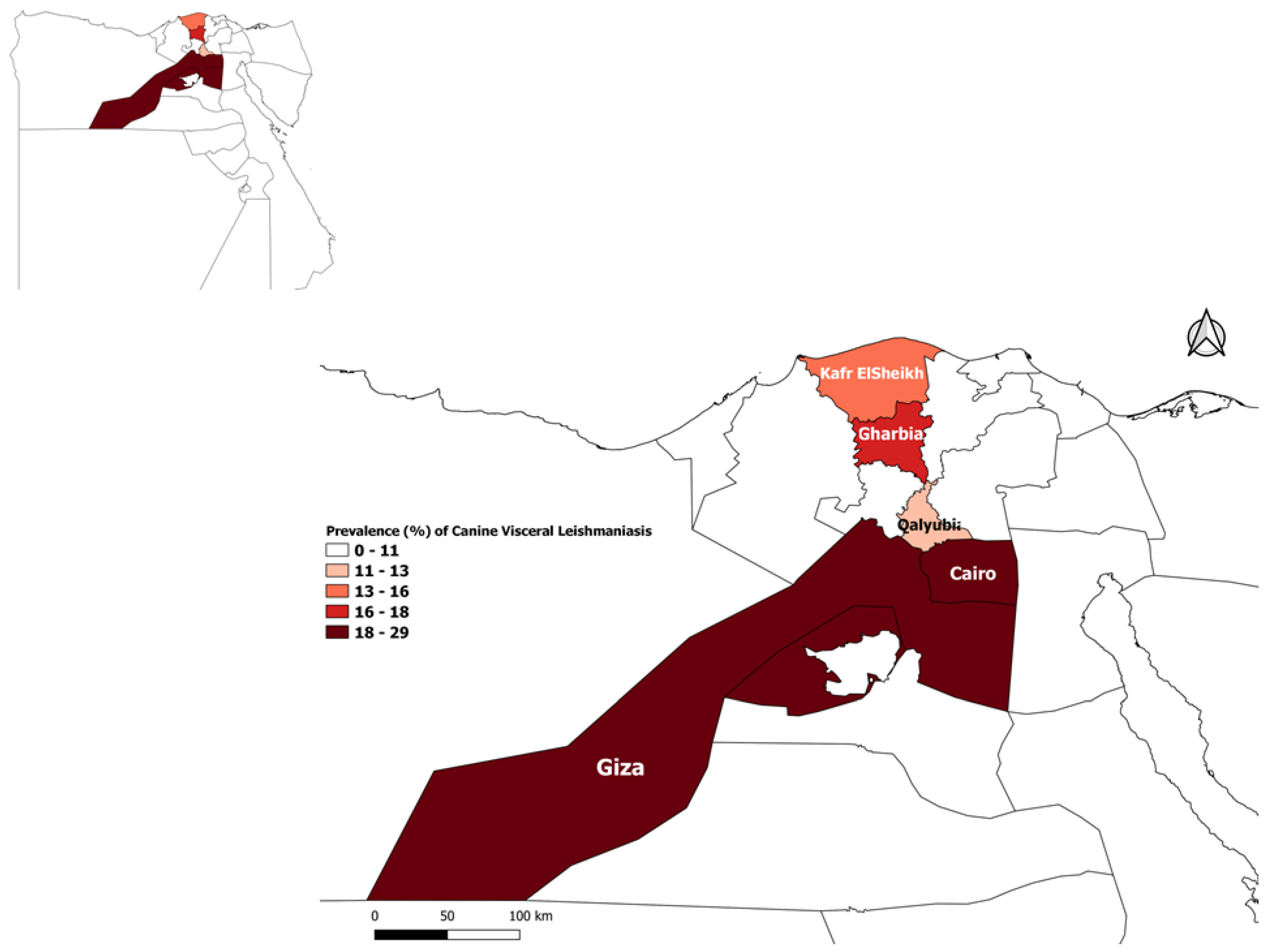
2.2. Study Area
2.3. Sampling and Data Collection
2.4. Statistical Analysis
3. Results
4. Discussion
5. Conclusions
Author Contributions
Funding
Institutional Review Board Statement
Informed Consent Statement
Data Availability Statement
Acknowledgments
Conflicts of Interest
References
- Abuowarda, M.; AbuBakr, H.O.; Ismael, E.; Shaalan, M.; Mohamed, M.A.; Aljuaydi, S.H. Epidemiological and genetic characteristics of asymptomatic canine leishmaniasis and implications for human Leishmania infections in Egypt. Zoonoses Public Health 2021, 68, 413–430. [Google Scholar] [CrossRef]
- Malmasi, A.; Janitabar, S.; Mohebali, M.; Akhoundi, B.; Maazi, N.; Aramoon, M.; Khorrami, N.; Seifi, H.A. Seroepidemiologic survey of canine visceral leishmaniasis in Tehran and Alborz Provinces of Iran. J. Arthropod Borne Dis. 2014, 8, 132. [Google Scholar] [PubMed]
- Cardoso, L.; Rodrigues, M.; Santos, H.; Schoone, G.J.; Carreta, P.; Varejão, E.; van Benthem, B.; Afonso, M.O.; Alves-Pires, C.; Semião-Santos, S.J. Sero-epidemiological study of canine Leishmania spp. infection in the municipality of Alijó (Alto Douro, Portugal). Vet. Parasitol. 2004, 121, 21–32. [Google Scholar] [CrossRef] [PubMed]
- Alvar, J.; Vélez, I.D.; Bern, C.; Herrero, M.; Desjeux, P.; Cano, J.; Jannin, J.; Boer, M.D.; Team, W.L.C. Leishmaniasis worldwide and global estimates of its incidence. PLoS ONE 2012, 7, e35671. [Google Scholar] [CrossRef] [PubMed]
- Tamponi, C.; Scarpa, F.; Carta, S.; Knoll, S.; Sanna, D.; Gai, C.; Pipia, A.P.; Dessì, G.; Casu, M.; Varcasia, A. Seroprevalence and risk factors associated with Leishmania infantum in dogs in Sardinia (Italy), an endemic island for leishmaniasis. Parasitol. Res. 2020, 120, 289–300. [Google Scholar] [CrossRef]
- Tabbabi, A. Review of leishmaniasis in the Middle East and North Africa. Afr. Health Sci. 2019, 19, 1329–1337. [Google Scholar] [CrossRef] [Green Version]
- Medkour, H.; Davoust, B.; Dulieu, F.; Maurizi, L.; Lamour, T.; Marié, J.-L.; Mediannikov, O. Potential animal reservoirs (dogs and bats) of human visceral leishmaniasis due to Leishmania infantum in French Guiana. PLoS Negl. Trop. Dis. 2019, 13, e0007456. [Google Scholar] [CrossRef]
- Dantas-Torres, F.; de Brito, M.E.F.; Brandão-Filho, S.P. Seroepidemiological survey on canine leishmaniasis among dogs from an urban area of Brazil. Vet. Parasitol. 2006, 140, 54–60. [Google Scholar] [CrossRef]
- Najafi, L.; Omidian, M.; Rezaei, Z.; Shahabi, S.; Ghorbani, F.; Arefkhah, N.; Mohebali, M.; Zaraei, Z.; Sarkari, B. Molecular and serological evaluation of zoonotic visceral leishmaniasis in dogs in a rural area of Fars province, southern Iran, as a source of Leishmania infantum infection. Vet. Med. Sci. 2021, 7, 1082–1089. [Google Scholar] [CrossRef]
- Solano-Gallego, L.; Miró, G.; Koutinas, A.; Cardoso, L.; Pennisi, M.G.; Ferrer, L.; Bourdeau, P.; Oliva, G.; Baneth, G. LeishVet guidelines for the practical management of canine leishmaniosis. Parasites Vectors 2011, 4, 1–16. [Google Scholar] [CrossRef] [Green Version]
- Esteva, L.; Vargas, C.; de León, C.V. The role of asymptomatics and dogs on leishmaniasis propagation. Math. Biosci. 2017, 293, 46–55. [Google Scholar] [CrossRef]
- Rosypal, A.C.; Bowman, S.S.; Epps, S.A.; El Behairy, A.; Hilali, M.; Dubey, J. Serological survey of dogs from Egypt for antibodies to Leishmania species. J. Parasitol. 2013, 99, 170–171. [Google Scholar] [CrossRef]
- Morsy, T.A.; Schnur, L.F.; Feinsod, F.M.; Salem, A.M.; Wahba, M.M.; El Said, S.M. Natural infections of Leishmania major in domestic dogs from Alexandria, Egypt. Am. J. Trop. Med. Hyg. 1987, 37, 49–52. [Google Scholar] [CrossRef]
- Bessat, M.; Okpanma, A.; Shanat, E. Leishmaniasis: Epidemiology, control and future perspectives with special emphasis on Egypt. J. Trop. Dis. 2015, 2, 1–10. [Google Scholar]
- Awadalla, H.; Mansour, N.; Mohareb, E. Further characterization of Leishmania isolates from children with visceral infection in Alexandria area, Egypt. Trans. R. Soc. Trop. Med. Hyg. 1987, 81, 915–917. [Google Scholar] [CrossRef]
- Faris, R.; Massoud, A.; El Said, S.; Gadallah, M.; Feinsod, F.; Saar, A.; Londner, M.; Rosen, G. The epidemiology of human visceral leishmaniasis in El Agamy (Alexandria Governorate), Egypt: Serosurvey and case/control study. Ann. Trop. Med. Parasitol. 1988, 82, 445–452. [Google Scholar] [CrossRef] [PubMed]
- Fryauff, D.J.; Modi, G.B.; Mansour, N.S.; Kreutzer, R.D.; Soliman, S. Epidemiology of Cutaneous Leishmaniasis at a Focus Monitored by the Multinational Force and Observers in the Northeastern Sinai Desert of Egypt. Am. J. Trop. Med. Hyg. 1993, 49, 598–607. [Google Scholar] [CrossRef] [PubMed]
- Hamadto, H.A.; El Fkahany, A.F.; Morsy, T.A.; Farrag, A.; MK, A.M. Re-evaluation of zoonotic cutaneous leishmaniasis status in North Sinai Governorate, Egypt. J. Egypt. Soc. Parasitol. 2003, 33, 687–694. [Google Scholar]
- Vélez, R.; Ballart, C.; Domenech, E.; Abras, A.; Fernández-Arévalo, A.; Gómez, S.A.; Tebar, S.; Muñoz, C.; Cairó, J.; Gállego, M. Seroprevalence of canine Leishmania infantum infection in the Mediterranean region and identification of risk factors: The example of North-Eastern and Pyrenean areas of Spain. Prev. Vet. Med. 2019, 162, 67–75. [Google Scholar] [CrossRef]
- Rombolà, P.; Barlozzari, G.; Carvelli, A.; Scarpulla, M.; Iacoponi, F.; Macrì, G. Seroprevalence and risk factors associated with exposure to Leishmania infantum in dogs, in an endemic Mediterranean region. PLoS ONE 2021, 16, e0244923. [Google Scholar] [CrossRef]
- Mahshid, M.; Baharak, A.; Iraj, S.; Sina, K.; Javad, K.; Mehdi, B. Seroprevalence of canine visceral leishmaniasis in southeast of Iran. J. Parasit. Dis. 2014, 38, 218–222. [Google Scholar] [CrossRef] [Green Version]
- Costa, M.M.; Penido, M.; Dos Santos, M.S.; Doro, D.; de Freitas, E.; Michalick, M.S.M.; Grimaldi, G.; Gazzinelli, R.T.; Fernandes, A.P. Improved canine and human visceral leishmaniasis immunodiagnosis using combinations of synthetic peptides in enzyme-linked immunosorbent assay. PLoS Negl. Trop. Dis. 2012, 6, e1622. [Google Scholar] [CrossRef] [PubMed]
- Pourhoseingholi, M.A.; Vahedi, M.; Rahimzadeh, M. Sample size calculation in medical studies. Gastroenterol. Hepatol. Bed Bench 2013, 6, 14. [Google Scholar] [PubMed]
- Gharekhani, J.; Pourmahdi Borujeni, M.; Sazmand, A. Seroprevalence of Visceral Leishmaniosis in Stray Dogs of Hamedan, West of Iran in 2018. J. Med Microbiol. Infect. Dis. 2020, 8, 71–75. [Google Scholar] [CrossRef]
- Abdeen, Z.A.; Sawalha, S.S.; Eisenberger, C.L.; Khanfar, H.M.; Greenblatt, C.L.; Yousef, O.; Schnur, L.F.; Azmi, K.; Warburg, A.; Bader, K.A. Epidemiology of visceral leishmaniasis in the Jenin District, West Bank: 1989–1998. Am. J. Trop. Med. Hyg. 2002, 66, 329–333. [Google Scholar] [CrossRef] [PubMed] [Green Version]
- Rab, M.; Frame, I.; Evans, D. The role of dogs in the epidemiology of human visceral leishmaniasis in northern Pakistan. Trans. R. Soc. Trop. Med. Hyg. 1995, 89, 612–615. [Google Scholar] [CrossRef]
- Dereure, J.; El-Safi, S.H.; Bucheton, B.; Boni, M.; Kheir, M.M.; Davoust, B.; Pratlong, F.; Feugier, E.; Lambert, M.; Dessein, A. Visceral leishmaniasis in eastern Sudan: Parasite identification in humans and dogs; host-parasite relationships. Microbes Infect. 2003, 5, 1103–1108. [Google Scholar] [CrossRef]
- Selim, A.; Almohammed, H.; Abdelhady, A.; Alouffi, A.; Alshammari, F.A. Molecular detection and risk factors for Anaplasma platys infection in dogs from Egypt. Parasites Vectors 2021, 14, 1–6. [Google Scholar] [CrossRef]
- Selim, A.; Manaa, E.; Khater, H. Seroprevalence and risk factors for lumpy skin disease in cattle in Northern Egypt. Trop. Anim. Health Prod. 2021, 53, 1–8. [Google Scholar] [CrossRef]
- Selim, A.; Manaa, E.A.; Waheed, R.M.; Alanazi, A.D. Seroprevalence, associated risk factors analysis and first molecular characterization of chlamydia abortus among Egyptian sheep. Comp. Immunol. Microbiol. Infect. Dis. 2021, 74, 101600. [Google Scholar] [CrossRef]
- Martín-Sánchez, J.; Morales-Yuste, M.; Acedo-Sánchez, C.; Barón, S.; Díaz, V.; Morillas-Márquez, F. Canine leishmaniasis in southeastern Spain. Emerg. Infect. Dis. 2009, 15, 795. [Google Scholar] [CrossRef]
- Sauda, F.; Malandrucco, L.; Macrì, G.; Scarpulla, M.; De Liberato, C.; Terracciano, G.; Fichi, G.; Berrilli, F.; Perrucci, S. Leishmania infantum, Dirofilaria spp. and other endoparasite infections in kennel dogs in central Italy. Parasite 2018, 25, 2–10. [Google Scholar] [CrossRef] [Green Version]
- Matos, M.M.; Filgueira, K.D.; Amora, S.; Suassuna, A.; Ahid, S.M.M.; Alves, N. Ocorrência da leishmaniose visceral em cães em Mossoró, Rio Grande do Norte. Ciênc. Anim. 2006, 16, 51–54. [Google Scholar]
- Selim, A.; Megahed, A.A.; Kandeel, S.; Abdelhady, A. Risk factor analysis of bovine leukemia virus infection in dairy cattle in Egypt. Comp. Immunol. Microbiol. Infect. Dis. 2020, 72, 101517. [Google Scholar] [CrossRef]
- Selim, A.; Radwan, A. Seroprevalence and molecular characterization of West Nile Virus in Egypt. Comp. Immunol. Microbiol. Infect. Dis. 2020, 71, 101473. [Google Scholar] [CrossRef] [PubMed]
- Selim, A.; Radwan, A.; Arnaout, F.; Khater, H. The Recent Update of the Situation of West Nile Fever among Equids in Egypt after Three Decades of Missing Information. Pak. Vet. J. 2020, 40, 390–393. [Google Scholar]
- Gálvez, R.; Miró, G.; Descalzo, M.; Nieto, J.; Dado, D.; Martín, O.; Cubero, E.; Molina, R. Emerging trends in the seroprevalence of canine leishmaniosis in the Madrid region (central Spain). Vet. Parasitol. 2010, 169, 327–334. [Google Scholar] [CrossRef] [PubMed]
- Živičnjak, T.; Martinković, F.; Marinculić, A.; Mrljak, V.; Kučer, N.; Matijatko, V.; Mihaljević, Ž.; Barić-Rafaj, R. A seroepidemiologic survey of canine visceral leishmaniosis among apparently healthy dogs in Croatia. Vet. Parasitol. 2005, 131, 35–43. [Google Scholar] [CrossRef]
- Selim, A.; Abdelhady, A. The first detection of anti-West Nile virus antibody in domestic ruminants in Egypt. Trop. Anim. Health Prod. 2020, 52, 3147–3151. [Google Scholar] [CrossRef]
- Selim, A.; Ali, A.-F. Seroprevalence and risk factors for C. burentii infection in camels in Egypt. Comp. Immunol. Microbiol. Infect. Dis. 2020, 68, 101402. [Google Scholar] [CrossRef]
- Selim, A.; Marawan, M.A.; Ali, A.-F.; Manaa, E.; AbouelGhaut, H.A. Seroprevalence of bovine leukemia virus in cattle, buffalo, and camel in Egypt. Trop. Anim. Health Prod. 2020, 52, 1207–1210. [Google Scholar] [CrossRef]
- Burnham, A.C.; Ordeix, L.; Alcover, M.M.; Martínez-Orellana, P.; Montserrat-Sangrà, S.; Willen, L.; Spitzova, T.; Volf, P.; Solano-Gallego, L. Exploring the relationship between susceptibility to canine leishmaniosis and anti-Phlebotomus perniciosus saliva antibodies in Ibizan hounds and dogs of other breeds in Mallorca, Spain. Parasites Vectors 2020, 13, 1–15. [Google Scholar] [CrossRef] [PubMed]
- Solano-Gallego, L.; Llull, J.; Ramis, A.; Fernández-Bellon, H.; Rodriguez, A.; Ferrer, L.; Alberola, J. Longitudinal study of dogs living in an area of Spain highly endemic for leishmaniasis by serologic analysis and the leishmanin skin test. Am. J. Trop. Med. Hyg. 2005, 72, 815–818. [Google Scholar] [CrossRef] [PubMed]
- Cortes, S.; Vaz, Y.; Neves, R.; Maia, C.; Cardoso, L.; Campino, L. Risk factors for canine leishmaniasis in an endemic Mediterranean region. Vet. Parasitol. 2012, 189, 189–196. [Google Scholar] [CrossRef] [PubMed]
- Silva, J.C.F.D.; Costa, R.T.D.; Siqueira, A.M.; Coelho, G.L.L.M.; Costa, C.A.D.; Mayrink, W.; Vieira, E.P.; Silva, J.C.D. Epidemiology of canine visceral leishmaniasis in the endemic area of Montes Claros Municipality, Minas Gerais State, Brazil. Vet. Parasitol. 2003, 111, 161–173. [Google Scholar] [CrossRef]
- Belo, V.S.; Struchiner, C.J.; Werneck, G.L.; Barbosa, D.S.; de Oliveira, R.B.; Neto, R.G.T.; da Silva, E.S. A systematic review and meta-analysis of the factors associated with Leishmania infantum infection in dogs in Brazil. Vet. Parasitol. 2013, 195, 1–13. [Google Scholar] [CrossRef] [PubMed]
- Lopes, P.M.; Sorte, E.D.C.B.; Gasparetto, N.D.; Oliveira, C.M.; Almeida, A.D.B.P.F.D.; Sousa, V.R.F. Seroprevalence and risk factors associated with visceral leishmaniasis in dogs in Jaciara, State of Mato Grosso. Rev. Soc. Bras. Med. Trop. 2014, 47, 791–795. [Google Scholar] [CrossRef] [PubMed] [Green Version]
- Coura-Vital, W.; Marques, M.J.; Veloso, V.M.; Roatt, B.M.; Aguiar-Soares, R.D.D.O.; Reis, L.E.S.; Braga, S.L.; Morais, M.H.F.; Reis, A.B.; Carneiro, M. Prevalence and factors associated with Leishmania infantum infection of dogs from an urban area of Brazil as identified by molecular methods. PLoS Negl. Trop. Dis. 2011, 5, e1291. [Google Scholar] [CrossRef] [Green Version]
- De Almeida Leal, G.G.; Carneiro, M.; da Costa Pinheiro, A.; Marques, L.A.; Ker, H.G.; Reis, A.B.; Coura-Vital, W. Risk profile for Leishmania infection in dogs coming from an area of visceral leishmaniasis reemergence. Prev. Vet. Med. 2018, 150, 1–7. [Google Scholar] [CrossRef]


| Factor | No of Examined Dogs | No of Positive | No of Negative | % | 95% CI | Statistics |
|---|---|---|---|---|---|---|
| Location | ||||||
| Cairo | 95 | 27 | 68 | 28.4 | 20.33–38.19 | |
| Giza | 115 | 33 | 82 | 21.7 | 15.2–30.1 | χ2 = 4.179 df = 4 p = 0.382 |
| Qalyubia | 75 | 9 | 66 | 18.7 | 11.5–28.9 | |
| Kafr ElSheikh | 80 | 12 | 68 | 18.8 | 11.7–28.7 | |
| Gharbia | 85 | 15 | 70 | 17.6 | 11–27.1 | |
| Age | ||||||
| 6–12 months | 30 | 1 | 29 | 3.3 | 0.2–19.1 | χ2 = 11.483 df = 4 p = 0.02 * |
| 1–2 years | 65 | 9 | 56 | 13.8 | 6.0–25.2 | |
| 2–4 years | 230 | 60 | 170 | 26.1 | 20.6–32.3 | |
| 4–6 years | 95 | 21 | 74 | 22.1 | 14.5–32.0 | |
| >6 years | 30 | 5 | 25 | 16.7 | 6.3–35.4 | |
| Sex | ||||||
| Male | 280 | 67 | 213 | 23.9 | 19.1–29.5 | χ2 = 2.975 df = 1 p = 0.08 |
| Female | 170 | 29 | 141 | 17.1 | 11.9–23.7 | |
| Breed | ||||||
| German Shepherd | 170 | 31 | 139 | 18.2 | 13.2–24.7 | χ2 = 2.891 df = 2 p = 0.236 |
| Rott Weiler | 100 | 27 | 73 | 27 | 19.3–36.4 | |
| Mongrel | 180 | 38 | 142 | 21.1 | 15.7–27.6 | |
| Hair length | ||||||
| Long | 150 | 21 | 129 | 14 | 9.1–20.8 | χ2 = 7.210 df = 1 p = 0.007 * |
| Short | 300 | 75 | 225 | 25 | 20.3–30.4 | |
| Veterinary care | ||||||
| Yes | 260 | 32 | 228 | 12.3 | 8.7–17.1 | χ2 = 29.891 df = 1 p = 0.0001 * |
| No | 190 | 64 | 126 | 33.7 | 27.0–40.9 | |
| Application of insecticides | ||||||
| Yes | 270 | 32 | 238 | 11.9 | 8.4–16.5 | χ2 = 36.158 df = 1 p = 0.0001 * |
| No | 180 | 64 | 116 | 35.6 | 28.7–43.1 | |
| Floor of shelter | ||||||
| Paved | 310 | 45 | 265 | 14.5 | 10.8–19.1 | χ2 = 27.594 df = 1 p = 0.0001 * |
| Soil | 140 | 51 | 89 | 36.4 | 28.5–45.0 | |
| Variable | B a | SE b | OR c | 95% CI d | p Value |
|---|---|---|---|---|---|
| Age | |||||
| 1–2 years | 1.602 | 1.094 | 4.96 | 0.6–42.3 | 0.143 |
| 2–4 years | 2.485 | 1.041 | 12.00 | 1.6–92.3 | 0.017 * |
| 4–6 years | 2.249 | 1.060 | 9.48 | 1.2–75.8 | 0.034 * |
| >6 years | 1.678 | 1.146 | 5.35 | 0.6–50.6 | 0.143 |
| Sex | |||||
| Male | 0.655 | 0.269 | 1.93 | 1.1–3.3 | 0.015 * |
| Breed | |||||
| German Shepherd | 0.462 | 0.320 | 1.59 | 0.9–2.9 | 0.15 |
| Hair length | |||||
| Short | 0.729 | 0.285 | 2.07 | 1.2–3.6 | 0.011 * |
| Veterinary Care | |||||
| No | 0.994 | 0.387 | 2.7 | 1.3–5.8 | 0.010 * |
| Application of Insecticides | |||||
| No | 1.127 | 0.378 | 3.09 | 1.5–6.5 | 0.003 * |
| Floor of shelter | |||||
| Soil | 0.353 | 0.373 | 1.42 | 0.7–2.9 | 0.343 |
Publisher’s Note: MDPI stays neutral with regard to jurisdictional claims in published maps and institutional affiliations. |
© 2021 by the authors. Licensee MDPI, Basel, Switzerland. This article is an open access article distributed under the terms and conditions of the Creative Commons Attribution (CC BY) license (https://creativecommons.org/licenses/by/4.0/).
Share and Cite
Selim, A.; Shoulah, S.; Abdelhady, A.; Alouffi, A.; Alraey, Y.; Al-Salem, W.S. Seroprevalence and Risk Factors Associated with Canine Leishmaniasis in Egypt. Vet. Sci. 2021, 8, 236. https://0-doi-org.brum.beds.ac.uk/10.3390/vetsci8100236
Selim A, Shoulah S, Abdelhady A, Alouffi A, Alraey Y, Al-Salem WS. Seroprevalence and Risk Factors Associated with Canine Leishmaniasis in Egypt. Veterinary Sciences. 2021; 8(10):236. https://0-doi-org.brum.beds.ac.uk/10.3390/vetsci8100236
Chicago/Turabian StyleSelim, Abdelfattah, Salma Shoulah, Abdelhamed Abdelhady, Abdulaziz Alouffi, Yasser Alraey, and Waleed S. Al-Salem. 2021. "Seroprevalence and Risk Factors Associated with Canine Leishmaniasis in Egypt" Veterinary Sciences 8, no. 10: 236. https://0-doi-org.brum.beds.ac.uk/10.3390/vetsci8100236