
Vagally Associated Second Degree Atrio-Ventricular Block in a Dog with Severe Azotemia and Evidence of Sympathetic Overdrive


Abstract
:1. Introduction
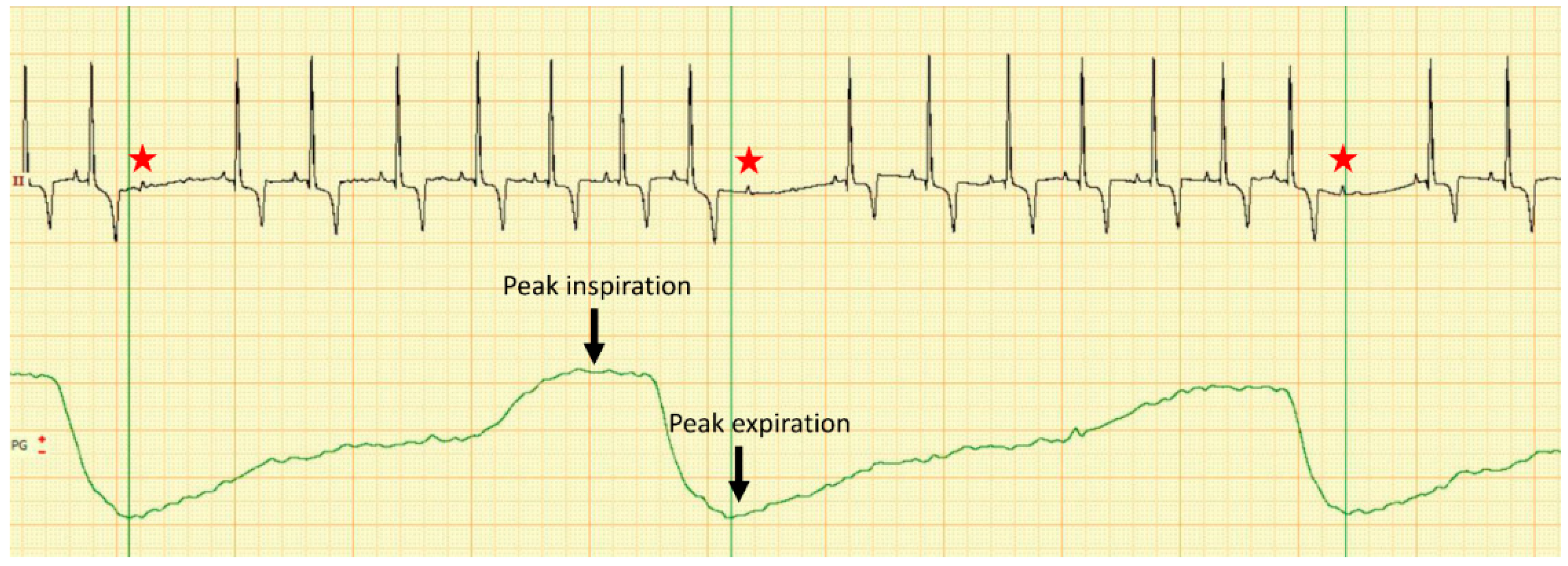
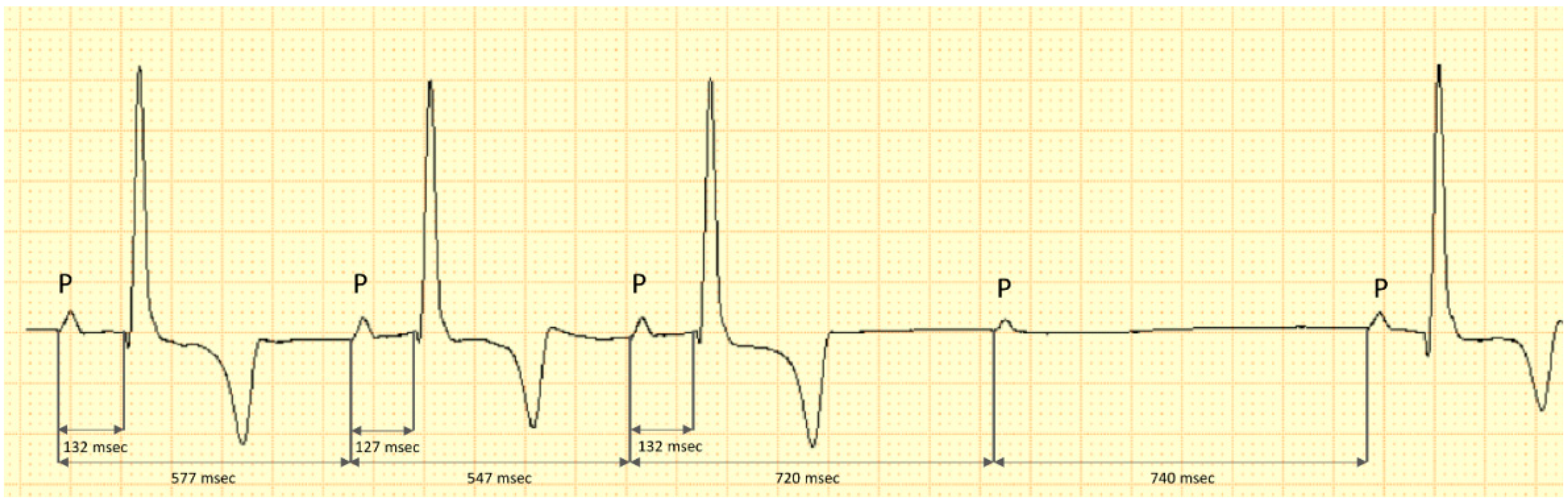
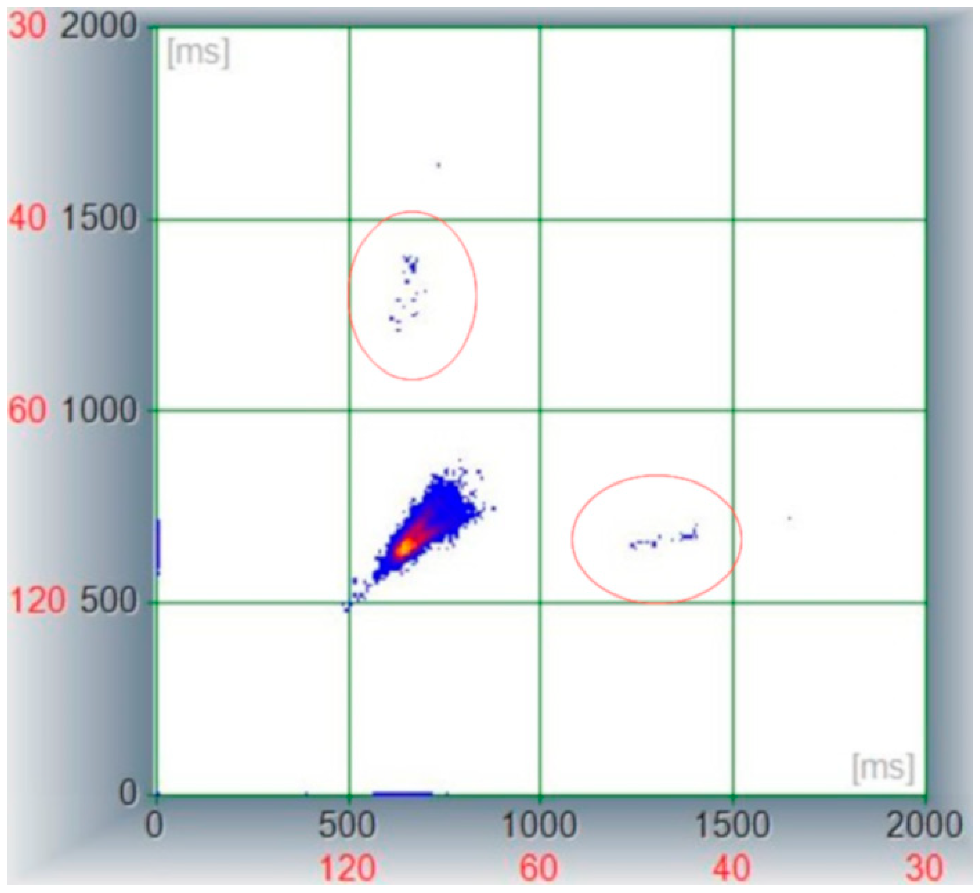
2. Case Description
3. Discussion
4. Conclusions
Author Contributions
Funding
Institutional Review Board Statement
Informed Consent Statement
Data Availability Statement
Conflicts of Interest
References
- Oehnell, R.F.; Andersson, B. A-V-block with Wenckebach periods; periodicity related to the respiratory movements. Cardiologia 1947, 12, 316–318. [Google Scholar] [CrossRef] [PubMed]
- Katoh, T.; Kinoshita, S.; Ueji, I.; Sasaki, Y.; Tsujimura, Y. Apparent bradycardia-dependent advanced second-degree atrioventricular block. J. Electrocardiol. 2002, 35, 153–158. [Google Scholar] [CrossRef] [PubMed]
- Hoffmann, D.A.; Atamanuk, A.N.; Ruiz, G.A.; Chirife, R.; Tentori, M.C.; Salzberg, S. Symptomatic, Inspiration-Induced Atrioventricular Block: Treated with Pacemaker Implant. Pacing Clin. Electrophysiol. PACE 2017, 40, 900–903. [Google Scholar] [CrossRef] [PubMed]
- Santilli, R.A.; Perego, M. Electrocardiography of the Dog and Cat; EDRA LSWR: Milano, Italy, 2014. [Google Scholar]
- Alboni, P.; Holz, A.; Brignole, M. Vagally mediated atrioventricular block: Pathophysiology and diagnosis. Heart 2013, 99, 904–908. [Google Scholar] [CrossRef]
- Suggs, L.D.; Tonnessen, J.B.; Pavri, B.B. Differential Effects of Vagal Activation on the Sinus and Atrioventricular Nodes: Report of 2 Cases. JACC Case Rep. 2020, 2, 1748–1752. [Google Scholar] [CrossRef]
- Thomas, W.P.; Gaber, C.E.; Jacobs, G.J.; Kaplan, P.M.; Lombard, C.W.; Moise, N.S.; Moses, B.L. Recommendations for standards in transthoracic two-dimensional echocardiography in the dog and cat. Echocardiography Committee of the Specialty of Cardiology, American College of Veterinary Internal Medicine. J. Vet. Intern. Med. 1993, 7, 247–252. [Google Scholar] [CrossRef] [PubMed]
- Boswood, A.; Haggstrom, J.; Gordon, S.G.; Wess, G.; Stepien, R.L.; Oyama, M.A.; Keene, B.W.; Bonagura, J.; MacDonald, K.A.; Patteson, M.; et al. Effect of Pimobendan in Dogs with Preclinical Myxomatous Mitral Valve Disease and Cardiomegaly: The EPIC Study-A Randomized Clinical Trial. J. Vet. Intern. Med. Am. Coll. Vet. Intern. Med. 2016, 30, 1765–1779. [Google Scholar] [CrossRef]
- Marchesotti, F.; Vezzosi, T.; Tognetti, R.; Marchetti, F.; Patata, V.; Contiero, B.; Zini, E.; Domenech, O. Left atrial anteroposterior diameter in dogs: Reference interval, allometric scaling, and agreement with the left atrial-to-aortic root ratio. J. Vet. Med. Sci. Jpn. Soc. Vet. Sci. 2019, 81, 1655–1662. [Google Scholar] [CrossRef] [Green Version]
- Cornell, C.C.; Kittleson, M.D.; Della Torre, P.; Haggstrom, J.; Lombard, C.W.; Pedersen, H.D.; Vollmar, A.; Wey, A. Allometric scaling of M-mode cardiac measurements in normal adult dogs. J. Vet. Intern. Med. Am. Coll. Vet. Intern. Med. 2004, 18, 311–321. [Google Scholar] [CrossRef]
- IRIS. IRIS Staging of CKD Modified 2019. Available online: http://www.iris-kidney.com/guidelines/staging.html (accessed on 16 February 2022).
- Tarvainen, M.P.; Niskanen, J.P.; Lipponen, J.A.; Ranta-Aho, P.O.; Karjalainen, P.A. Kubios HRV--heart rate variability analysis software. Comput. Methods Programs Biomed. 2014, 113, 210–220. [Google Scholar] [CrossRef]
- Bogucki, S.; Noszczyk-Nowak, A. Short-term heart rate variability (HRV) in healthy dogs. Pol. J. Vet. Sci. 2015, 18, 307–312. [Google Scholar] [CrossRef] [PubMed]
- Romito, G.; Guglielmini, C.; Poser, H.; Baron Toaldo, M. Lorenz Plot Analysis in Dogs with Sinus Rhythm and Tachyarrhythmias. Animals 2021, 11, 1645. [Google Scholar] [CrossRef] [PubMed]
- Moise, N.S.; Flanders, W.H.; Pariaut, R. Beat-to-Beat Patterning of Sinus Rhythm Reveals Non-linear Rhythm in the Dog Compared to the Human. Front. Physiol. 2019, 10, 1548. [Google Scholar] [CrossRef] [PubMed]
- Quarti-Trevano, F.; Seravalle, G.; Dell’Oro, R.; Mancia, G.; Grassi, G. Autonomic Cardiovascular Alterations in Chronic Kidney Disease: Effects of Dialysis, Kidney Transplantation, and Renal Denervation. Curr. Hypertens. Rep. 2021, 23, 10. [Google Scholar] [CrossRef] [PubMed]
- Ranpuria, R.; Hall, M.; Chan, C.T.; Unruh, M. Heart rate variability (HRV) in kidney failure: Measurement and consequences of reduced HRV. Nephrol. Dial. Transplant. Off. Publ. Eur. Dial. Transpl. Assoc. Eur. Ren. Assoc. 2008, 23, 444–449. [Google Scholar] [CrossRef] [PubMed] [Green Version]
- Vita, G.; Bellinghieri, G.; Trusso, A.; Costantino, G.; Santoro, D.; Monteleone, F.; Messina, C.; Savica, V. Uremic autonomic neuropathy studied by spectral analysis of heart rate. Kidney Int. 1999, 56, 232–237. [Google Scholar] [CrossRef] [Green Version]
- Saftencu, P.M.; Mocanu, D.; Baisan, R.A.; Solcan, G.; Musteață, M. Heart rate variability in dogs with chronic kidney failure. In Proceedings of the Research Communications of the 26th ECVIM-CA Congress, Goteborg, Sweden, 8–10 September 2016; p. 249. [Google Scholar]
- Alfonso, A.; Le Sueur, A.N.V.; Geraldes, S.S.; Guimaraes-Okamoto, P.T.C.; Tsunemi, M.H.; Santana, D.F.; Ribeiro, V.R.F.; Melchert, A.; Chiacchio, S.B.; Lourenco, M.L.G. Heart Rate Variability and Electrocardiographic Parameters Predictive of Arrhythmias in Dogs with Stage IV Chronic Kidney Disease Undergoing Intermittent Haemodialysis. Animals 2020, 10, 1829. [Google Scholar] [CrossRef]
- Perez-Riera, A.R.; Barbosa-Barros, R.; Daminello-Raimundo, R.; de Abreu, L.C.; Nikus, K. Current aspects of the basic concepts of the electrophysiology of the sinoatrial node. J. Electrocardiol. 2019, 57, 112–118. [Google Scholar] [CrossRef]
- Berntson, G.G.; Bigger, J.T., Jr.; Eckberg, D.L.; Grossman, P.; Kaufmann, P.G.; Malik, M.; Nagaraja, H.N.; Porges, S.W.; Saul, J.P.; Stone, P.H.; et al. Heart rate variability: Origins, methods, and interpretive caveats. Psychophysiology 1997, 34, 623–648. [Google Scholar] [CrossRef]
- Vanderlei, L.C.; Pastre, C.M.; Hoshi, R.A.; Carvalho, T.D.; Godoy, M.F. Basic notions of heart rate variability and its clinical applicability. Rev. Bras. Cir. Cardiovasc. 2009, 24, 205–217. [Google Scholar] [CrossRef] [Green Version]
- Dikow, R.; Kihm, L.P.; Zeier, M.; Kapitza, J.; Tornig, J.; Amann, K.; Tiefenbacher, C.; Ritz, E. Increased infarct size in uremic rats: Reduced ischemia tolerance? J. Am. Soc. Nephrol. JASN 2004, 15, 1530–1536. [Google Scholar] [CrossRef] [PubMed]
- Dikow, R.; Hardt, S.E. The uremic myocardium and ischemic tolerance: A world of difference. Circulation 2012, 125, 1215–1216. [Google Scholar] [CrossRef] [PubMed] [Green Version]
- Orii, M.; Hirata, K.; Tanimoto, T.; Ota, S.; Shiono, Y.; Yamano, T.; Matsuo, Y.; Ino, Y.; Yamaguchi, T.; Kubo, T.; et al. Comparison of cardiac MRI and 18F-FDG positron emission tomography manifestations and regional response to corticosteroid therapy in newly diagnosed cardiac sarcoidosis with complet heart block. Heart Rhythm 2015, 12, 2477–2485. [Google Scholar] [CrossRef] [PubMed]
- Shaffer, F.; Ginsberg, J.P. An Overview of Heart Rate Variability Metrics and Norms. Front. Public Health 2017, 5, 258. [Google Scholar] [CrossRef] [Green Version]


| HRV Measurements | Resulted Values | Reference Ranges [13] | |
|---|---|---|---|
| Time domain | SDNN (ms) | 54 | 208.86 ± 77.1 |
| rMSSD (ms) | 46 | 259 ± 120.17 | |
| pNN50% | 5.06 | 71.84 ± 13.96 | |
| Frequency domain | LF band (ms2) | 212 | 1501.24 ± 736.32 |
| HF band (ms2) | 704 | 5845.45 ± 2914.20 | |
| LF/HF | 0.3 | 0.28 ± 0.11 |
Publisher’s Note: MDPI stays neutral with regard to jurisdictional claims in published maps and institutional affiliations. |
© 2022 by the authors. Licensee MDPI, Basel, Switzerland. This article is an open access article distributed under the terms and conditions of the Creative Commons Attribution (CC BY) license (https://creativecommons.org/licenses/by/4.0/).
Share and Cite
Baisan, R.A.; Turcu, A.C.; Condurachi, E.I.; Vulpe, V. Vagally Associated Second Degree Atrio-Ventricular Block in a Dog with Severe Azotemia and Evidence of Sympathetic Overdrive. Vet. Sci. 2022, 9, 223. https://0-doi-org.brum.beds.ac.uk/10.3390/vetsci9050223
Baisan RA, Turcu AC, Condurachi EI, Vulpe V. Vagally Associated Second Degree Atrio-Ventricular Block in a Dog with Severe Azotemia and Evidence of Sympathetic Overdrive. Veterinary Sciences. 2022; 9(5):223. https://0-doi-org.brum.beds.ac.uk/10.3390/vetsci9050223
Chicago/Turabian StyleBaisan, Radu Andrei, Andreea Cătălina Turcu, Eusebiu Ionuț Condurachi, and Vasile Vulpe. 2022. "Vagally Associated Second Degree Atrio-Ventricular Block in a Dog with Severe Azotemia and Evidence of Sympathetic Overdrive" Veterinary Sciences 9, no. 5: 223. https://0-doi-org.brum.beds.ac.uk/10.3390/vetsci9050223