Published online Jan 28, 2013. doi: 10.3748/wjg.v19.i4.604
Revised: December 14, 2012
Accepted: December 25, 2012
Published online: January 28, 2013
Emphysematous cholecystitis is a rare variant of acute cholecystitis with a high mortality rate. The combination of emphysematous cholecystitis and pneumoperitoneum is also rare. We herein describe a case of emphysematous cholecystitis with massive gas in the abdominal cavity. A 77-year-old male presented with epigastric pain and lassitude lasting for one week. A computed tomography scan demonstrated massive gas in the abdominal cavity. Gas was also detectable inside the gallbladder. Massive ascites as well as a pleural effusion were also detected. Under the diagnosis of perforation of the digestive tract, we performed emergency surgery. Beyond our expectations, the perforation site was not in the alimentary tract, but rather in the gallbladder. We then diagnosed the patient with emphysematous cholecystitis with perforation, and performed cholecystectomy. A pathological examination of the resected gallbladder revealed necrosis in the mucosa and thinning of the wall. Cultures of the ascites detected Clostridium perfringens, a gas-producing microorganism.
- Citation: Miyahara H, Shida D, Matsunaga H, Takahama Y, Miyamoto S. Emphysematous cholecystitis with massive gas in the abdominal cavity. World J Gastroenterol 2013; 19(4): 604-606
- URL: https://www.wjgnet.com/1007-9327/full/v19/i4/604.htm
- DOI: https://dx.doi.org/10.3748/wjg.v19.i4.604
Emphysematous cholecystitis is a type of acute cholecystitis characterized by the presence of intramural and/or intraluminal gas that may develop into gangrene or perforation of the gallbladder. The morbidity and mortality rates of emphysematous cholecystitis are considerable[1]. The disease begins with acute cholecystitis followed by ischemia or gangrene of the gallbladder wall and an infection caused by gas-producing bacteria.
Emphysematous cholecystitis is an uncommon variant of acute cholecystitis. Emphysematous cholecystitis occurring in association with a pneumoperitoneum is very rare. Modini et al[2] reported the 16th case of emphysematous cholecystitis with a pneumoperitoneum in the English-language literature in 2008. Thereafter, only one case was reported[3]. We herein report the 18th known case. What is of much note is that, among these cases, the finding of macroscopic perforation of the gallbladder was made in only eight patients[4].
This report presents a case of emphysematous cholecystitis causing a pneumoperitoneum with the finding of macroscopic perforation of the gallbladder.
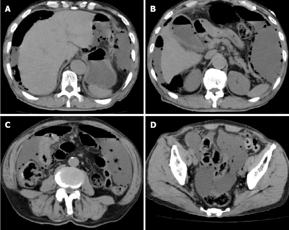
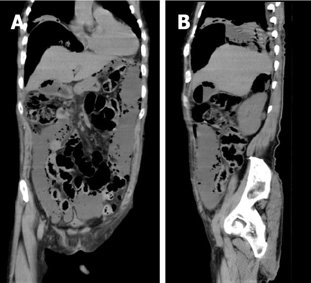
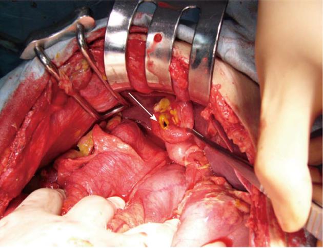
A 77-year-old male was transported to our hospital in September 2011 because he was found falling down in his home with epigastric pain and lassitude lasting for one week. There was nothing particular in the patient’s history. His vital signs were as follows: pulse: 115 beats/min, blood pressure: 151/86 mmHg, respirations: 36 breaths/min, saturation: 92% on room air, and temperature: 37.7 °C. The patient’s abdomen was distended, and there was local tenderness in the upper abdomen without muscular defense. A laboratory examination showed the following values: leukocyte count: 16.6 × 10³/μL, hemoglobin: 14.4 g/dL, hematocrit: 42.4%, platelet count: 16.5 ×10⁴/μL; serum values: sodium: 130 mEq/L, potassium: 3.8 mEq/L, blood urea nitrogen: 88 mg/dL, creatinine: 3.6 mg/dL, alkaline phosphatase: 251 U/L, lactic dehydrogenase: 371 U/L, aspirate aminotransferase: 32 U/L, alanine aminotransferase: 25 U/L, total bilirubin: 0.4 mg/dL, γ-glutamyl trans ferase: 28 U/L, glucose: 207 mg/dL. Plain abdominal radiography showed the presence of intestinal gas. A computed tomography (CT) scan demonstrated massive gas in the abdominal cavity (Figure 1). Gas was also detectable inside the gallbladder (Figures 1B and 2). Massive ascites as well as a pleural effusion were also detected (Figure 1C and D). Under the diagnosis of perforation of the digestive tract, emergency surgery was performed. In the abdominal cavity, there was massive yellow-brown purulent ascites that did not include any saburra. Contrary to our expectations, the perforation site was not in the alimentary tract, but rather in the gallbladder (Figure 3). We therefore diagnosed the patient to have emphysematous cholecystitis with perforation, and performed cholecystectomy. No gallstones were detected in the gallbladder. Tazobactam/piperacillin was given pre- and post-operation. The patient’s postoperative course was uneventful, and he was discharged healthy 28 d after undergoing surgery. Cultures of the ascites detected Clostridium perfringens. A pathological examination of the resected gallbladder revealed necrosis in the mucosa and thinning of the wall.
Emphysematous cholecystitis is a type of acute cholecystitis characterized by the presence of gas in the gallbladder wall. The disease begins with acute cholecystitis followed by ischemia or gangrene of the gallbladder wall and infection caused by gas-producing bacteria. Whereas the mortality rate of uncomplicated acute cholecystitis is approximately 1.4%, that of acute emphysematous cholecystitis is 15%-20% due to the increased incidence of gallbladder wall gangrene and perforation[5]. Therefore, prompt diagnosis and treatment are essential. The most common symptoms are right upper quadrant pain, low-grade fever, nausea and vomiting. Peritoneal signs may be present, and masses in the right upper quadrant may be palpated in as many as half of patients. CT scanning is the most sensitive test for detecting emphysematous cholecystitis. The presence of gas within the gallbladder wall and lumen is easily confirmed on CT scans.
Emphysematous cholecystitis is more common in males than females (7:3), and 40% of affected patients have diabetes mellitus. In our case, there was no history of diabetes mellitus. After the operation, his blood sugar was decreased down to a normal level, and HbA1c was within normal range.
The presence of a concomitant pneumoperitoneum, which may occur following gallbladder perforation, is rarely found. Most patients with a concomitant pneumoperitoneum are in unstable condition. Therefore, the first choice of treatment in such cases is emergency exploratory laparotomy, followed by cholecystectomy, under a correct intraoperative diagnosis. Another method of treatment, involves initial percutaneous cholecystostomy with a strict intravenous antibiotics regimen, followed by subsequent cholecystectomy during a second stage[4]. In severely ill patients in particular, percutaneous cholecystostomy with broad-spectrum antibiotics may be an alternative choice of treatment[5]. In our case, we did not diagnose the patient with emphysematous cholecystitis preoperatively due to the huge amounts of gas in the abdominal cavity. Compared with previous cases, the amount of gas in our case was very large, thus suggesting not an acute stage, but a sub-acute stage and continuous infection with gas-producing bacteria. If the correct diagnosis could be done preoperatively, laparoscopic surgery may be the alternative treatment.
The bacteria most frequently cultured in this setting include anaerobes, such as Clostridia welchii or Clostridia perfringens. The bacteria second most frequently cultured include aerobes, such as Escherichia coli[5]. In our case, cultures of the ascites detected Clostridium perfringens. Zeebregts et al[4] reported that, in six of 14 cases, Clostridium perfringens was detected on cultures.
In conclusion, we herein reported a case presenting with a very large amount of gas in the abdominal cavity due to gallbladder perforation. We first suspected the possibility of upper gastrointestinal perforation and performed emergency surgery. However, we found a perforation not in the gastrointestinal tract, but rather in the gallbladder. Pneumoperitoneums are nearly always due to perforation of the gastrointestinal tract, however, although unusual, they may also be caused by emphysematous cholecystitis.
P- Reviewers Aurello P, Toso C, Foelsch UR S- Editor Gou SX L- Editor A E- Editor Xiong L
| 1. | Shrestha Y, Trottier S. Images in clinical medicine. Emphysematous cholecystitis. N Engl J Med. 2007;357:1238. [PubMed] [Cited in This Article: ] |
| 2. | Modini C, Clementi I, Simonelli L, Antoniozzi A, Assenza M, Ciccarone F, Bartolucci P, Ricci G, Petroni R. Acute emphysematous cholecystitis as a cause of pneumoperitoneum. Chir Ital. 2008;60:315-318. [PubMed] [Cited in This Article: ] |
| 3. | Kanehiro T, Tsumura H, Ichikawa T, Hino Y, Murakami Y, Sueda T. Patient with perforation caused by emphysematous cholecystitis who showed flare on the skin of the right dorsal lumbar region and intraperitoneal free gas. J Hepatobiliary Pancreat Surg. 2008;15:204-208. [PubMed] [DOI] [Cited in This Article: ] [Cited by in Crossref: 9] [Cited by in F6Publishing: 10] [Article Influence: 0.6] [Reference Citation Analysis (0)] |
| 4. | Zeebregts CJ, Wijffels RT, de Jong KP, Peeters PM, Slooff MJ. Percutaneous drainage of emphysematous cholecystitis associated with pneumoperitoneum. Hepatogastroenterology. 1999;46:771-774. [PubMed] [Cited in This Article: ] |
| 5. | Wu JM, Lee CY, Wu YM. Emphysematous cholecystitis. Am J Surg. 2010;200:e53-e54. [PubMed] [DOI] [Cited in This Article: ] [Cited by in Crossref: 18] [Cited by in F6Publishing: 20] [Article Influence: 1.4] [Reference Citation Analysis (0)] |


