Published online Feb 16, 2012. doi: 10.4253/wjge.v4.i2.28
Revised: January 16, 2012
Accepted: February 6, 2012
Published online: February 16, 2012
The small bowel has long been considered a black box for endoscopists because of its long length and the presence of multiple complex loop. Most of the small bowel is inaccessible by traditional endoscopic means. In addition, radiographic studies have significant limitations with regard to diagnostic yield, and surgery is an invasive alternative. This limitation was overcome through the development of balloon enteroscopy that becomes established throughout the world for diagnostic and therapeutic examinations of the small bowel. The single-balloon enteroscope (SBE) system (Olympus, Tokyo, Japan) was introduced into the commercial market in 2007. Several study demonstrated its efficacy and safety. Early reports on the use of single-balloon enteroscopy have suggested a high diagnostic yield and similar therapeutic potential to that of the double-balloon endoscope. SBE is viable technique for in the management of small bowel disease. Technically, it is easy to perform, may be efficient, and in the literature data available, seems to provide high diagnostic and therapeutic yield.
- Citation: Manno M, Barbera C, Bertani H, Manta R, Mirante VG, Dabizzi E, Caruso A, Pigo F, Olivetti G, Conigliaro R. Single balloon enteroscopy: Technical aspects and clinical applications. World J Gastrointest Endosc 2012; 4(2): 28-32
- URL: https://www.wjgnet.com/1948-5190/full/v4/i2/28.htm
- DOI: https://dx.doi.org/10.4253/wjge.v4.i2.28
The small bowel has long been considered a black box for endoscopists because of its long length and the presence of multiple complex loop. Endoscopy using standard gastroscopes can reach up to the second or third portion of the duodenum while push enteroscopy can reach the ligament of Treitz and approximately about 80 cm beyond. Colonoscopy can reach 10 cm to 20 cm beyond the ileocecal valve. Most nonsurgical endoscopic techniques were unsatisfactory, and managing small-bowel diseases often required surgical intraoperative enteroscopy. Thus, the development of capsule endoscopy (CE) and double-balloon enteroscopy (DBE) has permitted the observation of the entire small bowel. The diagnostic yield of the balloon-enteroscopy for relevant pathologic findings can be of 70%-80%; in addition, it allows histologic sampling and endoscopic therapy that it can be performed in more than 50% of patients.
The single-balloon enteroscope (SBE) system (Olympus, Tokyo, Japan) was developed in 2006 and was introduced into the commercial market in 2007. It is easier to use respect to double-balloon enteroscope, avoiding attaching the enteroscope balloon to the distal tip of the scope encountered and the requirement of inflating and deflating two balloons.
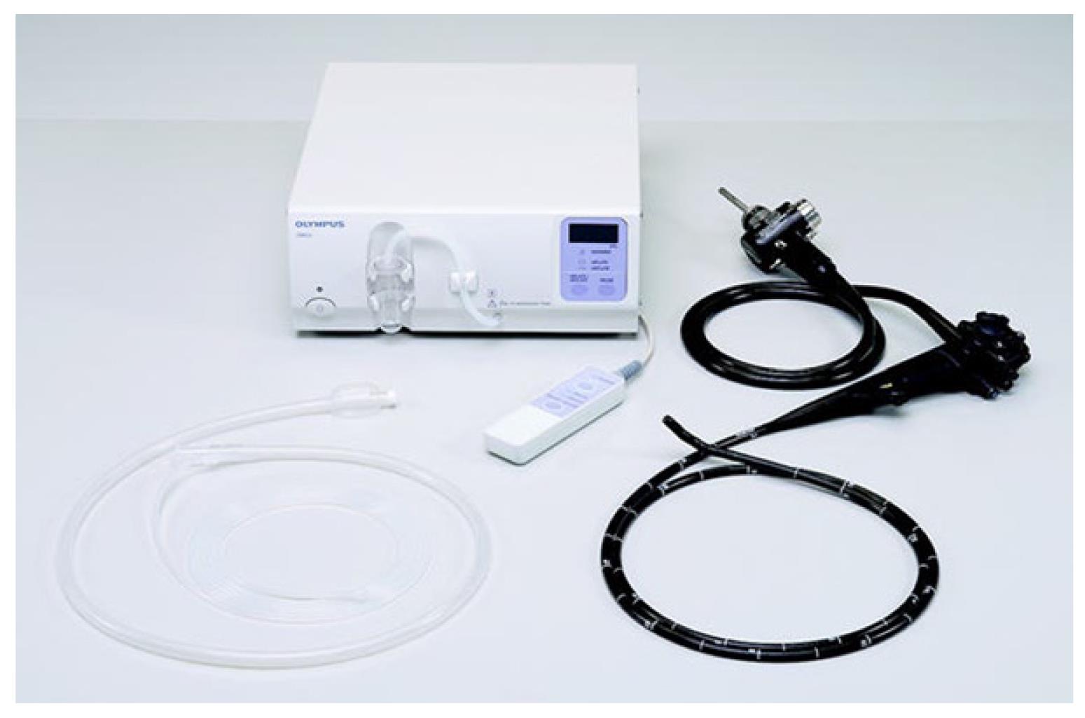
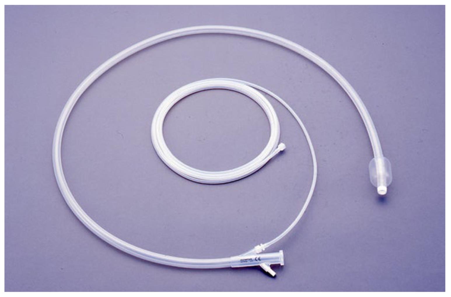
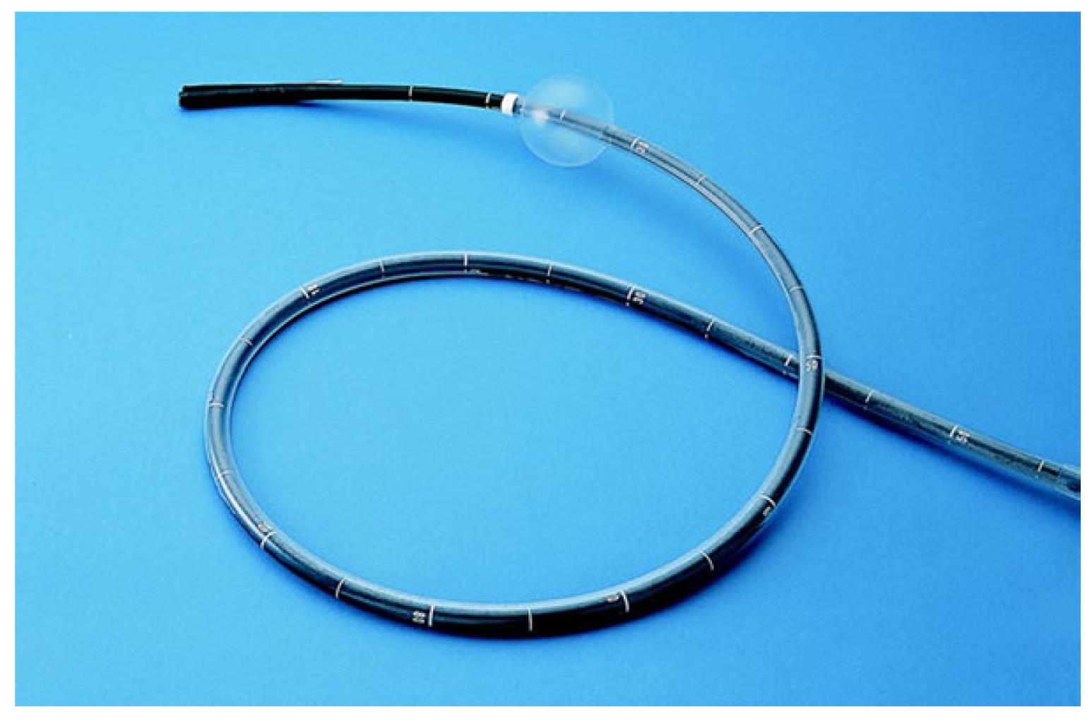
The single-balloon enteroscope system consists of the SIF-Q180 enteroscope, an overtube balloon control unit (OBCU Olympus Balloon Control Unit) and a disposable silicone splinting tube with balloon (ST-SB1) (Figures 1-3).
The enteroscope is a high-resolution video endoscope that works with Olympus EVIS processors and EVIS EXERA II system. The outer diameter is of 9.2 mm with working length of 2000 mm, while the operating channel size is of 2.8 mm.
The splinting tube is an overtube with an inflatable balloon fixed to the distal, radiopaque tip, both in latex-free silicone. The inner diameter of the tube is 11 mm, the outer diameter is 13.2 mm, the working length is 1320 mm, and the total length is 1400 mm. The addition of a small amount of water through a small port on the proximal end of the splinting tube activates the hydrophilic coating avoiding friction between the overtube and the enteroscope. Additional water can be flushed into a dedicated port throughout the procedure, in order to reduce friction or to wash away debris collected between the enteroscope and the splinting tube. The balloon is inflated and deflated by a balloon control unit with a safety pressure setting range from -6.0 kPa to +5.4 kPa. The overtube balloon control unit has one button for inflation, one button for deflation, and a third control for the pause/cancel feature.
No bowel preparation is generally recommended in most cases for single-balloon enteroscopy by the oral approach, except a minimum of 12 h fasting while the standard 4 L of a polyethylene glycol (PEG) preparation is used for retrograde approach.
For retrograde SBE, conscious sedation as for colonoscopy is sufficient in most cases. For anterograde approach deep monitored sedation with propofol or general anesthesia with intubation is recommended[1].
Because of length of the procedure, large volumes of air are usually insufflated that can lead to failure of the procedure. Carbon dioxide (CO2), unlike standard air, is rapidly absorbed from the bowel. A randomized, double blind trial showed that insufflation with CO2 is safe, reduces patient discomfort, and significantly improves intubation depth[2,3].
Fluoroscopy can be helpful during the initial 10 to 20 SBE cases to observe advancement and reduction of the enteroscope and as an aid to determine when looping is present and how to solve it.
In addition, for some patients with surgically modified anatomy and for those undergoing therapeutic procedures such as dilations, fluoroscopic guidance is recommended.
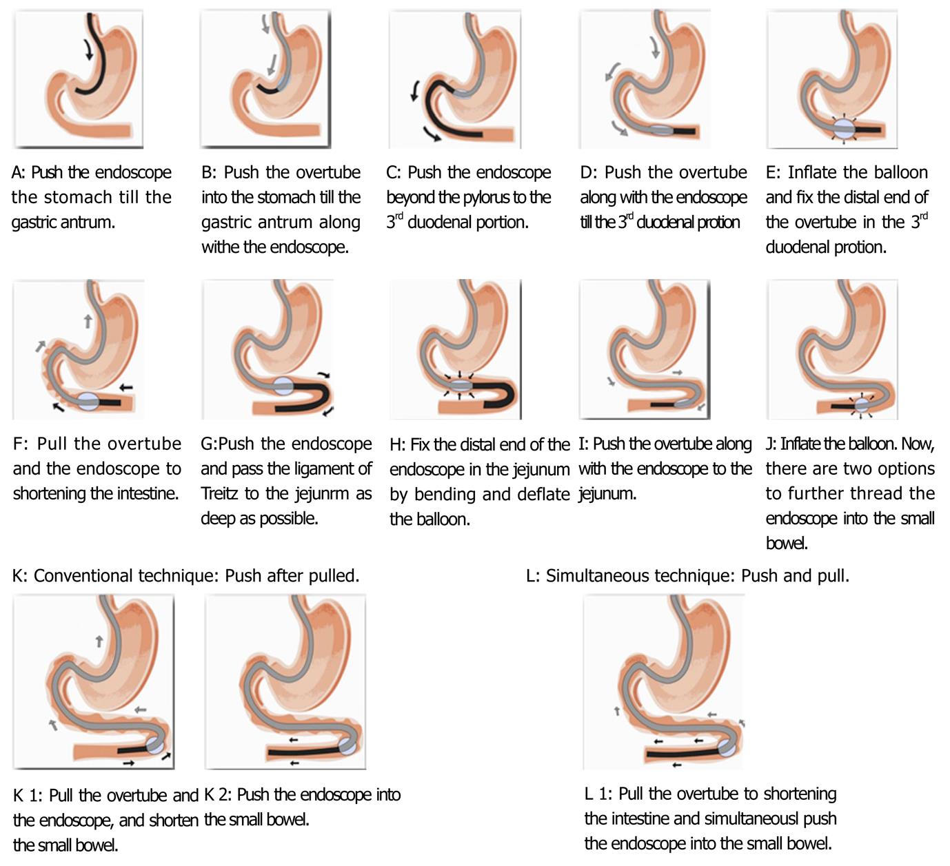
There are two techniques in order to advance the enteroscope into the small bowel that can be alternately used in the same exam. With the “conventional technique”, the enteroscope is initially passed into the esophagus with the same technique used for a standard gastroscopy. The enteroscope is pushed into the duodenum until lacking of forward advancement. By angulating the tip of the enteroscope in order to hook the intestine, the overtube is gently advanced over the enteroscope to the point of maximal insertion, located at 155 cm, where a white line is present on the scope. In order to shorten the intestine and to advance into the small bowel, the endoscope and the overtube, with inflated balloon, are withdrawn together. Then, the endoscope is pushed maximally into the small bowel. The balloon still inflated allows the pushing force of the operator applied to the endoscope to be transmitted to the distal end of the endoscope without further stretching of the intestine. The repetition of these manoeuvres permits progression of the endoscope (Figure 4)[4].
Hartmann and colleagues[5] described an alternative method of single-balloon enteroscopy insertion (“simultaneous” technique) that consists of withdrawing only the inflated overtube in order to shorten the intestine, and simultaneously pushing the endoscope as deep as possible into the small bowel. In a study published by author and his colleagues[6], the alternative technique allowed a lower mean procedure time while the depth of insertion did not differ significantly between the conventional and alternative techniques.
To estimate the depth of insertion, the method proposed by May et al[7] can be used, recording data on a standardized documentation sheet. This method was validated with an animal model during training courses and demonstrated to be accurate. For each advancement of the enteroscope, the distance is added, usually ranging between 20 cm and 40 cm. Any portion of an advancement that is lost during a reduction manoeuvre is then subtracted.
When advancement is no longer possible, it is recommended to mark the area with a tattoo of India ink, injected with a sclerotherapy needle, or to position a clip. This marker can then be visualized during subsequent balloon-assisted enteroscopy performed with the opposite approach.
Once the point of maximal insertion is reached, the enteroscope can be gradually withdrawn. The overtube is gently withdrawn toward the proximal end of the enteroscope without moving the enteroscope. Then, the overtube balloon is inflated and the enteroscope slowly withdrawn until it reaches the distal end of the overtube.
The retrograde approach is a little bit difficult than the oral approach, even for expert endoscopists, with a longer learning curve (20 to 30 cases). It is recommended to start reduction manoeuvres when looping is first appreciated in the colon. Initial attempts at ileocecal valve intubation should occur with the patient in the left lateral or supine positions in order to achieve an ideal location of the ileocecal valve between the 3 and 9 o’clock positions. In case of failure, it is recommended to change patient position[8].
Single-balloon enteroscopy, such as DBE, allow the possibility of suction and flushing via the instrument channel, sampling biopsies, and therapeutic interventions such as argon plasma coagulation, injection, positioning of clips, polypectomy, dilation, and foreign-body extraction, even when inserted distally into the small intestine.
Several study demonstrated its efficacy and safety. Early reports on the use of SBE since its appearance in 2006 involving the prototype XSIF Q160 and its commercially available successor, the SIF Q180, have suggested a high diagnostic yield and similar therapeutic potential to that of the DBE. Ohtsuka and colleagues[9] helped to develop the single-balloon enteroscope in cooperation with Olympus Medical Systems and are credited with the first comparison of single-balloon with double- balloon enteroscopy. They performed 102 procedures in 65 patients with suspected small intestinal disease. Seventy-nine of the procedures were done with the single-balloon enteroscopy system and 11 with double-balloon enteroscopy. Examination time for anterograde insertion was 65.3 min for single-balloon enteroscopy and 74 min for anterograde DBE. Single-balloon enteroscopy retrograde insertion averaged 57.5 min and 56.3 min for retrograde DBE. There were no complications. The investigators concluded it was easy to set up the SBE system and perform the procedure with a single operator. Although not reported, they felt they were able to achieve a high diagnostic yield with the SBE, using their experience with the double-balloon enteroscopy system as a point of reference.
Ramchandani and colleagues[10] studied 60 patients with suspected small bowel disease using the prototype single-balloon enteroscopy system. All patients underwent anterograde examinations; 10 underwent anterograde and retrograde procedures. Mean procedure time was 63 min. The mean depth of insertion was 260 cm beyond the ligament of Treitz. Total enteroscopy was possible in 5 out of 10 cases (50%). Diagnostic yield in cases of obscure gastrointestinal bleeding, chronic abdominal pain, and malabsorption syndrome were 77%, 61%, and 63%, respectively.
Tsujikawa et al[4] evaluated 41 patients using the SBE and found it easy to perform, due to the single balloon, and safe to examine the deep small intestine with useful diagnostic and therapeutic capabilities. Other authors with larger series of patients concluded that SBE demonstrated a high diagnostic yield with the real possibility of useful therapeutic interventions[10-13].
Recently, Domagk et al[14] published a randomized international multicenter study comparing two balloon-assisted enteroscopy systems: DBE vs SBE. A total of 130 patients were included over 12 mo: 65 with DBE and 65 with the SBE technique. Patient and procedure characteristics were comparable between the two groups. Mean oral intubation depth was 253 cm with DBE and 258 cm with SBE, showing non-inferiority of SBE vs DBE. Complete visualization of the small bowel was achieved in 18% and 11% of procedures in the DBE and SBE groups, respectively. Mean anal intubation depth was 107 cm in the DBE group and 118 cm in the SBE group. Diagnostic yield and mean pain scores during and after the procedures were similar in the two groups. No adverse events were observed during or after the examinations. This first head-to-head comparison trial of DBE vs SBE, comparing the Fujinon DBE system with the Olympus SBE system, demonstrated no difference with respect to the insertion depths. Diagnostic yield, rate of complications between the two systems, and patient discomfort scores during and after the procedures were comparable.
In addition, SBE seems to be a safe procedure. However, scrupulous care is required when passing a small-intestinal lesion or in patients with known adhesions or strictures. Relevant complications, i.e., perforation, in diagnostic balloon-assisted enteroscopy can be expected in approximately 1% of cases. As in conventional endoscopy, the risk is higher in therapeutic enteroscopy, at around 3% to 4%[15].
Among the 622 SBE procedures published to date, only two perforations (0.32%) were occurred: one during diagnostic SBE (in a postoperative case of ulcerative colitis), and one during therapeutic enteroscopy (balloon dilation)[4,13,14,16-19].
In contrast to per oral DBE, no acute pancreatitis has been reported following SBE.
SBE is an effective endoscopic tool for the evaluation of the small bowel. Technically, it is easy and safe to perform and it provides similar diagnostic and therapeutic yield when compared with DBE.
Peer reviewers: Nobumi Tagaya, Associate Professor, Second Department of Surgery, Dokkyo Medical University, 880 Kitakobayashi, Mibu, Tochigi 321-0293, Japan; José Luiz Sebba Souza, MD, Clinics Hospital, University of São Paulo, 255 Eneas de Carvalho Ave. 9 th Floor, Room 9159, São Paulo, Brazil
S- Editor Yang XC L- Editor A E- Editor Yang XC
| 1. | Pohl J, Delvaux M, Ell C, Gay G, May A, Mulder CJ, Pennazio M, Perez-Cuadrado E, Vilmann P. European Society of Gastrointestinal Endoscopy (ESGE) Guidelines: flexible enteroscopy for diagnosis and treatment of small-bowel diseases. Endoscopy. 2008;40:609-618. [PubMed] [Cited in This Article: ] |
| 2. | Hirai F, Beppu T, Nishimura T, Takatsu N, Ashizuka S, Seki T, Hisabe T, Nagahama T, Yao K, Matsui T. Carbon dioxide insufflation compared with air insufflation in double-balloon enteroscopy: a prospective, randomized, double-blind trial. Gastrointest Endosc. 2011;73:743-749. [PubMed] [DOI] [Cited in This Article: ] [Cited by in Crossref: 46] [Cited by in F6Publishing: 49] [Article Influence: 3.8] [Reference Citation Analysis (0)] |
| 3. | Domagk D, Bretthauer M, Lenz P, Aabakken L, Ullerich H, Maaser C, Domschke W, Kucharzik T. Carbon dioxide insufflation improves intubation depth in double-balloon enteroscopy: a randomized, controlled, double-blind trial. Endoscopy. 2007;39:1064-1067. [PubMed] [DOI] [Cited in This Article: ] [Cited by in Crossref: 82] [Cited by in F6Publishing: 89] [Article Influence: 5.2] [Reference Citation Analysis (0)] |
| 4. | Tsujikawa T, Saitoh Y, Andoh A, Imaeda H, Hata K, Minematsu H, Senoh K, Hayafuji K, Ogawa A, Nakahara T. Novel single-balloon enteroscopy for diagnosis and treatment of the small intestine: preliminary experiences. Endoscopy. 2008;40:11-15. [PubMed] [Cited in This Article: ] |
| 5. | Hartmann D, Eickhoff A, Tamm R, Riemann JF. Balloon-assisted enteroscopy using a single-balloon technique. Endoscopy. 2007;39 Suppl 1:E276. [PubMed] [Cited in This Article: ] |
| 6. | Manno M, Mussetto A, Conigliaro R. Preliminary results of alternative “simultaneous” technique for single-balloon enteroscopy. Endoscopy. 2008;40:538; author reply 539. [PubMed] [DOI] [Cited in This Article: ] [Cited by in Crossref: 5] [Cited by in F6Publishing: 5] [Article Influence: 0.3] [Reference Citation Analysis (0)] |
| 7. | May A, Nachbar L, Schneider M, Neumann M, Ell C. Push-and-pull enteroscopy using the double-balloon technique: method of assessing depth of insertion and training of the enteroscopy technique using the Erlangen Endo-Trainer. Endoscopy. 2005;37:66-70. [PubMed] [DOI] [Cited in This Article: ] [Cited by in Crossref: 162] [Cited by in F6Publishing: 172] [Article Influence: 9.1] [Reference Citation Analysis (0)] |
| 8. | Mehdizadeh S, Han NJ, Cheng DW, Chen GC, Lo SK. Success rate of retrograde double-balloon enteroscopy. Gastrointest Endosc. 2007;65:633-639. [PubMed] [DOI] [Cited in This Article: ] [Cited by in Crossref: 58] [Cited by in F6Publishing: 60] [Article Influence: 3.5] [Reference Citation Analysis (0)] |
| 9. | Ohtsuka K, Kashida H, Kodama K, Ukegawa J, Kanie H, Mizuno K, Kudo Y, Takemura O, Kudo S. Observation and treatment of small bowel diseases using single balloon endoscope. Gastrointest Endosc. 2008;67:AB271. [Cited in This Article: ] |
| 10. | Ramchandani M, Reddy DN, Gupta R, Lakhtakia S, Tandan M, Rao GV, Darisetty S. Diagnostic yield and therapeutic impact of single-balloon enteroscopy: series of 106 cases. J Gastroenterol Hepatol. 2009;24:1631-1638. [PubMed] [DOI] [Cited in This Article: ] [Cited by in Crossref: 98] [Cited by in F6Publishing: 84] [Article Influence: 5.6] [Reference Citation Analysis (0)] |
| 11. | Frantz DJ, Dellon ES, Grimm IS, Morgan DR. Single-balloon enteroscopy: results from an initial experience at a U.S. tertiary-care center. Gastrointest Endosc. 2010;72:422-426. [PubMed] [DOI] [Cited in This Article: ] [Cited by in Crossref: 39] [Cited by in F6Publishing: 43] [Article Influence: 3.1] [Reference Citation Analysis (0)] |
| 12. | Upchurch BR, Sanaka MR, Lopez AR, Vargo JJ. The clinical utility of single-balloon enteroscopy: a single-center experience of 172 procedures. Gastrointest Endosc. 2010;71:1218-1223. [PubMed] [DOI] [Cited in This Article: ] [Cited by in Crossref: 58] [Cited by in F6Publishing: 62] [Article Influence: 4.4] [Reference Citation Analysis (0)] |
| 13. | Aktas H, de Ridder L, Haringsma J, Kuipers EJ, Mensink PB. Complications of single-balloon enteroscopy: a prospective evaluation of 166 procedures. Endoscopy. 2010;42:365-368. [PubMed] [DOI] [Cited in This Article: ] [Cited by in Crossref: 72] [Cited by in F6Publishing: 76] [Article Influence: 5.4] [Reference Citation Analysis (0)] |
| 14. | Domagk D, Mensink P, Aktas H, Lenz P, Meister T, Luegering A, Ullerich H, Aabakken L, Heinecke A, Domschke W. Single- vs. double-balloon enteroscopy in small-bowel diagnostics: a randomized multicenter trial. Endoscopy. 2011;43:472-476. [PubMed] [DOI] [Cited in This Article: ] [Cited by in Crossref: 126] [Cited by in F6Publishing: 137] [Article Influence: 10.5] [Reference Citation Analysis (0)] |
| 15. | May A. Balloon enteroscopy: single- and double-balloon enteroscopy. Gastrointest Endosc Clin N Am. 2009;19:349-356. [PubMed] [DOI] [Cited in This Article: ] [Cited by in Crossref: 18] [Cited by in F6Publishing: 18] [Article Influence: 1.2] [Reference Citation Analysis (0)] |
| 16. | Kawamura T, Yasuda K, Tanaka K, Uno K, Ueda M, Sanada K, Nakajima M. Clinical evaluation of a newly developed single-balloon enteroscope. Gastrointest Endosc. 2008;68:1112-1116. [PubMed] [DOI] [Cited in This Article: ] [Cited by in Crossref: 107] [Cited by in F6Publishing: 118] [Article Influence: 7.4] [Reference Citation Analysis (0)] |
| 17. | Mensink PB, Haringsma J, Kucharzik T, Cellier C, Pérez-Cuadrado E, Mönkemüller K, Gasbarrini A, Kaffes AJ, Nakamura K, Yen HH. Complications of double balloon enteroscopy: a multicenter survey. Endoscopy. 2007;39:613-615. [PubMed] [DOI] [Cited in This Article: ] [Cited by in Crossref: 261] [Cited by in F6Publishing: 273] [Article Influence: 16.1] [Reference Citation Analysis (0)] |
| 18. | Manno M, Barbera C, Dabizzi E, Mussetto A, Conigliaro R. Safety of single-balloon enteroscopy: our experience of 72 procedures. Endoscopy. 2010;42:773; author reply 774. [PubMed] [DOI] [Cited in This Article: ] [Cited by in Crossref: 12] [Cited by in F6Publishing: 12] [Article Influence: 0.9] [Reference Citation Analysis (0)] |
| 19. | Riccioni ME, Urgesi R, Cianci R, Spada C, Nista EC, Costamagna G. Single-balloon push-and-pull enteroscopy system: does it work? A single-center, 3-year experience. Surg Endosc. 2011;25:3050-3056. [PubMed] [DOI] [Cited in This Article: ] [Cited by in Crossref: 25] [Cited by in F6Publishing: 24] [Article Influence: 1.8] [Reference Citation Analysis (0)] |


