Published online Aug 16, 2012. doi: 10.4253/wjge.v4.i8.356
Revised: June 29, 2012
Accepted: August 8, 2012
Published online: August 16, 2012
The demarcation line between the cancerous lesion and the surrounding area could be easily recognized with flexible spectral imaging color enhancement (FICE) system compared with conventional white light images. The characteristic finding of depressed-type early gastric cancer (EGC) in most cases was revealed as reddish lesions distinct from the surrounding yellowish non-cancerous area without magnification. Conventional endoscopic images provide little information regarding depressed lesions located in the tangential line, but FICE produces higher color contrast of such cancers. Histological findings in depressed area with reddish color changes show a high density of glandular structure and an apparently irregular microvessel in intervening parts between crypts, resulting in the higher color contrast of FICE image between cancer and surrounding area. Some depressed cancers are shown as whitish lesion by conventional endoscopy. FICE also can produce higher color contrast between whitish cancerous lesions and surrounding atrophic mucosa. For nearly flat cancer, FICE can produce an irregular structural pattern of cancer distinct from that of the surrounding mucosa, leading to a clear demarcation. Most elevated-type EGCs are detected easily as yellowish lesions with clearly contrasting demarcation. In some cases, a partially reddish change is accompanied on the tumor surface similar to depressed type cancer. In addition, the FICE system is quite useful for the detection of minute gastric cancer, even without magnification. These new contrasting images with the FICE system may have the potential to increase the rate of detection of gastric cancers and screen for them more effectively as well as to determine the extent of EGC.
- Citation: Osawa H, Yamamoto H, Miura Y, Yoshizawa M, Sunada K, Satoh K, Sugano K. Diagnosis of extent of early gastric cancer using flexible spectral imaging color enhancement. World J Gastrointest Endosc 2012; 4(8): 356-361
- URL: https://www.wjgnet.com/1948-5190/full/v4/i8/356.htm
- DOI: https://dx.doi.org/10.4253/wjge.v4.i8.356
Modest changes in morphology and color of the mucosa are important factors for the diagnosis of early gastric cancer (EGC). The morphological characteristics of EGC include mild elevation and shallow depression of the mucosa, as well as discontinuity with surrounding mucosa and areas of uneven surface. For changes in color, pale redness or fading is important. However, these endoscopic features have not been sufficient to achieve an accurate diagnosis for EGC. The flexible spectral imaging color enhancement (FICE) system was developed as a selection system of the narrow band image and introduced in 2005 as a novel image-processing tool for video endoscopy[1-4]. FICE enhances the contrast of mucosal surface without the use of dyes. Additionally, FICE provides optimal band image with the same light intensity as conventional endoscope, implying that the FICE system can display clear images even without magnification. Indeed, FICE can facilitate detection of changes in EGC without magnification and confirm accurately the diagnosis of cancer with low or half magnification[5-7].
Endoscopic submucosal dissection (ESD) was developed in the past 10 years as a more reliable method of endoscopic resection than endoscopic mucosal resection (EMR)[8-10]. In Japan, ESD has now been officially approved as an endoscopic treatment for EGC, and the mainstream of therapy for EGC has shifted from EMR to ESD. However, the accurate diagnosis of extent of gastric cancer is needed to achieve complete resection by ESD. FICE is very useful for the diagnosis of extent without or with magnification.
In this report, advance in endoscopic diagnosis for the extent of EGCs will be reviewed particularly focusing on FICE images.
FICE was developed with the aim of enhancing the capillary pattern and the pit patterns in endoscopic images. FICE technology is based on the selection of spectral transmittance with a dedicated wavelength. In contrast to narrow band imaging, in which the bandwidth of the spectral transmittance is narrowed by optical filters, FICE is based on a new spectral estimation technique that eliminates the need for optical filters. FICE takes an ordinary endoscopic image from a video processor and arithmetically processes the reflected photons to reconstitute virtual images for a choice of different wavelengths. Because the spectra of pixels are known, it is possible to perform imaging on a single wavelength. Such single-wavelength images are randomly selected, and assigned to red (R), green (G), and blue (B) to build and display an FICE-enhanced color image.
Endoscopes used with FICE system include EG-590ZW for routine and magnifying observation, EG-590WR for routine observation and EG-530 NW for transnasal observation, all of which are developed by Fujifilm Corporation (Kanagawa, Japan) for the upper gastrointestinal tract and need an electronic endoscope system (FTS4400 and 4450, Fujifilm). The EG-590ZW scope can magnify endoscopic images optically up to 135-fold[5-7] through the use of a zoom attachment. It is easy to change wavelengths in each endoscopic procedure, because the system allows selection of a setting from up to ten possible settings. Osawa et al[5] and Yoshizawa et al[6,7] at the Jichi Medical University in Japan selected the best setting that enhances the demarcation lines more strongly between cancerous lesions and surrounding areas in the case of EGC without magnification. For most cases of gastric cancer, the best images were obtained with use of a specific combination of the following three wavelengths: 470 nm for blue (B), 500 nm for green (G), and 550 nm for red (R) that also produces a brighter image of various types of gastric cancers, that is, depressed, flat and elevated type.
Chromoendoscopy is often carried out by spraying dyes such as indigo carmine after thoroughly washing out the mucus and is useful to determine the extent of the tumor by clearer detection of the morphological feature of depressed or protruded cancerous margin. By contrast, FICE is often carried out not only by such detection of morphological feature but also higher color contrast between cancer and surrounding atrophic mucosa. Therefore, tumor margin may be easily identified in FICE images even without magnification.
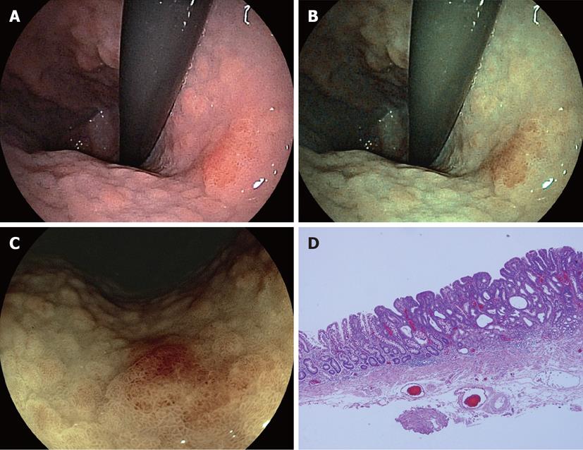
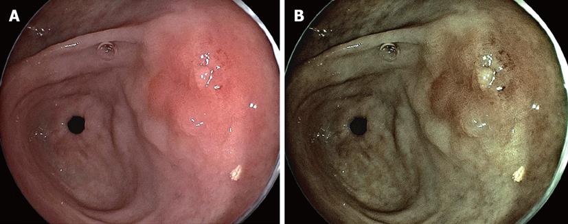
The characteristic finding of depressed-type EGC in most cases was revealed as reddish lesions distinct from the surrounding yellowish non-cancerous area without magnification. Compared with conventional white light images, the demarcation line between the cancerous lesion and the surrounding area could be easily recognized with FICE system (Figure 1). Conventional endoscopic images provide little information regarding depressed lesions located in the tangential line, but FICE produces higher color contrast of such cancers, even with the small caliber-size scope for transnasal route[11] (EG 530NW) (Figure 1A and B). Histological findings in depressed area with reddish color changes show a high density of glandular structure and an apparently irregular microvessel in intervening parts between crypts. Such histological features differ from those of surrounding mucosa and may cause higher color contrast of FICE images between them.
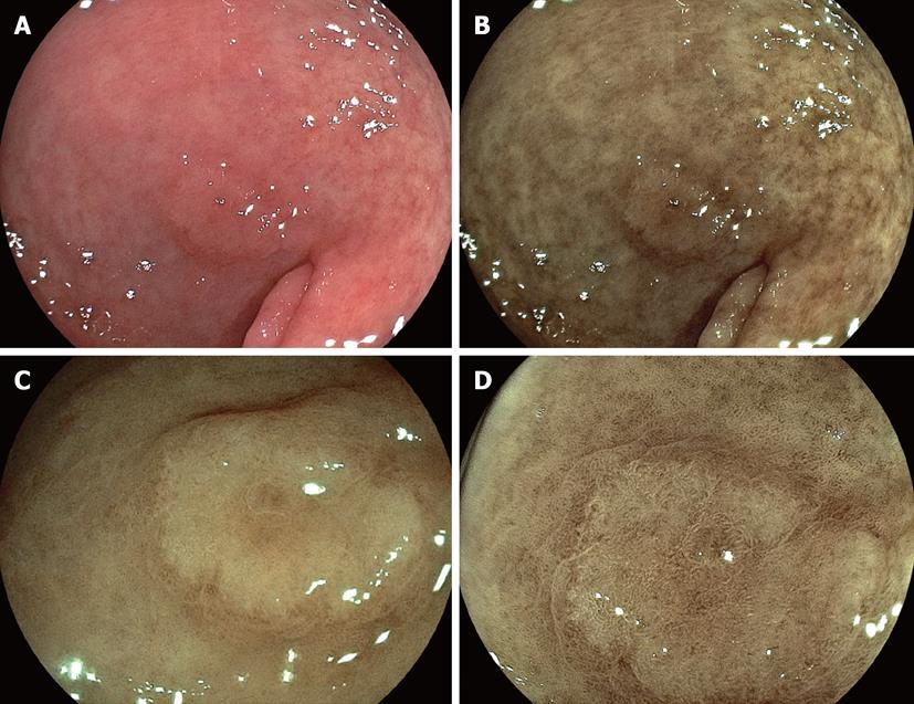
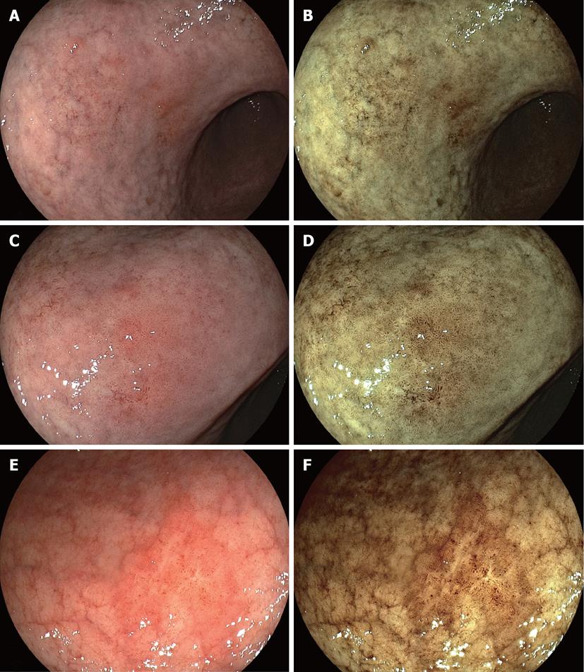
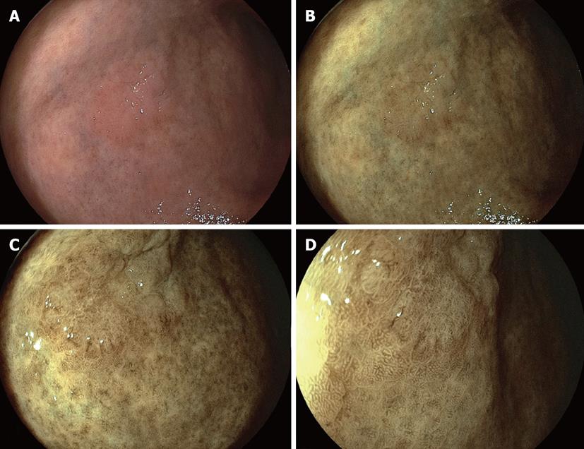
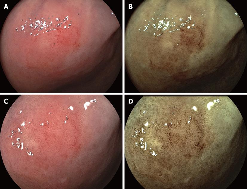
Some depressed cancers are shown as whitish lesion by conventional endoscopy (Figure 2). FICE also can produce higher color contrast between whitish cancerous lesions and surrounding atrophic mucosa. For nearly flat cancer, FICE can produce an irregular structural pattern of cancer distinct from that of the surrounding mucosa, leading to a clear demarcation (Figure 3). On the other hand, most elevated-type EGCs are detected easily as yellowish lesions with clearly contrasting demarcation (Figure 4). In some cases, a partially reddish change is accompanied on the tumor surface similar to depressed type cancer (Figure 5)[5]. In addition, the FICE system is quite useful for the detection of minute gastric cancer, even without magnification. These new contrasting images with the FICE system may have the potential to increase the rate of detection of gastric cancers and screen for them more effectively as well as to determine the extent of EGC.
Magnified FICE images are quite useful for an accurate diagnosis for EGC and determination of extent of EGC[5-7]. The irregular microstructural or nonstructural pattern is clearly identified on the tumor surface with magnification, despite the morphological types of EGC (Figures 1-4 and 6). Such patterns were found in none of the surrounding mucosa, resulting in the accurate demarcation line[5-7]. In addition, the irregular microvascular pattern of lesions is clearly visualized with half magnification (Figure 6). These findings were also helpful to confirm a definitely endoscopic diagnosis for EGC and are quite useful for marking in the noncancerous mucosa around the tumor after the determination of tumor margin, even in lesions with a blurred tumor margin by conventional images. Thus, en-bloc specimens with free lateral margins can be obtained by ESD. Low or half magnification in the FICE system allows endoscopists to more easily maintain the proper distance between the tip of endoscope and the gastric mucosa and to observe broad areas that include both cancerous and surrounding noncancerous portions simultaneously on the same endoscopic images.
Histological types of gastric cancer do not influence the diagnosis of its extent and therefore an extremely high accuracy of demarcation line is maintained in most cases. However, an unclear demarcation line is evident in a few cases of EGC. The extent of gastric cancer with more than 20 mm in diameter may be misdiagnosed in a few cases even by an experienced endoscopists using FICE. Also, it is difficult to diagnose the extent of cancers with similar structural pattern to the surrounding area or accompanied by flat invasion, even though such lesions are observed with magnification.
In summary, even though targeted areas of the mucosa can be removed precisely by ESD, a complete resection cannot be expected without determining the extent of EGCs. The diagnostic accuracy of extent of gastric cancer using FICE is superior to that using conventional white light image. It is noted that endoscopic diagnosis of EGC with FICE system can be performed even with non-magnified images and half magnified images. FICE can yield higher color contrast between cancerous lesion and the surrounding area and reveal an irregular structural pattern in cancerous lesions without magnification. In addition, FICE can also produce a microvascular patterns with magnification. These findings contribute to the precise determination of extent of EGC.
Peer reviewer: Santiago Vivas, MD, Gastroenterology Unit, Hospital Universitario de León, Altos de Nava s/n, 24008 León, Spain
S- Editor Song XX L- Editor A E- Editor Zheng XM
| 1. | Miyake Y, Sekiya T, Kubo S, Hara T. A new spectrophotometer for measuring the spectral reflectance of gastric mucous membrane. J Photogr Sci. 1989;37:134-138. [Cited in This Article: ] |
| 2. | Knyrim K, Seidlitz H, Vakil N, Classen M. Perspectives in "electronic endoscopy". Past, present and future of fibers and CCDs in medical endoscopes. Endoscopy. 1990;22 Suppl 1:2-8. [PubMed] [Cited in This Article: ] |
| 3. | Tsumura N, Tanaka T, Haneishi H, Miyake Y. Optimal design of mosaic color filters for the improvement of image quality in electronic endoscopes. Opt Commun. 1998;145:27-32. [DOI] [Cited in This Article: ] |
| 4. | Haneishi H, Shiobara T, Miyake Y. Color correction for colorimetric color reproduction in an electronic endoscope. Opt Commun. 2003;114:57-63. [DOI] [Cited in This Article: ] |
| 5. | Osawa H, Yoshizawa M, Yamamoto H, Kita H, Satoh K, Ohnishi H, Nakano H, Wada M, Arashiro M, Tsukui M. Optimal band imaging system can facilitate detection of changes in depressed-type early gastric cancer. Gastrointest Endosc. 2008;67:226-234. [PubMed] [DOI] [Cited in This Article: ] [Cited by in Crossref: 59] [Cited by in F6Publishing: 61] [Article Influence: 3.8] [Reference Citation Analysis (0)] |
| 6. | Yoshizawa M, Osawa H, Yamamoto H, Satoh K, Nakano H, Tsukui M, Sugano K. Newly developed optimal band imaging system for the diagnosis of early gastric cancer. Digest Endosc. 2008;20:194-197. [DOI] [Cited in This Article: ] [Cited by in Crossref: 3] [Cited by in F6Publishing: 2] [Article Influence: 0.1] [Reference Citation Analysis (0)] |
| 7. | Yoshizawa M, Osawa H, Yamamoto H, Kita H, Nakano H, Satoh K, Shigemori M, Tsukui M, Sugano K. Diagnosis of elevated-type early gastric cancers by the optimal band imaging system. Gastrointest Endosc. 2009;69:19-28. [PubMed] [DOI] [Cited in This Article: ] [Cited by in Crossref: 26] [Cited by in F6Publishing: 30] [Article Influence: 2.0] [Reference Citation Analysis (0)] |
| 8. | Gotoda T, Yamamoto H, Soetikno RM. Endoscopic submucosal dissection of early gastric cancer. J Gastroenterol. 2006;41:929-942. [PubMed] [DOI] [Cited in This Article: ] [Cited by in Crossref: 485] [Cited by in F6Publishing: 489] [Article Influence: 27.2] [Reference Citation Analysis (0)] |
| 9. | Yamamoto H, Yahagi N, Oyama T. Mucosectomy in the colon with endoscopic submucosal dissection. Endoscopy. 2005;37:764-768. [PubMed] [DOI] [Cited in This Article: ] [Cited by in Crossref: 66] [Cited by in F6Publishing: 72] [Article Influence: 3.8] [Reference Citation Analysis (0)] |
| 10. | Yamamoto H. Endoscopic submucosal dissection of early cancers and large flat adenomas. Clin Gastroenterol Hepatol. 2005;3:S74-S76. [PubMed] [DOI] [Cited in This Article: ] [Cited by in Crossref: 81] [Cited by in F6Publishing: 90] [Article Influence: 4.7] [Reference Citation Analysis (0)] |
| 11. | Osawa H, Yamamoto H, Miura Y, Ajibe H, Shinhata H, Yoshizawa M, Sunada K, Toma S, Satoh K, Sugano K. Diagnosis of depressed-type early gastric cancer using small-caliber endoscopy with flexible spectral imaging color enhancement. Dig Endosc. 2012;24:231-236. [PubMed] [Cited in This Article: ] |


