Boosting ATM activity alleviates aging and extends lifespan in a mouse model of progeria
- Shenzhen University Health Science Center, China
- Guangzhou Institutes of Biomedicine and Health, Chinese Academy of Sciences, China
- University of Hong Kong, China
- Tongji University School of Medicine, China
- Shanghai Tenth People's Hospital, Tongji University School of Medicine, China
- Tongji University, China
Abstract
DNA damage accumulates with age (Lombard et al., 2005). However, whether and how robust DNA repair machinery promotes longevity is elusive. Here, we demonstrate that ATM-centered DNA damage response (DDR) progressively declines with senescence and age, while low dose of chloroquine (CQ) activates ATM, promotes DNA damage clearance, rescues age-related metabolic shift, and prolongs replicative lifespan. Molecularly, ATM phosphorylates SIRT6 deacetylase and thus prevents MDM2-mediated ubiquitination and proteasomal degradation. Extra copies of Sirt6 extend lifespan in Atm-/- mice, with restored metabolic homeostasis. Moreover, the treatment with CQ remarkably extends lifespan of Caenorhabditis elegans, but not the ATM-1 mutants. In a progeria mouse model with low DNA repair capacity, long-term administration of CQ ameliorates premature aging features and extends lifespan. Thus, our data highlights a pro-longevity role of ATM, for the first time establishing direct causal links between robust DNA repair machinery and longevity, and providing therapeutic strategy for progeria and age-related metabolic diseases.
https://doi.org/10.7554/eLife.34836.001eLife digest
As cells live and divide, their genetic material gets damaged. The DNA damage response is a network of proteins that monitor, recognize and fix the damage, which is also called DNA lesions. For example, an enzyme called ATM senses when DNA is broken and then begins a process that will get it repaired, while another enzyme known as SIRT6 participates in the actual mending process.
When organisms get older, the repair machinery becomes less efficient, and the number of DNA lesions and errors increases. The accumulation of DNA damage may cause the ‘symptoms’ of old age – from cancer, to wrinkles and the slowing down of the body’s chemical processes. In fact, individuals with defective ATMs (who thus struggle to repair their DNA) age abnormally fast; conversely, SIRT6 promotes longevity. If declining repair mechanisms cause aging, would boosting the DNA damage response slow down this process?
Chloroquine is a drug used to combat malaria, but it can also enhance the activity of ATM without damaging DNA. Qian, Liu et al. show that chloroquine helps cells remove broken DNA and keep dividing for longer. In animals, the drug increases the lifespan of worms and prolongs the lives of mice who have mutations that make them age quicker.
Qian, Liu et al. also demonstrate that ATM works by chemically altering the pro-longevity enzyme SIRT6. These changes make SIRT6 more stable, and keep it safe from cellular processes that destroy it. In addition, mice that are genetically engineered to lack ATM can survive for longer if they also produce extra SIRT6. These experiments show that enhancing the DNA damage response can slow down aging, thus linking the DNA repair machinery to longevity.
Progeria is a group of rare genetic conditions with inefficient DNA repair; people with progeria age fast and die young. The results by Qian, Liu et al., if confirmed in humans, could provide a new way of treating these diseases.
https://doi.org/10.7554/eLife.34836.002Introduction
A variety of metabolic insults can generate DNA lesions in mammalian cells, which, if incorrectly repaired, can lead to somatic mutations and cell transformation (Vijg, 2014). If unrepaired, such lesions can accumulate and constantly activate the DNA damage response (DDR), a unique feature and mechanism of senescence (Halliwell and Whiteman, 2004; Tanaka et al., 2006). Ataxia telangiectasia mutated (ATM), a serine/threonine protein kinase, is one of the key regulators of DDR (Guleria and Chandna, 2016). Upon DNA damage, self-activated ATM phosphorylates downstream transducers and effectors, promoting DNA repair (Bakkenist and Kastan, 2003; Paull, 2015). H2AX is a well-documented phosphorylation target of ATM; phosphorylated H2AX at S139 (γH2AX) is widely recognized as a hallmark of DNA damage (Burma et al., 2001). Accompanied by decline in DNA repair function, γH2AX-enriched DNA damage foci accumulate in senescent cells and in tissues from aged animals (Maslov and Vijg, 2009), supporting causal links between defective DDR and aging. In human fibroblasts, a dramatic decline of homologous recombination (HR) efficiency, attributable to defective recruitment of Rad51, has been observed (Li et al., 2016). Similar defects in HR also have been observed in Hutchinson-Gilford progeria syndrome (HGPS), which is predominantly caused by a LMNA C1024T mutation (Liu et al., 2005).
In addition to DNA damage accumulation, inherited loss-of-function mutations in essential components of DNA repair machinery accelerate aging in humans and mice (Hoeijmakers, 2009b). Patients suffering from ataxia telangiectasia (A-T) develop prominent aging features in their second decades (Boder and Sedgwick, 1958; Shiloh and Lederman, 2017). Werner syndrome, Bloom’s syndrome and Rothmund-Thomson syndrome are all progeria syndromes caused by mutations of genes that directly regulate DNA repair (Balajee et al., 1999; Cooper et al., 2000; Lebel et al., 1999; Li and Comai, 2000). Homozygous disruption of Atm in mice causes many premature aging features of A-T, such as growth retardation, infertility, neurodegeneration, immunodeficiency and cancer predisposition (Barlow et al., 1996). Mouse models deficient in DNA repair factors, including DNA-PKcs, Ku70, Ku80, DNA ligase IV, Artemis or Ercc1 etc., phenocopy premature aging features (Hasty, 2005; Hoeijmakers, 2009a), supporting the suggestion that defects in DNA repair accelerate aging. However, whether and how robust DNA repair machinery promotes longevity is poorly understood.
Metabolic disturbance is another antagonistic hallmark of aging (López-Otín et al., 2013). Although DNA repair deficiency is implicated in aging and age-related diseases including metabolic disorders (López-Otín et al., 2016; Shimizu et al., 2014), the mechanistic link between decreased DNA repair machinery and metabolic reprogramming during aging is poorly understood. Notably, in response to oxidative stress, ATM phosphorylates Hsp27, shifting glucose metabolism from glycolysis to the pentose phosphate pathway (PPP) (Cosentino et al., 2011; Krüger and Ralser, 2011). Inactivating ATM enhances glucose and glutamine consumption by inhibiting P53 and upregulating c-MYC (Aird et al., 2015). However, the role of ATM in age-onset metabolic disturbances is as yet unclear.
Here, we identified a progressive decline in ATM-centered DNA repair machinery during aging, along with shunted glucose metabolism to glycolysis. DNA damage-free activation of ATM by chloroquine (CQ) promotes DNA damage clearance, rescues age-related metabolic shift, and alleviates cellular senescence. Mechanistically, ATM phosphorylates and stabilizes pro-longevity protein SIRT6. Extra copies of Sirt6 attenuate metabolic abnormality and extend lifespan in Atm-/- mice. Importantly, long-term treatment of CQ restores metabolic reprogramming and extends the lifespan of nematodes and a progeria mouse model.
Results
ATM activation alleviates replicative senescence
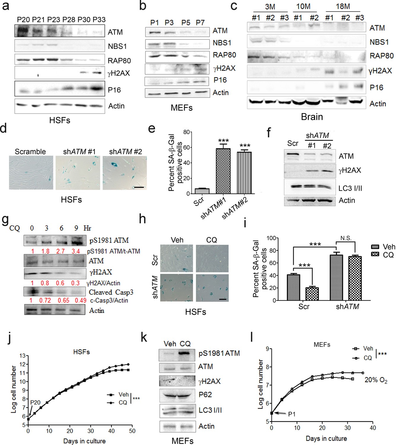
In searching for genes/pathways that drive senescence, we employed human primary endothelial cells, which underwent replicative senescence at passage 21, with increased p21 expression and β-galactosidase activity (Figure 1—figure supplement 1a–b). By RNAseq analysis, a gradual decline of ATM-centered DNA repair machinery was identified (Figure 1—figure supplement 1c–e). Western blotting analysis confirmed progressively downregulated protein levels of ATM and its downstream target NBS1 and RAP80 in senescent human skin fibroblasts (HSFs) (Figure 1a). Mouse embryonic fibroblasts (MEFs) with limited growth capacity and senescent phenotypes when cultured in vitro (Parrinello et al., 2003; Samper et al., 2003; Sherr and DePinho, 2000), and brain tissues from aged mice also showed progressive decline of ATM, NBS1, and RAP80 (Figure 1b–c). Concomitantly, upregulation of γH2AX, indicating accumulated DNA damage, and an increase in p16Ink4a were observed in senescent HSFs, MEFs, and aged brain tissues (Figure 1a–c). Knocking down ATM via shRNA accelerated senescence in HSFs, evidenced by increased β-galactosidase activity (Figure 1d–e), enlarged morphology (Figure 1—figure supplement 2a), accumulated γH2AX (Figure 1f), and reduced cell proliferation (Figure 1—figure supplement 2b). These data indicate that ATM decline retards DDR and drives senescence.
Figure 1 with 2 supplements see all
ATM activation by chloroquine alleviates senescence.
(a) Immunoblots showing protein levels of ATM, NBS1, and RAP80 in human skin fibroblasts (HSFs). A gradually increased level of p16 indicates cellular senescence, while elevated γH2AX level indicates accumulated DNA damage. (b) Immunoblots showing protein levels of ATM, NBS1, and RAP80 in mouse embryonic fibroblasts (MEFs). (c) Immunoblots showing protein levels of ATM, NBS1, and RAP80 in brain tissues isolated from 3-, 10-, and 18-month-old male mice. (d) SA-β-Gal staining in HSFs treated with sh-ATM or scramble shRNA. Scale bar, 100 µm. (e) Quantification of SA-β-Gal-positive staining of (d) from five views randomly captured for each group. Data represent means ± SEM. ***p<0.001. (f) Immunoblots showing increased γH2AX and unaffected LC3I/II in HSFs treated with sh-ATM or scramble shRNA. (g) Immunoblots showing protein levels of pS1981 ATM, γH2AX, and cleaved caspase-3 in HSFs treated with 10 μM of CQ for indicated time. (h) SA-β-Gal staining in HSFs expressing either scramble or ATM shRNA treated with 1 μM CQ or DMSO (12 hr). Scale bar, 100 µm. (i) Quantification of SA-β-Gal-positive staining of (h) from five views randomly captured for each group. Data represent means ± SEM. ***p<0.001; ‘N.S.’ indicates no significant difference. (j) HSFs at passage 20 were continuously cultured with 1 μM CQ or DMSO, and cell number was calculated at each passage. Data represent means ± SEM. ***p<0.01. (k) Immunoblots showing protein levels of γH2AX, p62, and LC3 in MEFs treated with 1 μM CQ or DMSO. Note that CQ had little effect on the expression levels of p62 and LC3. (l) MEFs at passage one were continuously cultured in 20% O2 with 1 μM CQ or DMSO, and cell number was determined at each passage. Data represent means ± SEM. ***p<0.01.
-
Figure 1—source data 1
Statistical analysis for SA-β-Gal positive staining.
- https://doi.org/10.7554/eLife.34836.006
-
Figure 1—source data 2
Statistical analysis for EdU positive staining.
- https://doi.org/10.7554/eLife.34836.007
Other than DNA damage, ATM is activated by chloroquine (CQ), an antimalarial drug that modulates chromatin confirmation (Bakkenist and Kastan, 2003). We confirmed that a low dose of CQ increased the level of pS1981 auto-phosphorylation of ATM but not γH2AX (Figure 1—figure supplement 2c). We then investigated whether activating ATM by CQ can ameliorate senescence. As shown, the CQ treatment activated ATM (pS1981), promoted clearance of DNA damage (γH2AX), and inhibited apoptosis (cleaved Casp3) in HSFs (Figure 1g). Also, the CQ treatment suppressed β-galactosidase activity, which was abrogated if ATM was knocked down (Figure 1h–i). Importantly, CQ treatment extended the replicative lifespan of HSFs (Figure 1j). Likewise, CQ treatment activated Atm, cleared up accumulated DNA damage, suppressed β-galactosidase activity (Figure 1k and Figure 1—figure supplement 2d–e), and prolonged replicative lifespan in MEFs (Figure 1l). Although both 10 μM and 1 μM of CQ activated ATM, a dose-dependent toxicity assay showed that 1 μM is suitable for long-term treatment (Figure 1—figure supplement 2f–g). Of note, ATM KD or low dose of CQ applied in this study had little effect on basal autophagic activity (Figure 1f,j and Figure 1—figure supplement 2g). Collectively, CQ activates ATM to alleviate replicative senescence.
An ATM-SIRT6 axis underlies age-associated metabolic reprogramming
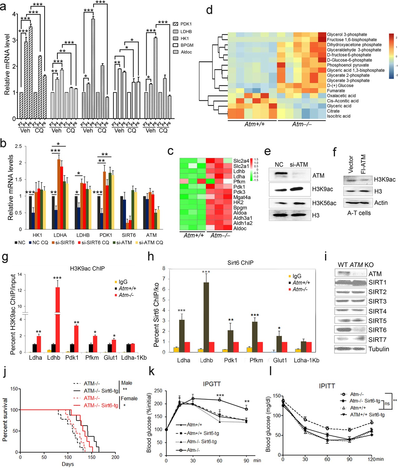
A-T patients lacking functional ATM display features of premature aging, accompanied by insulin resistance and glucose intolerance (Bar et al., 1978; Espach et al., 2015). Senescent cells exhibit impaired mitochondrial respiration, but enhanced glycolysis producing more lactate (Hagen et al., 1997; Lenaz et al., 2000). As such, we wondered whether ATM decline triggers an age-associated metabolic shift. Levels of glycolytic genes LDHB and PDK1 were dramatically increased in senescent MEFs and HSFs (Figure 2a and Figure 2—figure supplement 1a), and in liver tissues from Atm-/- mice (Figure 2—figure supplement 1b). Significantly, activating ATM via CQ suppressed senescence-associated glycolysis (Figure 2a and Figure 2—figure supplement 1a). Similarly, the inhibitory effect on glycolysis was diminished when ATM was depleted in HepG2 cells (Figure 2b). These data suggest a role for ATM in inhibiting glycolysis.
Figure 2 with 2 supplements see all
ATM-SIRT6 axis regulates age-related metabolic reprogramming.
(a) Quantitative RT-PCR analysis of mRNA levels of indicated glycolytic genes in different passages of MEFs with or without treatment of CQ. Data represent means ± SEM. *p<0.05, **p<0.01, ***p<0.001. (b) Quantitative RT-PCR analysis of mRNA levels of indicated glycolytic genes in Scramble (NC), si-SIRT6 or si-ATM HepG2 cells incubated with or without CQ (10 μM, 6 hr). Data represent means ± SEM. *p<0.05, **p<0.01, ***p<0.001. (c) Heatmap representation of RNA-Seq data (GSE109280) showing relative changes of glycolytic genes in Atm-/- MEF cells. The transcript levels are qualified in reads per kilobase of exon per million mapped sequence reads (RPKM), which is a normalized measure of exonic read density. Red and green indicate up- and downregulation, respectively. (d) Heatmap showing relative levels of metabolites in Atm+/+ and Atm-/- MEF cells of p53 null background, analyzed by LC-MS. Red and blue indicate up- and downregulation, respectively. (e) Immunoblots showing protein levels of H3K9ac and H3K56ac in ATM-deficient HepG2 cells. (f) Immunoblots showing levels of H3K9ac in A-T cells reconstituted with Flag-ATM. (g) ChIP analysis showing enrichment of H3K9ac at the promoter regions of indicated genes in Atm+/+ and Atm-/- MEFs. Data represent means ± SEM of three independent experiments. *p<0.05, **p<0.01, ***p<0.001. (h) ChIP analysis showing enrichment of Sirt6 at the promoter regions of indicated genes in Atm+/+ and Atm-/- MEFs. Data represent means ± SEM of six independent experiments. *p<0.05, **p<0.01, ***p<0.001. (i) Immunoblots showing protein levels of sirtuins in wild-type (WT) and ATM knockout (KO) HEK293 cells. (j) Kaplan-Meier survival of Atm-/- and Atm-/-;Sirt6-Tg male (n = 11 in each group) and female (n = 9 in each group) mice. **p<0.01. (k) Results of glucose tolerance tests in Atm+/+, Atm-/-, and Atm-/-;Sirt6-Tg mice. Data represent means ± SEM, n = 6. **p<0.01, ***p<0.001. (l) Results of insulin tolerance tests in Atm+/+, Atm-/-, and Atm-/-;Sirt6-Tg mice. Data represent means ± SEM, n = 6. **p<0.01. ‘ns’ indicates no significant difference.
-
Figure 2—source data 1
Differently expressed mRNA profiles of Atm-/- MEF cells.
- https://doi.org/10.7554/eLife.34836.011
-
Figure 2—source data 2
Differentially expressed Metabolites in Atm KO MEFs.
- https://doi.org/10.7554/eLife.34836.012
-
Figure 2—source data 3
Differently expressed mRNA profiles of Sirt6-/- MEF cells.
- https://doi.org/10.7554/eLife.34836.013
To examine how ATM regulates glycolysis, we performed RNA-Seq in Atm-/- MEF cells, and revealed a significant upregulation of glycolytic pathways (Figure 2c, and Figure 2—source data 1). Specific genes were validated by q-PCR (Figure 2—figure supplement 1c). As p53 is critical in glycolysis (Kruiswijk et al., 2015; Schwartzenberg-Bar-Yoseph et al., 2004), we further analyzed metabolomics of Atm-/- and control MEFs in p53 null background. As shown, the metabolic profile exhibited a clear shift, i.e. mitochondrial electron transport chain and intermediates of TCA cycle were reduced, while intermediates of glycolysis were elevated (Figure 2d, Figure 2—figure supplement 1d–e and Figure 2—source data 2). The data suggest that ATM deficiency enhances anaerobic glycolysis in a p53-independent manner.
Sirt6 deacylase is able to shunt energy metabolism away from anaerobic glycolysis to the TCA cycle via H3K9ac-mediated local chromatin remodeling (Sebastián et al., 2012; Zhong et al., 2010). We noted that the level of H3K9ac was enhanced in cells depleted ATM (Figure 2e). Re-expressed ATM in A-T cells suppressed H3K9ac level (Figure 2f). ChIP analysis showed that H3K9ac was enriched at the promoter regions of glycolytic genes in Atm-/- cells (Figure 2g), where the relative occupancy of SIRT6 was abolished (Figure 2h). Consistent with increased H3K9ac, SIRT6 protein level was dramatically downregulated in Atm-/- mouse livers, and ATM-deficient HepG2, U2OS and HEK293 cells (Figure 2—figure supplement 1f–i). In contrast, protein levels of other sirtuins were not much affected in ATM KO HEK293 cells (Figure 2i), and mRNA levels of all sirtuins remained unchanged (Figure 2—figure supplement 1j). Moreover, transcriptomic analysis and q-PCR data illustrated that Sirt6 depletion upregulated a similar cluster of genes essential for glycolysis (Figure 2—figure supplement 2a–b and Figure 2—source data 3). More importantly, the hyper-activated glycolytic pathway caused by ATM deficiency was completely restored by ectopic SIRT6 in HepG2 cells (Figure 2—figure supplement 2c). The CQ treatment upregulated SIRT6 level and reduced H3K9ac level, especially at the regulatory regions of glycolytic genes (Figure 2—figure supplement 2d–e). Knocking down SIRT6 abolished the inhibitory effect of CQ on glycolysis (Figure 2b). Additionally, ATM depletion in HEK293 cells, HSFs, and MEFs, significantly downregulated SIRT6 protein level, with little effect on SIRT1 or SIRT7 (Figure 2i and Figure 2—figure supplement 2f–g). Thus, these data suggest that ATM decline triggers an age-associated metabolic shift via SIRT6-mediated chromatin remodeling.
Other than metabolic abnormality, depleting Sirt6 leads to premature aging features and shortened lifespan (Mostoslavsky et al., 2006a), whereas extra copies of Sirt6 promote longevity in male mice (Kanfi et al., 2012). Given that Sirt6 was destabilized in Atm null mice, we wondered whether the Sirt6 transgene could rescue premature aging phenotypes and shortened lifespan in Atm-/- mice. To this end, we generated Sirt6 transgenic mice by microinjection, and bred them with Atm-/- mice. The overexpression of Sirt6 was demonstrated by western blotting (Figure 2—figure supplement 2h). Significantly, ectopic Sirt6 restored the elevation of serum lactate, and extended lifespan of Atm-/- mice of both genders (Figure 2j and Figure 2—figure supplement 2i). Importantly, Atm-/-;Sirt6-tg mice exhibited improved glucose tolerance and decreased insulin resistance (Figure 2k–l). Given that little difference was observed in glucose metabolism between young wild-type (WT) and Sirt6-transgenic mice (Kanfi et al., 2012), these data suggest a contributory role of the Atm-Sirt6 axis in the age-associated metabolic reprogramming.
ATM phosphorylates and stabilizes SIRT6
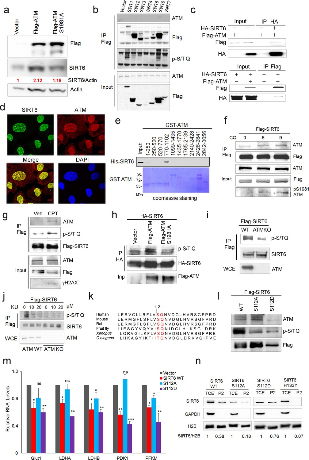
Next, we examined how ATM regulates SIRT6. Significantly, overexpression of ATM increased SIRT6 level, but this was abolished when ATM was S1981A-mutated to block dimeric ATM dissociation (Bakkenist and Kastan, 2003; Berkovich et al., 2007) (Figure 3a). Moreover, in addition to CQ, hypotonic buffer (20 mM NaCl), low glucose (LG), DNA-damaging agent camptothecin (CPT), and doxorubicin (Dox) all activated ATM and concomitantly increased SIRT6 protein level (Figure 3—figure supplement 1a–c), which was abrogated in ATM-depleted cells (Figure 3—figure supplement 1b–c). These data implicate a direct regulation of SIRT6 stability by ATM kinase activity. To confirm this, we first performed co-immunoprecipitation (Co-IP) in cells transfected with various FLAG-sirtuins. Interestingly, ATM was predominantly associated with SIRT6 among seven sirtuins (Figure 3b). The interaction was further confirmed at both ectopic and endogenous levels (Figure 3c and Figure 3—figure supplement 1d). Immunofluorescence microscopy showed co-localization of SIRT6 and ATM protein in the nucleus (Figure 3d). A domain mapping experiment indicated that the C-terminal domain was required for SIRT6 binding to ATM (Figure 3—figure supplement 1e). To determine whether ATM physically binds to SIRT6, 10 consecutive recombinant GST-ATM proteins were obtained and the binding to purified His-SIRT6 was analyzed. As shown (Figure 3e), His-SIRT6 bound predominantly to GST-ATM-4 (residues 770–1102) and relatively weakly to GST-ATM-1 (residues 1–250); both belong to the N-terminal HEAT repeat domain of ATM.
Figure 3 with 1 supplement see all
ATM interacts with and phosphorylates SIRT6.
(a) Immunoblots showing protein levels of SIRT6 in HEK293 cells expressing Flag-ATM or Flag-ATM S1981A. (b) Immunoblots showing endogenous ATM and p-S/TQ motif in anti-Flag immunoprecipitates in HEK293 cells transfected with empty vector or Flag-sirtuins. (c) Immunoblots showing Flag-ATM and HA-SIRT6 in anti-HA (upper) or anti-Flag (lower) immunoprecipitates in HEK293 cells transfected with indicated constructs. (d) Representative photos of immunofluorescence staining of SIRT6 and ATM in U2OS cells, showing co-localization in the nucleus. Scale bar, 50 µm. (e) GST pull-down assay showing bacterially expressed His-SIRT6 predominantly bound to GST-ATM fragment 4 (770–1102), the N-terminal HEAT-repeat of ATM. (f) Immunoblots showing the increased binding capacity of ATM and SIRT6 under the treatment of (10 µM) CQ for the indicated time. (g) Immunoblots showing ATM and p-S/TQ in anti-Flag immunoprecipitates in HEK293 cells expressing Flag-SIRT6 treated with CPT (0.4 µM) or DMSO. (h) Immunoblots showing level of p-S/TQ SIRT6 in HEK293 cells co-transfected with HA-SIRT6 and Flag-ATM, Flag-ATM S1981A, or empty vector. (i) Immunoblots showing p-S/T Q SIRT6 in WT or ATM KO HEK293 cells. (j) Immunoblots showing p-S/T Q level of SIRT6 in ATM WT or KO HEK293 cells treated with DMSO and KU55933 (10 or 20 µM, 2 hr). (k) Alignment of protein sequence of human SIRT6 and orthologues in mouse, rat, fruit fly, Xenopus, and C. elegans. A conserved S112 Q113 motif was highlighted. (l) Immunoblots showing p-S/T Q level of Flag-SIRT6, Flag-SIRT6 S112A, or Flag-SIRT6 S112D in HEK293 cells. (m) Quantitative RT-PCR analysis of mRNA levels of indicated glycolytic genes in sh-SIRT6 HepG2 cells re-expressing SIRT6, SIRT6 S112A, or 112D mutation. Data represent means ± SEM. *p<0.05, **p<0.01, ***p<0.001. ‘ns’ indicates no significant difference. (n) Immunoblots showing SIRT6 protein level in total cell extract (TCE) and chromatin-enriched fractions (P2). Densitometry analysis was performed to determine the relative ratio of SIRT6/H2B within chromatin fractions.
We next examined whether ATM phosphorylates SIRT6. Firstly, we found that CQ or CPT treatment significantly enhanced the binding of SIRT6 to ATM (Figure 3f and Figure 3—figure supplement 1f), whereas the S1981A mutant blocked such association (Figure 3—figure supplement 1g). ATM preferentially phosphorylates the S/T-Q motif. In the presence of CPT, an increased p-S/TQ level of SIRT6 was identified (Figure 3g). Of note, lambda protein phosphatase (λPP) diminished the p-S/TQ level of SIRT6 (Figure 3—figure supplement 1h). Likewise, the p-S/TQ level of SIRT6 was elevated in cells treated with low glucose, which activates ATM by ROS generation (Assaily et al., 2011; Sarre et al., 2012) (Figure 3—figure supplement 1i). Moreover, ectopic ATM significantly increased the p-S/TQ level of SIRT6, but this was abolished in the case of S1981A mutant (Figure 3h). Consistently, a pronounced reduction of p-S/TQ level of SIRT6 was observed in cells lacking ATM or treated with KU55933, a selective and specific ATM kinase inhibitor (Berkovich et al., 2007; Hickson et al., 2004) (Figure 3i–j). The decrease in p-S/TQ level was primarily attributable to loss of ATM, as it was restored by ectopic FLAG-ATM in a dose-dependent manner (Figure 3—figure supplement 1j). Indeed, SIRT6 has one evolutionarily conserved S112Q113 motif (Figure 3k). We therefore constructed S112A and S112D mutants, which resemble hypo- and hyper-phosphorylated SIRT6 respectively. As shown, these mutations almost abolished the pS/T-Q level of FLAG-SIRT6 (Figure 3l). The in vitro kinase assay showed that ATM could phosphorylate GST-SIRT6, but not S112A (Figure 3—figure supplement 1k). Furthermore, compared with SIRT6 S112A, ectopic S112D exhibited a much higher inhibitory effect on glycolytic gene expression in sh-SIRT6 HepG2 cells (Figure 3m and Figure 3—figure supplement 1l), and enhanced chromatin association of SIRT6 (Figure 3n). Collectively, the data suggest that ATM directly phosphorylates SIRT6 at Serine 112.
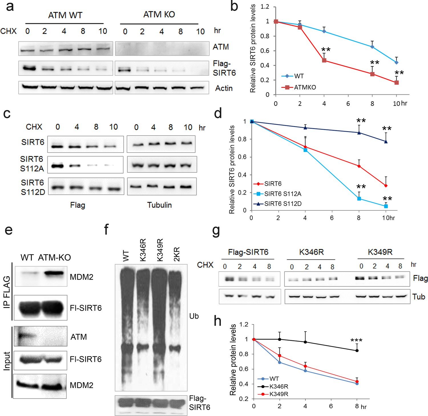
We next examined whether ATM is involved in regulating SIRT6 protein stability. Notably, compared with WT or vehicle control, the degradation rate of ectopic and endogenous SIRT6 was largely increased in ATM KO HEK293 cells, Atm-/- MEFs, and cells incubated with KU55933 in the presence of cycloheximide (CHX) (Figure 4a–b and Figure 4—figure supplement 1a–c). Recently, MDM2 was demonstrated to ubiquitinate SIRT6 and promote its proteasomal degradation (Thirumurthi et al., 2014). We therefore examined the polyubiquitination level of SIRT6. As shown, the ubiquitination level of FLAG-SIRT6 in ATM KO cells was significantly elevated compared with WT (Figure 4—figure supplement 1d). While S112A mutant markedly enhanced the polyubiquitination level of SIRT6, S112D had little effect (Figure 4—figure supplement 1e). Moreover, S112A accelerated SIRT6 degradation, whereas S112D retarded it (Figure 4c–d), indicating that the Ser112 phosphorylation by ATM regulates SIRT6 ubiquitination and thus protein stability. Indeed, ectopic MDM2 enhanced the polyubiquitination level of FLAG-SIRT6 (Figure 4—figure supplement 1f). In the case of ATM depleted or SIRT6 S112A mutant, the binding capacity of SIRT6 to MDM2 was enhanced (Figure 4e and Figure 4—figure supplement 1g). In searching for key residues that are polyubiquitinated by MDM2, we identified two clusters of lysine residues, i.e. K143/145 and K346/349, which are conserved across species. We then generated KR mutations of these residues, and found that K346/349R remarkably reduced the polyubiquitination level of SIRT6 (Figure 4—figure supplement 1h). Individual KR mutation showed that K346R significantly blocked MDM2-mediated ubiquitination and degradation of SIRT6, whereas K349R hardly affected it (Figure 4f–g). More importantly, K346R restored the increased ubiquitination and accelerated protein degradation of SIRT6 S112A (Figure 4—figure supplement 1i–j). Collectively, these data indicate that K346 is subject to MDM2-mediated ubiquitination, which is inhibited by ATM-mediated S112 phosphorylation.
Figure 4 with 1 supplement see all
ATM prevents ubiquitination and degradation of SIRT6.
(a) Immunoblots showing protein levels of Flag-SIRT6 in WT and ATM KO HEK293 cells treated with CHX (50 µg/ml) for indicated periods of time. (b) Quantification of protein levels in (a) by ImageJ. Data represent means ± SEM of three independent experiments. **p<0.01. (c) Immunoblots showing protein levels of Flag-SIRT6, S112A, and S112D in the presence of CHX (50 μg/ml) for indicated periods of time. (d) Quantification of protein levels in (c) by ImageJ. Data represent means ± SEM of three independent experiments. ***p<0.001. (e) Immunoblots showing increased binding capacity between SIRT6 and MDM2 in ATM KO HEK293 cells. (f) Immunoblots showing ubiquitination of Flag-SIRT6, K346R, K349R, and K346/349R (2KR) in HEK293 cells. Note that 2KR and K346R abrogated the ubiquitination of Flag-SIRT6. (g, h) Upper, immunoblots showing protein levels of Flag-SIRT6, K346R, and K349R in the presence of CHX (50 μg/ml) for indicated periods of time. Lower, quantification of protein levels by ImageJ. Data represent means ± SEM of three independent experiments. ***p<0.001.
Activating ATM via CQ promotes longevity
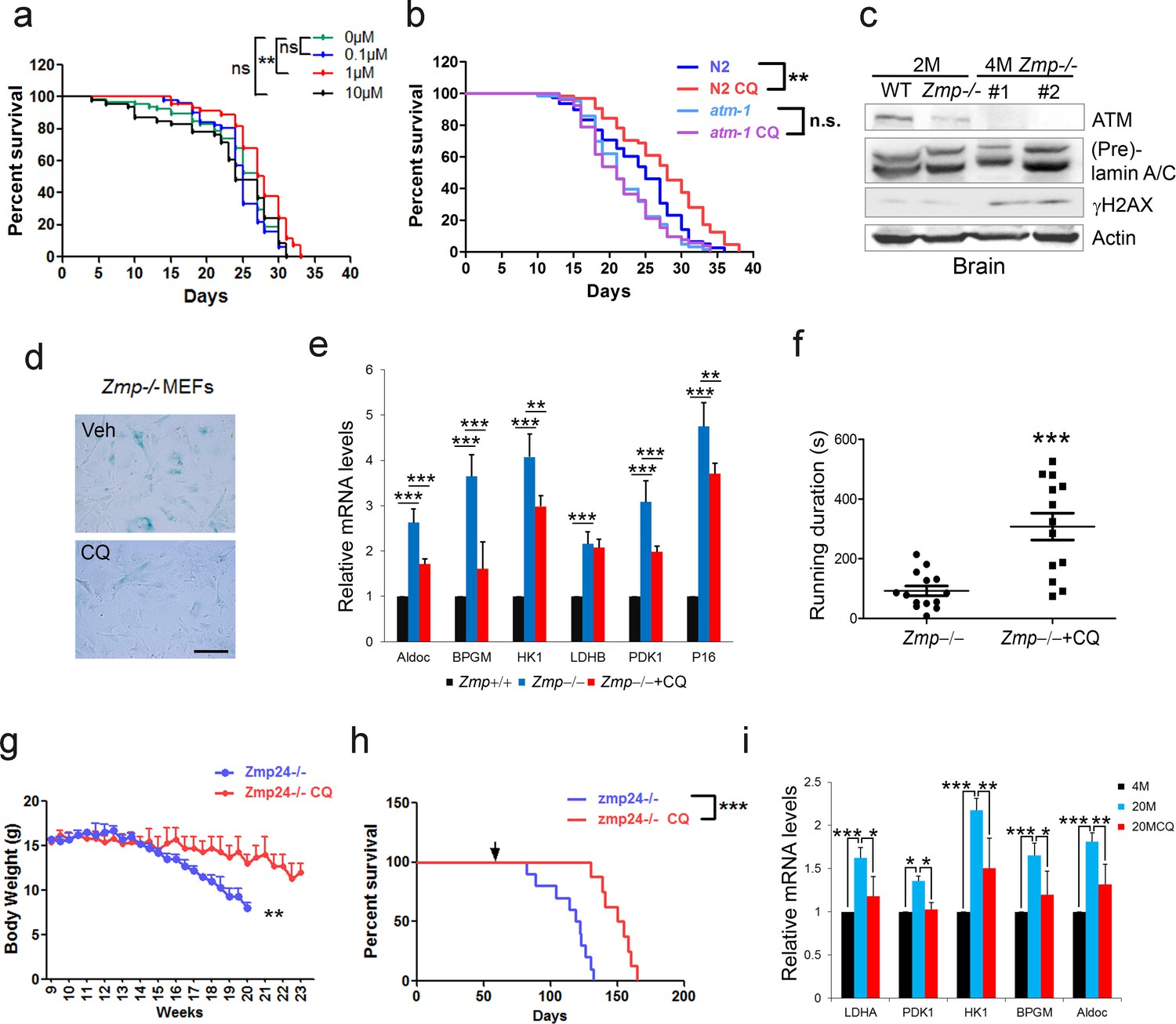
The cellular data suggest a pro-longevity function of ATM. We then tested it at organismal level. We employed Caenorhabditis elegans, which have a short lifespan of approximate 30 days. Nematodes deficient for atm-1, an orthologue of mammalian ATM, and WTs were exposed to various doses of CQ (see Materials and methods). Significantly, the period treatment with CQ (1.0 µM) extended the median lifespan (~14%) of C. elegans (Figure 5a). The lifespan-extending effect was abolished in atm-1 KO (Figure 5b) or in SIRT6 homolog sir-2.4 KD nematodes (Figure 5—figure supplement 1a–b). The data suggest that CQ promotes longevity in an ATM- and SIRT6- dependent manner. We further examined the beneficial effect of CQ in a HGPS model, i.e. Zmpste24-/- mice, which has a shortened lifespan of 4–6 months (Pendás et al., 2002) and impaired ATM-mediated DNA repair signaling (Liu et al., 2013a). We found that the level of Atm was dramatically reduced in Zmpste24-/- MEFs and tissues (Figure 5c and Figure 5—figure supplement 1c). Significantly, CQ treatment activated Atm, stabilized Sirt6, decreased the accumulated DNA damage, inhibited glycolysis, and alleviated senescence in Zmpste24-/- cells (Figure 5d–e and Figure 5—figure supplement 1d–e). The CQ treatment also delayed body weight decline, increased running endurance, and prolonged lifespan in Zmpste24-/- mice (Figure 5f–h), but had no significant effect on the lifespan of Atm-/- mice (Figure 5—figure supplement 1f).
Figure 5 with 2 supplements see all
CQ extends lifespan in an ATM- dependent manner.
(a) Survival analysis of C. elegans treated with the indicated dosage of CQ. **p<0.01. NS indicates no significant difference. (b) Survival analysis of wild-type and atm-1 null C. elegans cultured in medium with or without 1 µM CQ. (c) Immunoblots showing protein levels of Atm and γH2AX in brain tissues of Zmpste24+/+ (2 months), Zmpste24-/- (2 months), and Zmpste24-/- (4 months) mice. (d) Representative images showing SA-β-Gal staining in Zmpste24-/- MEFs with or without CQ treatment. Scale bar, 100 µm. (e) Quantitative RT-PCR analysis of mRNA levels of p16Ink4a and indicated glycolytic genes in liver tissues of Zmpste24+/+, saline-treated, and CQ-treated Zmpste24-/- mice. Mice were treated for 8 weeks with two weekly intraperitoneal injections of CQ at 3.5 mg/kg. Data represent means ± SEM. *p<0.05, **p<0.01, ***p<0.001. (f) Maximum running duration in saline- and CQ-treated Zmpste24-/- mice. Data represent means ± SEM. ***p<0.001. (g) Body weight of saline- and CQ-treated male Zmpste24-/- mice. Data represent means ± SEM. **p<0.01. (h) Kaplan-Meier survival curves of saline-treated (n = 10) and CQ-treated (n = 8) Zmpste24-/- mice. ***p<0.001. (i) Quantitative RT-PCR analysis of mRNA levels of indicated glycolytic genes in the liver tissues of 4-month-old, saline-treated 12-month-old (n = 3), and CQ-treated 20-month-old (n = 3) mice. Data represent means ± SEM. *p<0.05, **p<0.01, ***p<0.001.
-
Figure 5—source data 1
Lifespan analysis of chloroquine's effect on nematodes.
- https://doi.org/10.7554/eLife.34836.021
-
Figure 5—source data 2
Lifespan analysis of chloroquine's effect on mice.
- https://doi.org/10.7554/eLife.34836.022
Physiologically aged mice frequently develop aging-associated metabolic disorders, with high glucose and lactate (Houtkooper et al., 2011). Given that ATM declines with age, and activation of ATM by CQ inhibits glycolysis in senescent cells and Zmpste24-/- mice, we intraperitoneally administrated 12-month-old ‘old’ male mice with low-dose CQ (3.5 mg/kg) twice a week. Remarkably, compared with the saline-treated group, CQ treatment inhibited glycolysis, lowered serum lactate level, and attenuated body weight decline (Figure 5i and Figure 5—figure supplement 1g–h), implicating potential benefits of CQ in physiologically aged mice. Generally, these data demonstrate a lifespan-extending benefit of ATM activation by CQ.
Discussion
DNA damage accumulates with age, and defective DDR and DNA repair accelerates aging. However, whether boosting DNA repair machinery promotes healthiness and longevity is still unknown. DNA damage stimulates DDR, but if persistent, it instead leads to senescence. Therefore, if enhancing DDR efficacy possibly promotes longevity, it must be DNA damage free. The antimalarial drug CQ can intercalate into the internucleosomal regions of chromatin, unwind DNA helical twist, and thus activate ATM without causing any DNA damage (Bakkenist and Kastan, 2003; Krajewski, 1995). We demonstrate that long-term treatment with CQ activates ATM, improves DNA repair, restores age-related metabolic shift, alleviates cellular senescence, and extends lifespan of nematodes and Zmpste24 null mice. Mechanistically, ATM phosphorylates the longevity gene SIRT6 (Tasselli et al., 2017), and prevents MDM2-mediated ubiquitination and proteasomal degradation of SIRT6. To our knowledge, this is the first study to establish direct causal links between robust DNA repair machinery and longevity. In support of this notion, DNA repair efficacy has been shown to be enhanced in long-lived naked mole rat (MacRae et al., 2015), and human longevity has been shown to be associated with single nucleotide polymorphisms (SNPs) in DNA repair genes/pathways (Debrabant et al., 2014; Soerensen et al., 2012). Interestingly, the heterozygous rather than homozygous status of a SNP, albeit both enhance the transcription of ATM, is associated with longevity in Chinese and Italian populations (Chen et al., 2010; Piaceri et al., 2013). Therefore, in future study, it would be worthwhile evaluating whether Atm can promote longevity in model organisms, and, if so, how many extra copies are required.
Accumulation of DNA damage and metabolic disturbance are common denominators of aging (López-Otín et al., 2013; Moskalev et al., 2013). Metabolic reprogramming from the TCA cycle to glycolysis is prominent in both physiological and pathological aging (Feng et al., 2016; Shimizu et al., 2014). Why senescent cells become glycolytic is poorly understood. The crosstalk between cellular metabolism and DDR is not well elucidated. Upon genotoxic stress, ATM represses the rapamycin-sensitive mammalian target of the rapamycin (mTORC1) pathway (Alexander et al., 2010), but activates the pentose phosphate pathway (PPP) (Cosentino et al., 2011), suggesting that cell metabolism may be key downstream of DDR signaling. Moreover, it is recognized that deficiency in DNA repair machinery such as ATM, WRN, and Ercc1, accelerates aging and causes severe metabolic disorders (Garinis et al., 2008; White and Vijg, 2016). In this study, we showed that boosting ATM activity using a low dose of CQ enhances genomic stability, attenuates age-onset metabolic reprogramming, alleviates senescence, and extends lifespan in mice. The data demonstrate for the first time that enhanced DNA repair machinery (ATM-SIRT6 axis) promotes longevity. Considering that ATM and SIRT6 function not only in maintenance of genome integrity but also as homeostatic protein modifiers, the pro-longevity role of ATM is most likely benefited from enhanced DNA repair and metabolic homeostasis, but it is difficult to determine which is more important.
Recently, Bohr’s group identified that increased consumption of NAD+ by an early DDR factor poly (ADP-ribose) polymerase (PARP1), owing to accumulated DNA damage, accelerated aging in Atm mutant mice (Fang et al., 2016). NAD+ serves as a cofactor of sirtuins, including SIRT1 and SIRT6. Therefore, this work establishes a linear causal link between deficient DDR, DNA damage accumulation, consumption of NAD+, decline in sirtuin activity, and aging. Moreover, administration of nicotinamide mononucleotide or nicotinamide riboside ameliorates age-related function decline and extends lifespan in mice (Mills et al., 2016; Zhang et al., 2016). Here, we found that ATM decline during aging causes DNA damage accumulation and enhances glycolysis, both of which consume most of the NAD+, providing an explanation for the low NAD+ level in Atm-/- mice and physiologically aged mice.
Closely resembling normal aging, HGPS has attracted numerous efforts in understanding of molecular mechanisms and developing therapeutic strategies (De Sandre-Giovannoli et al., 2003; Eriksson et al., 2003). We and others have found that HGPS and Zmpste24 null cells undergo premature senescence because of defective chromatin remodeling (Ghosh et al., 2015; Krishnan et al., 2011; Liu et al., 2013a; Liu et al., 2013b), delayed DDR, and impaired DNA repair (Liu et al., 2005; Liu et al., 2013b; Varela et al., 2005). Specifically, Atm-Kap-1 signaling is compromised (Liu et al., 2013a), and SIRT6 protein level and deacetylase activity are reduced in progeria cells (Endisha et al., 2015; Ghosh et al., 2015). Here we showed that Atm is significantly downregulated, which explains the reduced SIRT6, delayed DDR, and metabolic shift in progeria cells and mice. It would be interesting to investigate whether ectopic Atm or Sirt6 could rescue progeroid features in these mice. Nevertheless, the activation of ATM via CQ remarkably improves glucose homeostasis, DNA damage clearance, and running endurance, and extends lifespan in progeria mice. It would be worthwhile evaluating the pro-longevity benefits of CQ in physiological aging.
CQ is an FDA-approved and clinically used medicine for treatment of malaria (2015). Via activation of ATM, long-term treatment of CQ protects against atherosclerosis, improves insulin sensitivity, and rescues glucose tolerance in type 2 diabetes (T2D) (Emami et al., 1999; Razani et al., 2010; Schneider et al., 2006). The lysosomotropic property of CQ also makes it a potent inhibitor of autophagy (Yang et al., 2013). The application of CQ for antimalarial treatment (500 mg/week, maximum 0.8 µM in plasma) and for cancer therapy (100–500 mg/day) (Kimura et al., 2013) is attributed to its inhibitory action on autophagy. Of note, CQ also the attenuates inflammatory response by inhibiting autophagy (Szatmári-Tóth et al., 2016; Whelan et al., 2017; Wu et al., 2018), requiring a high dosage of 50 mg/kg for mice. In the current study, we used a low dose of CQ to activate ATM, i.e. 1–10 μM for cell line and 3.5 mg/kg twice a week for mice (Schneider et al., 2006). The results showed that at such low doses, CQ has no toxicity and little effect on basal autophagic activity. Moreover, a low dose of CQ prolongs lifespan in progeroid mice, but exhibits little effect on Atm KO background, supporting an ATM-dependent pro-longevity function of CQ. Unfortunately, we could not test the CQ effect in Sirt6-/- mice with only 1-month lifespan (Mostoslavsky et al., 2006b). Here, we addressed the pro-longevity benefits of CQ-activated ATM, most likely attributable to improved DNA repair and glucose metabolism. Given that ATM also displays anti-inflammatory function (Erttmann et al., 2017; Shoelson, 2006), we could not rule out an anti-inflammatory effect in lifespan extension observed in CQ-treated mice.
In conclusion, our data establish direct causal links between robust DNA repair machinery and longevity. In line with DNA damage theory of aging, we propose that DNA damage activates DDR; however, its constant activation causes senescence; defective ATM-SIRT6 axis underlies premature aging, exemplified by HGPS and A-T mouse models, which are rescued by treatment of CQ and Sirt6 transgene, respectively; in physiological aging, DNA damage-free activation of ATM by CQ stabilizes SIRT6, thus promoting longevity in nematodes and most likely also in mice (Figure 5—figure supplement 2). Our findings provide a novel therapeutic strategy for HGPS, and could facilitate clinical trials of CQ as an effective treatment for age-related diseases.
Materials and methods
Key resources table
| Reagent type (species) or resource | Designation | Source or reference | Identifiers | Additional information |
|---|---|---|---|---|
| Gene (human) | ATM | National Center for Biotechnology Information | Gene ID: 472 | |
| Gene (mouse) | Atm | National Center for Biotechnology Information | Gene ID: 11920 | |
| Gene (Caenorhabditis elegans) | atm-1 | National Center for Biotechnology Information | Gene ID: 3565793 | |
| Gene (human) | SIRT6 | National Center for Biotechnology Information | Gene ID: 51548 | |
| Gene (mouse) | Sirt6 | National Center for Biotechnology Information | Gene ID: 50721 | |
| Gene (Caenorhabditis elegans) | sir-2.4 | National Center for Biotechnology Information | Gene ID: 182284 | |
| Gene (mouse) | Zmpste24 | National Center for Biotechnology Information | Gene ID: 230709 | |
| Gene (mouse) | p53 | National Center for Biotechnology Information | Gene ID: 230710 | |
| Cell line (human) | HEK293 | ATCC | Catalog number: ATCC CRL-1573; RRID:CVCL_0042 | |
| Cell line (human) | HepG2 | ATCC | Catalog number: ATCC HB-8065; RRID:CVCL_0027 | |
| Cell line (human) | U2OS | ATCC | Catalog number: ATCC HTB-96; RRID:CVCL_0042 | |
| Cell line (mouse) | Atm-/-; p53-/- MEF | from Dr. Yosef Shiloh (Tel Aviv University, Israel) | ||
| Cell line (mouse) | Sirt6-/- MEF | from Dr. Raul Mostoslavsky (Massachusetts General Hospital Cancer center, USA) | ||
| Antibody | ATM | Abcam (Cambridge, UK) | Cat# ab78; RRID:AB_306089 | Applications: WB;Immunofluorescence |
| Antibody | SIRT6 | Abcam (Cambridge, UK) | Cat# ab62739; RRID:AB_956300 | Applications: WB; Immunofluoresce;Chromatin immunoprecipitation |
| Antibody | γH2AX | Abcam (Cambridge, UK) | Cat# ab81299; RRID:AB_1640564 | Applications: WB |
| Antibody | p21 | Santa Cruz Biotechnology | Cat# sc-6246; RRID:AB_628073 | Applications: WB |
| Antibody | MDM2 | Santa Cruz Biotechnology | Cat# sc-965; RRID:AB_627920 | Applications: WB |
| Antibody | p-ATM (Ser1981) | EMD Millipore | Cat# 05–740; RRID:AB_309954 | Applications: WB |
| Antibody | H3K9ac | EMD Millipore | Cat# 07–352; RRID:AB_310544 | Applications: WB; Chromatin immunoprecipitation |
| Antibody | p-S/T Q | Cell Signaling Technology (Beverly, MA) | Cat #9607S; RRID:AB_10889739 | Applications: WB |
| Antibody | cleaved caspase-3 | Cell Signaling Technology (Beverly, MA) | Cat #9661; RRID:AB_2341188 | Applications: WB |
| Antibody | HA | Sigma-Aldrich | Cat# H3663; RRID:AB_262051 | Applications: WB |
| Antibody | Flag | Sigma-Aldrich | Cat# F1804; RRID: AB_262044 | Applications: WB |
| Antibody | LC3B | Sigma-Aldrich | Cat# L7543; RRID:AB_796155 | Applications: WB |
| Transfected construct (human) | Flag-His-ATM wt | Addgene (Cambridge, MA) | Cat #31985 | |
| Transfected construct (human) | Flag-SIRT6 | Addgene (Cambridge, MA) | Cat #13817 | |
| Transfected construct (human) | Flag-His-ATM S1981A | Addgene (Cambridge, MA) | Cat #32300 | |
| Commercial assay or kit | Senescence beta- galactosidase staining Kit | Cell Signaling Technology (Beverly, MA) | Cat #9860 | |
| Commercial assay or kit | Lactate Colorimetric Assay Kit | BioVision | Cat #K667-100 | |
| Commercial assay or kit | Click-iT EdU Alexa Fluor 488 Kit | Invitrogen | Cat #C10425 | |
| Chemical compound, drug | Cycloheximide | Sigma-Aldrich | Cat #66-81-9 | |
| Chemical compound, drug | MG-132 | Sigma-Aldrich | Cat #474787 | |
| Chemical compound, drug | Chloroquine | Sigma-Aldrich | Cat #C6628 |
Mice
Zmpste24-/- mice and Atm-/- mice have been described previously (Barlow et al., 1996; Pendás et al., 2002). Sirt6-transgenic mice (Sirt6-tg) of C57BL/6J background were constructed by injecting cloned mSirt6 cDNA with CAG promoter into fertilized eggs. Primers for genotyping of Sirt6 transgenic allele were as follows: forward: 5’-CTGGTTATTGTGCTGTCTCATCAT-3’; reverse: 5’-CCGTCTACGTTCTGGCTGAC-3’. Atm-/- mice were crossed to Sirt6-tg mice to get Atm-/-;Sirt6-tg mice. Chloroquine (CQ) experiments were conducted as described (Schneider et al., 2006). Briefly, 12-month-old wild-type C57BL/6J male mice, 2-month-old Zmpste24-/-, and Atm-/- male mice were administered with CQ (Sigma, St. Louis, MO) in 0.9% saline twice per week at 7 mg/kg body weight, and the control group was treated with saline alone. At least 8 weeks after treatment of CQ, mice were subjected to functional tests. Body weight and lifespan was recorded. The survival rate was analyzed using the Kaplan–Meier method and statistical comparison was performed using the Log-rank Test. Mice were housed and handled in the laboratory animal research center of Shenzhen University. All experiments were performed in accordance with the guidelines of the Institutional Animal Care and Use Committee (IACUC). The protocols were approved by the Animal Welfare and Research Ethics Committee of Shenzhen University (Approval ID: 201412023).
C. elegans survival assay
Request a detailed protocolC. elegans nematode survival assay was performed according to standard protocols (Kenyon et al., 1993). Briefly, wild-type and atm-1 null nematodes (100 to 150 per group) synchronized to prefertile young adult stage were exposed to NGM plates containing the indicated dosage of CQ. After 1-day incubation, animals were transferred to fresh incubation plates without CQ for another 2 days. This procedure was repeated every 3 days. Nematodes that showed no response to gentle stimulation were recorded as dead. The survival data were analyzed using the Kaplan–Meier method and statistical comparison was performed using the Log-rank Test.
Cell lines
Request a detailed protocolHEK293 (CRL-1573), HepG2 (HB-8065), and U2OS (HTB-96) cells were purchased from ATCC. Human skin fibroblasts HSFs (F2-S) and primary MEFs were prepared as described previously (Liu et al., 2005). Immortalized Atm-/-; p53-/- and Sirt6-/- MEFs were provided as a kind gift from Dr. Yosef Shiloh (Tel Aviv University, Israel) and Dr. Raul Mostoslavsky (Massachusetts General Hospital Cancer center, USA), respectively. These cell lines were authenticated by short tandem repeat (STR) profile analysis and genotyping, and were mycoplasma free. Cells were cultured in Gibco DMEM (Life Technologies, USA) with 10% fetal bovine serum (FBS), 100 U/ml penicillin and streptomycin (P/S) at 37°C in 5% CO2 and atmospheric oxygen conditions. For CQ experiments, cells were maintained in the medium containing 1 μM chloroquine for 12 hr, and then grown in new fresh medium for 48 hr.
Plasmids
Human Flag-SIRT6, pcDNA3.1 Flag-ATM, Flag-ATM S1981A, and pcDNA3 human MDM2 were all purchased from Addgene (Cambridge, MA). Flag-SIRT6 with amino acid substitution mutations (S112A, S112D, K346R/K349R) were generated by PCR-based mutagenesis using pcDNA3-Flag-SIRT6 as a template and a QuikChange II site-directed mutagenesis kit (Agilent Technologies), following the manufacturer’s instructions. Primer sequences for amino acid mutations of SIRT6 were as follows: SIRT6 S112A: (forward) 5'-cgtccacgttctgggcgaccaggaagcgga-3', (reverse) 5’-tccgcttcctggtcgcccagaacgtggacg-3’; SIRT6 S112D: (forward) 5'-ccgtccacgttctggtcgaccaggaagcggag-3', (reverse) 5'-ctccgcttcctggtcgaccagaacgtggacgg-3'; SIRT6 K346R: (forward) 5'-ggccttcacccttctggggggtctgtg-3', (reverse) 5'-cacagaccccccagaagggtgaaggcc-3'; SIRT6 K349R: (forward) 5'-gccttggccctcacccttttggggggt-3', (reverse) 5'-accccccaaaagggtgagggccaaggc-3. HA-tagged human SIRT6 plasmid was amplified from the respective cDNAs and constructed into pKH3-HA vector. To express four truncated forms of SIRT6 protein, HA-SIRT6 plasmid as a template was constructed by PCR-based deletion.
Protein extraction and western blotting
Request a detailed protocolFor whole cell protein extraction, cells were suspended in five volumes of suspension buffer (20 mM Tris-HCl, pH 7.5, 150 mM NaCl, 1 mM EDTA, 1 mM DTT, protease inhibitor cocktail), and then five volumes of 2X SDS loading buffer were added and incubated at 98°C for 6 min. Mice tissues were homogenized with 1 ml of ice-cold tissue lysis buffer (25 mM TrisHCl, pH 7.5, 10 mM Na3VO4, 100 mM NaF, 50 mM Na4P2O7, 5 mM EGTA, 5 mM EDTA, 0.5% SDS, 1% NP-40, protease inhibitor cocktail). After homogenization and sonication, lysates were centrifuged at 16,000 g for 15 min. The clean supernatant was carefully transferred to new tubes. Protein concentrations were determined using a bicinchoninic acid (BCA) assay method (Pierce, Rockford, IL) and were normalized with lysis buffer for each sample. Samples were denatured in 1X SDS loading buffer by boiling at 98°C for 6 min. Proteins were separated by loading to SDS-polyacrylamide gels, and then were transferred to PVDF membrane (Millipore). The protein levels were determined by immunoblotting using respective antibodies. The ImageJ program was used for densitometric analysis of immunoblotting, and the quantification results were normalized to the loading control.
Antibodies
Rabbit anti-SIRT6 (ab62739), ATM (ab78), SIRT1 (ab12193), γH2AX (ab81299), RAP80 (ab52893), Kap-1 (ab10484), and p-KAP-1 (Ser824, ab70369) antibodies were obtained from Abcam (Cambridge, UK). Anti-lamin A/C (sc-20681), p21 (sc-6246), MDM2 (sc-965), and P53 (sc-6243) antibodies were purchased from Santa Cruz Biotechnology. Rabbit anti-γH2AX (05–636), p-ATM (Ser1981) (05–740), histone H3 (07–690), anti-H3K56ac (07–677), and H3K9ac (07–352) antibodies were sourced from EMD Millipore. Mouse anti-p-ATM (Ser1981) (#5883), p-S/TQ (#9607), ubiquitin (#3936), and cleaved caspase-3 (#9661) antibodies were purchased from Cell Signaling Technology (Beverly, MA). Mouse anti- HA, Flag, rabbit anti-LC3B, and P62 antibodies were obtained from Sigma-Aldrich. Anti-Nbs1 (NB100-143) antibody was purchased from Novus Biologicals. Mouse anti-actin, tubulin antibodies were obtained from Beyotime. Anti-pS112 SIRT6 monoclonal antibodies were prepared by Abmart generated from a specific phosphorylated peptide (peptide sequence CLRFVSPQNV).
Protein degradation assay
Request a detailed protocolHEK293 cells (WT and ATM-deficient cells) were transfected with Flag-SIRT6 alone or together with Mdm2. 48 hr later, the cells were treated with 50 μg/ml of cycloheximide (CHX, Sigma-Aldrich), a translation inhibitor. For endogenous SIRT6 protein degradation assay, ATM wild-type and null MEFs were grown in 6 cm plates, and were treated with 50 mg/ml CHX for indicated time points. Cells were collected and the protein levels were determined by western blotting, the subsequent quantification was performed with ImageJ software.
In vivo ubiquitination assay
Request a detailed protocolIn vivo ubiquitination assay was performed by transfecting HEK293 cells in 6 cm dishes with 1 μg Myc-ubiquitin, 2 μg Flag-SIRT6 or its mutations, and/or 1 μg MDM2 vector. 48 hr after transfection, cells were lysed in the buffer (25 mM Tris-HCl pH 8.0, 250 mM NaCl, 10 mM Na3VO4, 1 mM EDTA, 10% glycerol, protease inhibitor cocktail, and 0.1 mM phenylmethylsulphonyl fluoride), and then incubated with Flag-M2 beads (Sigma-Aldrich) overnight at 4°C. Beads were washed with lysis buffer three times, bound proteins were eluted by adding 1.5 × SDS loading buffer. The ubiquitin levels were analyzed by immunoblotting.
In vitro kinase assay
Request a detailed protocolHEK293T cells were transfected with 10 μg of FLAG-ATM and then treated with CPT. Activated ATM was immune-purified from the cell extracts with FLAG beads (Sigma, M8823). GST-SIRT6 or the S112A mutant was purified from bacteria. Kinase reactions were initiated by incubating purified ATM with GST-SIRT6 in the kinase buffer with or without 1 mM ATP for 120 min at 30°C. After reaction, proteins were blocked by SDS loading buffer. The membrane was then subjected to western blotting with antibodies against p-S/TQ.
Immunoprecipitation
Request a detailed protocolCells under indicated treatments were totally lysed in lysis buffer containing 20 mM HEPES, pH 7.5, 150 mM NaCl, 10 mM Na3VO4, 10% glycerol, 2 mM EDTA, protease inhibitor cocktail, and 0.1 mM phenylmethylsulphonyl fluoride. After sonication and centrifugation, the supernatant was collected and incubated with H3K9ac (Millipore, 2 μg/sample) overnight at 4°C with a gentle rotation. Protein A/G agarose (Pierce, 10 μl/sample) were added to the tubes and rotated at 4°C for 2 hr. Beads were precipitated by centrifugation at 1000 g for 15 s and washed three times with cold lysis buffer. The pellet was resuspended in 1.5 × SDS loading buffer and incubated at 98°C for 6 min. The supernatants were collected and used for western blotting.
GST pull-down assay
Request a detailed protocolA series of GST fusion proteins of truncated ATM, which together spanned the full length of ATM, were constructed into pGEX4T-3 vector. For GST pull-down, bacterially expressed 6 × His tagged SIRT6 was separately incubated with various GST-ATM fragments in a buffer of 150 mM NaCl, 20 mM Tris-HCl [pH 7.5], 5 mM MgCl2, 0.2 mM EDTA, 10% glycerol, 0.2% NP-40, and protease inhibitors (Roche Complete). GST-fusion proteins were then precipitated by adding Glutathione Sepharose fast flow (GE Healthcare). After washing twice with TEN buffer (0.5% Nonidet P-40, 20 mM Tris-HCl [pH 7.4], 0.1 mM EDTA, and 300 mM NaCl), glutathione agarose beads were analyzed by western blotting and coomassie staining.
RNA interference and shRNA lentiviral infection
Request a detailed protocolBriefly, cells were transfected with small interfering RNAs (siRNAs) for 48 hr using Lipofectamine 3000 (Invitrogen, USA) according to the manufacturer’s instructions. The siRNAs targeting human ATM, SIRT6, and HDM2 were purchased (GenePharma, China) with sequences as follows, si-ATM#1: 5’-AAUGUCUUUGAGUAGUAUGUU-3’ (Zhou et al., 2003); Si-ATM#2: 5’-AAGCACCAGUCCAGUAUUGGC-3’ (Zhang et al., 2005); si-SIRT6#1: 5’-AAGAAUGUGCCAAGUGUAAGA-3’; si-SIRT6#2: 5’-CCGGCTCTGCACCGTGGCTAA-3’; si-HDM2#1: 5’-AACGCCACAAATCTGATAGTA-3’; si-HDM2#2: 5’-AATGCCTCAATTCACATAGAT-3’. A scrambled siRNA sequence was used as control. Lentiviral shRNA constructs were generated in a pGLVH1 backbone (GenePharma, China), and virus was produced in HEK293 cells. To deplete ATM in HSF cells and SIRT6 in HepG2 cells, lentiviral infection was performed in the presence of 5 μg/ml polybrene. Two days later, the infected HSF cells or HepG2 cells were selected with 2 μg/ml puromycin. To downregulate sir-2.4 expression, the NL2099 worms were exposed to incubation plates containing HT115 bacteria with sir-2.4 RNAi vector.
CRISPR/Cas9-mediated genome editing
Request a detailed protocolGene mutagenesis by the CRISPR/Cas9 system was conducted as described (Ran et al., 2013). The following gRNAs targeting human ATM, SIRT6 were constructed in pX459 vector (Addgene, #48139). sgATM F: 5’-CACCGATATGTGTTACGATGCCTTA-3’, R: 5’- AAACTAAGGCATCGTAACACATATC-3’. HEK293 cells were transfected with pX459 or pX459-gRNA using Lipofetamine 3000 Transfection Reagent according to the manufacturer's instructions. After 2-day culture, cells were selected with 2 μg/ml puromycin, six colonies were picked and grown to establish stable cell lines. The targeted mutations were identified by western blotting, and PCR-based sequencing.
EdU (5-ethynyl-2’-deoxyuridine) incorporation assay
Request a detailed protocolEdU incorporation assays were conducted in HSF cells to estimate cell proliferation using the Click-iT EdU Alexa Fluor 488 Kit (Invitrogen, USA). HSF cells, infected by the respective lentiviruses containing shNC and shATM, were cultured in a six-well plate containing the coverslips in the presence of 10 μM EdU for 12 hr. Cells were fixed in 3.7% formaldehyde followed by 0.5% Triton X-100 permeabilization, and then stained with Alexa Fluor picolyl azide. Five random views were captured to calculate the positive staining rate for each group.
Growth curves and SA-β-gal assays
Request a detailed protocolCell population doublings were monitored using a Coulter Counter. SA-β-galactosidase assay in primary cells was performed using Senescence β-galactosidase staining Kit (#9860, CST) according to the manufacturer's instructions. Five views were captured randomly to calculate the positive staining rate for each group.
RNA preparation and Real-Time qPCR
Request a detailed protocolTotal RNA was extracted from cells or mouse tissues using Trizol reagent RNAiso Plus (TaKaRa, Japan) following the phenol–chloroform extraction method. Purified total RNA was used to obtain cDNA using PrimeScript RT Master Mix (Takara, Japan) following this method: 37°C for 30 min, and 85°C for 5 s. The gene expression was analyzed with the CFX Connected Real-Time PCR Detection System (BioRad) with SYBR Ex Taq Premixes (Takara, Japan). Gene expression levels were normalized to actin.
Glucose tolerance test
Request a detailed protocolMice were fasted overnight (6 p.m. to 9 a.m.), and D-glucose (2.5 g/kg body weight) was administrated intraperitoneally. Blood glucose levels were determined from tail vein blood using a glucometer (Onetouch ultravue, Johnson, USA) at 0, 30, 60, 90, and 120 min after D-glucose injection.
Insulin tolerance test
Request a detailed protocolMice were fasted for 6 hr (8 a.m. to 2 p.m.), and recombinant human insulin (0.75 U/kg body weight) was administered intraperitoneally. Blood glucose levels were determined in tail vein blood using a glucometer (Onetouch ultravue, Johnson) at 0, 30, 60, 90, and 120 min after insulin injection.
Lactate assay
Request a detailed protocolMouse serum was five-fold diluted, and lactate concentration was determined with the Lactate Colorimetric Assay Kit (BioVision).
Endurance running test
Request a detailed protocolZmpste24-/- mice were treated for 8 weeks with chloroquine or saline before running on a Rota-Rod Treadmill (YLS-4C, Jinan Yiyan Scientific Research Company, Shandong, China) to test the effect of chloroquine on fatigue resistance. Mice were placed on the rotating lane, and the speed was gradually increased to 10 r/min. When mice were exhausted and safely dropped from the rotating lane, the time latency to fall was automatically recorded.
Metabolite analysis
Request a detailed protocolWild-type and ATM KO cells were grown in normal medium for 24 hr, and methanol-fixed cell pellets were analyzed by a two liquid chromatography-tandem mass spectrometry (LC-MS) method as described (Luo et al., 2007).
Immunofluorescence microscopy
Request a detailed protocolThe cells were fixed using 4% paraformaldehyde at room temperature for 15 min, permeabilized by 0.5% Triton X-100 at room temperature for 10 min, blocked using 10% FBS/PBS, and then incubated with primary antibodies diluted in PBS containing 2% BSA overnight at 4°C. The primary antibodies were detected using an Alexa-488-conjugated anti-mouse secondary antibody (Invitrogen). The nuclei were stained using DAPI in anti-fade mounting medium. Images were captured using a Zeiss LSM880 confocal/multiphoton microscope.
ChIP assay
Request a detailed protocolCells were fixed in 1% formaldehyde for 10 min at room temperature. The crosslinking reaction was quenched with 0.125 M glycine. After washing with PBS, cells were lysed with lysis buffer (50 mM Tris·HCl pH 8.0, 2 mM EDTA, 15 mM NaCl, 1% SDS, 0.5% deoxycholate, protease inhibitor cocktail, 1 mM PMSF), followed by sonication and centrifugation. The supernatant was collected and precleared in dilution buffer (50 mM Tris-HCl pH 8.0, 2 mM EDTA, 150 mM NaCl, 1% Triton X-100) with protein A/G Sepharose and pre-treated salmon DNA. The precleared samples were incubated overnight with H3K9ac antibody (2 μg/sample, Millipore) or appropriate control IgGs (Santa Cruz), and protein A/G Sepharose (Invitrogen). After washing sequentially with a series of buffers, the beads were heated at 65°C to reverse the crosslink. DNA fragments were purified and analyzed. Real-time PCR was performed with primers as described (Zhong et al., 2010):
LDHB-ChIP-5’: AGAGAGAGCGCTTCGCATAG
LDHB-ChIP-3’: GGCTGGATGAGACAAAGAGC
ALDOC-ChIP-5’: AAGTGGGGCACTGTTAGGTG
ALDOC-ChIP-3’: GTTGGGGATTAAGCCTGGTT
PFKM-ChIP-5’: TTAAGACAAAGCCTGGCACA
PFKM-ChIP-3’: CAACCACAGCAATTGACCAC
LDHA-ChIP-5’: AGGGGGTGTGTGAAAACAAG
LDHA-ChIP-3’: ATGGCTTGCCAGCTTACATC
LDHA-ChIP-1Kb-5’: TGCAAGACAAGTGTCCCTGT
LDHA-ChIP-1Kb-3’: GAGGGAATGAAGCTCACAGC
Statistical analysis
Request a detailed protocolStatistical analyses were conducted using two-tailed Student's t-test between two groups. All data are presented as mean ± S.D. or mean ± S.E.M. as indicated, and a p value < 0.05 was considered statistically significant.
Data availability
Sequencing data have been deposited in GEO under accession code GSE109280.
-
Boosting ATM activity alleviates ageing and extends lifespan in a mouse model of progeria.Publicly available at the NCBI Gene Expression Omnibus (accession no: GSE109280).
References
-
The Werner syndrome protein is involved in RNA polymerase II transcriptionMolecular Biology of the Cell 10:2655–2668.https://doi.org/10.1091/mbc.10.8.2655
-
Extreme insulin resistance in ataxia telangiectasia: defect in affinity of insulin receptorsThe New England Journal of Medicine 298:1164–1171.https://doi.org/10.1056/NEJM197805252982103
-
Ataxia-telangiectasia; a familial syndrome of progressive cerebellar ataxia, oculocutaneous telangiectasia and frequent pulmonary infectionPediatrics 21:526–554.
-
ATM phosphorylates histone H2AX in response to DNA double-strand breaksJournal of Biological Chemistry 276:42462–42467.https://doi.org/10.1074/jbc.C100466200
-
A functional single nucleotide polymorphism in promoter of ATM is associated with longevityMechanisms of Ageing and Development 131:636–640.https://doi.org/10.1016/j.mad.2010.08.009
-
Ku complex interacts with and stimulates the Werner proteinGenes & Development 14:907–912.
-
Human longevity and variation in DNA damage response and repair: study of the contribution of sub-processes using competitive gene-set analysisEuropean Journal of Human Genetics 22:1131–1136.https://doi.org/10.1038/ejhg.2013.299
-
Insulin-sparing effect of hydroxychloroquine in diabetic rats is concentration dependentCanadian Journal of Physiology and Pharmacology 77:118–123.https://doi.org/10.1139/y98-146
-
ATM protein kinase signaling, type 2 diabetes and cardiovascular diseaseCardiovascular Drugs and Therapy 29:51–58.https://doi.org/10.1007/s10557-015-6571-z
-
Reprogramming of energy metabolism as a driver of agingOncotarget 7:15410–15420.https://doi.org/10.18632/oncotarget.7645
-
DNA damage and ageing: new-age ideas for an age-old problemNature Cell Biology 10:1241–1247.https://doi.org/10.1038/ncb1108-1241
-
Measuring reactive species and oxidative damage in vivo and in cell culture: how should you do it and what do the results mean?British Journal of Pharmacology 142:231–255.https://doi.org/10.1038/sj.bjp.0705776
-
The impact of DNA damage, genetic mutation and cellular responses on cancer prevention, longevity and aging: observations in humans and miceMechanisms of Ageing and Development 126:71–77.https://doi.org/10.1016/j.mad.2004.09.036
-
DNA damage, aging, and cancerNew England Journal of Medicine 361:1475–1485.https://doi.org/10.1056/NEJMra0804615
-
DNA damage, aging, and cancerNew England Journal of Medicine 361:1475–1485.https://doi.org/10.1056/NEJMra0804615
-
The metabolic footprint of aging in miceScientific Reports 1:134.https://doi.org/10.1038/srep00134
-
p53 in survival, death and metabolic health: a lifeguard with a licence to killNature Reviews Molecular Cell Biology 16:393–405.https://doi.org/10.1038/nrm4007
-
The Werner syndrome gene product co-purifies with the DNA replication complex and interacts with PCNA and topoisomerase IJournal of Biological Chemistry 274:37795–37799.https://doi.org/10.1074/jbc.274.53.37795
-
Mitochondrial bioenergetics in agingBiochimica et Biophysica Acta (BBA) - Bioenergetics 1459:397–404.https://doi.org/10.1016/S0005-2728(00)00177-8
-
Functional interaction between Ku and the werner syndrome protein in DNA end processingJournal of Biological Chemistry 275:28349–28352.https://doi.org/10.1074/jbc.C000289200
-
Impaired DNA double-strand break repair contributes to the age-associated rise of genomic instability in humansCell Death & Differentiation 23:1765–1777.https://doi.org/10.1038/cdd.2016.65
-
Genomic instability in laminopathy-based premature agingNature Medicine 11:780–785.https://doi.org/10.1038/nm1266
-
Genome instability, cancer and agingBiochimica et Biophysica Acta (BBA) - General Subjects 1790:963–969.https://doi.org/10.1016/j.bbagen.2009.03.020
-
The role of DNA damage and repair in aging through the prism of Koch-like criteriaAgeing Research Reviews 12:661–684.https://doi.org/10.1016/j.arr.2012.02.001
-
Oxygen sensitivity severely limits the replicative lifespan of murine fibroblastsNature Cell Biology 5:741–747.https://doi.org/10.1038/ncb1024
-
Mechanisms of ATM ActivationAnnual Review of Biochemistry 84:711–738.https://doi.org/10.1146/annurev-biochem-060614-034335
-
Ataxia-telangiectasia mutated (ATM) genetic variant in Italian centenariansNeurological Sciences 34:573–575.https://doi.org/10.1007/s10072-012-1188-5
-
Genome engineering using the CRISPR-Cas9 systemNature Protocols 8:2281–2308.https://doi.org/10.1038/nprot.2013.143
-
p53 is required for chloroquine-induced atheroprotection but not insulin sensitizationJournal of Lipid Research 51:1738–1746.https://doi.org/10.1194/jlr.M003681
-
Reactive oxygen species are produced at low glucose and contribute to the activation of AMPK in insulin-secreting cellsFree Radical Biology and Medicine 52:142–150.https://doi.org/10.1016/j.freeradbiomed.2011.10.437
-
Ataxia-telangiectasia (A-T): An emerging dimension of premature ageingAgeing Research Reviews 33:76–88.https://doi.org/10.1016/j.arr.2016.05.002
-
DNA damage response and metabolic diseaseCell Metabolism 20:967–977.https://doi.org/10.1016/j.cmet.2014.10.008
-
Banking on ATM as a new target in metabolic syndromeCell Metabolism 4:337–338.https://doi.org/10.1016/j.cmet.2006.10.009
-
SIRT6: novel mechanisms and links to aging and diseaseTrends in Endocrinology & Metabolism 28:168–185.https://doi.org/10.1016/j.tem.2016.10.002
-
Somatic mutations, genome mosaicism, cancer and agingCurrent Opinion in Genetics & Development 26:141–149.https://doi.org/10.1016/j.gde.2014.04.002
-
Do DNA double-strand breaks drive aging?Molecular Cell 63:729–738.https://doi.org/10.1016/j.molcel.2016.08.004
-
World Health Organ Tech Rep Ser1–546, The Selection and Use of Essential Medicines, World Health Organ Tech Rep Ser.
-
Application and interpretation of current autophagy inhibitors and activatorsActa Pharmacologica Sinica 34:625–635.https://doi.org/10.1038/aps.2013.5
-
DNA damage-mediated apoptosis induced by selenium compoundsJournal of Biological Chemistry 278:29532–29537.https://doi.org/10.1074/jbc.M301877200
Article and author information
Author details
Funding
Natural Science Foundation of Guangdong Province (2016A030310064)
- Minxian Qian
National Natural Science Foundation of China (81501206)
- Minxian Qian
National Natural Science Foundation of China (81501210)
- Mingyan Zhou
Natural Science Foundation of Guangdong Province (2015A030308007)
- Baoming Qin
Shenzhen Science and Technology Innovation Commission (JCYJ20140418095735645)
- Zimei Wang
Research Grant Council of Hong Kong (HKU2/CRF/13G)
- Zhongjun Zhou
National Natural Science Foundation of China (81422016)
- Baohua Liu
Ministry of Science and Technology of the People's Republic of China (2017YFA0503900)
- Baohua Liu
Natural Science Foundation of Guangdong Province (2014A030308011)
- Baohua Liu
Shenzhen Science and Technology Innovation Commission (CXZZ20140903103747568)
- Baohua Liu
National Natural Science Foundation of China (91439133)
- Baohua Liu
National Natural Science Foundation of China (81571374)
- Baohua Liu
Ministry of Science and Technology of the People's Republic of China (2016YFC0904600)
- Baohua Liu
Shenzhen Science and Technology Innovation Commission (JCYJ20160226191451487)
- Baohua Liu
Research Grant Council of Hong Kong (773313)
- Baohua Liu
Discipline Construction Funding of Shenzhen
- Baohua Liu
The funders had no role in study design, data collection and interpretation, or the decision to submit the work for publication.
Acknowledgements
We thank Dr. Yosef Shiloh (Tel Aviv University, Israel) for Atm-/-; p53-/- MEF cells and Dr. Raul Mostoslavsky (Massachusetts General Hospital Cancer center, USA) for Sirt6-/- MEFs. This project is supported by research grants from the National Natural Science Foundation of China (81422016, 91439133, 81571374, 81501206, 81501210), National Key R and D Program of China (2017YFA0503900, 2016YFC0904600), Research Grant Council of Hong Kong (773313, HKU2/CRF/13G), Natural Science Foundation of Guangdong Province (2014A030308011, 2015A030308007, 2016A030310064) and Shenzhen Science and Technology Innovation Commission (CXZZ20140903103747568, JCYJ20160226191451487 and JCYJ20140418095735645) and Discipline Construction Funding of Shenzhen (2016).
Ethics
Animal experimentation: Mice were housed and handled in the laboratory animal research center of Shenzhen University. All experiments were performed in accordance with the guidelines of the Institutional Animal Care and Use Committee (IACUC). The protocols were approved by the Animal Welfare and Research Ethics Committee of Shenzhen University (Approval ID: 201412023).
Version history
- Received: January 5, 2018
- Accepted: April 16, 2018
- Accepted Manuscript published: May 2, 2018 (version 1)
- Version of Record published: May 17, 2018 (version 2)
Copyright
© 2018, Qian et al.
This article is distributed under the terms of the Creative Commons Attribution License, which permits unrestricted use and redistribution provided that the original author and source are credited.
Metrics
-
- 5,128
- views
-
- 783
- downloads
-
- 54
- citations
Views, downloads and citations are aggregated across all versions of this paper published by eLife.
Download links
A two-part list of links to download the article, or parts of the article, in various formats.
Downloads (link to download the article as PDF)
Open citations (links to open the citations from this article in various online reference manager services)
Cite this article (links to download the citations from this article in formats compatible with various reference manager tools)
Boosting ATM activity alleviates aging and extends lifespan in a mouse model of progeria
eLife 7:e34836.
https://doi.org/10.7554/eLife.34836
Further reading
-
- Biochemistry and Chemical Biology
- Structural Biology and Molecular Biophysics
Vitamin B6 deficiency has been linked to cognitive impairment in human brain disorders for decades. Still, the molecular mechanisms linking vitamin B6 to these pathologies remain poorly understood, and whether vitamin B6 supplementation improves cognition is unclear as well. Pyridoxal 5’-phosphate phosphatase (PDXP), an enzyme that controls levels of pyridoxal 5’-phosphate (PLP), the co-enzymatically active form of vitamin B6, may represent an alternative therapeutic entry point into vitamin B6-associated pathologies. However, pharmacological PDXP inhibitors to test this concept are lacking. We now identify a PDXP and age-dependent decline of PLP levels in the murine hippocampus that provides a rationale for the development of PDXP inhibitors. Using a combination of small-molecule screening, protein crystallography, and biolayer interferometry, we discover, visualize, and analyze 7,8-dihydroxyflavone (7,8-DHF) as a direct and potent PDXP inhibitor. 7,8-DHF binds and reversibly inhibits PDXP with low micromolar affinity and sub-micromolar potency. In mouse hippocampal neurons, 7,8-DHF increases PLP in a PDXP-dependent manner. These findings validate PDXP as a druggable target. Of note, 7,8-DHF is a well-studied molecule in brain disorder models, although its mechanism of action is actively debated. Our discovery of 7,8-DHF as a PDXP inhibitor offers novel mechanistic insights into the controversy surrounding 7,8-DHF-mediated effects in the brain.
-
- Biochemistry and Chemical Biology
- Stem Cells and Regenerative Medicine
Molecules that facilitate targeted protein degradation (TPD) offer great promise as novel therapeutics. The human hepatic lectin asialoglycoprotein receptor (ASGR) is selectively expressed on hepatocytes. We have previously engineered an anti-ASGR1 antibody-mutant RSPO2 (RSPO2RA) fusion protein (called SWEETS) to drive tissue-specific degradation of ZNRF3/RNF43 E3 ubiquitin ligases, which achieved hepatocyte-specific enhanced Wnt signaling, proliferation, and restored liver function in mouse models, and an antibody–RSPO2RA fusion molecule is currently in human clinical trials. In the current study, we identified two new ASGR1- and ASGR1/2-specific antibodies, 8M24 and 8G8. High-resolution crystal structures of ASGR1:8M24 and ASGR2:8G8 complexes revealed that these antibodies bind to distinct epitopes on opposing sides of ASGR, away from the substrate-binding site. Both antibodies enhanced Wnt activity when assembled as SWEETS molecules with RSPO2RA through specific effects sequestering E3 ligases. In addition, 8M24-RSPO2RA and 8G8-RSPO2RA efficiently downregulate ASGR1 through TPD mechanisms. These results demonstrate the possibility of combining different therapeutic effects and degradation mechanisms in a single molecule.


